Article

Morphological, Molecular, and Pathogenic Characterization of Neofabraea alba, a Postharvest Pathogen of Apple in Serbia

1
Faculty of Agriculture, University of Novi Sad, Trg Dositeja Obradovića 8, 21000 Novi Sad, Serbia
2
Institute of Field and Vegetable Crops, Maksima Gorkog 30, 21000 Novi Sad, Serbia
3
Institute of Pesticides and Environmental Protection, Banatska 31 b, Zemun, 11080 Belgrade, Serbia
*
Author to whom correspondence should be addressed.
Agronomy 2022, 12(9), 2015; https://doi.org/10.3390/agronomy12092015
Submission received: 14 July 2022 / Revised: 19 August 2022 / Accepted: 19 August 2022 / Published: 26 August 2022
(This article belongs to the Special Issue Research Progress on Pathogenicity of Fungus in Crop)

Abstract

:
Neofabraea spp. is one of the major causal agents of the postharvest decay of apple fruit. Bull’s eye rot can cause fruit rot, as well as tree canker. In Serbia, during February and March, 2017 and 2018, 56.3% of collected stored rotten apple fruits were found to be infected with Neofabraea spp. Using polymerase chain reaction, phylogenetic analysis, and morphological characterization, Neofabraea alba was identified as the causal agent of Bull’s eye rot. Twenty-five selected isolates were analyzed by DNA sequencing of the internal transcribed spacer and 16 S mitochondrial ribosomal RNA. The phylogenetic analyses provided information on the distance between Neofabraea spp., while merged phylogenetic tree was required to give insight and clarify the relationships between isolates of Neofabraea alba. N. alba demonstrated pathogenicity on apple fruit as well as on branches, with significantly different aggressiveness among the isolates. Isolates were able to produce larger cankers and acervuli on apple branches with abundant sporulation All identified isolates were separated into two morphotypes and described based on their macromorphological characteristics on culture media. It was noticed that different culture media, pH, and temperature could affect macromorphological characteristics and development. Representative isolates were able to grow at a temperature range of 0 to 25 °C, above which only one isolate was able to grow on culture media. All isolates showed growth at a pH range of 2–10, with significant differences detected between isolates at different pH values. The type of cultivation medium significantly affected the size and shape of conidia, while the presence of the microconidia was recorded within isolates. The aim of this study was to identify the causal agents of Bull’s eye rot on stored apples in Serbia in the period 2017–2018 and characterize the obtained isolates based on pathogenic, morphological, physiological, and molecular criteria.

1. Introduction

Apple is one of the most widely grown fruit crops in Serbia. In 2020, the harvested area was 26,360 ha, with a production of 489,426 tons (http://www.fao.org/fastat/en/#data, (accessed on 18 April 2022)). Yield is threatened by numerous fungal pathogens during the growing season, as well as after harvest, during storage, and marketing. Neofabraea spp. is one of the major causes of the postharvest decay of apple and pear fruit [1,2,3]. Neofabraea spp. can cause Bull’s eye rot of apple fruits, as well as anthracnose canker of trees. The disease can be caused by various species, including Neofabraea alba (E. J. Guthrie) Verkley (anamorph: Phlyctema vagabunda Desm.), Neofabraea malicorticis H. S. Jacks. (anamorph: Cryptosporiopsis curvispora (Peck)), Neofabraea perennans Kienholz (anamorph: C. perennans (Zeller and Childs) Wollenw.), Neofabraea sp. nov. (anamorph: C. kienholzii Seifert, Spotts and Levesque), and two species recently described as apple pathogens Neofabraea actinidiae and Neofabraea brasiliensis [4]. N. alba has been identified in European countries such as Poland, the Czech Republic, Italy, the Netherlands, and Spain as a causal agent of Bull’s eye rot [3,5,6,7,8]. In Serbia, the disease was first recorded in 2016 [9]. In 2018, after investigating the prevalence of rot pathogens on apple fruits in storage facilities in Serbia, some previously considered less-dominant pathogens such as Fusarium spp. and Neofabraea spp. were identified, with Neofabraea spp. founded to be one of the most prevalent apple pathogens, with an incidence of 82% in diseased apple fruits (cultivar Jonagold) [10,11]. In order to optimize the disease management protocols, precise pathogen identification is crucial. However, it can be challenging to identify Neofabraea spp. at the species level because they produce similar symptoms on apple fruits and are often morphologically similar when grown in vitro. Additionally, their morphological traits may be inconsistent [12]. Previous research showed that symptoms on naturally infected apple fruits and the colony appearance were not sufficient for reliable diagnosis [13]. Neofabraea species were first incorrectly described as a part of Pezicula genus. However, after phylogenetic analyses, four species that cause apple disease were recognized as separate evolutionary lineages [14]. Several studies confirmed that, besides conventional identification methods based on isolation, pathogenicity conformation, morphological, and ecological characterization, the use of molecular tools for precise species identification is indispensable. Distinguishing between Neofabraea species and establishing phylogenetic relationships is possible with the genetic analyses of ITS nuclear DNA, mitochondrial rDNA, the β-tubulin gene, and elongation factor (EF1α), as well as with species specific primers [7,14,15,16,17]. In order to distinguish the species within the genus of Neofabraea that are threatening apple production in Serbian orchards, morphological, physiological, and molecular investigation of the pathogen was conducted. The aims of this study were: (a) to isolate and identify dominant Bull’s eye rot causal agents that occur in apple storages in Serbia as dominant rot causing agents; (b) to characterize collected isolates using morphological and molecular tools; (c) to determine the physiological and pathogenic features of the isolates.

2. Materials and Methods

2.1. Collection and Storage of Isolates

In February and March 2017 and 2018, after 6–9 months of cold storage under normal or ultra-low oxygen atmosphere, fruits of 13 different apple cultivars (Idared, Golden Delicious, Granny Smith, Braburn, Fuji, Jonagold, Jonagored, Red Delicious, Kiku, Mutsu, Pink Lady, Red Chief, Red Jonaprince) with typical symptoms of Bull’s eye rot were collected from storage facilities and local markets in the northern part of Serbia (the region of Vojvodina). Direct isolations from diseased tissue were performed from fruits, previously surface sterilized in 70% ethanol and rinsed with sterile distilled water. A small section (Ø 3 mm) of decayed tissue was plated on potato-dextrose agar medium (PDA; 200 g potato, 20 g dextrose, 17 g agar, and 1 L H2O) and incubated at 25 °C for 7 days. After isolation, a collection of hyphal tip purified isolates was established and maintained on PDA slants at 4 °C. Based on variations of morphological features, for identification and further characterization, 25 Neofabraea-like isolates were selected for identification and characterization and transferred to fresh malt extract agar (MEA; 20 g malt extract, 18 g agar, and 1 L H2O).

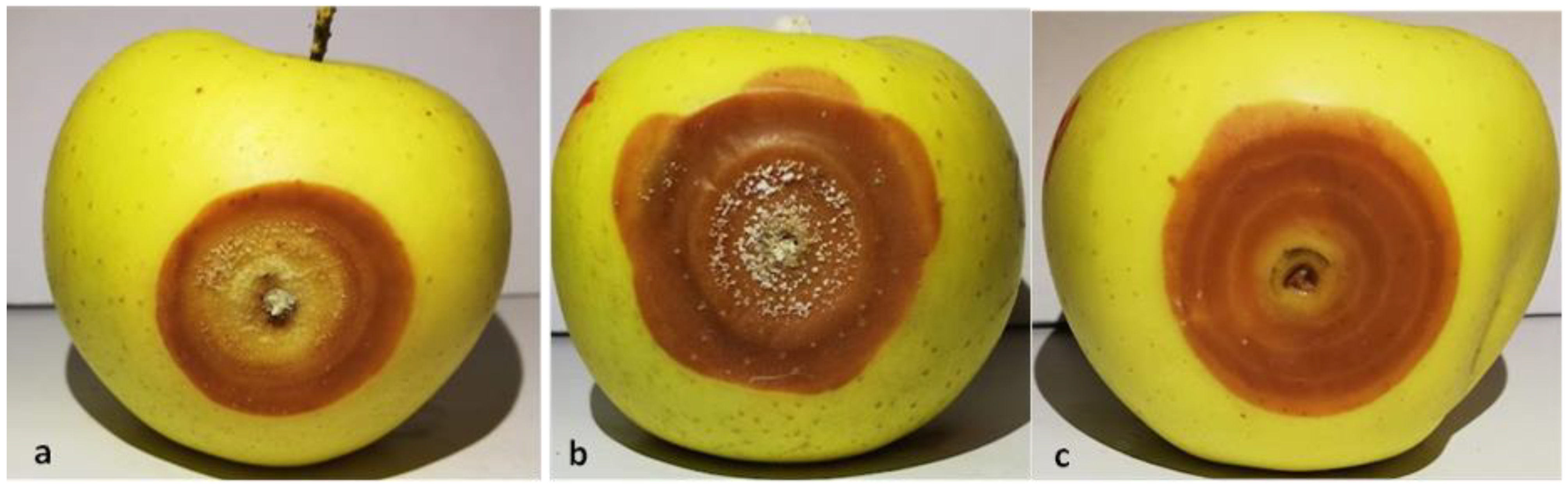
2.2. Pathogenicity Test

Fruits of cv. ‘Golden Delicious’, grown in the Vojvodina region and harvested at commercial maturity, uniform in size and without physical injuries or apparent infections, were used for pathogenicity test ex situ. Fruits of cv. ‘Golden Delicious’ were selected on the basis of previously recorded high susceptibility to Neofabraea (unpublished data). Pathogenicity in situ was studied on apple branches in the orchard. The fruits were surface sterilized (70% ethanol, 30 s), wounded aseptically on both sides of the equatorial plane with a cork borer (Ø 3 mm), and artificially inoculated with 21-day-old mycelial plugs (Ø 3 mm) of 25 selected Neofabraea-like isolates. Fruit inoculated the same way with sterile PDA plugs served as a negative control. For each isolate, two fruit with two inoculation sites on opposite sides of the fruit were used. Inoculated fruits were placed in plastic chambers with a non-hermetic seal and incubated at room temperature. The diameter of developed necrotic lesions (mm) was measured after 14, 21, and 28 days. From the developed lesions, the pathogen was reisolated on MEA medium and derived reisolates were compared with the original isolates used for inoculation.
In situ pathogenicity of Neofabraea-like isolates was tested in October 2019 (Karadjordjevo 45°51′25″ N; 19°35′29″ E) in a three-year-old apple orchard on the trees of cv. ‘Red Delicious’. Inoculation was performed as described by Henriquez et al. (2006), with a slight modification [18]. Two-year-old branches were first rinsed with sterile water and surface-disinfected with 70% ethanol, and then a 21-day-old mycelial plug (Ø 3 mm) was placed under a V-shaped cut (the depth of secondary xylem). Three inoculations per isolate and negative control with sterile PDA plugs were performed. After inoculation, the cut was covered by sterile cheesecloth and sealed with parafilm and aluminum folium for one month. The cover was removed, and the canker progression was observed once a month for one year (the length of canker was measured and expressed in mm/month). To confirm the pathogen identity, one-year post inoculation, conidia were washed from cankers and microscopically examined (A Krüss optronic, Hamburg Germany). For each sporulated isolate, 50 conidia were measured.

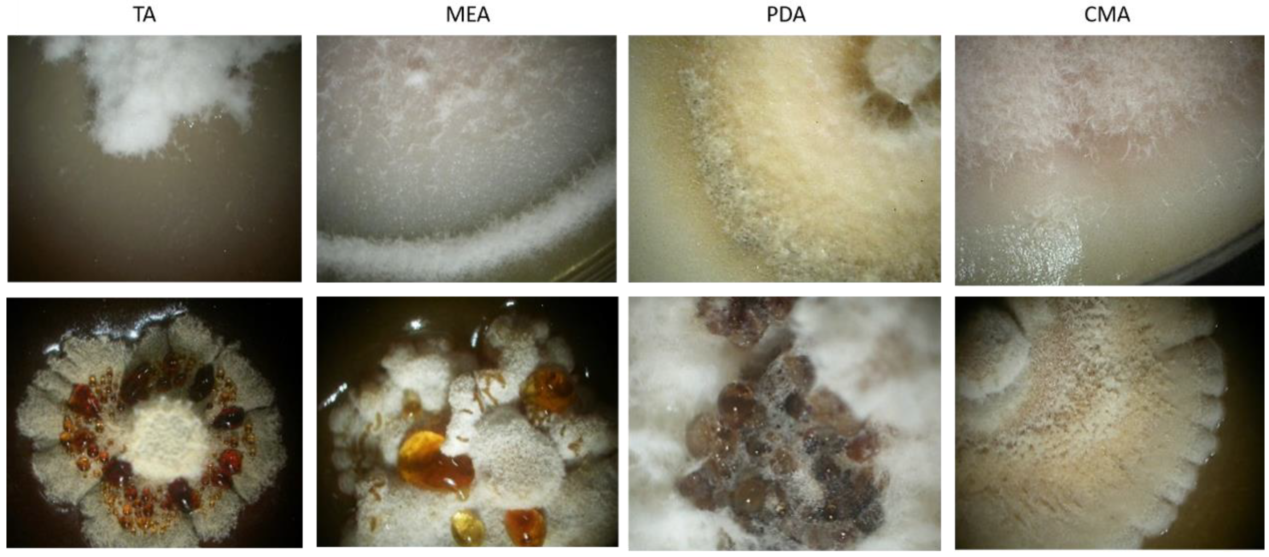
2.3. Morphological Characterization

Immediately after hyphal tip purification, 25 isolates, previously identified as Neofabraea spp. were morphologically characterized after 21 days of incubation on PDA at 20 °C in darkness. Based on colony appearance on PDA, the isolates were grouped into morphotypes. Representative isolates of each group, originating from different cultivars, were selected for further in-depth morphological and physiological studies in radial growth experiments at 20 °C, if not specified differently. To determine additional macromorphological features, quantitative growth rate and sporulation ability were recorded. Selected isolates from both morphological groups were grown on four different media: corn meal agar (CMA; 17 g corn meal, 17 g agar, 1 L H2O), PDA2, MEA, and TA (200 mL tomato juice, 2 gr CaCO3, 17 g agar, 800 mL H2O) for 4 months under two light regimes: 12 h near UV light/12 h darkness and continuous darkness (24 h). The experiment was conducted in four replicates per isolate. After 4 months, conidia size was observed with a microscope (BTC, Lalitpur-Nepal) and recorded with a camera (Nikon Coolpix 4500). For each sporulated isolate, 50 conidia were measured.

2.4. Physiological Characterization

The effects of five culture media (MEA, PDA, TA, PCA (potato-carrot agar; 250 g potato, 200 g carrot, 17 g agar, 1 L of H2O)) and OA (oatmeal agar: 30 g oatmeal, 17 g agar, 1 L H2O)), eight temperatures (0, 5, 10, 15, 20, 25, 30, 35 °C) and nine pH levels (2, 3, 4, 5, 6, 7, 8, 9, 10) of MEA medium on the mycelial growth rate of representative isolates of each group were studied. The pH level of the MEA medium was adjusted with 0.1 M KOH and 0.1 M HCl (adjusted after sterilization of culture media). Mycelial plugs (Ø 3 mm) of 21-day-old selected Neofabraea-like isolates were transferred in the center of each petri plate. Colony diameters were measured along two axes perpendicular to one another at the 7th and 21st day post inoculation, followed by subtraction of the diameter of the inoculation plug. The experiment was conducted in three replicates for each treatment.

2.5. DNA Extraction, Amplification, Sequencing and Analysis

Monohyphal cultures of 25 Neofabraea-like isolates were maintained on tomato agar medium. After 21-day incubation at 20 °C, fungal cultures were prepared for DNA extraction. Aerial mycelium was scrubbed of the medium and total DNA was extracted using a DNeasy Plant Mini Kit (Qiagen, Hilden, Germany), following the manufacturer’s instructions. DNA amplicons were generated using the following primer pairs: ITS1 and ITS4, NS1 and NS2 [19]. They were used for the amplification of the sequences of internal transcribed spacer region (ITS) and portions of the 16S mitochondrial rRNA (mtSSU). The PCR reactions were carried out in 25 μL volumes containing 30 ng DNA, 1× TaqBuffer (Thermo-Fisher Scientific, Waltham, USA-Massachusetts), 0.2 μM each of forward and reverse primers, 0.2 mM of each nucleotide, 1.5 mM of MgCl2, and 1.2 units of TaqDNA Polymerase (Thermo-Fisher Scientific Waltham, USA-Massachusetts). The amplification protocol for ITS was: initial denaturation for 5 min at 97 °C, followed by 37 cycles with 94 °C for 30s, 60 °C for 60 s, and 72 °C for 45 s. Final elongation was at 72 °C for 5 min. The conditions for mtSSU were: 5 min denaturation at 94 °C, followed by 36 cycles of 95 °C for 30 s, 48 °C for 50 s, 72 °C for 90 s, and a final elongation step of 7 min at 72 °C. Amplicons were separated in a 1.5% agarose gel in 1× TBE buffer, stained with DNA Stain G (Serva, Heidelberg, Germany), and visualized under UV light. The amplification products showing clear bands were purified with a QIAEX II Gel Extraction Kit (Qiagen, Hilden, Germany) and sent for Sanger sequencing to Macrogen Inc. (Macrogen Europe B.V., Amsterdam, The Netherlands). To identify the isolates at the species level, all sequences of each target genomic region were subjected to BLASTn analysis (National Center for Biotechnology Information, Bethesda, MD, USA) [20]. Phylogenetic analyses were performed using ITS and mtSSU sequences of 19 isolates. Phylogenetic analyses were performed using the software MEGA 7 (Molecular Evolutionary Genetic Analyses) [21]. Joined phylogenetic tree included ITS and mtSSU sequences of 19 isolates from this study, as well as sequences of reference isolates obtained from the GenBank database (Table 1). The alignment was performed using the ClustalW algorithm, and multisequence alignments were concatenated. The sequence of Pezicula corticola (Table 1) was used as an outgroup. To test clade stability, ML (maximum likelihood) analyses were assessed by bootstrapping with 1000 replications.

2.6. Statistical Analyses

The obtained data were checked for homogeneity and subjected to one- and two-way analyses of variance (MANOVA/ANOVA) using statistical package TIBCO Software Inc. Separation and group means were compared by Duncan’s test at p < 0.05.

3. Results

3.1. Collection of Isolates

In 2017, symptoms resembling Bull’s eye rot were observed in apple fruit at 13 storage facilities in the northern part of Serbia. From 269 collected symptomatic fruits, 103 Neofabraea-like isolates were derived, suggesting Neofabraea as rot causal agent on 38.3% of collected fruits. In 2018, fruit with Bull’s eye rot-suspected symptoms were detected in 418 fruit, originating from four out of five storage facilities (Table 2). In total, 387 fruit were found to be infected with Neofabraea spp., comprising 56.3% of collected apple fruit in both years. From symptomatic fruits, 206 Neofabraea isolates were successfully isolated, with the highest percentage derived from cv. ‘Golden delicious’, 46.6%, and 23.3% from cv. ‘Idared’. After establishing a collection of monohyphal cultures, 25 isolates with white-creamy to brown aerial mycelia, representing all morphological appearances, were selected for further studies.

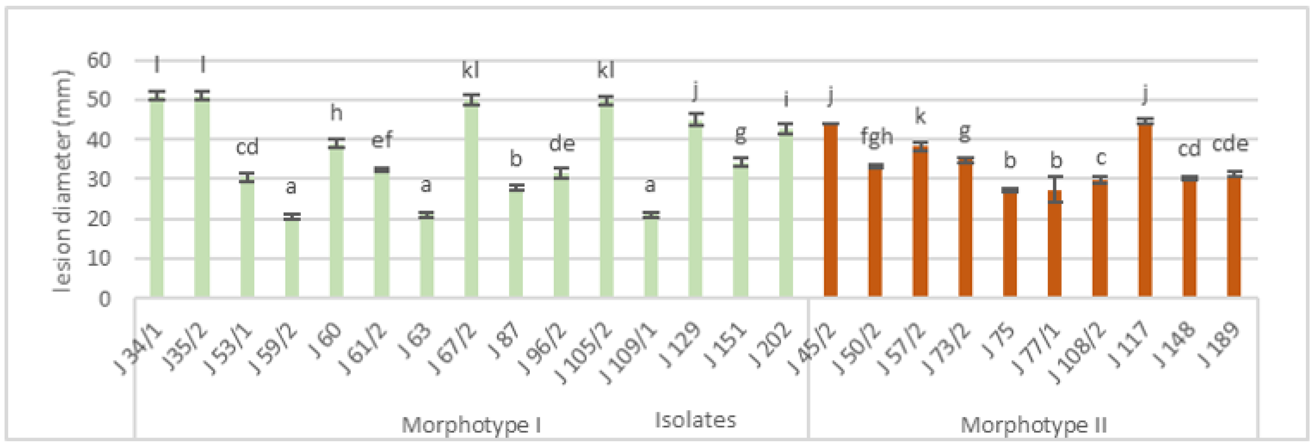
3.2. Pathogenicity Test

All tested Neofabraea-like isolates were pathogenic to apple fruits, producing typical Bull’s eye rot symptoms. Diameters of necrotic lesions were measured at 14, 21, and 28 days after artificial inoculation. The trial was terminated after 28 days. For representation, the data after 28 days of incubation were chosen and are presented in Figure 1. Neofabraea isolates caused three types of symptoms: yellowish lesions with brown margins; typical dark brown lesions, flat or slightly sunken; and brown lesions with concentric circles (Figure 2). Differences in aggressiveness among isolates was observed. Isolates J34/1 and J35/2 were highly aggressive and caused lesions 51.0 mm in diameter after 28 days of incubation, while the isolate J59/2 was the least aggressive (lesion diameter 20.5 mm). After 21 days of incubation, isolates started to produce conidiomata in the center of necrotic lesions and, by the termination of the experiment, 48% of studied isolates formed conidiomata on inoculated fruits. Two types of conidiomata on necrotic lesions were observed: small subepidermal conidiomata with white and creamy to dark brown color and large erumpent conidiomata with white mycelia in some cases (isolates J96/2, J129, and J67/2). The isolates from morphotype I formed larger lesions (38.5 mm on average) and 57.1% produced conidiomata, while isolates from morphotype II were less aggressive (30.9 mm on average) and formed conidiomata in 32.2% of cases, mainly subepidermal and small in size.
Isolates tested in the orchard were chosen based on their aggressiveness (isolates J34/1, J96/2, J67/2, and J151) and differences of symptoms on apple fruits (separated earlier in research, isolates J148 and J129). The length of canker, visibility of acervuli, and size of conidia are presented in Table 3. Two types of cankers were observed (Figure 3): the first type of canker, caused by isolates J148, J129, and J151, was shorter, but penetrated through the callus, sunken and depressed with visible fiber strings and isolated from healthy tissue without visible acervuli; the second type of canker, caused by isolates J34/1, J67/2, and J96/2, was longer and with variation in surface tissue color from orange to brown, intermittently. In second-type canker, surface tissue peeled off and revealed black acervuli. The development of apple canker progression and Neofabraea aggressiveness in a one-year study was observed (Figure 4).

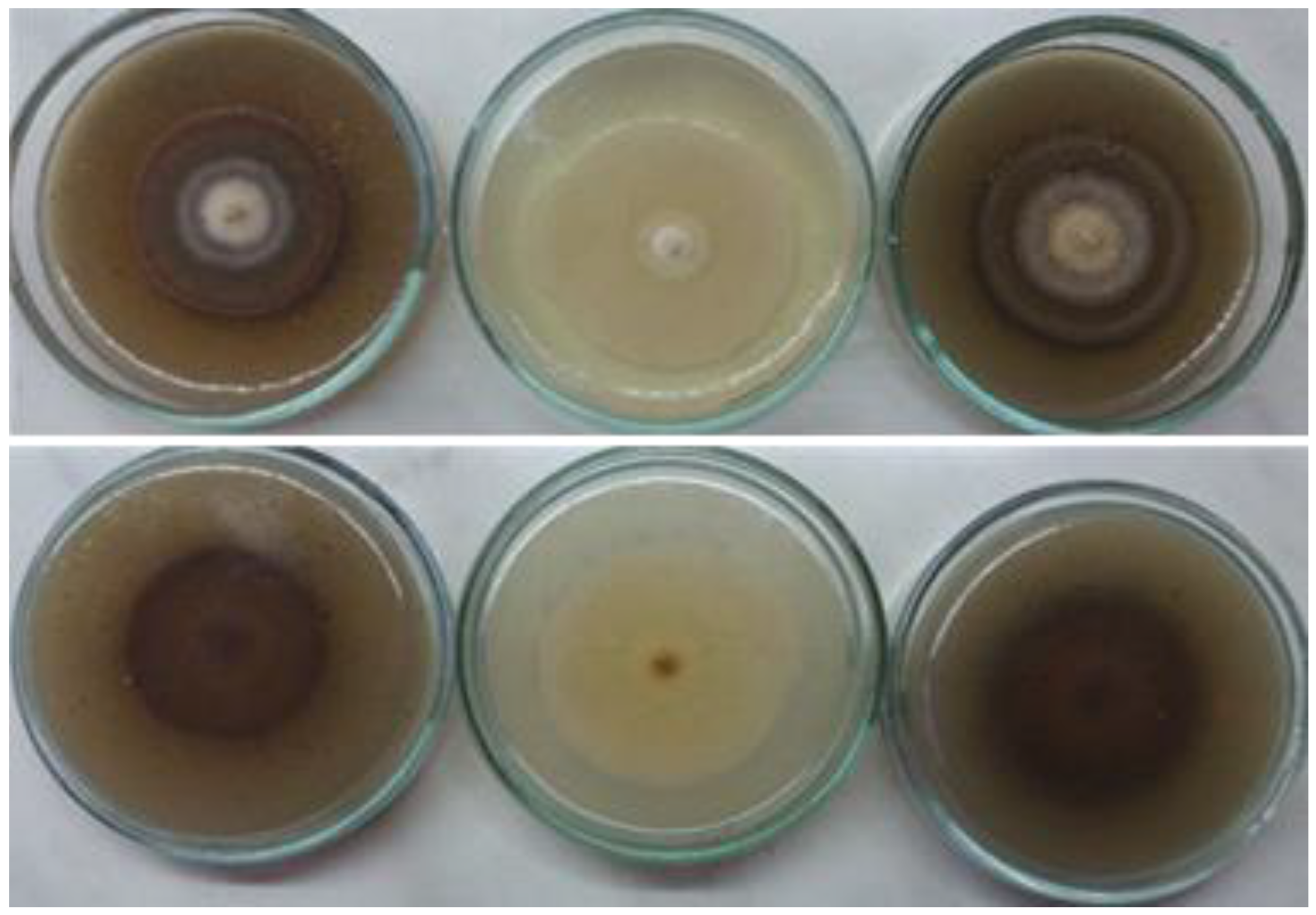
3.3. Morphological Characterization

After morphological characterization, based on the characteristics of the colony, appearance of aerial mycelium, and color, isolates were separated into two main morphotypes (Table 4). Morphotype I, with white to creamy-yellowish or sandy mycelia without pigmentation, and morphotype II, with greyish to dark brown mycelia with dark brown pigment in substrate. Afterwards, four isolates with slight differentiation in colony appearance per group were selected for further macro-morphological investigation. Morphotype I (60% of identified isolates) showed larger colonies (31.9 mm average) than morphotype II (28.1 average). Morphotype I isolates had white-creamy to grey-sandy mycelium, sometimes with brown spots (different pigmentation on aerial mycelium). The isolates were mainly circular and with a uniform appearance without raised substrate; some isolates formed fewer uniform colonies and raised substrate. Neofabraea sp. isolates started changing appearance in colony color from the same inoculum source after multiple transferring (Figure 5). After 21 days of incubation, there were no visible conidiomata or present conidia. Morphotype II isolates produced less aerial mycelium and were unevenly shaped, with raised substrate and with slightly darker color, ranging from dark grey and toasted almond to brown or cinnamon, with frequently present white margins. In some cases, pigmentation in the substrate was recorded. After an additional trial for inducing sporulation, the highest percentage of sporulated isolates was observed on MEA (85.7%) and the lowest on TA (35.7%). Morphotype I isolates preferred MEA, while isolates grouped in morphotype II preferred PDA for producing conidia.
After 4 months of incubation, UV light did not show any impact on the overall production of conidiomata in isolates plated on PDA or MEA, but there was an impact in the case of isolate J108/2 plated on TA media. In addition, after two months of incubation, some changes in colony morphology were recorded; one isolate from morphotype I started to change color from white-creamy to dark brown (J35/1), while some isolates from morphotype II started producing more aerial mycelium, becoming lighter in color. Overall, after 8 weeks of incubation isolates from morphotype II formed visible black erupt conidiomata on the surface of aerial mycelium. On MEA and TA, large watery droplets, amber to yellowish in color, were visible on the mycelial surface, without conidia (Figure 6).
On the top of the aerial mycelium, black acervuli were formed, within some isolates immersed in mycelia and massive leakage of slimy conidial masses (white to creamy color). A difference in the appearance and size of conidia in different culture media was observed. Conidial size varied in relation to culture media. The conidial size of both morphotypes was smaller in the case of isolates plated on MEA and larger in the case of PDA. The shape of conidia was influenced by different media; on PDA, conidia was similar to conidia produced on tree branches. On MEA and TA within isolates J35/1, J60, and J151, thinner, aseptate, straight, hyaline, rounded at apex, and smaller in size (5–8.5 × 2–2.5 µm) conidia were observed. The morphological characteristics of both morphotypes suggested that the isolates belonged to Neofabraea genus and possibly to the species Neofabraea alba. Therefore, the isolates were preliminary identified as N. alba.

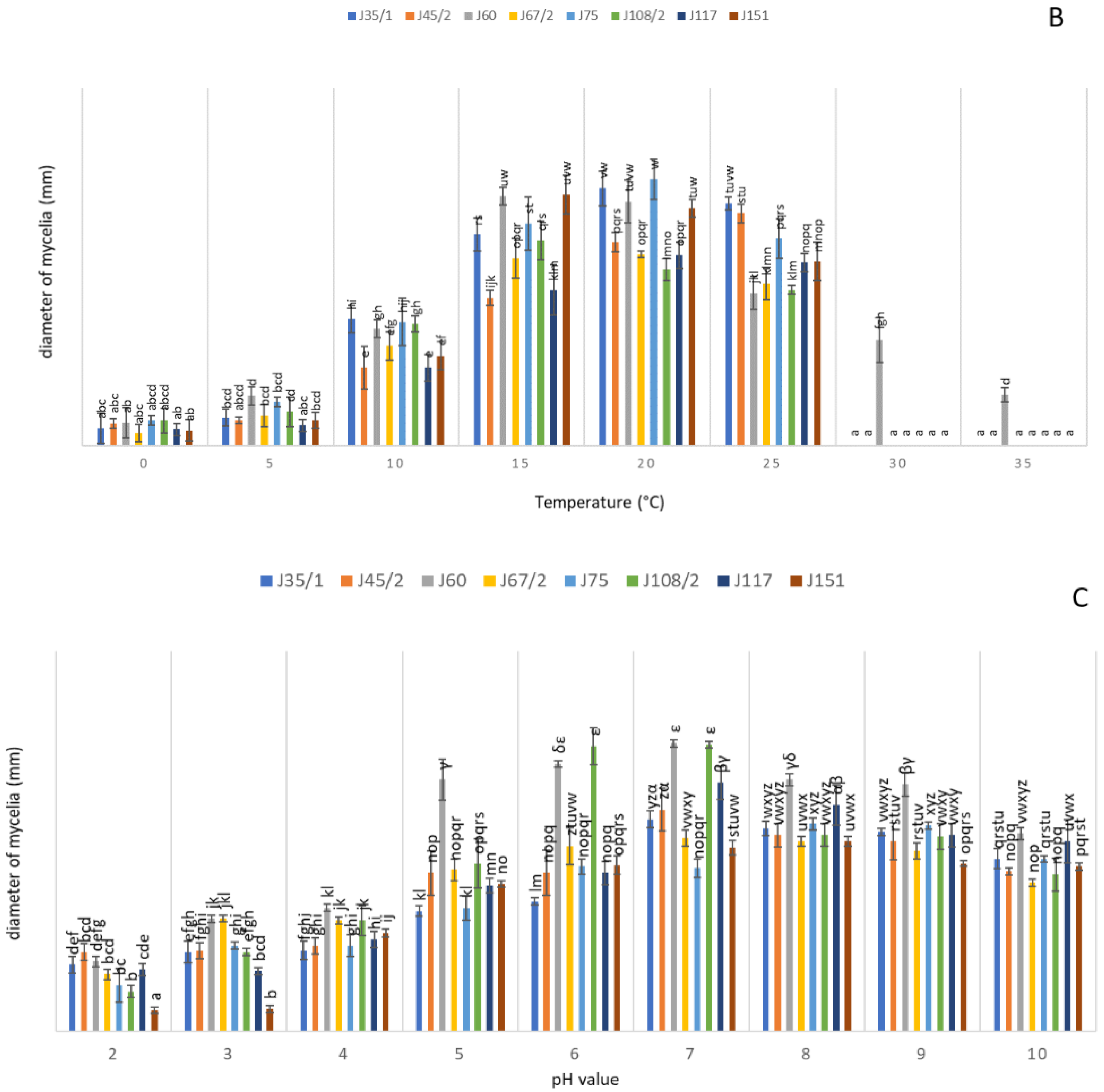
3.4. Physiological Characterization

All tested Neofabraea isolates were able to grow on MEA at a pH range of 2–10. For most of the isolates, a maximal growth rate was recorded at pH 7, except for isolates J75 and J108/2, which had a maximal growth rate at pH 6. Isolates exhibited slower growth at pH levels lower than 5. The lowest growth rate was recorded at the most acidic medium (pH 2). In addition, differences in the appearance of aerial mycelium and pigmentation were observed on acidic versus alkaline medium. Significant difference in radial growth rate at different temperatures among isolates was observed. Eight selected isolates of Neofabraea were able to grow at low temperatures, but failed to grow at high temperatures (30 °C and 35 °C), with the exception of isolate J60, which was able to grow at all studied temperatures. The temperature optimum for four isolates was 20 °C, for J45/2 it was 25 °C, and for J60 and J108/2 it was 15 °C. Only isolate J117 exhibited the same growth rate at 20 and 25 °C. The most favorable medium for growth was OA, followed by PCA medium and MEA. The slowest growth was recorded on PDA and TA media with significant differences among isolates. Isolate J67/2 preferred PCA medium for growth relative to other isolates and culture media. Lack of aerial mycelium was noted on the culture media that provided the fastest growth of mycelia (Figure 7).

3.5. Molecular Identification and Characterization

Twenty-five selected Neofabraea spp. isolates were identified at the species level by amplification and sequencing of the internal transcribed spacer (ITS) region of ribosomal DNA (rDNA) and portions of the 16S mitochondrial rRNA (mtSSU). The final length of ITS sequences ranged from 301 to 495 bp (base pairs) and from 302 to 625 bp for mtSSU, respectively. There was a similarity of over 99% with several N. alba sequences deposited in GenBank (KT963910.1 KT963903.1, KT963897.1, MH924828.1, etc.) for both ITS and mtSSU sequences. The jointed phylogenetic tree was separated in two clades, one which obtained all N. alba sequences, and another consisting of sequences of N. malicorticis, N. perennans, and N. sp. nov. The ML analyses showed N. alba sequences separated in two groups, with a bootstrap value of 99%. The first group obtained sequences of isolates obtained in this study and GenBank isolates, while another group was composed only of identified isolates obtained in this study (J63, J129, J202, J59/2, J34/1, J148, J151). The most distant isolate in the second group was J59/2, followed by J63, presented in a separated branch (Figure 8). After analyzing separated phylogenetic trees of ITS and mtSSU sequences and concatenated tree, we concluded that the merged tree separated N. alba isolates more precisely and gave more insight into the groups formed within the specie, but with less available reference isolates in the GenBank database (data unpublished).

4. Discussion

Bull’s eye rot is becoming increasingly significant in all fruit-growing areas in Europe, as well as in Serbia, where production of apple has a major economic importance. The first appearance of N. alba in Serbia is unknown, but preliminary research conducted in 2016 proved its existence [9]. In this study, a wide range of Neofabraea spp. isolates were derived from symptomatic apple fruits collected from warehouses and local markets. Observed Bull’s eye rot symptoms developed after latent infections of fruit through lenticels. Lesions grew very slowly, and after several months in storages they became visible. During February and March, infected fruits exhibited depressed, circular, flat or slightly sunken lesions, light to dark brown with a yellow center, with a clear border between decayed and healthy tissue. In some cases, conidiomata accompanied with mycelia were observed on the surface of the lesions. Several species of the genus Neofabraea cause Bull’s eye rot disease. In general, symptoms caused by N. alba and other Neofabraea species are difficult to distinguish, although the lesions caused by N. alba in some studies were slightly lighter in color and less zonate than those caused by N. malicorticis [22]. Symptoms on apple fruits originating from Serbia, caused by N. alba, were similar to the literature description, and could be grouped into three types. Only symptoms caused by isolate J108/2 and a few other isolates were slightly different; the lesions were brown and with more concentric circles. After morphological and molecular characterization, the selected isolates were identified as N. alba, with a high percentage of similarity with reference sequences obtained from the GenBank. ITS region was accepted as a suitable fungal marker for identification, with a noted lack of precision to separate closely related species. In our survey, where Neofabraea spp. sequences were analyzed, it was not sufficiently informative, and multi-sequence analyses was required. This is in line with the results of other authors [16,23]. The joined phylogenetic tree indicates the more precise distinction between N. alba isolates and other specie sequences, compared to separated phylogenetic analyses. The most distant isolate (J59/2) from the second group in the first cluster was previously described as the least aggressive pathogen on apple fruit, followed by J63 (in a separate branch), which has been in the same group of significance. The relationship between morphotypes and the genetic distance within some isolates was noted. Almost all isolates from the second group in the first cluster belonged to morphotype I (except isolate J148). In future analyses, expanding the research on more reliable gene analyses such as β-tubulin and EF-1α is required. Nevertheless, for more clarifying phylogenetic research and high identification success rate, the construction of a larger phylogenetic tree is necessary, which is a condition with an extensive GenBank database, especially for newly described species such as N. kienholzii, N. brasiliensis, and N. actinidiae. All studied isolates exhibited pathogenicity on apple fruits, as well as on apple branches, with differences in aggressiveness. A relationship was observed within some isolates between the aggressiveness on the fruit and that on the branches. Differences between the two types of symptoms on branches were followed by aggressivity, the ability to form conidiomata on the surface, and sporulation. In our survey, we observed that isolates from morphotype I manifest greater aggressiveness on branches. In previous studies, N. alba was described as a weak pathogen with saprophytic growth on apple trees, uncapable of producing conidia and with mild pathogenicity [24]. However, the inoculation experiment in this study showed that N. alba isolates were capable of causing larger cankers, with the ability to produce conidia, similar to N. malicorticis and N. perennans. In Henriquez et al. (2006), larger cankers were recorded on Granny Smith apple trees than on pear branches, with growth beyond the inoculation point. N. alba followed a similar pattern, with abundant sporulation and smaller cankers compared to N. perennans. It was concluded that the growth of the canker was affected by the period of inoculation, host, and cultivar. Our findings are in agreement with other recent studies conducted in Czechia and Oregon, USA [18,25].
In Europe, apple cultivars ‘Golden Delicious’ and ‘Pink Lady’ are particularly susceptible to the disease [26,27]. Our results showed that Neofabraea spp. frequently occurred in Serbian warehouses in February and March in the apple cultivars ‘Golden Delicious’ and ‘Idared’, which coincided with the facts that these two cultivars were susceptible to the pathogen and that they were dominant cultivars in the production. The pathogen was proven to be of economic importance, but it has not yet been studied in-depth. The main reasons for the previous lack of interest in the genus of Neofabraea could be widely-accepted observation of their weak pathogenicity, slow mycelial growth, or the absence of sporulation in in vitro experiments. In our study, some methods for inducing sporulation were useful, but still partially effective, depending on the isolate, cultivation period, and the medium used. Not all isolates were able to produce conidia in the examined culture media, and the most effective medium was MEA, which was suitable for conidia production after 2 months, yet only for five out of seven of the examined isolates, and without the presence of conidiomata. However, after 4 months of incubation, all studied isolates produced conidia on MEA. PDA medium was significantly less effective (50% of isolates and replicates produced conidia), but isolates that sporulated formed erumpt acervuli with abundant slimy conidial masses. Everett et al. (2017) reported that UV light on PDA and TA media did not induce conidia formation, while CMA medium was the most effective [28]. In our case, the least favorable culture media were TA and CMA, with either low intensity or complete lack of sporulation. The influence of temperature was an important factor that favored sporulation on some media, and limited it on others. For instance, some isolates grown on TA could not produce conidia, even after 4 months at 20 °C, although in the research of Cameldi et al. (2017) N. alba isolates grown on TA medium produced conidia at 15 °C [29]. In our opinion, an interaction between temperature and culture medium could also affect conidial production, suggesting that it requires further investigation. We also found that different culture media can affect the macroscopic characteristics of the isolates, as the color of the colony, mycelial density, and the presence of conidiomata. Furthermore, the medium can also influence size, shape, and volume of conidia of N. alba isolates. Recent studies revealed that the temperature of incubation can change the size and shape of conidia [29]. In this study we concluded that substrates can also affect the microscopic characteristics of the isolates. Differences among conidia produced on tree branches and on different culture media in vitro were observed. Conidia formed in different substrates were compared, and it was concluded that microconidia were not found in nature. However, smaller conidia (microconidia) were produced in some cases and only by isolates J35/1, J60, and J151 on TA and MEA media (grouped in morphotype I). Microconidia were also found in one isolate of N. alba studied by Carneiro et al. (2022), as well as in the research conducted by Verkley (1999) and S Rooney-Latham (2013) [23,30,31], while in other studies there was no record of microconidia [5,8,25,29,32]. Immediately after isolation and hyphal tip purification, identified isolates were grouped into two main morphotypes, with dominant morphotype I comprising 60% of the isolates. However, we found that isolates were able to change appearance with multiple transfers from the same inoculum source, making grouping of isolates based on the colony appearance questionable. This high variability was previously noticed by Spotts et al. (2009) in distinguishing isolates of the closely related species Cryptosporiopsis kienholzii (Neofabraea, Dermataceae), but not in N. alba [12]. The growth rate of N. alba isolates was variable for each pH level. All isolates were able to grow at all studied pH levels, from pH 2 to pH 10. The fastest growth was noted at pH 7 for most of the isolates, except for J75 and J108/2. Furthermore, the isolate J60 showed the highest growth rate, compared to other isolates. Hortova et al. (2014) reported that N. alba isolates were not able to grow on highly acidic medium (pH 2), which differs from the results in our study [25]. Appearance of the aerial mycelium was different comparing alkaline and acidic media; by increasing pH level, the substrate mycelium was less aerial and slimier, with an even radial shape. In terms of temperature, lower temperatures reduced mycelial growth, but temperatures of 30 °C and 35 °C suppressed the growth of all isolates, except J60. The optimal temperature for the growth of isolates J60 and J108/2 was 15 °C and 20 °C for all other isolates, which agrees with earlier studies with isolates of N. alba and N. malicorticis [25,29,33]. N. alba isolates preferred OA medium, which provided the fastest mycelial growth of all tested isolates, while the least suitable were PDA and TA media. The appearance of isolates and their colonial morphology varied depending on the culture media used. N. alba exhibited a different appearance and color of mycelia on different media. It also had the ability to change colors through incubation time, or at different temperatures, making separation from other species in the genus difficulties. According to the results of our study, it can be concluded that environmental factors have a great impact on morphological variability. The OA and PCA media allowed a higher growth rate and induced a regular circular shape with less aerial mycelium and rings of different colors above the medium. Malt extract agar was found to be a great medium for decreasing the variability of colonies with colors from white-creamy to orange, as well as for abundant aerial mycelium and intensive growth into substrate, resulting in a raised center of the colony. Tomato agar and PDA both allow lower growth rate of isolates, with high color variability and the possibility to produce pigment and conidiomata. There was a similarity in the research noticed among isolates of Neofabraea and Phlyctema, where the effect of temperature, pH, and culture media on the growth rates and appearance of N. alba were noted [8,23,25].
In addition, extended research concerning the identification and distinguishing of Neofabraea spp., as well as the definition of structure population in Serbia, is required. It is necessary to establish a method to induce sporulation that could favor all N. alba isolates, but this would require fast and abundant sporulation for more detailed studies. The ITS and mtSSU regions were suitable for the identification of N. alba isolates, but for extensive phylogenetic research more precise and sensitive diagnostic tools, such as β-tubulin, EF-1α, and species-specific primers are needed. Additionally, we can say that dividing N. alba isolates based on morphological characteristics is not sufficient enough due to its ability to change its characteristics, according to examined factors. Our research confirms the ability of N. alba isolates to produce microconidia, and further analyses will be directed toward factors that influence their formation. N. alba isolates have proven to be virulent on branches and fruits, as well as tolerable due to extreme ecological factors (such as the temperature and pH of the substrate), so our major goal is directed at control and target alternative methods to prevent the spreading and development of necrosis.

Author Contributions

Conceptualization, J.V. and V.S.; methodology, V.S., N.N. and M.G.; software, M.P. and J.V.; validation, B.T., J.V. and V.S.; investigation, J.V.; writing—original draft preparation, J.V.; writing—review and editing, B.T., M.G. and J.H.; supervision, V.S. All authors have read and agreed to the published version of the manuscript.

Funding

This work was funded by the Serbian Ministry of Education, Science, and Technological Development (Project No. 451-03-68/2022-14/200117).

Data Availability Statement

The data and materials are available upon reasonable request from the corresponding author.

Conflicts of Interest

The authors declare no conflict of interest.

References

  1. Maxin, P.; Weber, R.W.; Pedersen, H.L.; Williams, M. Control of a wide range of storage rots in naturally infected apples by hot-water dipping and rinsing. Postharvest Biol. Technol. 2012, 70, 25–31. [Google Scholar] [CrossRef]
  2. Köhl, J.; Wenneker, M.; Haas, B.H.G.-D.; Anbergen, R.; de Geijn, H.M.G.-V.; der Plas, C.H.L.-V.; Pinto, F.A.M.F.; Kastelein, P. Dynamics of post-harvest pathogens Neofabraea spp. and Cadophora spp. in plant residues in Dutch apple and pear orchards. Plant Pathol. 2018, 67, 1264–1277. [Google Scholar] [CrossRef]
  3. Wenneker, M.; Pham, K.T.K.; Boekhoudt, L.C.; De Boer, F.A.; Van Leeuwen, P.J.; Hollinger, T.C.; Thomma, B.P.H.J. First Report of Neofabraea kienholzii Causing Bull’s Eye Rot on Pear (Pyrus communis) in the Netherlands. Plant Dis. 2017, 101, 634. [Google Scholar] [CrossRef]
  4. Bogo, A.; Comparin, C.; Valdebenito-Sanhueza, R.; Ritschel, P.; Casa, R.; Da Silva, F.; Everhart, S. Characterization of Neofabraea actinidiae and N. brasiliensis as causal agents of apple bull’s-eye rot in southern Brazil. Can. J. Plant Pathol. 2018, 40, 229–237. [Google Scholar] [CrossRef]
  5. Soto-Alvear, S.; Lolas, M.; Rosales, I.M.; Chávez, E.R.; Lattore, B.A. Characterization of the bull’s eye rot of apple in Chile. Plant Dis. 2013, 97, 485–490. [Google Scholar] [CrossRef] [PubMed]
  6. Michalecka, M.; Bryk, H.; Poniatowska, A.; Pulawska, J. Identification of Neofabraea species causing bull’s eye rot of apple in Poland and their direct detection in apple fruit using multiplex PCR. Plant Pathol. 2016, 65, 643–654. [Google Scholar]
  7. Pešicová, K.; Kolarik, M.; Hortová, B.; Novotny, D. Diversity and identification of Neofabraea species causing bull’s eye rot in the Czech Republic. Eur. J. Plant Pathol. 2017, 147, 683–693. [Google Scholar] [CrossRef]
  8. Romero, J.; Raya, M.C.; Roca, L.F.; Moral, J.; Trapero, A. First Report of Neofabraea vagabunda Causing Branch Cankers on Olives in Spain. Plant Dis. 2016, 100, 527. [Google Scholar]
  9. Vico, I.; Duduk, N.; Vasić, M.; Zebeljan, A.; Radivojević, D. Bull’s eye rot of apple fruit caused by Neofabraea alba. Acta Hortic. 2016, 1139, 733–738. [Google Scholar] [CrossRef]
  10. Krsmanović, S.; Grahovac, M.; Medić, J.; Stojšin, V.; Petreš, M. Struktura populacije prouzrokovača truleži uskladištenih plodova jabuke. Plant Dr. 2018, 46, 563–569. [Google Scholar]
  11. Petreš, M.; Kalajdžić, J.; Milić, B.; Magazin, N.; Stankov, A.; Vukotić, J.; Grahovac, M. Effect of hot water treatments on apple fruit rot caused by Fusarium spp. J. Plant Dis. Prot. 2020, 127, 651–655. [Google Scholar] [CrossRef]
  12. Spotts, R.A.; Seifert, K.A.; Wallis, K.M.; Sugar, D.; Xiao, C.L.; Serdani, M.; Henriquez, J.L. Description of Cryptosporiopsis kienholzii and species profiles of Neofabraea in major pome fruit growing districts in the Pacific Northwest USA. Mycol. Res. 2009, 113, 1301–1311. [Google Scholar] [CrossRef]
  13. Gariepy, T.D.; Rahe, J.E.; Lèvesque, C.A.; Spotts, R.A.; Sugar, D.L.; Henriquez, J.L. Neofabraea species associated with bull’s eye rot and canker of apple and pear in the Pacific Northwest. Can. J. Plant Pathol. 2005, 27, 118–124. [Google Scholar] [CrossRef]
  14. Abeln, E.C.A.; Pagter, M.A.; Verkley, G.J.M. Phylogeny of Pezicula, Dermea and Neofabraea inferred from partial sequences of the nuclear ribosomal RNA gene cluster. Mycologia 2000, 92, 685–693. [Google Scholar] [CrossRef]
  15. de Jong, S.N.; Lèvesque, C.A.; Verkley, G.J.M.; Abeln, E.C.A.; Rahe, J.E.; Braun, P.G. Phylogenetic relationships among Neofabraea species causing tree cankers and bull’s eye rot of apple based on DNA sequencing of ITS nuclear rDNA, mitochondrial rDNA, and β-tubulin gene. Mycol. Res. 2001, 105, 658–669. [Google Scholar] [CrossRef]
  16. de Jong, S.N. The Development of Specific, Non-Discriminating PCR Primers for the Detection of Pezicula malicorticis and P. perennans Agents of Anthracnose Canker, Perennial Canker, and Bull’s-Eye Rot of Apple. Master’s Thesis, Department of Biological Sciences, Simon Fraser University, Burnaby, BC, Canada, 1998. [Google Scholar]
  17. Gariepy, T.D.; Levesque, C.A.; de Jong, S.N.; Rahe, J.E. Species specific identification of the Neofabraea pathogen complex associated with pome fruits using PCR and multiplex DNA amplification. Mycol. Res. 2003, 107, 528–536. [Google Scholar] [CrossRef]
  18. Henriquez, J.L.; Sugar, D.; Spotts, R.A. Induction of Cankers on Pear Tree Branches by Neofabraea alba and N. perennans, and Fungicide Effects on Conidial Production on Cankers. Plant Dis. 2006, 90, 481–486. [Google Scholar] [CrossRef]
  19. White, T.J.; Bruns, T.; Lee, T.J. Amplification and direct sequencing of fungal ribosomal RNA genes for phylogenetics. In PCR Protocols, a Guide to Methods and Applications; Innis, M.A., Gelfand, D.H., Sninsky, J.J., White, T.J., Eds.; Academic Press: San Diego, CA, USA, 1990; pp. 315–322. [Google Scholar]
  20. Boratyn, G.M.; Camacho, C.; Cooper, P.S.; Coulouris, G.; Fong, A.; Ma, N.; Madden, T.L.; Matten, W.T.; McGinnis, S.D.; Marezhuk, Y.; et al. BLAST: A more efficient report with usability improvements. Nucleic Acids Res. 2013, 41, 29–33. [Google Scholar] [CrossRef]
  21. Kumar, S.; Stecher, G.; Tamura, K. MEGA7: Molecular Evolutionary Genetics Analysis version 7.0 for bigger datasets. Mol. Biol. Evol. 2016, 33, 1870–1874. [Google Scholar] [CrossRef]
  22. Snowdon, A.L. Pome fruits. In A Colour Atlas of Post-Harvest Diseases and Disorders of Fruits and Vegetables; Snowdon, A.L., Ed.; Wolfe Scientific Ltd.: London, UK, 1990; Volume 1, pp. 170–218. [Google Scholar]
  23. Carneiro, G.A.; Walcher, M.; Storti, A.; Baric, S. Phylogenetic diversity and Phenotypic characterization of Phlyctema vagabunda (syn. Neofabraea alba) and Neofabraea kienholzii causing postharvest bull’s eye rot of apple in northern Italy. Plant Dis. 2022, 106, 451–463. [Google Scholar] [CrossRef]
  24. Corke, A.T.K. Bitter rot of apples II. Seasonal variations in the development and sporulation of cankers of Gloeosporium spp. inoculated into apple branches. J. Hortic. Sci. 1956, 31, 272–283. [Google Scholar] [CrossRef]
  25. Hortova, B.; Novotny, D.; Erban, T. Physiological characteristics and pathogenicity of eight Neofabraea isolates from apples in Czechia. Eur. J. Hortic. Sci. 2014, 79, 327–334. [Google Scholar]
  26. Cameldi, I.; Neri, F.; Ventrucci, D.; Ceredi, G.; Muzzi, E.; Mari, M. Influence of harvest date on bull’s eye rot of Cripps Pink apples and control chemical strategies. Plant Dis. 2016, 100, 2287–2293. [Google Scholar] [CrossRef]
  27. Neri, F.; Mari, M.; Brigati, S.; Bertolini, P. Control of Neofabraea alba by plant volatile compounds and hot water. Postharvest Biol. Technol. 2009, 51, 425–430. [Google Scholar] [CrossRef]
  28. Everett, K.; Pushparajah, I.; Fisher, B.M.; Wood, P. A simple method for conidial production and establishing latent infections of apples by Phlyctema vagabunda (syn: Neofabraea alba). N. Z. Plant Prot. 2017, 70, 106–111. [Google Scholar] [CrossRef]
  29. Cameldi, I.; Neri, F.; Menghini, M.; Pirondi, A.; Nanni, I.M.; Collina, M.; Mari, M. Characterization of Neofabraea vagabunda isolates causing apple bull’s eye rot in Italy (Emilia-Romagna region). Plant Pathol. 2017, 66, 1432–1444. [Google Scholar] [CrossRef]
  30. Verkley, G.J.M. A monograph of Pezicula and its anamorphs. Stud. Mycol. 1999, 44, 1–176. [Google Scholar]
  31. Henriquez, J.L. Studies on the Etiology and Epidemiology of Bull’s Eye Rot of Pears. Ph.D. Thesis, Oregon State University, Ann Arbor, MI, USA, 2003. [Google Scholar]
  32. Rooney-Latham, S.; Gallegos, L.L.; Vossen, P.M.; Gubler, W.D. First report of Neofabraea alba causing fruit spot on olive in North America. Plant Dis. 2013, 97, 1384. [Google Scholar] [CrossRef] [PubMed]
  33. Trouillas, F.P.; Nouri, M.T.; Lawrence, D.P.; Moral, J.; Travadon, R.; Aegerter, B.J.; Danielle, L. Identification and Characterization of Neofabraea kienholzii and Phlyctema vagabunda Causing Leaf and Shoot Lesions of Olive in California. Plant Dis. 2019, 103, 3018–3030. [Google Scholar] [CrossRef]
Figure 1. Pathogenicity of identified Neofabraea isolates ex situ on inoculated apple fruit after 28 days of incubation at room temperature. The same letters represent no significant differences according to Duncan’s test (p < 0.05).
Agronomy 12 02015 g001
Figure 2. Different decay symptoms on apple fruit inoculated with Neofabraea sp. after incubation of 35 days at room temperature: (a) Isolate J129—lesion with yellowish center and brownish margins; (b) Isolate J96/2—brownish lesion; and (c) Isolate J108/2—brownish lesion with concentric circles.
Agronomy 12 02015 g002
Figure 3. Two type of symptoms caused by Neofabraea sp. on apple branches: (a) developed canker caused by isolate J129; (b) canker with visible conidiomata under surface of branches caused by isolate J67/2. Pictures were taken 10 months post inoculation.
Agronomy 12 02015 g003
Figure 4. Neofabraea sp.: canker progression on inoculated apple branches.
Agronomy 12 02015 g004
Figure 5. Colony differences of Neofabraea alba isolate J45/2, grown on oatmeal agar plates (OA) at 20 °C for 14 days in multiple transfers from the same inoculum source.
Agronomy 12 02015 g005
Figure 6. Colony and sporulation of two Neofabraea sp. isolates on different culture media after 2 months of incubation. The first row represents isolate J60 (morphotype I), while the second row represent isolate J117 (morphotype II).
Agronomy 12 02015 g006
Figure 7. Effect of culture media (A), temperature (B), and pH (C) on growth of Neofabraea isolates. * Separation and group means were compared by Duncan’s test at p < 0.05; letters used for the definition of homogeneity belong to the English and Greek alphabet.
Agronomy 12 02015 g007aAgronomy 12 02015 g007b
Figure 8. Maximum-likelihood concatenated tree constructed using MEGA 7 software [21], based on the internal transcribed spacer (ITS) and 16S mitochondrial ribosomal RNA gene (mtSSU) sequences of Neofabraea spp. Pezicula corticola was used as an outgroup. The numbers at the branches represent bootstrap values obtained after 1000 replications.
Agronomy 12 02015 g008
Table 1. Species of fungi used in phylogenetic analyses, including isolate number, source, location, host, and reference number.
SpeciesIsolate NumberSourceLocationHostReference Number
ITSSSU
Neofabraea albaJ34/1VukotićSerbiaAppleOM761173OM514687
Neofabraea albaJ35VukotićSerbiaAppleOM491330OM514688
Neofabraea albaJ45/2VukotićSerbiaAppleOM638703OM677770
Neofabraea albaJ50/2VukotićSerbiaAppleOM491331OM514689
Neofabraea albaJ53/1VukotićSerbiaAppleOM638704OM677771
Neofabraea albaJ57/2VukotićSerbiaAppleOM761173OM514690
Neofabraea albaJ59/2VukotićSerbiaAppleOL889667OM677778
Neofabraea albaJ60VukotićSerbiaAppleOM491332OM514691
Neofabraea albaJ63VukotićSerbiaAppleOM761176OM514692
Neofabraea albaJ67/2VukotićSerbiaAppleOM491333OM514693
Neofabraea albaJ75VukotićSerbiaAppleOM491334OM514694
Neofabraea albaJ77/2VukotićSerbiaAppleOM638707OM677776
Neofabraea albaJ87VukotićSerbiaAppleOM761177OM514695
Neofabraea albaJ108/2VukotićSerbiaAppleOM491335OM514696
Neofabraea albaJ109/1VukotićSerbiaAppleOM63870OM677772
Neofabraea albaJ129VukotićSerbiaAppleOM761180OM677773
Neofabraea albaJ148VukotićSerbiaAppleOM761181OM514697
Neofabraea albaJ151VukotićSerbiaAppleOM761182OM677774
Neofabraea albaJ202VukotićSerbiaAppleOM761184OM677775
Neofabraea albaATCC 38338De JongPolandAppleAF281366AF281404
Neofabraea albaM822RomeroItalyAppleKT454381KT963895
Neofabraea albaCBS 452.64AbelnUKCanadaAF141190AF281448
Neofabraea albaCAL 135De JongCanadaAppleAF281372AF281419
Neofabraea albaDAOM 227091De JongCanadaAppleAF281368AF281436
Neofabraea albaOGT3CarneiroItalyAppleMW130163MW228886
Neofabraea alba19-DSS-FU-1-013CarneiroItalyAppleMW130184MW228852
Neofabraea malicorticisCAL 102De JongCanadaAppleAF281379AF281405
Neofabraea malicorticisDAOM 227083De JongCanadaAppleAF281385AF281406
Neofabraea perennansDAOM 227089De JongCanadaAppleAF281392AF281432
Neofabraea perennansRGR 90.0123De JongUSAAppleAF281396AF281449
Neofabraea kienholziiDAOM 227086De JongCanadaAppleAF281398AF281409
Pezicula corticolaCBS 259.31AbelnDenmarkAppleAF141179AF281443
Table 2. Occurrence of Bull’s eye rot in stored apple fruit during February and March in 2017 and 2018 in northern part of Serbia.
Warehouse LocationYearNumber of Fruits withBull’s Eye Rot-Like SymptomsNumber of Fruits Infected with Neofabraea spp.Incidence of Neofabraea spp. (%)
Tavankut2017361952.8
Jazak3638.3
Mala Remeta20945.0
Titel20315.0
Irig22627.3
Novi Sad29827.6
Ljutovo151066.7
Bački Vinogradi17952.9
Slankamen191263.2
Bačka Topola10550.0
Subotica201050.0
Hajdukovo15853.3
Horgoš10110.0
Subtotal=1326910338.3
Tavankut201815010771.3
Jazak20210
Mala Remeta900.0
Titel14535.7
Kukujevci22517075.6
Subtotal = 541828467.9
Total = 18 68738756.3
Table 3. Length of cankers (mm) and visibility of acervuli and conidial size (µm) on two-year-old apple branches inoculated with Neofabraea isolates one-year post inoculation.
IsolatesApple Branches
Length of Canker (mm) aVisible AcervuliSize of Conidia (µm)
Mean (Length × Width)Range
J34/149.3 ± 6.4 b aYes18.4 × 3.17.5–25 × 2.5–5
J14819.3 ± 2.1 aNo14.2 × 2.77.5–25 × 2.5–5
J67/252.0 ± 8.0 bcYes17.6 × 3.07.5–27 × 2.5–5
J12914.0 ± 1.0 aNo16.8 × 2.910–26 × 2–5
J15120.0 ± 2.0 aNo19.9 × 3.411–26 × 2.5–5
J9660.0 ± 5.0 cYes19.5 × 2.612.5–25 × 2.8–5
a Data represent means and standard deviation; the same letters in the table represent no significant difference by Duncan’s test (p < 0.05).
Table 4. Identified Neofabraea sp. isolates from Serbia, including information on the location, host, cultivar, and morphotype of each isolate.
Isolate NumberYear of IsolationLocationCultivarMorphotype of Isolates *
J34/12017SlankamenRed chiefI
J35/12017SlankamenMutsuI
J45/22017IrigGranny SmithII
J50/22017IrigJonagoredII
J53/12017LjutovoGranny SmithI
J57/22017LjutovoGranny SmithII
J59/22017LjutovoGranny SmithI
J602017LjutovoGranny SmithI
J61/22017LjutovoGranny SmithI
J632017SuboticaPink LadyI
J67/22017SuboticaIdaredI
J73/22017IrigPink LadyII
J752017LjutovoGranny SmithII
J77/22017SuboticaIdaredII
J872017HajdukovoIdaredI
J96/22017TavankutGolden deliciousI
J105/22017Backi VinogradiIdaredI
J108/22017Backi VinogradiIdaredII
J109/12017Backi VinogradiIdaredI
J1172017Backa TopolaIdaredII
J1292018TavankutGolden deliciousI
J1482018TavankutGolden deliciousII
J1512018TavankutGolden deliciousI
J1892018KukujevciJonagoldII
J2022018KukujevciJonagoldI
* Morphotypes separated based on colony characteristics on PDA after 21 day of incubation in darkness.
Publisher’s Note: MDPI stays neutral with regard to jurisdictional claims in published maps and institutional affiliations.

