Article

LED Lighting Effects on Plant Growth and Quality of Pyrus communis L. Propagated In Vitro

1
Department of Agriculture and Forest Sciences (DAFNE), University of Tuscia, 01100 Viterbo, Italy
2
Department for Innovation in Biological, Agro-Food and Forest Systems, Tuscia University, 01100 Viterbo, Italy
*
Author to whom correspondence should be addressed.
Agronomy 2022, 12(10), 2531; https://doi.org/10.3390/agronomy12102531
Submission received: 14 August 2022 / Revised: 12 October 2022 / Accepted: 13 October 2022 / Published: 17 October 2022

Abstract

:
Fluorescent lights are typically used as light source in indoor horticultural production, including micropropagation. However, light emitting diodes (LEDs) have been recently used for plant growth under controlled environment. Major advantage of LEDs is wavelength specificity, that allows to adequately adjust the spectra according to plant needs. The possibility of using LED as primary light source for the micropropagation of Pyrus communis L. has been investigated in this work. It was proceeded to the optimization of a protocol of micropropagation using a LED lamp as the primary light source inside of the growth chamber with specific wavelengths, to reduce energy consumption and increase the quality of the micro propagated plants. Explants were maintained in a growth chamber and exposed to three different continuous spectrum LED lamps (AP67, NS1, G2) as a primary light source and fluorescent lamps (control) for 4 weeks. At the end of four weeks period, it was proceeded to the morphometric and biochemical analysis. Shoot and leaf growths were more influenced by LED lamps as compared to fluorescent lamps (control) in both cultivars. The results showed that the shoots of William and San Giovanni cultivars showed significant differences in morphological and physiological traits, as well as in chlorophyll, carotenoid, and MDA contents. Highest number of neo-formed shoots and nodes were observed in the plantlets of cv William under AP67 LED followed by NS1 and G2 LED lights respectively as compared to the white light (control), whereas same trend was observed in cv San Giovanni under AP67 LED, but it showed higher shoots and node numbers under control LED lamps as compared to both NS1 and G2. The photosynthetic pigments were significantly decreased in leaves of both cultivars when grown under LEDs as compared to the control fluorescent lamps. Moreover, the AP67 LED light had also significant effects on the protein and MDA contents in the leaves of both cultivars in comparison to all other treatments. This work underlines the importance of the modulation of light sources in relation with different species and varieties, allowing optimizing the proliferation phase and the efficiency of Pyrus communis L. micropropagation protocol. Moreover, this protocol can be improved with further studies to examine the response of the plantlets to the ex-vitro acclimatization because the possibility of using LED light for the micropropagation of pear can be considered as a valuable alternative for its ecologically sustainable production.

1. Introduction

Pear (Pyrus communis), belongs to the Rosaceae family, is a high-demand and very popular fruits because of its sweet taste and nutritional value. They are included among the best known and most highly consumed and economically important fruit trees in the world, along with apple, citrus, Prunus spp. fruits and grapes [1]. Pear is a major fruit crop in temperate regions of the world, considered a recalcitrant species in micropropagation, and characterized by weak multiplication, hyperhydricity, susceptible to phenolic oxidation and rooting ability highly genotype-dependent [2,3,4,5]. Among Pyrus taxa, 29 species are present and naturally occurring interspecific hybrids [6], and the most important cultivated species is the West European pear (P. communis L.), which is the primary focus of this study.
In horticulture and indoor farming, LEDs have several advantages e.g., they save energy, emit less heat, and have a long lifetime [7,8]. To exploit the spectral flexibility of LED lighting, the knowledge of the spectral effects on plant morphology and growth is required [9], and the energy consumption must be considered since it can vary between spectra depending on the LED types [10]. A higher energy consumption of red than of blue LEDs has been reported [10,11], even if the energy consumption of blue LEDs is higher than of red LEDs due to the higher energy level per photon need of shorter wavelengths [12].
Plants adapt to the signals exerted by the light quality and modify their biological cycles accordingly [13]. Light is one of the most important environmental factors for plants, representing the energy source in photosynthesis [14]. Quality of light affects various aspects of this process, such as chlorophyll synthesis [15,16], stomata density and conductance [15], gas exchange, water transport [17,18], as well as leaf anatomy [14]. Light is also the fundamental signal that regulates growth and development processes during the entire plant life cycle [19,20,21]. Different types of photoreceptors, such as cryptochromes and phytochromes, enable plants to perceive changes in the light quality [22,23]. They have distinct but, at the same time, overlapping functions; therefore, plant growth and development derive from a complex interaction of the numerous light signals mediated by these pigments [24]. Moreover, the plant responses vary according to species, genotype, organism age, irradiance, spectral quality, and temperature [20].
The traditional propagation technique of pear trees is exercised by grafting on quince, seedlings, or clonal selection of pear, which is considered largely unsatisfactory. This could be due to the lack of grafting affinity with some cultivars, heterozygosity of seedlings and excess growth, which poses to the sensitivity of the grafted plants in pear decline [25]. Therefore, specific protocols are required for establishment, multiplication, and rooting [26] of the different pear.
Micropropagation has been reported for several cultivars of pear [27,28]; however, effective in vitro propagation is a necessary precondition for the implementation of different biotechnological approaches in plant breeding. This creates new opportunities for the commercial use of the Pyrus communis L. on a larger scale. According to Bantis et al. [29] and Agati et al. [30] the content of polyphenols, particularly flavonoids, in plant tissues, are the key factors reported to act protectively against UV radiation which might explain the increased total phenolic formation under artificial light regimes. The literature is rich of works confirming that LED lights have several advantages compared to traditional light (such as incandescent, fluorescent, high-pressure sodium or metal-halide lamps) [31,32]. Regarding LED light use in micropropagation, there are several reports on their efficiency, that is strictly related to genotype, culture conditions, vegetative stage and in particular to the spectral composition of each LED system [33]. For example, the use of a purple LED was found to significatively influence the vegetative growth of in vitro Halia Bara plants, promoting the formation of shoots and leaves [34]. The use of LED illumination had been reported also in a few woody plant species, that is Eucalyptus urophylla [35], Cedrela fissilis [36], Pinus sylvestris [37] and Olea europea [38]. Therefore, the main objectives of this study were to determine how the growth of Pyrus communis L. shoots was affected by the light source (three different LED spectra vs. fluorescent lamps).

2. Materials and Methods

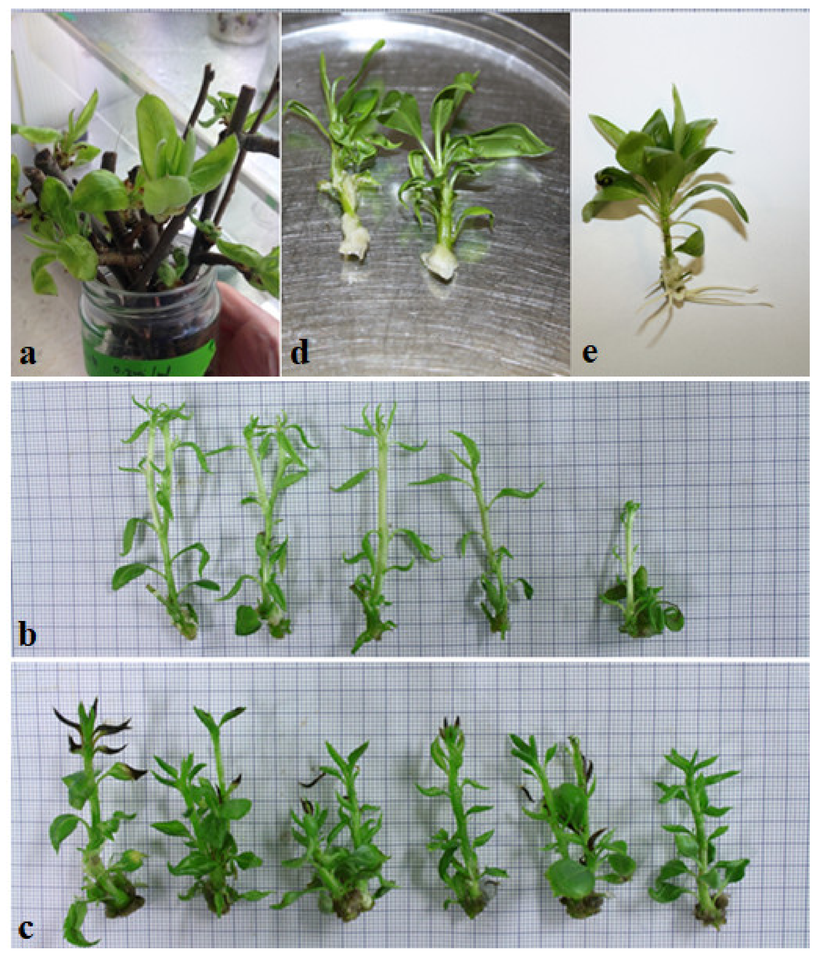
2.1. Plant Material, In Vitro Establishment, Rooting and Acclimatization

In field-growing plants of Pyrus communis L. varieties William and San Giovanni in situ preserved in the Experimental Farm of Tuscia University provided shoot tips for in vitro establishment of aseptic cultures.
Ten to fifteen cm long segments were collected, washed with running tap water for 5 min, and then surface sterilized in an aqueous solution containing 5% commercial bleach plus benomyl (Benlate ®) for 30 min and rinsed twice with sterile distilled water. Both ends of the segments were re-cut and placed in a glass jar containing 10 mL of forcing solution containing 200 mg/L 8-HQS plus 2% (w/v) sucrose (pH 5.6). After 7–8 days, the sprouts have been collected and the leaves removed. Uni-nodal segments were rinsed for 1 h in aqueous solution containing ascorbic acid 250 mg/L, citric acid 250 mg/L, GA3 5 mg/L and PPM® 0.1%. Then, the nodes were surface sterilized in a 20% commercial bleach plus few drops of Tween 20 for 30 min and the washed twice with sterile double-distilled water.
The culture establishments have been performed in 100 × 20 mm glass tube containing 5 mL of medium composed of half-strength MS medium, sucrose 20 mg/L, BAP 4.4 µM, NAA 0.054 µM and Plant Agar (Duchefa, NL) 0.55%. Medium pH has been adjusted to 5.8 with KOH 1M and autoclaved at 121 °C for 20 min. The medium used for the culture establishment was supplemented with 2,5 mg/L of CuSO4·5H2O as previously reported [39]. The contamination rates and number of buds sprouted have been collected (data not shown).
The cultures have been maintained in a growth chamber at 24 ± 1 °C with a 16-h photoperiod of 40-µmol m−2 s−1 provided by fluorescent lamps. Fifty explants for each media have been considered. The axillary shoots were multiplied on a Quoirin and LePoivre [40] medium (QL), supplemented with 4.9 µM N6-(2-Isopentenyl) adenine (2iP), 4.4 µM benzylaminopurine (BAP), 30 g/L sucrose and 0.55% of Plant Agar (Duchefa, NL). The pH was adjusted to 5.7. Multiplied shootlets were used for the experiment.
Two to three cm long individual shoots were excised at the end of a 4-week proliferation period on the proliferation media and culture conditions described above. The rooting media consisted of half-strength MS medium gelled with 0.6% Plant Agar and supplemented with 2% sucrose, indole-3-butyric acid (IBA) 1 µM.
Rooted shoots were transferred into plug trays (2 cm × 2 cm × 3.5 cm), containing 158 sterile BRIL Typical TYP 3 (Germany) peat substrate, and maintained in a growth chamber (temperature of 25 ± 4 °C, constant photoperiod at PPFD of 40 µmol m−2 s−1, and RH of 80%). The number of acclimatized plantlets was determined after eight weeks.

2.2. Culture Conditions and Light Quality

Axillary shoots of pears “William” and “San Giovanni” were cultured on the same medium described above, providing 100 mL of proliferation medium in each 500-mL jars. Each jar contained 15 explants, and three jars per treatment and variety were prepared.
Plant material was maintained in growth chamber equipped with four different LED light quality combinations (Valoya LED Grow Lights, Valoya Oy, Helsinky, Finland), compared with fluorescent lamp L36W/77 (Osram, Munich, Germany) as control. Briefly, the LED lamps used were AP67 (high composition in green and red), G2 (highest composition in red and far red) and NS1 (high composition in blue and green and low composition in far red). The spectral distribution and R:FR ratio of the light treatments are specified in Table 1. The environmental conditions inside the chambers consisted of 16 h photoperiod, 40 µmol m−2 s−2 photosynthetic photon flux density (PPFD) and an an air temperature 23 ± 1 °C. The experiments have been repeated two-fold.

2.3. Data Collection

At the end of the third subcultures, the morphological observations were carried out measuring the number of neo-formed shoots, node number and length of the main shoot. Shoot fresh weights (fw) have been determined weighting the explants and immediately oven-drying at 105 ± 1 °C until constant weight to determine the dry weight (dw).
The photosynthetic pigments were extracted from 100 mg of fresh leaves. The extraction has been done in 15 mL test tubes, adding 4 mL of methanol 100%, maintained 10 min in a water bath to 65 °C and then kept at 4 °C for 24 h. After centrifugation at 5000 × g for 5 min, the concentrations of total chlorophyll, chlorophyll a, chlorophyll b and carotenoid contents were determined spectrophotometrically [41] with a spectrophotometer EVO 60 (Thermo Fisher Scientific Inc. Waltham, MA, USA). Furthermore, Chl a/Chl b ratios were calculated. Five samples for each treatment were analyzed and repeated two time.
The lipid peroxidation has been expressed as malondialdehyde (MDA) content and was determined as TBA according to Astolfi et al. [42]. Briefly, fresh tissues (0.2 g) were homogenized in 10 mL of 0.25% TBA made in 10% TCA. The extracts were kept at 95 °C for 30 min and then cooled on ice. After centrifugation at 10,000× g for 10 min, the absorbance of the supernatant was measured at 532 nm. Correction of non-specific turbidity was made by subtracting the absorbance value taken at 600 nm. The level of lipid peroxidation was expressed as mmol g−1 fresh weight by using an extinction coefficient of 155 mM cm−1.

2.4. Statistical Analysis

All data were processed by XLSTAT integrates into Microsoft Excel. All the parameters were subjected to analysis of variance (ANOVA) and t-test. A Duncan post-hoc multiple range test was used for mean separation and to provide homogeneous groups for the means (at p ≤ 0.05). Furthermore, statistical elaboration consisted in the production of a correlation matrix (Pearson’s coefficient) to analyse intra-variable links and into a Principal Components Analysis (PCA) to investigate how varieties reacted differently to light treatments. First multicollinearity test has been done to verified high intercorrelations or inter-associations among the variables and to identified disturbance in the data. Later, Kaiser-Meyer-Olkin (KMO) has measured to assess the adequacy for each variable in the model and for the complete model and return information about if variables were suited for Multivariate Analysis (MA). In addition, variables with communality values <0.5 (Barlett’s test) could be removed. Finally, we run PCA applying a standardization using Pearson’s coefficient methods. The selection of Principal Component has been done following the latent root criterion (eigenvalues >1.0) [43].

3. Results

3.1. Morphological and Physiological Traits

The in vitro establishment of both pear cv San Giovanni e William was successfully achieved (Figure 1a). The shoots of William and San Giovanni cultivars showed significant differences in morphological and physiological traits measured at the end of the different light treatments used during micropropagation. (Figure 1b,c, Table 2). AP67 LED light positively affected the number of newly formed nodes in both varieties, although San Giovanni plantlets grown under fluorescent lamps did not much significant difference among number of nodes from that of plantlets grown under LEDs. Plantlets of variety William produced highest number of neo-formed shoots of 2.92 under AP67 LED followed by 2.15 in NS1 and 2.08 shoots under G2 LED light respectively, as compared to control treatment which formed 2.25 shoots. Whereas same trend was observed in San Giovanni which also produced higher 1.92 shoots under AP67 LED lights as compared to 1.68 and 1.09 shoots under G2 and NS1 treatments respectively. San Giovanni formed more shoots of 1.82 under control light as compared to G2 and NS1 which describes that both treatments did not show positive effect in this variety.
Considering the light effects, mean number of nodes in William plantlets significantly increased (13.50 and 12.51) under AP67 and NS1 LEDs respectively, when compared with fluorescent lamps, though G2 LED could not present a significant difference with control treatment. Whereas, San Giovanni plantlets were not significantly affected by LED lights, which showed lower node numbers when compared with control fluorescent lamps. Considering the cultivar factor, the new internode development in William plantlets was higher than in San Giovanni under all light treatments except the control conditions, in which San Giovanni showed increased node numbers (Table 3).
Plantlets of both varieties showed a similar average length of main shoots under fluorescent lamps (Table 2). Otherwise, significant differences in shoot length resulted between the two varieties by comparing the values obtained under each LED light. Particularly, William plantlets showed a significant increase in shoot length under LEDs, being G2 more effective than NS1 and AP67.
Whereas shoot of the cv San Giovanni grown under LEDs showed negative effects as compared to those growing under control fluorescent lamp, whereas they also showed the lowest average shoot length under LED light treatments. At the end of the experiment, the mean plantlet water content did not significantly differ among light treatments and varieties (data not shown). Furthermore, almost all the shoots differentiated calli (Figure 1d) or roots (Figure 1e) during the rooting phase, without differences due to the cv or the light treatments and, by the same way, they success in the acclimatization.

3.2. Photosynthetic Pigments

Chlorophyll contents in P. communis leaves were strongly influenced by the LED light treatment used. After four weeks of exposure to different light spectra, both varieties showed typical symptoms, such as decreased chlorophyll content. Total chlorophyll content was significantly lower in leaves of plants grown under LEDs when compared with control fluorescent lamps. No significant differences were observed between AP67 and NS1 LEDs for William plantlets, except under G2 light, in which total chlorophyll content in William leaves resulted lowest than in all treatments (Table 3). Same was the case for San Giovanni plantlets which showed higher contents of total chlorophyll under fluorescent lamps as compared to LEDs. Averages of chlorophyll a and b in both varieties also showed the negative effects when grown under LED lights as compared to fluorescent lamps treatment.
The ratio Chl a/Chl b showed a high variance inflation factor (VIF), perhaps due to the multicollinearity problem, and it was kept out from following multivariate analysis (MA), to avoid disturbance in the data. After, we had run the Kaiser-Meyer-Olkin’s (KMO) and the Bartlett’s test of sphericity to test if our variables were suited for MA. The measured sampling adequacy by KMO (0.6 as shown in Table 4) indicated that the sampling is adequate to MA, while Bartlett’s test revealed high similarity between total chlorophyll (Tc) and chlorophyll a (Chl a) content, so Tc was excluded by principal component analysis (PCA).
Carotenoid concentration was approximately the same in San Giovanni plantlets in control condition, as well as under all LEDs light spectra. Whereas William plantlets showed higher carotenoid content under fluorescent lamps. William plantlets showed an almost twofold increase in carotenoid contents under fluorescent lamps from that of both NS1 and G2 LED light spectra. Furthermore, William plantlets maintained approximately the higher carotenoid concentration irrespective of the light spectra, despite, it significantly declined in San Giovanni shoots with all used treatments (Table 3).

3.3. Protein and Malondialdehyde (MDA) Content

The results summarized in Table 3 show that different light qualities have significant effects on the MDA content in the leaves of William plantlets under AP67 light spectra when compared as than under control treatment (p  <  0.05). whereas, both NS1 and G2 light spectra produced lower MDA contents as compared to fluorescent lamps. The MDA content of San Giovanni plantlets under the NS1 and G2 treatment groups were significantly lower compared to AP67, contrarily it did not show much significance difference when compared with control treatment.

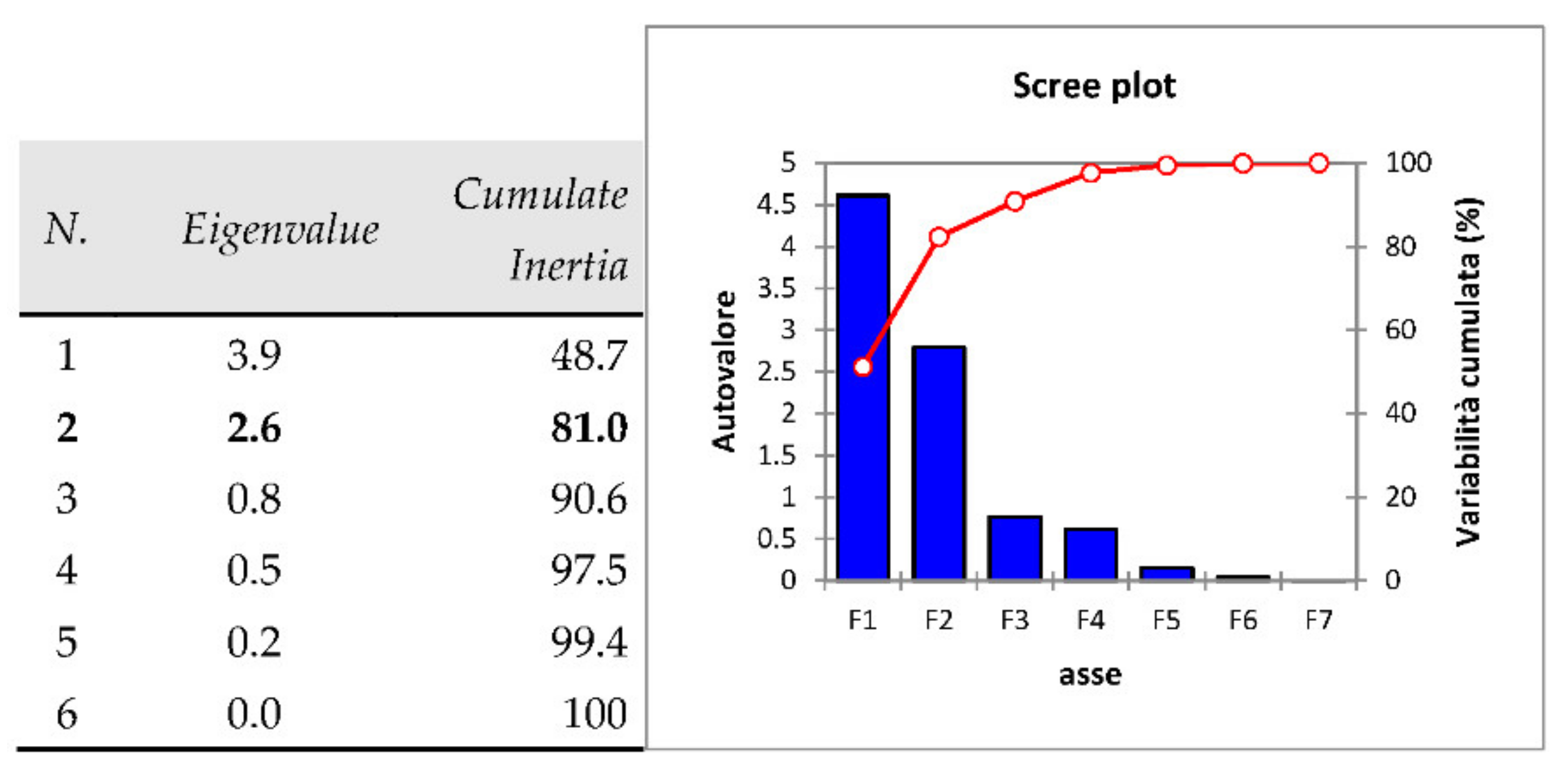
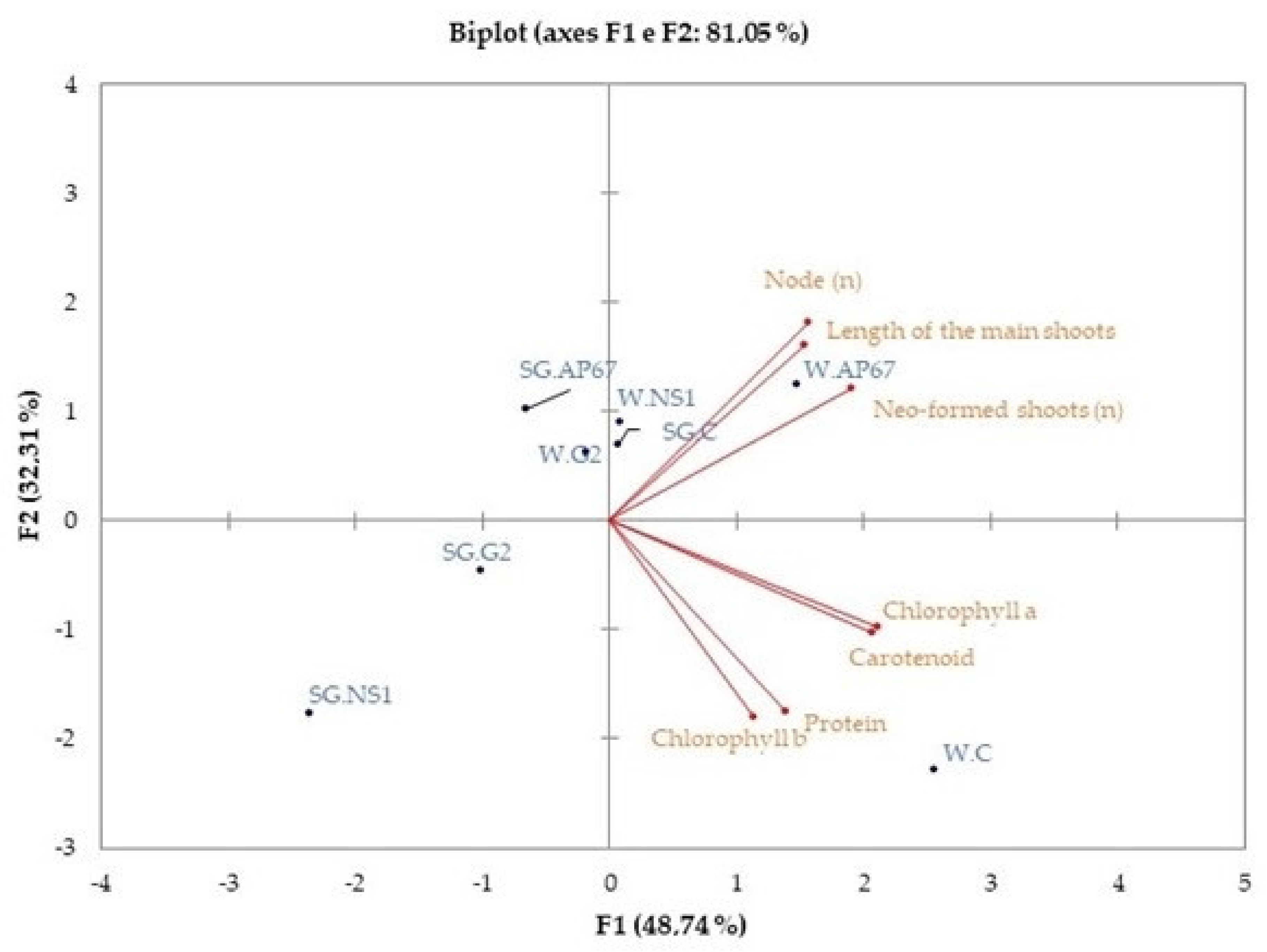
3.4. Principal Component Analysis (PCA)

In Figure 2 the set of the eigenvalues of PCA were reported, with the amount of inertia explained by each corresponding axis, and the cumulate inertia. So, we selected two principal axes of interest, by the latent root criterion (eigenvalues >1.0), that explained about 81% of the total variation (measured by the inertia), enough for our exploratory purposes.
High Pearson’s correlation coefficient was exhibited among Chl a, Carotenoid (C) and Protein (P). Significant correlations were also shown among Chl a, carotenoids, and neo-formed shoots (n) (0.6), while MDA was linked to node (n) (0.5) (Table 5).
The correlation plot of variables on to plane spanned by the PC1 and PC2 is reported in Figure 3. The two varieties reacted differently to light treatments. The cv William under control light conditions showed highest contents for chemical compounds such as Chl a, Chl b and carotenoids, on the contrary, all physical traits of shoots exhibited values lower than the average ones. Similar morphological traits of shoots were shown by cv William under the effect of spectrum G2, that reached the lowest values for all pigments content.
The cv William treated with the spectrum AP67 was related to high concentration of Chl a, carotenoids, and MDA and to highest values for all physical shoots’ parameters. Similar behavior has been shown by the same cv exposed to the lamp NS1.
The cv San Giovanni reacted to light treatments, as reported in Table 2 and Table 3 and explained by PCA in Figure 3. All biochemical and morphological traits reached the highest values compared to other San Giovanni thesis, excepted for protein content. This kind of morphological performances were confirmed by also by the cv San Giovanni under the light AP67, but Chl a and Chl b contents reduced by 33.8% and 71.7%, respectively, on the other hand protein increase of 29.6% respect to control.
The cv San Giovanni under the lamps NS1 and G2 showed reduced shoots growth. Under the light NS1 was also related to lowest Chl a, carotenoids, and MDA contents, contrary to the light G2 where Chl a content of cv San Giovanni was higher than NS1 and AP67 (+19.2%) but lower that control (−21.1%).

4. Discussions

In this experiment, the performance of micropropagated pear (Pyrus communis) cultivars William and San Giovanni was evaluated under different light treatments. Several morphological and physiological traits in both William and San Giovanni cultivars were influenced by different LED lights (AP67, NS1 and G2) as compared to the fluorescent lamps (control treatment). Statistical data related to physiological and morphological traits showed that AP67 LED positively affected shoot length, new internode length, and number of nodes in both cultivars. Moreover, San Giovanni plantlets were also sensitive to NS1 and G2 LED and to a minor extent, to AP67, in terms of shoot length and number of nodes. It concerns the stem elongation, which is considered as the main effect of the so-called shade avoidance syndrome (SAS) [44], and our results are substantially in agreement with those obtained in two hazelnut cultivars exposed to the same LED lights as in experiments conducted by [14], and they have explained that G2 LED positively affected shoot length, new internode length, and leaf area in both cultivars. Moreover, they also found that TGR hazelnut plantlets were sensitive to NS1 LED, and G2 LED clearly influenced the number of nodes developed during the acclimatization phase. Upon exposure to LED, in vitro-raised plantlets of pear have shown significant improvements in physiological and morphological traits. Green and red lights, either alone or in combination, have a significant influence on plant growth. In different previous studies [45,46,47,48,49,50,51,52,53], it also has been observed that a mixture of green and red LEDs enhanced plant growth, with increased shoot lengths, compared to monochromatic LEDs. Among the different lights used, AP67 LED emits the highest amount of green and red wavelengths (65 and 25% of the spectrum, respectively) and shows the lowest percentage. Moreover, William plantlets exhibited shoot elongation also when grown under G2 light, having the highest percentage of red wavelengths coupled with a high amount of red and far red. The significant difference in stem and new internode length between William and San Giovanni plantlets grown under each LED, being San Giovanni less responsive than William, is noteworthy. Similar cultivar specificity in morphological responses to a same light source have already been observed in hazelnut [14], basil [29], buckwheat [54], or lettuce [55].
Both the varieties reacted inversely to different light treatments. The cv William under controlled light conditions showed highest contents for chemical compounds such as Chl a, Chl b and C, on the contrary, all physical traits of shoots exhibited values lower than the average ones. Similar morphological traits of shoots were shown by cv William under the effect of spectrum G2, that reached the lowest values for all pigments content. Plants growing under artificial lighting often show modified photosynthetic processes because lamps do not usually mimic the sunlight spectrum. Therefore, plant biomass and metabolic products can be modified [56]. Therefore, the photosynthetic potential of plants along with their primary productions are directly influenced by the photosynthetic pigment contents, sequentially affected by different light qualities [57]. In experiments carried out both ex vitro and in vitro, significant differences were observed in the production of photosynthetic pigments in presence of green or red light, while the same pigments greatly decreased under spectra rich in red and far-red wavelengths [32,58]. In both pear cultivars, the total chlorophyll content was lowered under all tested LEDs, in respect to the fluorescent lamps. This result could not only depend on the high percentage of red and far-red wavelengths but could also be an effect of the highest amount of blue and red wavelengths.
In plant cells, the mechanisms regulating the carotenoid biosynthesis and accumulation are a complex phenomenon [59]. It has been suggested that light plays a key role in the biosynthesis of carotenoids through light signal sensing and downstream regulation [60]. However, with well-designed LED light spectrums, it has become possible to investigate effects of different monochromatic lights like red light and blue light with narrow spectra, on carotenoid metabolism in pear plantlets. Our results demonstrate that pear cultivar William is sensitive to a different LED photon flux applied in this study and fail to acclimate to such environments, in contrast to San Giovanni cultivar that did not show much significant difference among all the treatments. The light signal transduction of LED lights may be different from that of fluorescent lamps since plants usually have different photoreceptors, and these photoreceptors have both overlapping and distinct functions [61]. Biosynthesis of carotenoids is a complex pathway coordinated with the biogenesis of chlorophylls and proteins of the photosynthetic apparatus [62]. The content of the major carotenoids appears to be regulated in concert with the chlorophyll a, b content in pear cultivars as presented in Table 3. It is possible that carotenoid metabolism in both cultivars is regulated along with chlorophyll a, b, and total chlorophyll. Therefore, by controlling lighting conditions, it is possible to stimulate or inhibit the production of those compounds [63]. It was previously reported that blue LED light alone increased chlorophyll content in F. benjamina, Anthurium andraeanum Linden ex André, and Phalaenopsis sp. [64,65]. However, in the present experiment, the NS1 lamp, with an increased blue light content, did not improve the results. This could be explained by the fact that in the earlier studies, monochromatic lights were used, while in the present research, a combination of different wavelengths was applied. In particular, the presence of the green light portion of a spectrum impairs the blue light response [66].
In addition, soluble protein levels in William cultivar plantlets were relatively decreased under all tested LEDs, when compared to fluorescent lamps. Whereas an increasing trend was observed in San Giovanni cultivar under LEDs. However, leaf MDA content was lower in NS1 treated plantlets of both cultivars. While AP67 LED lights did not showed much significant difference as compared to fluorescent lamps in both William and San Giovanni cultivars. Therefore, it could be hypothesized that the different light treatments differently influence the phytochemical properties of both pear cultivars. Same results were achieved in previous studies conducted by [67], who also found decreased soluble proteins that suggests the plants growing under blue light and orange light undergo high environmental stress. Nevertheless, it can be assumed that light quality may affect the production of metabolites in the cultivars tested, although more experiments in this field are necessary.

5. Conclusions

In this work two pear cultivars, William and San Giovanni, were evaluated for the response of growth and morphogenesis under different LED lighting. A proper micropropagation protocol using LED system has been set up for both cultivars, for William, for reducing energy consumption and for increasing the quality of the process. In fact, the cv William showed an increased growth under LED AP67 (rich in green and red wavelengths) having a higher number of neo-formed shoots and node number. The cv San Giovanni also responded positively to LED AP67 but with no notable differences with the fluorescent light used as a control. Pigment analysis demonstrated that chlorophylls were negatively influenced by LED light. Protein analysis instead revealed that MDA was not significatively influenced by the treatment. The possibility of using LED light for the micropropagation of pear William should be considered as a valuable alternative as it can be considered more effective and more ecologically sustainable. This protocol should be improved with further studies to test the response of the plantlets to the ex-vitro acclimatization.

Author Contributions

Conceptualization: M.L., C.S., V.C.; Data Curation: C.S., M.A.B., E.B.; Formal Analysis: C.S. and E.B.; Funding Acquisition: V.C.; Investigation: M.L., M.A.B., C.S. and A.L.P.; Methodology: C.S.; Project Administration: C.S and V.C., Supervision: C.S. and V.C.; Writing—original draft: M.L., M.A.B. and C.S.; Writing—review and editing: C.S. and V.C. All authors have read and agreed to the published version of the manuscript.

Funding

The research was partially supported by MIUR (Ministry for Education, University and Research), Law 232/2016, “Department of Excellence” and by funding of Tuscia University DAFNE.RSA16CRI (Ricerca Scientifica Ateneo Cristofori).

Acknowledgments

The authors thank Bartolomeo Schirone for making available the growth chambers and LED lights. Thanks, are also due to Antonella Minandri for her technical assistance. We do not endorse any of the companies or product mentioned; names of the manufacturers and products are mentioned for information only.

Conflicts of Interest

The authors declare no conflict of interest.

References

  1. Lizárraga, A.; Fraga, M.; Ascasíbar, J.; González, M. In Vitro Propagation and Recovery of Eight Apple and Two Pear Cultivars Held in a Germplasm Bank. Am. J. Plant Sci. 2017, 8, 2238–2254. [Google Scholar] [CrossRef] [Green Version]
  2. Luziatelli, F.; Gatti, L.; Ficca, A.G.; Medori, G.; Silvestri, C.; Melini, F.; Muleo, R.; Ruzzi, M. Metabolites secreted by a plant-growth-promoting Pantoea agglomerans strain improved rooting of Pyrus communis L. cv dar gazi cuttings. Front. Microbiol. 2020, 11, 539359. [Google Scholar] [CrossRef] [PubMed]
  3. Aygun, A.; Dumanoglu, H. In vitro shoot proliferation and in vitro and ex vitro root formation of Pyruselae agrifolia Pallas. Front. Plant Sci. 2015, 6, 1–8. [Google Scholar] [CrossRef] [PubMed]
  4. Reed, B.M.; Wada, S.; DeNoma, J.; Niedz, R.P. Mineral nutrition influences physiological responses of pear in vitro. In Vitro Cell Dev. Biol. Plant 2013, 49, 699–709. [Google Scholar] [CrossRef]
  5. Bell, R.L.; Scorza, R.; Lomberk, D. Adventitious shoot regeneration of pear (Pyrus spp.) genotypes. Plant Cell Tissue Organ Cult. 2012, 108, 229–236. [Google Scholar] [CrossRef]
  6. USDA ARS. GRIN-Global. Available online: https://www.ars-grin.gov/ (accessed on 15 February 2021).
  7. Bantis, F.; Smirnakou, S.; Ouzounis, T.; Koukounaras, A.; Ntagkas, N.; Radoglou, K. Current status and recent achievements in the field of horticulture with the use of light-emitting diodes (LEDs). Sci. Hortic. 2018, 235, 437–451. [Google Scholar] [CrossRef]
  8. Singh, D.; Basu, C.; Meinhardt-Wollweber, M.; Roth, B. LEDs for energy efficient greenhouse lighting. Renew. Sustain. Energy Rev. 2015, 49, 139–147. [Google Scholar] [CrossRef] [Green Version]
  9. Olle, M.; Viršile, A. The effects of light-emitting diode lighting on greenhouse plant growth and quality. Agric. Food Sci. 2013, 22, 223–234. [Google Scholar] [CrossRef] [Green Version]
  10. Ahlman, L.; Bånkestad, D.; Wik, T. Using chlorophyll a fluorescence gains to optimize LED light spectrum for short term photosynthesis. Comput. Electron. Agric. 2017, 142, 224–234. [Google Scholar] [CrossRef]
  11. Pennisi, G.; Blasioli, S.; Cellini, A.; Maia, L.; Crepaldi, A.; Braschi, I.; Spinelli, F.; Nicola, S.; Fernandez, J.A.; Stanghellini, C.; et al. Unraveling the role of red:blue LED lights on resource use efficiency and nutritional properties of indoor grown sweet basil. Front. Plant Sci. 2019, 10, 305. [Google Scholar] [CrossRef]
  12. Schulze, P.S.C.; Barreira, L.A.; Pereira, H.G.C.; Perales, J.A.; Varela, J.C.S. Light emitting diodes (LEDs) applied to microalgal production. Trends Biotechnol. 2014, 32, 422–430. [Google Scholar] [CrossRef]
  13. Demotes-Mainard, S.; Péron, T.; Corot, A.; Bertheloot, J.; Gourrierec, J.G.-L.; Pelleschi-Travier, S.; Crespel, L.; Morel, P.; Huché-Thélier, L.; Boumaza, R.; et al. Plant responses to red and far-red lights, applications in horticulture. Environ. Exp. Bot. 2016, 121, 4–21. [Google Scholar] [CrossRef]
  14. Silvestri, C.; Caceres, M.E.; Ceccarelli, M.; Pica, A.L.; Rugini, E.; Cristofori, V. Influence of Continuous Spectrum Light on Morphological Traits and Leaf Anatomy of Hazelnut Plantlets. Front. Plant Sci. 2019, 10, 1318. [Google Scholar] [CrossRef]
  15. Liu-Gitz, L.; Britz, S.J.; Wergin, W.P. Blue light inhibits stomatal development in soybean isolines containing kaempferol- 3-O-2G-glycosylgentiobioside (K9), a unique flavonoid glycoside. Plant Cell Environ. 2000, 23, 883–891. [Google Scholar] [CrossRef] [Green Version]
  16. Ma, L.; Li, J.; Qu, L.; Chen, Z.; Zhao, H.; Deng, X.W. Light control of Arabidopsis development entails coordinated regulation of genome expression and cellular pathways. Plant Cell 2001, 13, 2589–2607. [Google Scholar] [CrossRef]
  17. Lee, S.H.; Tewari, R.K.; Hahn, E.J.; Pack, K.Y. Photon flux density and light quality induce changes in growth, stomatal development, photosynthesis and transpiration of Withania somnifera (L.) Dunal. plantlets. Plant Cell Tissue Organ Cult. 2007, 90, 141–151. [Google Scholar] [CrossRef]
  18. Savvides, A.; Fanourakis, D.; van Leperen, W. Co-ordination of hydraulic and stomatal conductances across light qualities in cucumber leaves. J. Exp. Bot. 2012, 63, 1135–1143. [Google Scholar] [CrossRef] [Green Version]
  19. Wang, J.; Wei, L.; Tong, Y.; Yang, Q. Leaf morphology, photosynthetic performance, chlorophyll fluorescence, stomatal development of lettuce (Lactuca sativa L.) exposed to different ratios of red light and blue light. Front. Plant Sci. 2016, 7, 250. [Google Scholar] [CrossRef] [Green Version]
  20. Smirnakou, S.; Ouzounis, T.; Radoglou, M. Continuous spectrum LEDs promote seedling quality traits and performance of Quercus ithaburensis var. macrolepis. Front. Plant Sci. 2017, 8, 188. [Google Scholar] [CrossRef] [Green Version]
  21. Zheng, L.; Van Labeke, M.C. Long-term effects of red- and blue light emitting diodes on leaf anatomy and photosynthetic efficiency of three ornamental pot plants. Front. Plant Sci. 2017, 8, 917. [Google Scholar] [CrossRef]
  22. Galvao, V.C.; Fankhauser, C. Sensing the light environment in plants: Photoreceptors and early signaling steps. Curr. Opin. Neurobiol. 2015, 34, 46–53. [Google Scholar] [CrossRef] [Green Version]
  23. Huché-Thélier, L.; Crespel, L.; Gourrierec, J.G.-L.; Morel, P.; Sakr, S.; LeDuc, N. Light signaling and plant responses to blue and UV radiations—Perspectives for applications in horticulture. Environ. Exp. Bot. 2016, 121, 22–38. [Google Scholar] [CrossRef]
  24. Ballaré, C.E.; Pierik, R. The shade-avoidance syndrome: Multiple signals and ecological consequences. Plant Cell Environ. 2017, 40, 2530–2543. [Google Scholar] [CrossRef]
  25. Kotb, O.M.; Abd EL-Latif, F.M.; Atawia, A.R.; Saleh, S.S.; EL-Gioushy, S.F. In vitro Propagation and Callus Induction of Pear (Pyrus communis) Cv. Le-Conte. Asian J. Biotechnol. Genet. Eng. 2020, 3, 1–10. [Google Scholar]
  26. Xu, J.; Wang, Y.; Zhang, Y.; Chai, T.Y. Rapid in Vitro Multiplication and ex Vitro Rooting of Malus zumi (Matsumura) Rehd. Acta Physiol. Plant 2008, 30, 129–132. [Google Scholar] [CrossRef]
  27. Erig, A.C.; Fortes, G.R. In Vitro Establishment of Pear (Pyrus spp.) Starting from Meristems and Buds. Cienc. Rural 2002, 32, 577–582. [Google Scholar] [CrossRef]
  28. Thakur, A.; Kanwar, J.S. Mircropropagation of “Wild Pear” Pyrus pyrifolia (Burm F) Nakai I Explant Establishment and Shoot Multiplication. Not. Bot. Horti Agrobot. Cluj-Napoca 2008, 36, 103–108. [Google Scholar]
  29. Bantis, F.; Ouzounis, T.; Radoglou, K. Artificial LED lighting enhances growth characteristics and total phenolic content of Ocimum basilicum, but variably affects transplant success. Sci. Hortic. 2016, 198, 277–283. [Google Scholar] [CrossRef] [Green Version]
  30. Agati, G.; Cerovic, Z.G.; Pinelli, P.; Tattini, M. Light-induced accumulation of ortho-dihydroxylated flavonoids as nondestructively monitored by chlorophyll fluorescence excitation techniques. Environ. Exp. Bot. 2011, 73, 3–9. [Google Scholar] [CrossRef]
  31. Nhut, D.T.; Takamura, T.; Watanabe, H. Responses of strawberry plantlets cultured in vitro under superbright red and blue light-emitting diodes (LEDs). Plant Cell Tissue Organ Cult. 2003, 73, 43–52. [Google Scholar] [CrossRef]
  32. Gupta, S.D.; Jatothu, B. Fundamentals and applications of light emitting diodes (LEDs) in in vitro plant growth and morphogenesis. Plant Biotechnol. Rep. 2013, 7, 211–220. [Google Scholar] [CrossRef]
  33. Cavallaro, V.; Pellegrino, A.; Muleo, R.; Forgione, I. Light and Plant Growth Regulators on In Vitro Proliferation. Plants 2022, 11, 844. [Google Scholar] [CrossRef] [PubMed]
  34. Gnasekaran, P.; Rahman, Z.A.; Chew, B.L.; Appalasamy, S.; Mariappan, V.; Subramaniam, S. Development of micropropagation system of Zingiber officinale var. rubrum Theilade using different spectrum light-emitting diode (LED) irradiation. Ind. Crops Prod. 2021, 170, 113748. [Google Scholar] [CrossRef]
  35. Miranda, N.A.; Xavier, A.; Otoni, W.C.; Gallo, R.; Gatti, K.C.; De Moura, L.C.; Souza, D.M.S.C.; Maggioni, J.H.; de Santos, S.S.O. Quality, and Intensity of Light in the In Vitro Development of Microstumps of Eucalyptus urophylla in a Photoautotrophic System. For. Sci. 2020, 66, 754–760. [Google Scholar] [CrossRef]
  36. Oliveira, T.D.R.D.; Aragão, V.P.M.; Moharana, K.C.; Fedosejevs, E.; Amaral, F.P.D.; de Sousa, K.R.; Thelen, J.J.; Venâncio, T.M.; Silveira, V.; Santa-Catarina, C. Light spectra affect the in vitro shoot development of Cedrela fissilis Vell. (Meliaceae) by changing the protein profile and polyamine contents. Biochim. Biophys. Acta BBA Proteins Proteom. 2020, 1868, 140529. [Google Scholar] [CrossRef]
  37. Smirnakou, S.; Ouzounis, T.; Radoglou, K. Effects of continuous spectrum LEDs used in indoor cultivation of two coniferous species Pinus sylvestris L. and Abies borisii-regis Mattf. Scand. J. For. Res. 2017, 32, 115–122. [Google Scholar] [CrossRef]
  38. Díaz-Rueda, P.; Cantos-Barragán, M.; Colmenero-Flores, J.M. Growth Quality and Development of Olive Plants Cultured In-Vitro under Different Illumination Regimes. Plants 2021, 10, 2214. [Google Scholar] [CrossRef]
  39. Silvestri, C.; Rugini, E.; Cristofori, V. The effect of CuSO4 for establishing in vitro culture, and the role nitrogen and iron sources in in vitro multiplication of Corylus avellana L. cv. tonda gentile romana. Plant Biosyst. 2020, 154, 17–23. [Google Scholar] [CrossRef]
  40. Quoirin, M.; Lepoivre, P.H. Improved media for in vitro culture of Prunus sp. In Symposium on Tissue Culture for Horticultural Purposes; ISHS: Gent, Belgium, 1977; Volume 78, pp. 437–442. [Google Scholar]
  41. Lichtenthaler, H.K. Chlorophylls and carotenoids: Pigments of photosynthetic biomembranes. Methods Enzymol. 1987, 148, 350–382. [Google Scholar]
  42. Astolfi, S.; Zuchi, S.; Passera, C. Effect of cadmium on H+ ATPase activity of plasma membrane vesicles isolated from roots of different S-supplied maize (Zea mays L.) plants. Plant Sci. 2005, 169, 361–368. [Google Scholar] [CrossRef]
  43. Hair, J.F.; Black, W.C.; Babin, B.J.; Anderson, R.E.; Tatham, R.L. Multivariate Data Analysis; Pearson University Press: Upper Saddle River, NJ, USA, 2006; Volume 6, p. 289. [Google Scholar]
  44. Smith, H. Physiological and ecological function within the phytochrome family. Ann. Rev. Plant Biol. 1995, 46, 289–315. [Google Scholar] [CrossRef]
  45. Nhut, D.T.; Takamura, T.; Watanabe, H.; Murakami, A.; Murakami, K.; Tanaka, M. Sugar-free micropropagation of Eucalyptus citriodora using light-emitting diode (LEDs) and film-rockwool culture system. Environ. Control Biol. 2002, 40, 147–155. [Google Scholar]
  46. Lian, M.L.; Murthy, H.N.; Paek, K.Y. Effects of light emitting diodes (LED) on the in vitro induction and growth of bulblets of Lilium oriental hybrid ‘Pesaro’. Sci. Hortic. 2002, 94, 365–370. [Google Scholar] [CrossRef]
  47. Duong, T.N.; Hong, L.T.A.; Watanabe, H.; Goi, M.; Tanaka, M. Efficiency of a novel culture system by using light-emitting diode (LED) on in vitro and subsequent growth of micro propagated banana plantlets. Acta Hortic. 2003, 616, 121–127. [Google Scholar]
  48. Kim, H.H.; Goins, G.D.; Wheeler, R.M.; Sager, J.C. Green light supplementation for enhanced lettuce growth under red and blue light emitting diodes. HortScience 2004, 39, 1617–1622. [Google Scholar] [CrossRef]
  49. Kim, H.H.; Goins, G.D.; Wheeler, R.M.; Sager, J.C. Stomatal conductance of lettuce grown under or exposed to different light qualities. Ann. Bot. 2004, 94, 691–697. [Google Scholar] [CrossRef] [Green Version]
  50. Kim, S.J.; Hahn, E.J.; Heo, J.W.; Paek, K.Y. Effects of LEDs on net photosynthetic rate, growth and leaf stomata of Chrysanthemum plantlets in vitro. Sci. Hortic. 2004, 101, 143–151. [Google Scholar] [CrossRef]
  51. Poudel, R.P.; Kataoka, I.; Mochioka, R. Effect of red-and blue-light emitting diodes on growth and morphogenesis of grapes. Plant Cell Tissue Organ Cult. 2008, 92, 147–153. [Google Scholar] [CrossRef]
  52. Shin, S.K.; Murthy, N.H.; Heo, W.J.; Hahn, J.E.; Paek, Y.K. The effect of light quality on the growth and development of in vitro cultured Doritaenopsis plants. Acta Physiol. Plant. 2008, 30, 339–343. [Google Scholar] [CrossRef]
  53. Li, H.; Xu, Z.; Tang, C. Effect of light-emitting diodes on growth and morphogenesis of upland cotton (Gossypium hirsutum L.) plantlets in vitro. Plant Cell Tissue Organ Cult. 2010, 103, 155–163. [Google Scholar] [CrossRef]
  54. Lee, S.W.; Seo, J.M.; Lee, M.K.; Chun, J.H.; Antonisamy, P.; Valan Arasu, M.; Suzuki, T.; Al-Dhabi, N.A.; Kim, S.J. Influence of different LED lamps on the production of phenolic compounds in common and Tartary buckwheat sprouts. Ind. Crops Prod. 2014, 54, 320–326. [Google Scholar] [CrossRef]
  55. Ouzounis, T.; Razi Parijkolaei, B.; Fretté, X.; Rosenqvist, E.; Ottosen, C.O. Predawn and high intensity application of supplemental blue light decreases the quantum yield of PSII and enhances the amount of phenolic acids, flavonoids, and pigments in Lactuca sativa. Front. Plant Sci. 2015, 6, 19. [Google Scholar] [CrossRef] [PubMed] [Green Version]
  56. Darko, E.; Heydarizadeh, P.; Schoefs, B.; Sabzalian, M.R. Photosynthesis under artificial light: The shift in primary and secondary metabolism. Philos. Trans. R. Soc. B 2014, 369, 20130243. [Google Scholar] [CrossRef] [Green Version]
  57. Gitelson, A.A.; Gritz, Y.; Merzlyak, M.N. Relationships between leaf chlorophyll content and spectral reflectance and algorithms for nondestructive chlorophyll assessment in higher plant leaves. J. Plant Physiol. 2003, 160, 271–282. [Google Scholar] [CrossRef] [PubMed]
  58. Muleo, R.; Morini, S. Light quality regulates shoot cluster growth and development of MM106 apple genotype in in vitro culture. Sci. Hortic. 2006, 108, 364–370. [Google Scholar] [CrossRef]
  59. Lu, S.; Li, L. Carotenoid metabolism: Biosynthesis, regulation, and beyond. J. Integr. Plant Biol. 2008, 50, 778–785. [Google Scholar] [CrossRef] [PubMed]
  60. Lamers, P.P.; Janssen, M.; De Vos, R.C.H.; Bino, R.J.; Wijffels, R.H. Exploring and exploiting carotenoid accumulation in Dunaliella salina for cell-factory applications. Trends Biotechnol. 2008, 26, 631–638. [Google Scholar] [CrossRef]
  61. Chory, J. Light signal transduction: An infinite spectrum of possibilities. Plant J. 2010, 61, 982–991. [Google Scholar] [CrossRef] [Green Version]
  62. Bohne, F.; Linden, H. Regulation of carotenoid biosynthesis genes in response to light in Chlamydomonas reinhardtii. Biochim. Biophys. Acta 2002, 1579, 26–34. [Google Scholar] [CrossRef]
  63. Ibrahim, M.H.; Jaafar, H.Z. Primary, secondary metabolites, H2O2, malondialdehyde and photosynthetic responses of Orthosiphon stimaneus Benth. To different irradiance levels. Molecules 2012, 17, 1159–1176. [Google Scholar] [CrossRef] [Green Version]
  64. Werbrouck, S.; Buyle, H.; Geelen, D.; Van Labeke, M.C. Effect or red-, far-red- and blue-light emitting diodes on in vitro growth of Ficus benjamina. Acta Hortic. 2012, 961, 533–538. [Google Scholar] [CrossRef] [Green Version]
  65. Bello-Bello, J.J.; Martinez-Estrada, E.; Caamal-Velazquez, J.H.; Morales-Ramos, V. Effect of LED light quality on in vitro shoot proliferation and growth of vanilla (Vanilla planifolia Andrews). Afr. J. Biotechnol. 2016, 15, 272–277. [Google Scholar]
  66. Folta, K.M.; Maruhnich, S.A. Green light: A signal to slow down or stop. J. Exp. Bot. 2007, 58, 3099–3111. [Google Scholar] [CrossRef]
  67. Saleem, M.H.; Rehman, M.; Zahid, M.; Imran, M.; Xiang, W.; Liu, L. Morphological changes and antioxidative capacity of jute (Corchorus capsularis, Malvaceae) under different color light-emitting diodes. Braz. J. Bot. 2019, 42, 581–590. [Google Scholar] [CrossRef]
Figure 1. Bud sprout of cv San Giovanni after ten days in forcing solution (a), different growth habits of cv William (b) and San Giovanni (c) after 4 weeks of culture under the same light quality (G2). Callus differentiation at the base of microshoots (d) and root differentiation (e).
Agronomy 12 02531 g001
Figure 2. Principal Component Analysis (PCA), eigenvalues, explained and cumulate inertia.
Agronomy 12 02531 g002
Figure 3. PCA representation of the factors levels on the plane spanned by the principal axes F1 and F2. Legend: SG.C = San Giovanni control; SG.AP67 = San Giovanni AP67; SG.G2 = San Giovanni G2; SG.NS1 = San Giovanni NS1; W.C = William control; W.AP67 = William AP67; W.G2 = William G2; W.NS1 = William NS1.
Agronomy 12 02531 g003
Table 1. Spectral distribution of the four light treatments.
LightSpectrum Composition (%)
400–500 nm500–600 nm600–700 nm700–800 nmR:FR Ratio
Fluorescent Lamp (C)34.824.136.74.45.7
AP67121657163.0
G28265253.1
NS1203935510.4
Table 2. Number of neo-formed shoots, node number, main internode length (cm) and length of the main shoots (mm) in William and San Giovanni shoots grown under different LED light conditions (AP67, G2 and NS1) and Fluorescent Lamps (Control). Data are represented as mean ± SD. Different lowercase letters on the columns for both varieties indicate significant differences among light treatments (Duncan’s test, p < 0.05). Different capital letters on the columns indicate significant differences among varieties (t-test).
VarietyLight QualityNumber of Neo-Formed ShootsNode NumberLength of the Main Shoots (mm)
WilliamControl2.25 ± 0.48 ab9.92 ± 1.7 b2.43 ± 0.36 b
AP672.92 ± 0.39 a13.50 ± 1.8 a2.94 ± 0.25 ab
NS12.15 ± 0.23 b12.51 ± 1.4 a2.96 ± 0.16 ab
G22.08 ± 0.50 b9.67 ± 1.5 b3.09 ± 0.46 a
William (avg) 2.35 ± 0.6 A11.31 ± 1.7 A2.85 ± 0.25 A
San GiovanniControl1.82 ± 0.13 a11.25 ± 3.2 a2.54 ± 0.49 a
AP671.92 ± 0.39 a10.51 ± 1.8 ab2.15 ± 0.33 ab
NS11.09 ± 0.20 b4.75 ± 2.4 c0.86 ± 0.36 c
G2 1.68 ± 0.29 a8.25 ± 3.5 b1.95 ± 0.71 b
San Giovanni (avg)1.63 ± 0.5 B8.94 ± 3.0 B1.88 ± 0.26 B
Table 3. Total chlorophyll, chlorophyll a, chlorophyll b and carotenoids (mg g−1), chl a and chl b ratio, total protein (mg g−1 f.w.) and MDA (µg g−1 f.w.) in William and San Giovanni shoots grown under different LED light conditions (AP67, G2 and NS1) and Fluorescent Lamps (Control). Data are represented as mean ± SD. Different lowercase letters on the columns for both varieties indicate significant differences among light treatments (Duncan’s test, p < 0.05). Different capital letters on the columns indicate significant differences among varieties (t-test).
Variety Light QualityTotal ChlorophyllChlorophyll a Chlorophyll bChl a/Chl bCarotenoidMDA (nmol g−1 f.w.)
WilliamControl1.91 ± 0.34 a1.23 ± 0.23 a0.68 ± 0.11 a1.81 ± 0.06 b0.33 ± 0.03 a3.72 ± 0.22 ab
AP671.05 ± 0.16 b0.82 ± 0.11ab0.23 ± 0.08 b3.53 ± 0.11 a 0.25 ± 0.02 b3.98 ± 0.17 a
NS11.04 ± 0.21 b0.70 ± 0.11 b0.34 ± 0.09 b2.19 ± 0.08 b0.16 ± 0.04 c3.11 ± 0.16 c
G20.89 ± 0.24 b0.57 ± 0.12 c0.32 ± 0.05 b1.93 ± 0.09 b0.16 ± 0.01 c3.52 ± 0.30 bc
William (average) 1.22 ± 0.46 A0.83 ± 0.29 A0.39 ± 0.19 A2.37 ± 0.09 A0.23 ± 0.08 A3.63± 0.29 A
San GiovanniControl1.17 ± 0.22 a0.71 ± 0.09 a0.46 ± 0.07 a1.80 ± 0.04 b0.16 ± 0.04 a4.88 ± 0.34 a
AP670.60 ± 0.18 b0.47 ± 0.08 b0.13 ± 0.07 b3.61 ± 0.12 a0.15 ± 0.02 a4.65 ± 0.18 a
NS10.89 ± 0.17 ab 0.47 ± 0.10 b0.36 ± 0.09 ab1.41 ± 0.11 b0.14 ± 0.03 a2.78 ± 0.24 c
G2 0.84 ± 0.21 ab0.56 ± 0.11 ab0.32 ± 0.08 ab1.77 ± 0.14 b0.16 ± 0.03 a3.17 ± 0.27 b
San Giovanni (average)0.88 ± 0.24 B0.55 ± 0.11 B0.32 ± 0.11 A2.15 ± 0.18 A0.15 ± 0.01 A3.81 ± 0.35 A
Table 4. KMO and Bartlett’s Test.
Kaiser-Meyer Olkin Measure of Sampling Adequacy0.6
Bartlett’s Test of SphericityApprox. Chi-Square5099.85
df36
Sign.0.05
Table 5. Correlation matrix, Pearson’s coefficient, and significance (p-value = 0.05).
VariablesChlorophyll aChlorophyll bCarotenoidProtein MDANumber of Neo-Formed ShootsNode Number Length of the Main Shoots
Chlorophyll a1
Chlorophyll b0.81
Carotenoid0.90.61
Protein0.70.60.81
MDA0.1−0.10.10.01
number of neo-formed shoots0.6−0.10.60.20.31
node number0.4−0.10.3−0.10.50.91
length of the main shoots0.40.00.3−0.10.40.80.91
Publisher’s Note: MDPI stays neutral with regard to jurisdictional claims in published maps and institutional affiliations.

