Article

The Effect of Different Drying Methods on Primary and Secondary Metabolites in Korean Mint Flower

1
Department of Crop Science, Chungnam National University, 99 Daehak-Ro, Yuseong-gu, Daejeon 34134, Korea
2
Department of Botany and Plant Pathology, Purdue University, 915 West State Street, West Lafayette, IN 47907, USA
3
Department of Plant Resources and Environment, Jeju National University, 102 Jejudaehak-ro, Jeju-si, Jeju 63243, Korea
4
Department of Smart Agriculture Systems, Chungnam National University, 99 Daehak-Ro, Yuseong-gu, Daejeon 34134, Korea
*
Authors to whom correspondence should be addressed.
Chang Ha Park and Hyeon Ji Yeo contributed equally to this work.
Agronomy 2021, 11(4), 698; https://doi.org/10.3390/agronomy11040698
Submission received: 12 March 2021 / Revised: 27 March 2021 / Accepted: 3 April 2021 / Published: 7 April 2021

Abstract

:
Edible flowers have been used in the food and beverage industries because of their high nutritional value, flavor, and scent. For the storage of edible flowers used in these industries, drying is a necessity to store the materials more easily and prevent the damage of metabolites in the flowers. However, drying may affect metabolite retention because drying conditions can differ according to the various methods. In this study, Agastache rugosa flowers were dried using four different methods (oven drying at 25 ± 1 °C, 50 ± 1 °C, 80 ± 1 °C, and freeze drying) and primary and secondary metabolites were analyzed using high-performance liquid chromatography (HPLC) and gas chromatography time-of-flight mass spectrometry (GC-TOF/MS). Freeze-dried flower samples contained higher levels of carotenoids (lutein, 13Z-β-carotene, β-carotene, and 9Z-β-carotene) and phenolics (rosmarinic acid, ferulic acid, and sinapic acid). Contrarily, the 80 °C oven-dried flower samples contained higher levels of most amino acids and flavonoids (including acacetin and tilianin) and at 25 °C and 50 °C contained higher levels of carbohydrates. Therefore, freeze-drying is a suitable method for retaining carotenoids and phenolics. In contrast, oven drying at 50 °C was highly recommended to retain amino acids and flavonoids.

1. Introduction

Korean mint (Agastache rugosa) is a medicinal plant native to east northern Asia and has been used as a traditional medicine to prevent fungal infections and vomiting and nausea [1]. Furthermore, Korean mint flower has been commercially used as sources for a tea and spice, and facial mask. This might be due to its pharmacological effects, including antimicrobial [2], anti-inflammatory and cardiovascular activities [3], antiviral [1] and otherwise [4] derived from the secondary metabolites identified in A. rugosa.
Agastache rugosa presents with various secondary metabolites, such as carotenoids, triterpenes, and flavonoids [5]. In particular, Korean mint flower has been reported to contain polyphenols (caffeic acid O-hexoside, 3-O-caffeoylquinic acid, caffeic acid, rosmarinic acid, 2,5-dihydroxycinnamic acid, hydroxycinnamic acids, emodin 6-O-glucoside, fraxiresinol hexoside, kaempferol 3-O-glucoside, phloretin-hexoside, calycosin, apigenin, genistein, and 6,7-dimethoxyquercetin 3-O-glucopyranoside) via LC/MS/MS analysis [6]. Furthermore, Park et al., 2019 described that total flavonoid and total phenolic contents were significantly higher in Korean mint flowers than those of leaves and stems [7], while Tuan et al., 2012 reported that the levels of rosmarinic acid, acacetin, and tilianin were significantly higher in Korean mint flowers than those of roots, stems, and leaves [5].
Carotenoids are a major group of natural products that represent more than 600 naturally occurring pigments found in most types of plant plastids [8,9]. The roles of plant carotenoids are determined primarily according to whether the plant tissue is photosynthetic. In photosynthetic plant tissue, pigments play an important role as structural components of plastid pigment–protein complexes, light harvesters for photosynthesis, and photoprotection against harmful reactive oxygen species. In non-photosynthetic tissues, the pigments contribute or determine the fruit, seed, and flower colors related to seed dispersal and pollination [10]. Furthermore, carotenoids function as precursors of abscisic acid, a plant hormone, associated with the developmental processes and regulation of stress [11]. Several carotenoids can only be obtained through consumption of food sources, because humans are unable to generate those using endogenous precursors and enzymes [12]. Other dietary carotenoids improve disease prevention; that is, the biological activities derived from carotenoids are correlated with a reduction in risk for cancers, immune disorders, chronic disease, degenerative diseases, eye diseases, and cardiovascular diseases [10,12,13].
Drying, as an important food preservation technique, is a traditional method most commonly used in the food industry. Drying is needed to reduce the water activity of the product to suppress microorganism growth and inhibit chemical reactions to extend the shelf-life of the product at room temperature. In addition, drying makes the weight of transportation lighter and reduces storage space [14]. The drying process can be carried out by various methods, namely, sun drying, shade drying, microwave drying, freeze drying, and many others [15], but enzymatic and non-enzymatic reactions that might occur during the drying process when performed under different conditions, lead to significant variation in the composition of secondary metabolites [16]. Therefore, the proper drying method should be selected to optimize the productivity of the target phytochemicals because all the developed drying methods are not appropriate for varied biological materials [15].
To date, no previous study has described the effect of different drying methods on the composition of diverse metabolites in Korean mint flower. Therefore, this study focused on profiling various metabolites, including amines, sugars, amino acids, organic acids, carotenoids, and flavones in A. rugosa flowers using GC-TOFMS and HPLC, and to determine the best drying method for the specific phytochemical. Furthermore, a comprehensive correlation between primary and secondary metabolites was described.

2. Materials and Methods

2.1. Chemicals

HPLC grade ethanol, methanol, water, chloroform, hexane, and ethyl acetate (EA) were purchased from Samchun Pure Chemical, Pyeongtaek, Korea. Methanol:Dichloromethane solution, potassium hydroxide, N-methyl-N-(trimethylsilyl)trifluoroacetamide, β-apo-8-carotenal, ribitol, methoxyamine hydrochloride, and pyridine were purchased from Sigma-Aldrich, Yongin, Korea.

2.2. Plant Materials

Agastache rugosa (Fisch. & C.A.Mey.) Kuntze was sown and grown in a glass greenhouse from March 2018 and cultivated from July to September 2018 at Chungnam National University, Daejeon, Korea (36°22′08.0″ N 127°21′14.2″ E) (Figure S1). The harvested A. rugosa flower inflorescences were washed with distilled water. Four drying methods were used: oven drying at 25 ± 1 °C, 50 ± 1 °C, 80 ± 1 °C (HB-502M, HANBAEK SCIENTIFIC CO., Daejeon, Korea), and freeze-drying (HC 3055, Bio-Medical Science Co., Ltd., Seoul, Korea). The harvested A. rugosa flower inflorescences were dried to a final moisture content below 10% and then ground in an electric grinder (wonder blender WB-1, SANPLATEC CORP, Osaka, Japan). All the chemical analyses were performed with three biological replicates and each biological replicate had a pool of 20 flower inflorescences harvested at the same flower development stage.

2.3. High-Performance Liquid Chromatography (HPLC) Analysis of Rosmarinic Acid, Tilianin, and Acacetin

HPLC analysis of rosmarinic acid, tilianin, and acacetin in A. rugosa flowers was performed according to the method described by Park et al. [7]. Fine powders (100 mg) of A. rugosa flowers dried using 25 °C oven drying, 50 °C oven drying, 80 °C oven drying, and freeze drying were added to 1 mL of aqueous methanol (80% v/v) and sonicated strongly at 37 °C for 1 h. After centrifugation at 15,000× g for 20 min, the supernatants were filtered through a 0.45 μm syringe filter into an HPLC vial. The HPLC equipment and operating conditions for the analysis were conducted according to the method described by Tuan et al. [5] (Table S1 and Figure S2). Three different flavones were identified, compared with retention time and through spike test, followed by quantification using the corresponding calibration curves.

2.4. Carotenoid Extraction and HPLC Analysis

HPLC analysis of lutein, zeaxanthin, β-cryptoxanthin, 13Z-β-carotene, α-carotene, β-carotene, and 9Z-β-carotene in A. rugosa was performed according to the method described by Chae et al. [17] (Table S1 and Figure S3). Fine powders (100 mg) of A. rugosa flowers dried using 25 °C oven drying, 50 °C oven drying, 80 °C oven drying, and freeze drying were soaked in 3 mL 0.1% (w/v) ascorbic acid in ethanol. After vortexing for 30 s and incubation at 85 °C in a water bath for 5 min, 120 μL of 80% (w/v) potassium hydroxide was added to the extracts, followed by vortexing for 30 s and incubation at 85 °C in a water bath for 10 min. The mixture was put on ice for 5 min and 0.1 mL of internal standard (β-apo-8-carotenal in ethanol; 25 μg/mL), 1.5 mL HPLC grade water, and 1.5 mL of hexane were added. After centrifugation at 1200× g and 4 °C for 10 min, the upper hexane layer was transferred to a fresh tube. For re-extraction, the centrifugation procedure was repeated. Nitrogen gas was used to dry the supernatants and then 0.25 mL of a 50:50 (v/v) methanol-dichloromethane solution was used to dissolve the dried supernatants. Each carotenoid was detected using our previous guidelines, through the combined use of retention time and co-elution with the internal standard, followed by a calculation using the corresponding calibration curves.

2.5. Hydrophilic Compound Extraction and GC-TOF-MS Analysis

GC-TOFMS analysis of hydrophilic compounds in A. rugosa flowers was performed according to the method described by Kim et al. [18]. Fine powders (50 mg) of A. rugosa flowers dried using 25 °C oven drying, 50 °C oven drying, 80 °C oven drying, and freeze drying were soaked in 1 mL of a chloroform-water-methanol mixture (1:1:2.5 v/v/v), followed by the addition of 60 µL of ribitol (0.2 g/L) as an internal standard. After mixing at 37 °C and 1200× g for 30 min and centrifugation at 13,000× g for 20 min, the supernatant was transferred and evaporated in a SpeedVac vacuum concentrator for 3 h, followed by derivatization by adding 80 μL of methoxyamine hydrochloride/pyridine (20 g L−1) and shaking at 37 °C and 1200× g for 2 h. Subsequently, 80 μL of N-methyl-N-(trimethylsilyl)trifluoroacetamide was added and the extract was heated at 37 °C for 30 min. Spinning down was performed and the extract was transferred into a GC vial. The GC-TOFMS equipment and operating conditions for the analysis were conducted according to the method described by Kim et al. [18] (Table S1 and Figure S4). Quantification was performed using selected ions, and Chroma-TOF software was used to locate the peaks [18,19].

2.6. Statistical Analysis

Duncan’s multiple range test (DMRT) was carried out using the Statistical Analysis System (SAS, system 9.4, 2013; SAS Institute, Inc., Cary, NC, USA). Heat map analysis and hierarchical cluster analysis (HCA) with Pearson correlations for 73 metabolites identified in these analyses were carried out using MetaboAnalyst 4.0 (http://www.metaboanalyst.ca/, accessed on 10 March 2021) with auto-scaling.

3. Results

3.1. Effect of the Different Drying Temperatures on Rosmarinic Acid, Tilianin, and Acacetin

Rosmarinic acid, tilianin, and acacetin were detected in the flower samples dried (Figure S5) using a freeze dryer and oven dryer at 25 °C, 50 °C, and 80 °C (Table 1). The freeze-dried sample contained the highest content of rosmarinic acid (2.95 ± 0.08 mg/g dw), and this value was about 1.32, 1.17, and 1.78 times higher than those of flower samples dried at 25 °C, 50 °C, and 80 °C in the oven dryer, respectively. In contrast, the 80 °C oven-dried samples possessed the highest content of acacetin, and the value was approximately 1.83, 2.87, and 2.75 times higher than those of freeze-dried, 25 °C oven-dried, and 50 °C oven-dried samples, respectively. Therefore, the freeze-drying method is suitable for the production of rosmarinic acid, whereas the 80 °C oven dry method can be selected for the production of tilianin and acacetin in A. rugosa.

3.2. Effect of the Different Drying Temperature on Carotenoids

Lutein, zeaxanthin, β-cryptoxanthin, 13Z-β-carotene, α-carotene, β-carotene, and 9Z-β-carotene were detected in the flower samples dried using a freeze dryer and oven dryer at 25 °C, 50 °C, and 80 °C (Table 2). The amounts of total carotenoids were significantly higher in the freeze-dried samples than in the 25 °C, 50 °C, and 80 °C oven-dried samples, and this value (187.07 ± 8.92 µg/g dw) was 1.72 times higher than the lowest level (108.51 ± 7.79 µg/g dw) observed in oven-dried samples at 80 °C. For the individual carotenoids, the contents of lutein, 13Z-β-Carotene, β-Carotene, and 9Z-β-Carotene had the highest levels in freeze-dried samples. In addition, zeaxanthin and α-carotene contents were higher in freeze-dried samples and 50 °C oven-dried samples. In contrast, β-cryptoxanthin levels were not significantly different. Therefore, the freeze-drying method is most suitable for the production of carotenoids (lutein, zeaxanthin, 13Z-β-carotene, α-carotene, β-carotene, and 9Z-β-carotene in A. rugosa.

3.3. Metabolic Profiling of Different Drying Temperatures of Agastache Rugosa

Heatmap analysis showed that 44 hydrophilic compounds were identified and quantified in A. rugosa flowers dried using a freeze dryer and oven dryer at 25 °C, 50 °C, and 80 °C (Figure 1). Specifically, 10 organic acids (oxalic acid, shikimic acid, threonic acid, urea, phosphoric acid, quinic acid, lactic acid, glyceric acid, glycolic acid, and pyruvic acid), four TCA cycle intermediates (fumaric acid, malic acid, succinic acid, and citric acid), one amine (ethanolamine), 18 amino acids (aspartic acid, glutamic acid, glutamine, pyroglutamic acid, proline, asparagine, glycine, β-alanine, threonine, valine, 4-aminobutyric acid, leucine, alanine, isoleucine, phenylalanine, methionine, serine, and tryptophan), and nine sugars (xylose, glucose, galactose, raffinose, sucrose, fructose, glycerol, inositol, and mannitol) were identified. Specifically, the levels of amino acids (alanine, leucine, valine, isoleucine, serine, threonine, β-alanine, 4-aminobutyric acid, phenylalanine, and methionine) were significantly higher in the flower samples oven dried at 80 °C, whereas the lowest levels of amino acids (alanine, leucine, valine, isoleucine, serine, threonine, β-alanine, 4-aminobutyric acid, phenylalanine, proline, glycine, asparagine, and tryptophan) were observed in freeze-dried flower samples, which contained the highest levels of aspartic acid, pyroglutamic acid, and glutamic acid. The contents of sugars, namely carbohydrates and sugar alcohols, were highest for the 25 °C oven-dried A. rugosa, which had the highest levels of glycerol, xylose, and fructose; 50 °C oven-dried flowers contained the highest levels of sucrose, inositol, and raffinose; while most sugar levels were low in freeze-dried and 80 °C oven-dried flowers. Furthermore, the levels of TCA intermediates (citric acid, fumaric acid, and succinic acid) were significantly higher in oven-dried flowers at 80 °C and ferulic acid and sinapinic acid levels were higher in freeze-dried flowers (Figure 1).
PCA was performed to investigate the variation in metabolic profiles among the Agastache rugosa flower samples dried using freeze-drying and oven drying at 25 °C, 50 °C, and 80 °C (Figure 2). Using to the PCA results, the score plot represents an overview of the differences between the flower samples dried by four different methods, and the loading plot facilitated correlation examination among the 54 compounds (Figure 2). In principal component 1 (PC1), the freeze-dried sample group and 80 °C oven-dried sample group were separated from the other groups since this separation was mainly attributed to secondary metabolites and amino acids. The significant metabolites of PC1 in the loading plot were 9Z-β-carotene, lutein, 13Z-β-carotene, rosmarinic acid, β-carotene, and sinapinic acid for which the eigenvector values were −0.18541, −0.17799, −0.17401, −0.17162, −0.17051, and −0.16225, respectively, and valine, 4-aminobutyric acid, threonine, succinic acid, glycolic acid, β-alanine, and isoleucine, for which the values were 0.18672, 0.18191, 0.17529, 0.17502, 0.17357, 0.17354, and 0.17305, respectively. Therefore, the PCA results revealed that freeze-dried flowers had higher levels of carotenoids and several phenolic compounds, 80 °C oven-dried flowers contained higher levels of most amino acids, and 25 °C and 50 °C flowers contained higher levels of sugars.

4. Discussion

In this study, secondary metabolites including carotenoids and phenolic compounds were detected in A. rugosa flowers dried using a freeze dryer and oven dryer at 25 °C, 50 °C, and 80 °C. These findings are consistent with those of previous studies reporting analysis of lutein, zeaxanthin, 13Z-β-carotene, α-carotene, and β-carotene in flowers, leaves, and roots of A. rugosa [17], and tilianin, acacetin, and rosmarinic acid in the flowers, leaves, stems, and roots of A. rugosa [5], and ferulic acid in flowers, leaves, and roots of A. rugosa [7].
This study showed that the freeze-dried A. rugosa flowers contained higher levels of carotenoids and phenolic compounds than the other drying treatments. Previously, Mediani et al. reported that freeze-dried P. niruri samples contained higher levels of phenolic compounds, as well as higher biological activity values, compared with P. niruri samples after air- and oven-drying treatments. Furthermore, this study reported the variation in metabolites according to the different drying treatments and extraction solvents [20]. A similar increase in carotenoids and chlorophylls was noted in freeze-dried Chinese cabbage and Nightshade plants compared with solar drying, microwave drying, oven drying, and sun drying [21], and in freeze-dried herbal tea composed of leaves of Apium graveolens, Averrhoa bilimbi, Centella asiatica, Mentha arvensis, Psidium guajava, Sauropus androgynous, Solanum nigrum, and Polygonum minus compared to drying at 50 °C and 70 °C [22]. Furthermore, Torres et al. reported an increase in volatiles (terpenes, sesquiterpenes, norisoprenoids, and C6 alcohols) and phenolics (anthocyanins and flavonols) in freeze-dried grape skin compared with 70 °C oven-dried grape skin [23], and Nunes et al. reported a slight increase in flavonoids and phenolic contents in freeze-dried guava powders compared with oven-dried guava powders [24]. However, the freeze-drying method did not confer any advantage to convection drying regarding the retention of β-carotene and lycopene in carrots [25], and 60 °C oven-dried wheatgrass samples exhibited higher contents of total phenolics and total flavonoids compared to freeze-dried samples [26]. In addition, Abdullah et al. reported that among leaves of Orthosiphon stamineus dried in an oven at 40 °C, under sunlight, and in the shade, sinensetin, belonging to the flavonoid group, was detected only in oven-dried samples, whereas rosmarinic acid was only detected in shade dried leaf samples [27]. Drying with airflow caused a decrease in the safranal production, while high-temperature drying enabled the retention of crocin pigments largely than saffron dried at intermediate temperatures [28].
Variations in metabolites according to the different drying methods used in the present study and of those in previous studies are not surprising. This may be due to enzymatic or non-enzymatic reactions that occur during the drying process of different drying methods [16]. Therefore, the proper methods should be selected for specific metabolites. This study suggests that freeze-drying is a suitable method to retain secondary metabolites, such as carotenoids and phenolic compounds, and that 80 °C oven drying is appropriate for retaining amino acids in A. rugosa flowers.

5. Conclusions

Dehydration is a method used to prevent the damage of metabolites during storage. However, the retention of metabolites varies according to the used drying method. Therefore, specific selection of drying methods is highly recommended for the retention of metabolites according to the purpose. This study showed that freeze-drying is suitable for the retention of secondary metabolites, such as carotenoids and phenolic compounds, in A. rugosa flowers. In contrast, 80 °C oven drying is recommended for retaining amino acids. Furthermore, oven drying at 25 °C and 50 °C could be used for the retention of carbohydrates.

Supplementary Materials

The following are available online at https://www.mdpi.com/article/10.3390/agronomy11040698/s1, Figure S1: Temperature conditions (A), relative humidity (B), and daylight hours (C) in Chungnam National University, Daejeon, Korea (36°22′08.0″ N 127°21′14.2″ E). Figure S2: Representative chromatogram of rosmarinic acid, tilianin, and acacetin obtained from freeze-dried Agastache rugosa. Peak: 1. Rosmarinic acid; 2. Tilianin; 3. Acacetin. Figure S3: Representative chromatogram of carotenoids obtained from freeze-dried Agastache rugosa. Peak: 1. Lutein; 2. Zeaxanthin; 3. trans-β-apo-8′-carotenal (internal standard); 4. β- Cryptoxanthin; 5. 13Z-β-Carotene; 6. α-Carotene; 7. β-Carotene; 8. 9Z-β-Carotene. Figure S4: Representative chromatogram of metabolites obtained from freeze-dried Agastache rugosa. Peak: 1, Pyruvic acid; 2, Lactic Acid; 3, Alanine; 4, Oxalic acid; 5, Glycolic acid; 6, Valine; 7, Urea; 8, Ethanolamine; 9, Phosphoric acid; 10, Glycerol; 11, Leucine; 12, Isoleucine; 13, Proline; 14, Glycine; 15, Succinic Acid; 16, Glyceric Acid; 17, Fumaric Acid; 18, Serine; 19, Threonine; 20, β-Alanine; 21, Malic acid; 22, Aspartic Acid; 23, Methionine; 24, Pyroglutamic Acid; 25, 4-Aminobutyric Acid; 26, Threonic acid; 27, Glutamic Acid; 28, Phenylalanine; 29, Xylose-1; 30, Xylose-2; 31, Asparagine; 32, Ribitol (internal standard) 33, Glutamine; 34, Shikimic acid; 35, Citric acid; 36, Quinic acid; 37, Fructose-1; 38, Fructose-2; 39, Galactose; 40, Glucose-1; 41, Glucose-2; 42, Mannitol; 43, Inositol; 44, Ferulic acid; 45, Tryptophan; 46, Sinapinic acid; 47, Sucrose; and 48, Raffinose. Figure S5: Agastache rugosa flower samples were dried using four different methods (oven drying at 25 ± 1 °C, 50 ± 1 °C, 80 ± 1 °C, and freeze drying). Table S1: HPLC and GC-TOFMS analysis methods.

Author Contributions

Y.S.C. and S.U.P. conceived and designed the experiments. C.H.P., H.J.Y. and C.P. performed the experiments and analyzed the data. C.H.P. and H.J.Y. wrote, reviewed, and edited the manuscript. All authors have read and agreed to the published version of the manuscript.

Funding

This research was supported by Basic Science Research Program through the National Research Foundation of Korea (NRF) funded by the Ministry of Education (2019R1A6A1A11052070).

Data Availability Statement

Not applicable.

Conflicts of Interest

The authors declare no conflict of interest.

References

  1. Wei, J.; Cao, P.; Wang, J.; Kang, W. Analysis of tilianin and acacetin in Agastache rugosa by high-performance liquid chromatography with ionic liquids-ultrasound based extraction. BMC Chem. 2016, 10, 1–9. [Google Scholar] [CrossRef] [PubMed] [Green Version]
  2. Shin, S. Essential oil compounds from Agastache rugosa as antifungal agents against Trichophyton species. Arch. Pharm. Res. 2004, 27, 295–299. [Google Scholar] [CrossRef] [PubMed]
  3. Hernández-Abreu, O.; Torres-Piedra, M.; García-Jiménez, S.; Ibarra-Barajas, M.; Villalobos-Molina, R.; Montes, S.; Rembao, D.; Estrada-Soto, S. Dose-dependent antihypertensive determination and toxicological studies of tilianin isolated from Agastache mexicana. J. Ethnopharmacol. 2013, 146, 187–191. [Google Scholar] [CrossRef]
  4. Zielińska, S.; Matkowski, A. Phytochemistry and bioactivity of aromatic and medicinal plants from the genus Agastache (Lamiaceae). Phytochem. Rev. 2014, 13, 391–416. [Google Scholar] [CrossRef] [Green Version]
  5. Tuan, P.A.; Park, W.T.; Xu, H.; Park, N.I.; Park, S.U. Accumulation of tilianin and rosmarinic acid and expression of phenylpropanoid biosynthetic genes in Agastache rugosa. J. Agric. Food Chem. 2012, 60, 5945–5951. [Google Scholar] [CrossRef]
  6. Desta, K.T.; Kim, G.S.; Kim, Y.H.; Lee, W.S.; Lee, S.J.; Jin, J.S.; El-Aty, A.M.A.; Shin, H.C.; Shim, J.H.; Shin, S.C. The polyphenolic profiles and antioxidant effects of Agastache rugosa Kuntze (Banga) flower, leaf, stem and root. Biomed. Chromatogr. 2016, 30, 225–231. [Google Scholar] [CrossRef] [PubMed]
  7. Park, C.H.; Yeo, H.J.; Baskar, T.B.; Park, Y.E.; Park, J.S.; Lee, S.Y.; Park, S.U. In vitro antioxidant and antimicrobial properties of flower, leaf, and stem extracts of Korean mint. Antioxidants 2019, 8, 75. [Google Scholar] [CrossRef] [PubMed] [Green Version]
  8. Kljak, K.; Grbeša, D. Carotenoid content and antioxidant activity of hexane extracts from selected Croatian corn hybrids. Food Chem. 2015, 167, 402–408. [Google Scholar] [CrossRef]
  9. Howitt, C.A.; Pogson, B.J. Carotenoid accumulation and function in seeds and non-green tissues. Plant Cell Environ. 2006, 29, 435–445. [Google Scholar] [CrossRef] [PubMed]
  10. Bartley, G.E.; Scolnik, P.A. Plant carotenoids: Pigments for photoprotection, visual attraction, and human health. Plant Cell 1995, 7, 1027. [Google Scholar]
  11. Koornneef, M. Genetic aspects of abscisic acid. In A Genetic Approach to Plant Biochemistry; Blonstein, A.D., King, P.J., Eds.; Springer: Vienna, Austria, 1986; pp. 35–54. [Google Scholar]
  12. Kopsell, D.A.; Kopsell, D.E. Accumulation and bioavailability of dietary carotenoids in vegetable crops. Trends Plant Sci. 2006, 11, 499–507. [Google Scholar] [CrossRef]
  13. Rao, A.V.; Rao, L.G. Carotenoids and human health. Pharmacol. Res. 2007, 55, 207–216. [Google Scholar] [CrossRef] [PubMed]
  14. Hamrouni-Sellami, I.; Rahali, F.Z.; Rebey, I.B.; Bourgou, S.; Limam, F.; Marzouk, B. Total phenolics, flavonoids, and antioxidant activity of sage (Salvia officinalis L.) plants as affected by different drying methods. Food Bioprocess Technol. 2013, 6, 806–817. [Google Scholar] [CrossRef]
  15. Walker, T.H.; Drapcho, C.M.; Chen, F. Bioprocessing technology for production of nutraceutical compounds. In Functional Food Ingredients and Nutraceuticals: Processing Technologies; Shi, J., Ed.; CRC Press: Boca Raton, Fl, USA, 2015; pp. 481–508. [Google Scholar]
  16. Capecka, E.; Mareczek, A.; Leja, M. Antioxidant activity of fresh and dry herbs of some Lamiaceae species. Food Chem. 2005, 93, 223–226. [Google Scholar] [CrossRef]
  17. Chae, S.C.; Lee, S.W.; Kim, J.K.; Park, W.T.; Uddin, M.R.; Kim, H.H.; Park, S.U. Variation of carotenoid content in Agastache rugosa and Agastache foeniculum. Asian J. Chem. 2013, 25, 4364–4366. [Google Scholar] [CrossRef]
  18. Kim, Y.B.; Kim, J.K.; Uddin, M.R.; Xu, H.; Park, W.T.; Tuan, P.A.; Li, X.; Chung, E.S.; Lee, J.-H.; Park, S.U. Metabolomics analysis and biosynthesis of rosmarinic acid in Agastache rugosa Kuntze treated with methyl jasmonate. PLoS ONE 2013, 8, e64199. [Google Scholar] [CrossRef] [PubMed] [Green Version]
  19. Park, C.H.; Park, S.Y.; Park, Y.J.; Kim, J.K.; Park, S.U. Metabolite Profiling and Comparative Analysis of Secondary Metabolites in Chinese Cabbage, Radish, and Hybrid Brassicoraphanus. J. Agric. Food Chem. 2020, 68, 13711–13719. [Google Scholar] [CrossRef] [PubMed]
  20. Mediani, A.; Abas, F.; Khatib, A.; Tan, C.P.; Ismail, I.S.; Shaari, K.; Ismail, A.; Lajis, N.H. Relationship between metabolites composition and biological activities of Phyllanthus niruri extracts prepared by different drying methods and solvents extraction. Plant Foods Hum. Nutr. 2015, 70, 184–192. [Google Scholar] [CrossRef] [PubMed]
  21. Managa, M.G.; Sultanbawa, Y.; Sivakumar, D. Effects of different drying methods on untargeted phenolic metabolites, and antioxidant activity in Chinese cabbage (Brassica rapa L. subsp. chinensis) and nightshade (Solanum retroflexum Dun.). Molecules 2020, 25, 1326. [Google Scholar] [CrossRef] [Green Version]
  22. Mahanom, H.; Azizah, A.H.; Dzulkifly, M.H. Effect of different drying methods on concentrations of several phytochemicals in herbal preparation of 8 medicinal plants leaves. Malays. J. Nutr. 1999, 5, 47–54. [Google Scholar] [PubMed]
  23. De Torres, C.; Díaz-Maroto, M.C.; Hermosín-Gutiérrez, I.; Pérez-Coello, M.S. Effect of freeze-drying and oven-drying on volatiles and phenolics composition of grape skin. Anal. Chim. Acta 2010, 660, 177–182. [Google Scholar] [CrossRef]
  24. Nunes, J.C.; Lago, M.G.; Castelo-Branco, V.N.; Oliveira, F.R.; Torres, A.G.; Perrone, D.; Monteiro, M. Effect of drying method on volatile compounds, phenolic profile and antioxidant capacity of guava powders. Food Chem. 2016, 197, 881–890. [Google Scholar] [CrossRef] [PubMed]
  25. Regier, M.; Mayer-Miebach, E.; Behsnilian, D.; Neff, E.; Schuchmann, H.P. Influences of drying and storage of lycopene-rich carrots on the carotenoid content. Dry. Technol. 2005, 23, 989–998. [Google Scholar] [CrossRef]
  26. Das, A.; Raychaudhuri, U.; Chakraborty, R. Effect of freeze drying and oven drying on antioxidant properties of fresh wheatgrass. Int. J. Food. Sci. Nutr. 2012, 63, 718–721. [Google Scholar] [CrossRef] [PubMed]
  27. Abdullah, S.; Shaari, A.R.; Azimi, A. Effect of drying methods on metabolites composition of misai kucing (Orthosiphon stamineus) leaves. APCBEE Procedia 2012, 2, 178–182. [Google Scholar] [CrossRef] [Green Version]
  28. Gregory, M.J.; Menary, R.C.; Davies, N.W. Effect of drying temperature and air flow on the production and retention of secondary metabolites in saffron. J. Agric. Food Chem. 2005, 53, 5969–5975. [Google Scholar] [CrossRef]
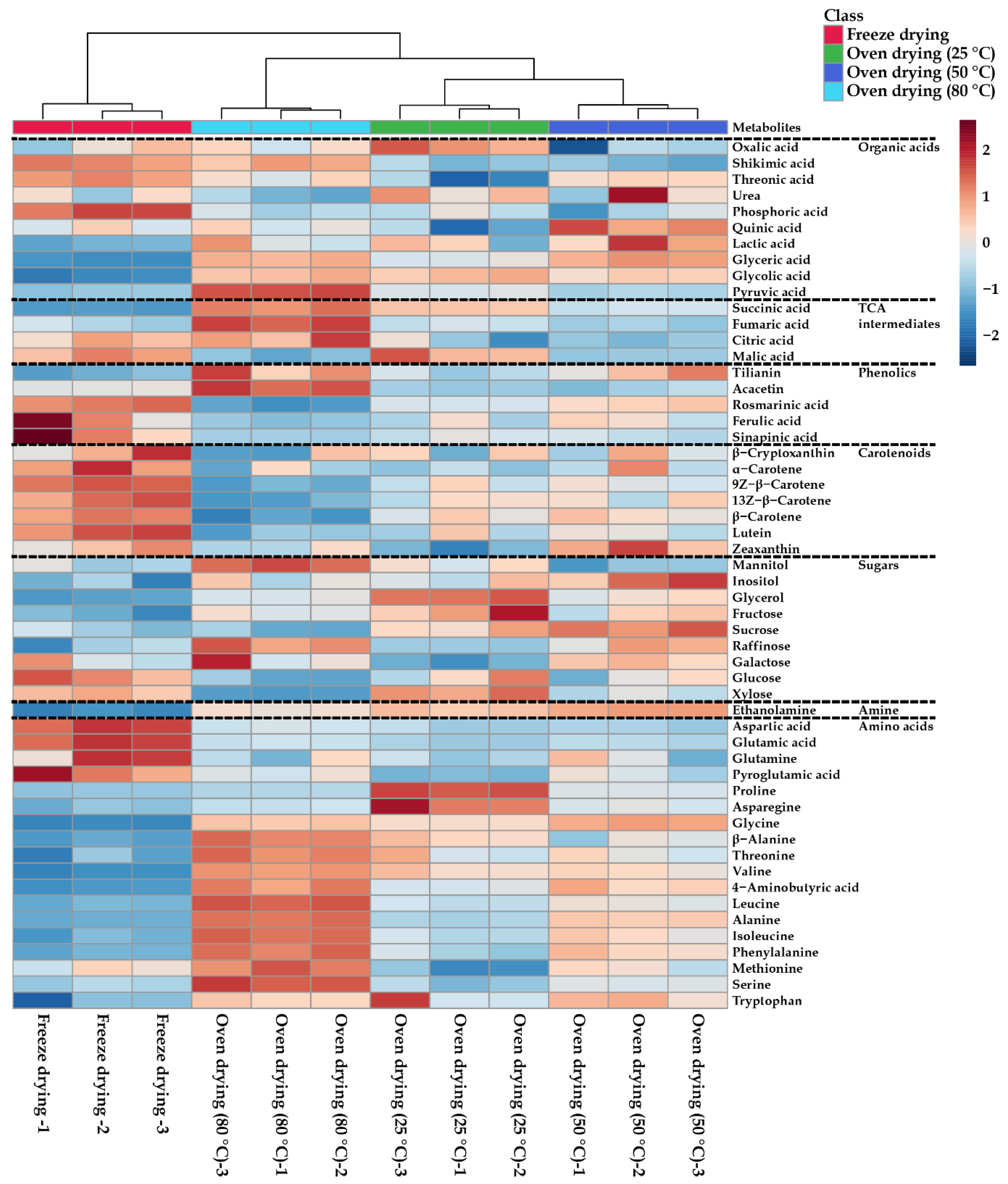
Figure 1. Heatmap represents the differences in A. rugosa flowers dried using freeze dryer and oven dryer at 25 °C, 50 °C, and 80 °C. Increasing and decreasing the contents of metabolites are shown by red and blue color, respectively.
Agronomy 11 00698 g001
Figure 2. (A) Score plots of principal components 1 and 2 of PCA results obtained from metabolites of Agastache rugosa at different drying temperatures. (B) Loading plots of principal components 1 and 2 of PCA results obtained from metabolites of Agastache rugosa at different drying methods.
Agronomy 11 00698 g002
Table 1. The contents of rosmarinic acid, tilianin, and acacetin after different drying conditions (mg/g dw).
Drying Methods
Freeze Drying25 °C Oven Drying50 °C Oven Drying80 °C Oven Drying
Rosmarinic acid2.95 ± 0.08 a 12.23 ± 0.02 c2.53 ± 0.07 b1.66 ± 0.06 d
Tilianin2.04 ± 0.10 c2.34 ± 0.15 b2.88 ± 0.31 a3.11 ± 0.33 a
Acacetin0.36 ± 0.01 b0.23 ± 0.01 c0.24 ± 0.05 c0.66 ± 0.04 a
Total5.34 ± 0.05 ab4.81 ± 0.17 b5.65 ± 0.44 a5.43 ± 0.42 a
1 Different letters in the same row indicate a significant difference (p < 0.05), applying a Duncan’s multiple range test.
Table 2. The contents of carotenoids in different drying conditions (µg/g dw).
Drying Methods
Freeze Drying25 °C Oven Drying50 °C Oven Drying80 °C Oven Drying
Lutein63.15 ± 3.35 a 148.14 ± 5.95 b49.16 ± 2.80 b41.83 ± 2.93 b
Zeaxanthin7.86 ± 0.62 ab5.87 ± 0.42 c8.37 ± 0.67 a6.90 ± 0.53 bc
β-Cryptoxanthin7.16 ± 0.64 a6.52 ± 0.60 a6.57 ± 0.52 a6.12 ± 0.73 a
13Z-β-Carotene9.69 ± 0.73 a7.45 ± 0.76 b7.54 ± 0.91 b5.27 ± 0.37 c
α-Carotene1.19 ± 0.12 a0.75 ± 0.07 b0.92 ± 0.21 ab0.79 ± 0.17 b
β-Carotene84.55 ± 3.61 a67.53 ± 5.76 b70.63 ± 4.88 b41.68 ± 3.78 c
9Z-β-Carotene 13.47 ± 0.39 a8.92 ± 1.31 b9.15 ± 0.64 b5.91 ± 0.51 c
Total187.06 ± 8.92 a145.19 ± 12.60 b152.34 ± 7.81 b108.51 ± 7.79 c
1 Different letters in the same row indicate a significant difference (p < 0.05), applying a Duncan’s multiple range test.
Publisher’s Note: MDPI stays neutral with regard to jurisdictional claims in published maps and institutional affiliations.