Article

Water as a Solvent of Election for Obtaining Oleuropein-Rich Extracts from Olive (Olea europaea) Leaves

1
Di3A, Dipartimento di Agricoltura, Alimentazione e Ambiente, University of Catania, via S. Sofia 100, 95123 Catania, Italy
2
CNR-ICB, Consiglio Nazionale delle Ricerche-Istituto di Chimica Biomolecolare, via Paolo Gaifami 18, 95126 Catania, Italy
3
Dipartimento di Scienze Agrarie, Alimentari e Forestali, University of Palermo, Viale delle Scienze Ed.4, 90128 Palermo, Italy
*
Author to whom correspondence should be addressed.
Agronomy 2021, 11(3), 465; https://doi.org/10.3390/agronomy11030465
Submission received: 8 February 2021 / Revised: 23 February 2021 / Accepted: 25 February 2021 / Published: 3 March 2021
(This article belongs to the Special Issue Phytochemicals of Edible Plants in Human Health)

Abstract

:
Leaves from Olea europaea represent one of the main by-products of the olive oil industry, containing a plethora of bioactive compounds with several promising activities for human health. An organic solvent-free extraction method was developed for the recovery of olive leaf phenols, which obtained an extract containing oleuropein in high amounts. A comparison of various extraction media is reported, together with the total phenolic content, DPPH (2,2-Diphenyl-1-picrylhydrazyl) content, ORAC (oxygen radical absorbance capacity), and polyphenol oxidase activity of the corresponding extracts. The polyphenol profiles and content of the most representative extracts have also been studied. Extraction solvent and temperature significantly influenced the phenolic content and antioxidant activity of the extracts, with hot water representing the solvent of election for the extraction of bioactive compounds from this matrix. All the extracts obtained showed reasonably high total phenol content (TPC) and good DPPH radical scavenging activity; among them, the water extract is characterized by desirable traits and could be used for many industrial applications and human consumption.

1. Introduction

The olive tree (Olea europaea L.) is an important crop in the Mediterranean area, it is considered a drought-tolerant crop and has developed physiological mechanisms to tolerate drought stress and grow under adverse climatic conditions, such as the regulation of gas exchange and an antioxidant system [1]. The olive tree and its products have a relevant importance in different fields. Recent research studies highlight that olive leaves have been a copious by-product of the olive oil industry (10% of the total weight of the harvested olives) and tree pruning (25 kg per olive tree) [2,3].
Olive leaves are a copious by-product of the olive oil industry and of olive tree pruning. They are also considered a cheap and natural source of phenolic compounds such as hydroxytyrosol, verbascoside, rutin, tyrosol, and oleuropein.
Oleuropein is an heterosidic ester of β-glucosylated elenolic acid and 3,4-dihydroxy-phenylethanol (hydroxytyrosol); it belongs to the chemical class of secoiridoids, which are present in all members of the Oleaceae family [4].
Oleuropein, dimethyloleuropein, ligstroside, and oleoside represent the predominant phenolic oleosides found in O. europaea [2,3], with oleuropein itself counting for up to 9% of the leaves’ dry weight matter [4,5,6,7]. Oleuropein and its derivatives have been widely studied for their antioxidant properties and health benefits, including antimicrobial and antiproliferative activities [8,9,10]. Other compounds identified in olive leaves are verbascoside and oleuroside [11,12], flavonoid glycosides (luteolin 7-O-glucoside, apigenin 7-O-glucoside, rutin [11,13,14], apigenin 7-O-rutinoside, luteolin 7-O-rutinoside, and luteolin 4-O-glucoside) [15], and flavonoid aglycones such as apigenin, quercetin, kaempferol, hesperidin, and luteolin [13,15]. Several phenolic acids (ferulic, caffeic, chlorogenic, p-coumaric, homovanillic, and vanillic) [13,15] were also found to be present in this matrix.
The polyphenol composition of olive leaves may vary according to many factors: cultivar, climatic conditions, stage of crop cycle, and agricultural practices [16]. The phenolic profile and content of leaves can also be influenced by endogenous enzymatic activities and extraction procedures. Different studies show that many factors affect the extraction efficiency, such as the type and volume of the solvent, temperature, pH, and number of extraction steps [17]. In the last few years, new extraction techniques have been investigated in order to reduce the volume of solvents used. With regard to extraction from olive leaves, organic solvents such as methanol [18], ethanol, hexane, ethyl acetate, or hydroalcoholic mixtures are commonly employed for their ability to extract both lipophilic and hydrophilic phenols [19] from this vegetable material. It was recently demonstrated that large amounts of oleuropein can be extracted from olive leaves using polar solvents such as a 20:80 acetonitrile/water mixture [20]. These extraction procedures often require laborious clean-up and mandatory concentration steps. Innovative extraction techniques, such as microwave and supercritical fluid extractions [21], superheated liquid, pressurized liquid, fractionation by solid-phase, dynamic ultrasound-assisted, and microwave-assisted extraction, have also been proposed for obtaining oleuropein and other phenolic compounds from olive leaves [22,23]; these methods aim to reduce extraction time and sample preparation costs. Non-conventional extraction techniques, such as microwave-assisted extraction (MAE), supercritical fluid extraction (SFE), and pressurized liquid extraction, have shown different extraction selectivity; in particular, MAE and conventional solvent extraction seem to be the most suitable choice for obtaining polar compounds, such as oleuropein derivatives, apigenin rutinoside, and luteolin glucoside, whilst SFE and pressurized liquid extraction seem to be more effective in extracting less polar compounds, such as apigenin, luteolin and diosmetin [18].
Because of the increasing interest in developing clean chemical procedures (so-called “green chemistry”), Paladino and Zuritz [24] investigated the possibility of extracting phenolic compounds from grape seeds using only distilled water as an extraction solvent at different temperatures and compared the extraction efficiency with that of traditional organic solvents.
Other authors employed boiling water to extract phenolic compounds from different plant materials, such as Salvia triloba L. leaves, Tiliaargentea flowers, green and black tea leaves, and grapes [25,26]. The use of water as an extraction medium is of relevant importance for avoiding toxic solvents, especially when the procedures have to be scaled up to an industrial level and the products targeted for human use (food ingredients or drugs). The aim of the present paper was to develop an environmentally friendly, fast, and cheap extraction method based on an organic solvent-free procedure to obtain large amounts of bioactive phenolic compounds from olive leaves. The proposed method was compared with the most popular solvent extraction procedures found in the literature. The effect of the extraction techniques was evaluated by studying qualitatively and quantitatively the phenolic composition of the resulting extracts and their radical scavenging activity, using both 2,2-Diphenyl-1-picrylhydrazyl (DPPH) and oxygen radical absorbance capacity (ORAC) assays. The presence of polyphenoloxidase in the olive leaf extracts obtained was also determined in order to assess its possible influence on the phenolic content.

2. Materials and Methods

2.1. Plant Material

Olea europaea ‘Biancolilla’ leaves were collected from olive trees during the pruning period. The samples were transported to the laboratory and dehydrated in a stove at 40 °C until at constant weight.

2.2. Chemicals and Standards

Unless otherwise stated, all solvents used in this study were high-purity laboratory products obtained from Carlo Erba (Milan, Italy). HPLC (high-performance liquid chromatography) -grade water, acetonitrile, and methanol were purchased from VWR (Milan, Italy). Pure luteolin, luteolin-7-O-glucoside and apigenin-7-O-glucoside were provided by Extrasynthese (Lyon, France). Rutin (quercetin-3-O-rutinoside), apigenin, caffeic acid, chlorogenic acid, p-coumaric acid, ferulic acid, fluorescein, hydroxytyrosol, oleuropein, 3,4-dihydroxyphenilacetic acid (DOPAC), 2,2-Diphenyl-1-picrylhydrazyl (DPPH), 2,2′-azobis (2-methylpropionamidine) dihydrochloride (AAPH) and 6-hydroxy-2,5,7,8-tetramethylchroman-2-carboxylic acid (Trolox) were provided by Sigma, (Sigma-Aldrich s.r.l., Milan, Italy).

2.3. Olive Leaf Extracts (OLEs) Preparation

The dried plant material was finely ground and suspended in a defined volume of the extraction solvent. The resulting heterogeneous mixtures were then homogenized at room temperature (25 °C) using an Ultra-Turrax IKA T-18 basic homogenizer (IKA-Werke GmbH & Co. KG, Staufen, Germany). The homogenates were filtered, and the clear supernatants were stored in the dark at −20 °C until analyzed. Different extraction treatments and conditions were tested as reported in Table 1.

2.4. Polyphenol Oxidase (PPO) Activity Assay

The polyphenoloxidase activity of the olive leaf extracts was tested according to the method reported by Ortega-García et al. [27] with some modifications. The assay was performed at 30 °C, with the standard reaction mixture containing DOPAC (500 mM) as a phenolic substrate, 0.1 M sodium phosphate with a pH of 6.2 as a buffer, and 50 μL of enzymatic extract in a total volume of 1 mL. The samples were read spectrophotometrically at 505 nm with a blank being used as a control. One unit of PPO activity is defined as the amount of enzyme which produces 1 μmol of product per min at 25 °C under assay conditions.

2.5. Total Phenolic Content and Radical Scavenging Activity

Total phenolic content and radical scavenging activity (RSA) were both evaluated spectrophotometrically as described by Palmeri et al. [28]. Total phenolic content was evaluated on all the OLEs. The total phenolic contents of the OLEs were expressed as caffeic acid equivalents in milligram per gram of dried leaves. RSA was evaluated by using a 2,2-Diphenyl-1-picrylhydrazyl (DPPH) assay and the RSA% was expressed as Trolox equivalent antioxidant capacity (TEAC). In addition, the DPPH radical scavenging capacity was evaluated in the OLEs obtained by different extractions.

2.6. Oxygen Radical Absorbance Capacity (ORAC) Assay

An ORAC assay measures the antioxidant inhibition of peroxyl radical-induced oxidation and thus reflects classical radical chain-breaking antioxidant activity by H atom transfer. In the basic assay, the peroxyl radicals, generated by 2,2′-azobis(2-amidinopropane) dihydrochloride (AAPH), react with a fluorescent probe to form a non-fluorescent product, which can be quantitated easily by fluorescence. An automated ORAC assay was carried out on a Wallac 1420 spectrofluorometric analyzer (Perkin Elmer, Turku, Finland; excitation wavelength = 485 nm and emission filter = 515 nm), based on a slightly modified procedure proposed by Ou et al. [29]. Fluorescein (116 nM) was the target molecule for free radical attack from AAPH (153 mM). The reaction was carried out in a 75 mM phosphate buffer (pH 7.4) at 37 °C. In total, 20 μL of OLE and 120 μL of fluorescein were mixed in the microplate and preincubated for 10 min. Then, 60 μL of AAPH solution was added, and the fluorescence was recorded for 60 min at excitation and emission wavelengths of 485 and 530 nm, respectively. A blank sample containing 20 μL of phosphate buffer and Trolox (10 μM) was used as a control. All solutions were freshly prepared prior to analysis. All samples were diluted with the buffer (1:200, v/v) prior to analysis and the ORAC values were expressed as mmol of Trolox equivalents (TE) per g of dried leaves using the standard curve established previously.

2.7. HPLC-DAD and HPLC-ESI-MS Analyses

High-performance liquid chromatographic (HPLC) analyses were carried out on an Ultimate 3000 “UHPLC focused” instrument equipped with a binary high-pressure pump, a photodiode array detector, a thermostatted column compartment, and an automated sample injector (Thermo Scientific, Milan, Italy). Collected data were processed through a Chromeleon chromatography information management system v. 6.80. Chromatographic analyses were carried out on a Gemini C18 column (250 × 4.6 mm, 5 μm particle size, Phenomenex, Italy) equipped with a guard column (Gemini C18 4 × 3.0 mm, 5 μm particle size, Phenomenex, Italy). OLEs were analyzed according to Gambacorta et al. [30] using solvent system A (2.5% formic acid in water) and solvent system B (acetonitrile/methanol 50:50). A linear gradient analysis was used as follows: 0 min: 5% B; 8 min: 30% B; 25 min: 60% B; 30 min: 80% B; then kept for 9 min at 80% B, for a total run time of 50 min. The diode array detector (DAD) was set in the range between 600 and 190 nm, recording the chromatographic runs at 280, 330, and 350 nm. In order to unambiguously identify the chromatographic signals and/or to confirm peak assignments, HPLC-ESI-MS analyses were also performed using the same conditions (solvents, elution program, guard column, column, injection volume, and flow) described above. OLEs were analyzed using a Waters instrument (Waters Italia S.p.A., Milan, Italy) consisting of a 1525 binary HPLC pump and a Micromass ZQ mass analyzer equipped with an ESI Z-spray source. Total ion current (TIC) chromatograms were acquired according to Siracusa et al. [31]. Quantification of hydroxytyrosol, hydroxytyrosolglucoside, ligstroside, oleuropein, and oleuropein aglycone was carried out at 280 nm using the calibration curves established with oleuropein (R2 = 0.9993) and hydroxytyrosol (R2 = 0.9992), respectively, whilst DOPAC was quantified at the same wavelength using its corresponding analytical standard (R2 = 0.9997). Apigenin-7-O-glucoside and apigenin were quantified at 330 nm using the calibration curve established with apigenin (R2 = 0.9995). Caffeic acid (R2 = 0.9998) was used to quantify caffeic acid, ferulic acid, chlorogenic acid, and verbascoside, whilst quantification of p-coumaric acid was done using the corresponding available standard (p-coumaric acid, R2 = 0.9999). Both calibration curves were built at 330 nm. Luteolin, luteolin 7-O-glucoside, and rutin were quantified at 350 nm using the calibration curves established with their corresponding analytical standards (luteolin R2 = 0.9999; luteolin-7-O-glucoside R2 = 09994; rutin R2 = 0.9999). Analyses were always carried out in triplicate.

2.8. Statistical Analysis

The statistical analysis was performed using INFOSTAT software version 2013. Initially, the data were analyzed by analysis of variance. When the differences were significant, a means comparison test (Fisher’s least significant difference (LSD) test) was applied. The criterion of significance was taken as p < 0.01.

3. Results

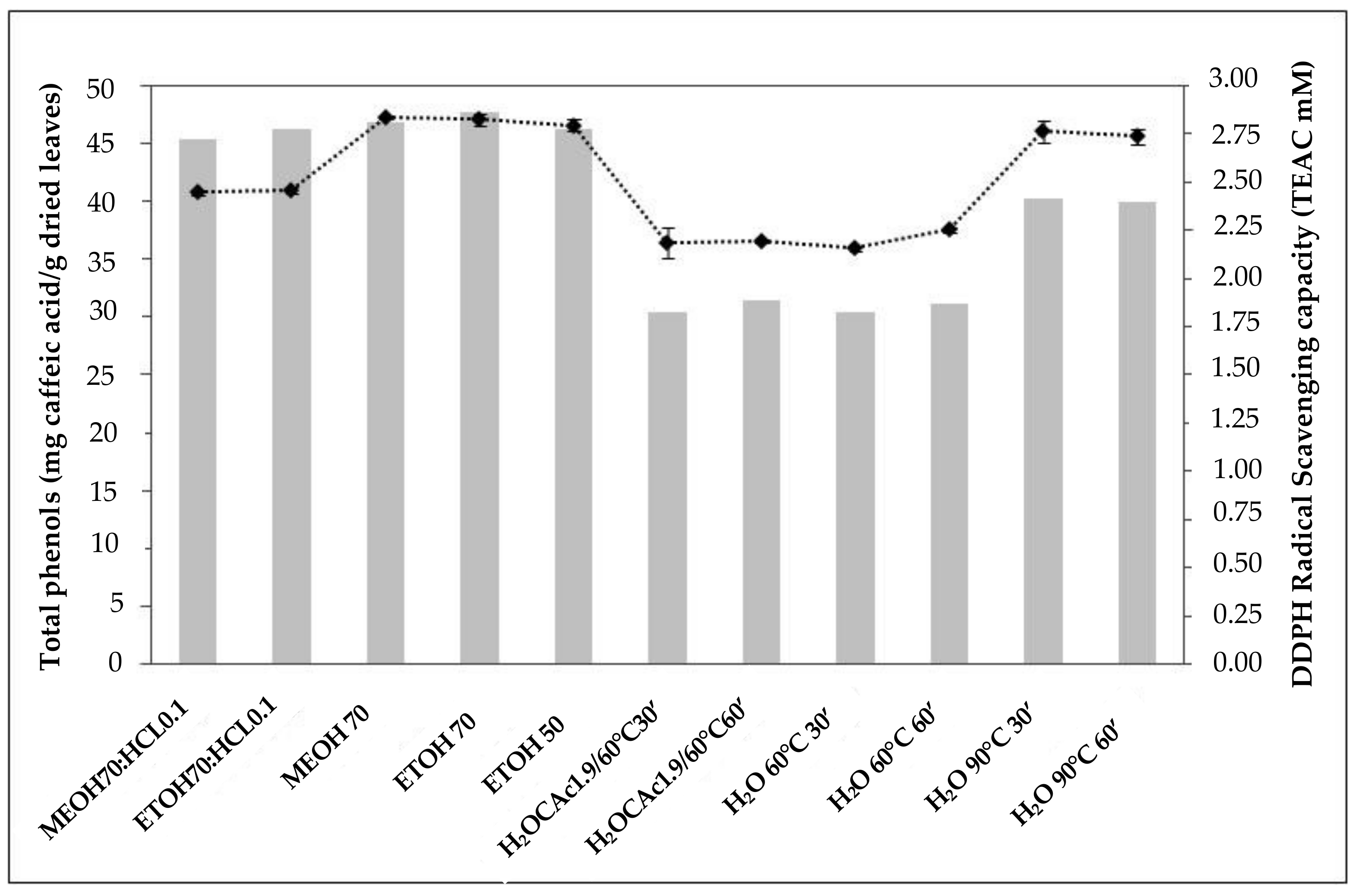
3.1. Effect of Different Extraction Media and Extraction Conditions on the TPC Content and Antiradical Capacity (DPPH Assay) of OLEs

The effect of the extraction techniques was evaluated mainly through the determination of the total phenolic content (TPC) of the different extracts obtained. The results, as reported in Table 2, showed that the TPC values found ranged from 30.44 to 47.75 mg caffeic acid/g dried leaves.
OLEEs at different concentrations (50%, 70%) and acidified with 0.1% HCl and OLEM showed the highest values of total phenols (47.75 ± 0.52, 46.29 ± 0.07, 46.29 ± 0.49, 46.83 ± 0.12), followed by acidified OLEM (45.41 ± 0.47) and OLEA at 90 °C (40.31 ± 0.08). The extract obtained using water at 60 °C for 30 min showed the lowest concentration of total phenolic compounds (30.44 ± 0.34).
The results also showed that the use of HCl in hydroalcoholic mixtures did not affect the TPC content of the extracts. The same behavior was observed when using an aqueous medium acidified with citric acid. A relevant effect of extraction conditions on the TPC content in aqueous extracts was observed only when 90 °C was used as the extraction temperature.
As reported in Table 2, the extraction procedure had a significant (p < 0.01) influence on antiradical capacity (AC) as measured by the DPPH assay. OLEA at 90 °C, hydroalcoholic extracts of OLEE (70%) and OLEM (70%), showed the highest scavenging activity, followed by acidified OLEM (70%), and OLEE and OLEA (60 °C). As observed for TPC content, the use of an acidic medium did not generally seem to have a positive effect on the radical scavenging activity of the extracts. Figure 1 shows that the extracts with a higher TPC content also displayed a higher AC, but the relationship between TPC and AC was also influenced by the different extraction treatments. The increment of extraction time from 30 to 60 min did not have a significant influence on the AC for OLEA (90 °C).

3.2. Effect of Different Extraction Mixtures and Conditions on PPO Activity

The results show that PPO activity is strongly inhibited for all hydroalcoholic treatments; at mild temperatures (60 °C) PPO is still active, while its activity is low at higher temperatures (Table 3).
As mentioned earlier, these data are of pivotal importance due to the role PPO plays in the degradation of phenolic compounds, especially in the oxidation of secoiridoids such as oleuropein [32].

3.3. Effect of Different Extraction Mixtures and Conditions on Antiradical Activity (ORAC Assay) of OLEs

In order to evaluate the ability of the aqueous, methanolic, and ethanolic extracts to quench different free radicals, we also determined their inhibitory capacity against peroxyl radicals by ORAC assay. Table 4 shows the TPC content values and antioxidant capacity of different leaf extracts as determined using DPPH and ORAC assays.
Ethanolic extract exhibited the highest ORAC value (2.17 mmol TE/g dried leaves), followed by methanolic extract (1.88 mmol TE/g dried leaves), and water extract (1.23 mmol TE/g dried leaves). However, the difference between the ORAC values of ethanolic and methanolic extracts was not statistically significant. Hydroalcoholic extracts were more efficient for flavonoids (Figure 2) than aqueous extract and they also showed higher total phenol content, whilst DPPH scavenging activity, expressed as % inhibition, was similar for all extracts. According to the results obtained, all extracts exhibited good antioxidant capacities, as measured by DPPH and ORAC methods.

3.4. Identification and Quantification of the Main Polyphenols Present in the Extracts through HPLC-DAD/ESI-MS

As previously mentioned, the differences in the antiradical activities registered for the different extracts tested may depend on their composition in terms of bioactive compounds. In order to determine the individual components present in the OLEs and their possible influence on the antioxidant potential, extracts obtained using water at 90 °C, 70% aqueous ethanol, and 70% aqueous methanol solutions were analyzed by means of HPLC-DAD/ESI-MS. Figure S1A–C shows the chromatograms corresponding to aqueous (A), 70% aqueous methanol (B), and 70% aqueous ethanol (C) extracts from olive leaves, whilst the corresponding quantitative data are listed in Table 5. Among the nearly 30 signals appearing in the chromatogram, 16 of them (peaks 1–16) were tentatively identified by comparing their relative retention times, UV-Vis, and MS data with those of the corresponding analytical standards when available; assignments were further corroborated by literature data [33,34]. As shown in Figure S1 A and reported in Table 5, oleuropein (peak 11, 46.25 mg/g dried leaves) and hydroxytyrosol glucoside (peak 1, 14.97 mg/g dried leaves) were the most abundant compounds present in the water extract, as extensively reported [33]. Ligstroside (peak 13) and verbascoside (peak 6) were also present in considerable amounts (9.68 mg/g and 5.313 mg/g, respectively).
The subclass of flavones was represented in this extract by the 7-O-glucoside derivatives of luteolin and apigenin (peak 10 and 12, 4.039 mg/g and 1.947 mg/g, respectively) but not by their corresponding aglycones. Similarly, Figure S1B,C show the chromatograms corresponding to the aqueous methanol and aqueous ethanol extracts of olive leaves. As reported in Table 5, oleuropein (peak 11, 39.40 mg/g and 36.45 mg/g dried leaves) was the most abundant compound identified in both alcoholic extracts, followed by luteolin 7-O-glucoside (peak 10, 8.12 mg/g and 8.93 mg/g dried leaves), and ligstroside (peak 13, 7.20 mg/g and 7.57 mg/g dried leaves). Hydroxytyrosol glucoside (peak 1 in Figure S1A), the second most abundant compound in water extract, was present here in a lesser amount (5.14 mg/g in aqueous methanol and 5.02 mg/g dried leaves in aqueous ethanol). The results obtained clearly showed that OLEA is the extract that is richer in oleuropein, ligstroside, verbascoside, hydroxytyrosol glucoside, and hydroxytyrosol. Hydroxycinnamic acids were also more abundant in this extract, whilst ethanolic and methanolic ones were richer in flavonoids (aglycones and glycosides), as also reported in Figure 2. These results indicate that the different extraction conditions had a significant influence on the content of secoiridoids and flavonoids in the extracts, and that water at 90 °C was more efficient in extracting more polar compounds such as oleuropein and its derivatives. On the other hand, the extract obtained using 70% aqueous ethanol exhibited the greatest amount of flavonoid glycosides.

4. Discussion

The TPC values found in our experiments are in accordance with what was reported by Ortega-García and Peragòn [35], who investigated the polyphenol content in leaves from different olive cultivars extracted by methanol-containing mixtures after an n-hexane pre-treatment to remove oil residues. The authors observed that the content depends on the cultivar and the fruit ripening stage, and their results ranged from 27.63 mg/g to 44.61 mg/g of dried leaves, expressed as caffeic acid content.
Studies on phenol extraction from different matrices using water at high temperatures as an extraction medium demonstrated that it is indeed an efficient method for the recovery of high-value natural bioactive compounds [12,36], and in some cases it was even more efficient than when compared to organic solvents [24,37]. This “high temperature effect” could be ascribed both to the nature of the vegetable matrix and to the structure of the bioactive compounds to be extracted [38]. It is also well known that high temperatures are able to deactivate endogenous enzymes such as oxidases, thus avoiding or minimizing phenolic degradation [39].
The main enzymatic oxidative activities, such as those of ascorbate peroxidase (AP), catalase (CAT), superoxidodismutase (SOD), and peroxidase (POD), increased both in the leaves and the roots after drought stress in relation to stress severity [1]. On the contrary, PPO activity decreased during the progression of stress. To our knowledge, there are few works regarding PPO determination in olive leaves, none of which are about Sicilian cultivars that show a low endogenous activity. PPO has been characterized by Ortega-García and Peragón [35] in the olive tree fruits and leaves of cv. Picual during ripening. The authors reported that the specific activity and catalytic efficiency of PPO changed in the leaves during fruit ripening; PPO from the leaves is different from that of the fruit for kinetic characteristics and tissue localization [27,40]. PPO shows a wide distribution in leaves, and this is probably related to the protection mechanism of the plant. PPO is involved in plant defense against pathogens and biotic and abiotic stress conditions. The induction of PPO expression has been related to plant tolerance against stress. An important function of the chemical defense of the plant for oleuropein and PPO in other Oleaceae was observed [41].
Differences in the content and type of individual polyphenols present in aqueous, methanolic, and ethanolic extracts determined the differences in their antioxidant properties. Antiradical activities could also be influenced by the mutual interactions occurring among the phenolic components present in these different matrices, as mentioned in previous studies [42]. The differences in antioxidant capacity as measured by DPPH and ORAC could then ultimately be due to the differences in the content and type of the individual polyphenols present in the different extracts.
There are very few data about the hot water extraction of polyphenols from olive leaves. In most of the extraction procedures, including in new extraction techniques, organic solvents or strong acidic media are frequently employed. In this work, we have obtained an aqueous extract from olive leaves particularly rich in oleuropein (46 mg/g). Methanolic olive leaf extracts from several Spanish and Italian varieties showed that the highest oleuropein content was that of 30.17 mg/g dried leaves in the “Frantoio” cultivar [35], whilst a more modest value of 14.35 mg/g dried leaves in the “Moraiolo” cultivar was obtained by using 50% ethanol as the extraction medium [43]. Supercritical fluid extraction (SFE) to recover bioactive compounds from “Koroneiki” olive leaves was employed. The extracts obtained by SFE, modified with 20% ethanol and subcritical water at 150 °C, exhibited the highest oleuropein content, 51 mg/g and 46 mg/g dried leaves, respectively [44]. Concerning the phenolic composition of olive leaves from Sicilian cultivars, there are few studies published. The main polyphenols in a methanolic extract from the “Biancolilla” cultivar were identified by Scognamiglio et al. [45]. In comparison with our extracts, the oleuropein content found was much less (8.7 mg/g dried leaves) and oleuropein derivatives were not identified. The aqueous extract obtained at a high temperature (90 °C) showed the highest contents of oleuropein and other secoiridoids like ligstroside, hydroxytyrosol, and hydroxytyrosol glucoside, whilst hydroalcoholic extracts showed high contents of flavonoid glycosides. The proposed procedure avoids the use of harsh organic solvents, minimizes extraction costs, and can be therefore used in the industry for the appropriate recycling of Olea europaea leaves.

Supplementary Materials

The following are available online at https://www.mdpi.com/2073-4395/11/3/465/s1, Figure S1 (A–C): HPLC chromatograms, visualized at 280 nm, of the olive leaves extract object of the study: A), water extract; B), methanolic extract; and C), ethanolic extract. Phenolic compounds tentatively identified (see text for details): 1, hydroxytyrosol-glucoside; 2, hydroxytyrosol; 3, DOPAC; 4, chlorogenic acid derivatives; 5, caffeic acid; 6, verbascoside; 7, p-coumaric acid; 8, rutin; 9, ferulic acid; 10, luteolin 7 -O- glucoside; 11, oleuropein; 12, apigenin 7-O-glucoside; 13, ligstroside; 14, oleuropein aglycone; 15, luteolin; 16, apigenin. Compounds with absorption maxima wavelengths different from 280 nm are visualized though their residual absorptions.

Author Contributions

Conceptualization, J.I.M. and G.S.; methodology, J.I.M. and L.S. and R.P.; formal analysis, J.I.M., E.S. and A.T.; data curation, J.I.M., E.S., A.T. and L.S.; writing—original draft preparation, J.I.M.; writing—review and editing, R.P. and L.S. and L.P.; supervision, G.S., R.P. and L.S. All authors have read and agreed to the published version of the manuscript.

Funding

This research received no external funding.

Institutional Review Board Statement

Not applicable.

Informed Consent Statement

Not applicable.

Data Availability Statement

Not applicable.

Conflicts of Interest

The authors declare no conflict of interest.

References

  1. Sofo, A.; Manfreda, S.; Fiorentino, M.; Dichio, B.; Xiloyannis, C. The olive tree: A paradigm for drought tolerance in Mediterranean climates. Hydrol. Earth Syst. Sci. 2008, 12, 293–301. [Google Scholar] [CrossRef] [Green Version]
  2. Soni, M.G.; Burdock, G.A.; Christian, M.S.; Bitler, C.M.; Crea, R. Safety assessment of aqueous olive pulp extract as an antioxidant or antimicrobial agent in foods. Food Chem. Toxicol. 2006, 44, 903–915. [Google Scholar] [CrossRef]
  3. Herrero, M.; Temirzoda, T.N.; Segura-Carretero, A.; Quirantes, R.; Plaza, M.; Ibañez, E. New possibilities for the valorization of olive oil by-products. J. Chromatogr. A 2011, 1218, 7511–7520. [Google Scholar] [CrossRef] [PubMed] [Green Version]
  4. Soler-Rivas, C.; Espin, J.C.; Wichers, H.J. Oleuropein and related compounds. J. Agric. Food Chem. 2000, 80, 1013–1023. [Google Scholar] [CrossRef]
  5. Gòmez-Rico, A.; Fregapane, G.; Salvador, M.D. Effect of Cultivar and Ripening on Minor Components in Spanish Olive Fruits and their corresponding Virgin Olive Oils. Food Res. Int. 2008, 41, 433–440. [Google Scholar] [CrossRef]
  6. Ragazzi, E.; Veronese, G.; Guiotto, A. Demetil oleuropeina, nuovo glucoside isolato da olive mature. Ann. Chim. 1973, 63, 13–20. [Google Scholar]
  7. Ryan, D.; Robards, K. Phenolic compounds in olives. Analyst 1998, 123, 31R–44R. [Google Scholar] [CrossRef]
  8. Bulotta, S.; Corradino, R.; Celano, M.; Maiuolo, J.; D’Agostino, M.; Oliverio, M.; Procopio, A.; Filetti, S.; Russo, D. Antioxidant and antigrowth action of peracetylated oleuropein in thyroid cancer cells. J. Mol. Endocrinol. 2013, 51, 181–189. [Google Scholar] [CrossRef] [Green Version]
  9. Casaburi, I.; Puoci, F.; Chimento, A.; Sirianni, R.; Ruggiero, C.; Avena, P.; Pezzi, V. Potential of olive oil phenols as chemopreventive and therapeutic agents against cancer: A review of in vitro studies. Mol. Nutr. Food Res. 2013, 57, 71–83. [Google Scholar] [CrossRef]
  10. Goulas, V.; Exarchou, V.; Troganis, A.N.; Psomiadou, E.; Fotsis, T.; Briasoulis, E. Phytochemicals in olive-leaf extracts and their antiproliferative activity against cancer and endothelial cells. Mol. Nutr. Food Res. 2009, 53, 600–608. [Google Scholar] [CrossRef]
  11. Japon-Lujan, R.; de Castro, M.D.L. Superheated liquid extraction of oleuropein and related biophenols from olive leaves. J. Chromatogr. A 2006, 1136, 185–191. [Google Scholar] [CrossRef] [PubMed]
  12. Kiritsakis, K.; Kontominas, M.G.; Kontogiorgis, C.; Hadjipavlou-Litina, D.; Moustakas, A.; Kiritsakis, A. Composition and Antioxidant Activity of Olive Leaf Extracts from Greek Olive Cultivars. J. Am. Oil Chem. Soc. 2010, 87, 369–376. [Google Scholar] [CrossRef]
  13. Altıok, E.; Baycın, D.; Bayraktar, O.; Ulku, S. Isolation of polyphenols from the extracts of olive leaves (Olea europaea L.) by adsorption on silk fibroin. Sep. Purif. Technol. 2008, 62, 342–348. [Google Scholar] [CrossRef] [Green Version]
  14. Savournin, C.; Baghdikian, B.; Elias, R.; Dargouth-Kesraoui, F.; Boukef, K.; Balansard, G. Rapid high-performance liquid chromatography analysis for the quantitative determination of oleuropein Olea europaea leaves. J. Agric. Food Chem. 2001, 49, 618–621. [Google Scholar] [CrossRef] [PubMed]
  15. Ryan, D.; Antolovich, M.; Prenzler, P.; Robards, K.; Lavee, S. Biotransformations of phenolic compounds in Olea europaea. L. Sci. Hortic. 2002, 92, 147–176. [Google Scholar] [CrossRef]
  16. Niaounakis, M.; Halvadakis, C.P. Olive Processing Waste Management: Literature Review and Patent Survey, 2nd ed.; Elsevier Waste Management Series; Elsevier: Amsterdam, The Netherlands, 2006; Volume 5. [Google Scholar]
  17. Abaza, L.; Taamalli, A.; Nsir, H.; Zarrouk, M. Olive Tree (Olea europaea L.) Leaves: Importance and Advances in the Analysis of Phenolic Compounds. Antioxidants 2015, 4, 682–698. [Google Scholar] [CrossRef]
  18. Taamalli, A.; Arràez-Roman, D.; Zarrouk, M.; Segura-Carretero, A.; Fernandez-Gutièrrez, A. The occurrence and bioactivity of polyphenols in Tunisian olive products and by-product: A review. J. Food Sci. 2012, 77, R83–R92. [Google Scholar] [CrossRef]
  19. Tsakona, S.; Galanakis, C.M.; Gekas, V. Hydro-ethanolic mixtures for the recovery of phenolic from Mediterranean plant materials. Food Bioprocess Technol. 2012, 5, 1384–1393. [Google Scholar] [CrossRef]
  20. Afaneh, I.; Yateem, H.; Al-Rimawi, F. Effect of Olive Leaves Drying on the Content of Oleuropein. Am. J. Anal. Chem. 2015, 6, 246–252. [Google Scholar] [CrossRef] [Green Version]
  21. Heemken, O.P.; Theobald, N.; Wenclawiak, B.W. Comparison of ASE and SFE with Soxhlet, sonication, and methanolic saponification extractions for the determination of organic micropollutants in marine particulate matter. Anal. Chem. 1997, 69, 2171–2180. [Google Scholar] [CrossRef]
  22. Ansari, M.; Kazemipour, M.; Fathi, S. Development of a simple green extraction procedure and HPLC method for determination of oleuropein in olive leaf extract applied to a multisource comparative study. J. Iran. Chem. Soc. 2011, 8, 38–47. [Google Scholar] [CrossRef]
  23. Procopio, A.; Alcaro, S.; Nardi, M.; Oliverio, M.; Ortuso, F.; Sacchetta, P.; Pieragostino, D.; Sindona, G. Synthesis, biological evaluation, and molecular modeling of oleuropein and its semisynthetic derivatives as cyclooxygenase inhibitors. J. Agric. Food Chem. 2009, 57, 11161–11167. [Google Scholar] [CrossRef]
  24. Paladino, S.; Zuritz, C. Antioxidant grape seed (Vitis vinifera L.) extracts: Efficiency of different solvents on the extraction process. Rev. Fac. Cienc. Agrar. 2011, 43, 187–199. [Google Scholar]
  25. Kähkönen, M.P.; Hopia, A.I.; Heinonen, M. Berry Phenolics and Their Antioxidant Activity. J. Agric. Food Chem. 2001, 49, 4076–4082. [Google Scholar] [CrossRef]
  26. Yildirim, A.; Mavi, A.; Oktay, M.; Kara, A.A.; Algur, O.F.; Bilaloggu, V. Comparison of antioxidant and antimicrobial activities of tilia (Tilia argentea Desf Ex DC), sage and black tea (Camellia sinensis) extracts. J. Agric. Food Chem. 2000, 48, 5030–5034. [Google Scholar] [CrossRef] [PubMed]
  27. Ortega-García, F.; Blanco, S.; Peinado, M.A.; Peragon, J. Polyphenol oxidase and its relationship with oleuropein concentration in fruits and leaves of olive (Olea europaea) cv. ‘Picual’ trees during fruit ripening. Tree Physiol. 2008, 28, 45–54. [Google Scholar] [CrossRef]
  28. Palmeri, R.; Monteleone, J.I.; Spagna, G.; Restuccia, C.; Raffaele, M.; Vanella, L.; Li Volti, G.; Barbagallo, I. Olive leaf extract from sicilian cultivar reduced lipid accumulation by inducing thermogenic pathway during adipogenesis. Front. Pharmacol. 2016, 7, 1–10. [Google Scholar] [CrossRef] [PubMed] [Green Version]
  29. Ou, B.; Hampsch-Woodill, M.; Prior, R.L. Development and validation of an improved oxygen radical absorbance capacity assay using fluorescein as the fluorescent probe. J. Agric. Food Chem. 2001, 49, 4619–4626. [Google Scholar] [CrossRef]
  30. Gambacorta, G.; Faccia, M.; Previtali, M.A.; Pati, S.; La Notte, E.; Baino, A. Effect of olive maturation and stoning on quality indices and antioxidant content of extra virgin olive oil (cv. Coratina) during storage. J. Food Sci. 2010, 75, C229–C235. [Google Scholar] [CrossRef]
  31. Siracusa, L.; Patanè, C.; Avola, G.; Ruberto, G. Polyphenols as chemotaxonomic markers in Italian “long storage” tomato genotypes. J. Agric. Food Chem. 2012, 60, 309–314. [Google Scholar] [CrossRef] [PubMed]
  32. Romero-Segura, C.; García-Rodríguez, R.; Sánchez-Ortiz, A.; Sanz, C.; Pérez, A.G. The role of olive β-glucosidase in shaping the phenolic profile of virgin olive oil. Food Res. Int. 2012, 1, 191–196. [Google Scholar] [CrossRef]
  33. Obied, H.K.; Allen, M.S.; Bedgood, D.R., Jr.; Prenzler, P.D.; Robards, K. Investigation of Australian olive mill waste for recovery of biophenols. J. Agric. Food Chem. 2005, 53, 9911–9920. [Google Scholar] [CrossRef]
  34. Rahmanian, N.; Mahdi Jafari, S.; Wani, T. Bioactive profile, dehydration, extraction and application of the bioactive components of olive Leaves. Trends Food Sci. Technol. 2015, 42, 150–172. [Google Scholar] [CrossRef]
  35. Ortega-García, F.; Peragón, J. Phenol Metabolism in the Leaves of the Olive Tree (Olea europaea L.) cv. Picual, Verdial, Arbequina, and Frantoio during Ripening. J. Agric. Food Chem. 2010, 58, 12440–12448. [Google Scholar] [CrossRef]
  36. Hajiaghaalipour, F.; Sanusi, J.; Kanthimathi, M.S. Temperature and time of steeping affect the antioxidant properties of white, green and black tea infusions. J. Food Sci. 2016, 81, H246–H254. [Google Scholar] [CrossRef] [PubMed]
  37. Pereira, A.P.; Ferreira, I.; Marcelino, F.; Valentão, P.; Andrade, P.B.; Seabra, R.; Estevinho, L.; Benta, A.; Pereira, J.A. Phenolic Compounds and Antimicrobial Activity of Olive (Olea europaea L. Cv. Cobrançosa) Leaves. Molecules 2007, 12, 1153–1162. [Google Scholar] [PubMed]
  38. Ahmad-Qasem, M.H.; Ahmad-Qasem, B.H.; Barrajón-Catalán, E.; Micol, V.; Carcel, J.A.; García-Pérez, J.V. Drying and storage of olive leaf extracts. Influence on polyphenols stability. Ind. Crops Prod. 2016, 79, 232–239. [Google Scholar] [CrossRef]
  39. Chism, G.W.; Haard, N.F. Characteristics of edible plant tissues. In Food Chemistry; Fennema, O.R., Ed.; Dekker: New York, NY, USA, 1996; pp. 943–1011. [Google Scholar]
  40. Brahmi, F.; Mechri, B.; Dabbou, S.; Dhibi, M.; Hammami, M. The efficacy of phenolics compounds with different polarities as antioxidants from olive leaves depending on seasonal variations. Ind. Crops Prod. 2012, 38, 146–152. [Google Scholar] [CrossRef]
  41. Konno, K.; Hirayama, C.; Yasui, H.; Nakamura, M. Enzymatic activation of oleuropein: A protein crosslinker used as a chemical defense in the privet tree. Proc. Natl. Acad. Sci. USA 1999, 96, 9159–9164. [Google Scholar] [CrossRef] [Green Version]
  42. Benavente-Garcıa, O.; Castillo, J.; Lorente, J.; Ortuno, A.; Del Rio, J.A. Antioxidant activity of phenolics extracted from Olea europaea L. leaves. Food Chem. 2000, 68, 457–462. [Google Scholar] [CrossRef]
  43. Briante, R.; Patumi, M.; Terenziani, S.; Bismuto, E.; Febbraio, F.; Nucci, R. Olea europea L. leaf extract and derivatives: Antioxidant properties. J. Agric. Food Chem. 2002, 50, 4934–4940. [Google Scholar] [CrossRef] [PubMed]
  44. Xynos, N.; Papaefstathiou, G.; Psychis, M.; Argyropoulou, A.; Aligiannis, N.; Skaltsounis, A.L. Development of a green extraction procedure with super/subcritical fluids to produce extracts enriched in oleuropein from olive leaves. J. Supercrit. Fluids 2012, 67, 89–93. [Google Scholar] [CrossRef]
  45. Scognamiglio, M.; D’Abrosca, B.; Pacifico, S.; Fiumano, V.; De Luca, P.F.; Monaco, P.; Fiorentino, A. Polyphenol characterization and antioxidant evaluation of Olea europaea varieties cultivated in Cilento National Park (Italy). Food Res. Int. 2012, 46, 294–303. [Google Scholar] [CrossRef]
Figure 1. Total phenolic content and DPPH scavenging radical activity of different types of olive leaf extracts.
Agronomy 11 00465 g001
Figure 2. Relationship between the ORAC values and the flavonoid content of the different extracts.
Agronomy 11 00465 g002
Table 1. Extraction conditions of the olive leaf extracts.
Extraction MixtureTemperature (°C)Time (min)
MeOH:H2O:HCl (70:29.9:0.1)2530
EtOH:H2O:HCl (70:29.9:0.1)2530
MeOH:H2O (70:30)2530
EtOH:H2O (70:30)2530
EtOH:H2O (50:50)2530
H2O:Citric acid (98.1:1.9)6030 and 60
H2O6030 and 60
H2O9030 and 60
Table 2. Total polyphenol content and DPPH activity of the different olive leaves.
Extraction Mixture/SolventTemperature (°C)Time (min)T.P. 1
(mg/g) 2
DPPH
(TEAC mM) 3
MeOH:H2O:HCl (OLEM; 70:29.9:0.1)253045.41 ± 0.47d 42.45 ± 0.01c
EtOH:H2O:HCl (OLEE; 70:29.9:0.1)253046.29 ± 0.49e2.46 ± 0.02c
MeOH:H2O (OLEM; 70:30)253046.83 ± 0.12e2.84 ± 0.01d
EtOH:H2O (OLEE; 70:30)253047.75 ± 0.52e2.83 ± 0.02d
EtOH:H2O (OLEE; 50:50)253046.29 ± 0.07e2.80 ± 0.03d
H2O:Citric acid (OLEA; 98.1:1.9)603030.45 ± 0.16a2.19 ± 0.08ab
H2O:Citric acid (OLEA; 98.1:1.9)606031.51 ± 0.06b2.20 ± 0.01ab
H2O (OLEA)603030.44 ± 0.34a2.16 ± 0.01a
H2O (OLEA)606031.12 ± 0.22ab2.25 ± 0.01b
H2O (OLEA)903040.31 ± 0.08c2.77 ± 0.06d
H2O (OLEA)906040.01 ± 0.76c2.76 ± 0.04d
1 Total phenol contents, 2 mg caffeic acid/g dried leaves, 3 Trolox equivalent antioxidant capacity mM, 4 different letters indicate significant differences (p ≤ 0.01).
Table 3. Extraction conditions and PPO activity of the olive leaf extracts.
Extraction Mixture/SolventPPO Activity (U/g)
MeOH:H2O:HCl.(70:29.9:0.1)1.76 × 10⁻³ ± 0.03
EtOH:H2O:HCl (70:29.9:0.1)1.41 × 10⁻³ ± 0.03
MeOH:H2O (70:30)2.61 × 10⁻³ ± 0.05
EtOH:H2O (70:30)1.98 × 10⁻³ ± 0.02
EtOH:H2O (50:50)4.31 × 10⁻³ ± 0.02
H2O:Citric acid (98.1:1.9)1.30 × 10⁻³ ± 0.03
H2O:Citric acid (98.1:1.9)2.00 × 10⁻³ ± 0.03
H2O (60 °C; 30′)7.74 × 10⁻³ ± 0.03
H2O (60 °C; 60′)7.18 × 10⁻³ ± 0.02
H2O (90 °C; 30′)2.45 × 10⁻³ ± 0.02
H2O (90 °C; 60′)1.59 × 10⁻³ ± 0.05
Table 4. Antioxidant capacity of olive leaf extracts obtained by different methods using water at 90 °C, 70% methanol, and 70% ethanol.
Extraction Medium, ConditionsT.P.
(mg/g) 1
DPPH
(% Inhibition)
ORAC
(mmol TE/g) 2
H2O, 90°C, 30′40.31 a 388.901.23 a
MeOH:H2O (70:30), 25°C, 30′46.83 b91.201.88 b
EtOH:H2O (70:30), 25°C, 30′47.75 b90.852.17 b
1 mg caffeic acid/g dried leaves; 2 mmol Trolox equivalent/g dried leaves; 3 different letters indicate significant differences (p ≤ 0.01).
Table 5. Composition of olive leaf extracts obtained with different extraction mixtures.
PeakCompoundOLEAOLEMOLEE
mg/g amg/g amg/g a
1hydroxytyrosol glucoside14.97b b5.137a b5.018a b
2hydroxytyrosol1.769b b0.138a b0.143a b
3dihydroxyphenylacetic acid (DOPAC)0.489b b0.042a b0.031a b
4chlorogenic acid0.167n.d.cn.d.
5caffeic acid0.157b b0.104a b0.099a b
6verbascoside5.313c b2.465a b2.669b b
7p-coumaric acid0.006n.d. cn.d. c
8rutin1.245a b1.539b b1.893c b
9ferulic acid0.066b b0.007a b0.009a b
10luteolin 7-O-glucoside4.039a b8.120b b8.937c b
11oleuropein46.25c b39.40b b36.35a b
12apigenin 7-O-glucoside1.947a b5.157b b5.370c b
13ligstroside9.684b b7.200a b7.568a b
14oleuropein aglyconen.d. c0.0700.072
15luteolinn.d. c0.2810.237
16apigeninn.d. c0.0260.023
total 86.10269.6868.42
a mg compound/g dried vegetable material. b different letters indicate significant differences (p < 0.01). c not determined, see text for detail.
Publisher’s Note: MDPI stays neutral with regard to jurisdictional claims in published maps and institutional affiliations.

