Article

Establishment of an Efficient Micropropagation System for Humulus lupulus L. cv. Cascade and Confirmation of Genetic Uniformity of the Regenerated Plants through DNA Markers

1
Institute of Advanced Horticulture Research of Transylvania, University of Agricultural Sciences and Veterinary Medicine Cluj-Napoca, Manastur St. 3-5, 400372 Cluj-Napoca, Romania
2
Faculty of Horticulture, University of Agricultural Sciences and Veterinary Medicine Cluj-Napoca, Manastur St. 3-5, 400372 Cluj-Napoca, Romania
*
Author to whom correspondence should be addressed.
Agronomy 2021, 11(11), 2268; https://doi.org/10.3390/agronomy11112268
Submission received: 18 October 2021 / Revised: 3 November 2021 / Accepted: 8 November 2021 / Published: 10 November 2021
(This article belongs to the Special Issue New Frontiers in Micropropagation)

Abstract

:
The demand for virus-free hop planting material has increased in the last few years due to its multipurpose uses. The present study aimed to establish an effective protocol for clonal propagation of cv. Cascade using only the cytokinins as PGRs in all stages of micropropagation: (i) in vitro culture initiation using single-node micro-cuttings inoculated on modified Murashige and Skoog (MSm) medium solidified with Plant agar and supplemented with 0.5 mg L−1 6-benziyladenine (BA) with 76% recorded viability of nodal explants; (ii) in vitro multiplication of multinodal shoots on MSm medium gelled with Plant agar and supplemented with different types and concentrations of cytokinins: 2 mg L−1 kinetin (KIN), 0.7 mg L−1 1-(2-Chloro-4-pyridyl)-3-phenylurea) (1 CPPU), 2 mg L−1 meta-topoline (mT) and 0.5 mg L−1 BA, which was the best variant for shoot proliferation (9.48 ± 0.78 shoots/explant); (iii) rooting and acclimatization with the best results obtained by ex vitro rooting and acclimatization of plants in the same stage in perlite (96.00 ± 0.60% acclimatized rooted plants with 100% survival under greenhouse conditions). The true-to-type nature of in vitro raised plants with the mother plant was assessed by Random Amplified Polymorphic DNA (RAPD) and Start Codon Target Polymorphism (SCoT) molecular markers, and then their genetic uniformity were confirmed.

1. Introduction

Humulus lupulus L., commonly known as hop, is a dioecious perennial, climbing plant, belonging to the Cannabaceae family native to Northern temperate climates [1]. Hop plants have a long and proven history of their use in herbal medicine, mainly due to their sedative, tonic and calming effect [2], but its importance in beer production as a main ingredient remains of great value [3]. Female hop inflorescences (hops, cones or strobiles) have been used in traditional medicine to treat neurological and gynecological disorders [4,5]. Moreover, Humulus lupulus L. is also known for its estrogenic activities due to the presence of 8-prenylnaringenin, which is considered one of the most potent phytoestrogens known to date [6,7]. Nowadays, supplements including hop phytoestrogens are recommended for the treatment of menopausal symptoms and also as an adjuvant to bone health [8]. Furthermore, recent reports have highlighted the antiproliferative, anticancer [9,10], anti-inflammatory [11,12] and antimicrobial [13] properties of hops due to the presence of phenolic compounds including terpenes, humulones (α-acids), lupulones (β-acids) and prenylated flavonoids secreted by lupulin glands [13]. For example, treatments with iso-α-acids extracted from hops proved to be efficient in relieving the symptoms of various diseases such as liver steatosis and fibrosis, as well as obesity and diabetes [14,15].
Throughout the centuries, out of all the herbs, hops have been selected and used to add flavor and preserve alcoholic beverages, serving as the essential raw material for the beer industry [16]. In contrast to its great economic value, hops have special requirements regarding their cultivation. Thus, areas where hop plants can be grown are limited by various factors such as day length and temperature to complete their life cycle. [17]. The natural range of the wild hop extends from roughly 35° to 55° degrees north latitude from Western Europe, east to Siberia and Japan, and across North America, except in highlands and deserts. Most of the hop cultivation areas are located in Europe (e.g., Germany, Czech Republic, Poland, Slovenia, United Kingdom) and in the United States in the Northern Hemisphere, as well as in Australia and New Zealand in the Southern Hemisphere [18]. Brewers have used different varieties of hops because of their specific and unique aromatic characteristics determined by the content in iso-α-acids [13,19]. For example, cv. Cascade was developed in the United States by the Department of Agriculture (USDA) in 1956 and has been used for commercial field production since 1976 [20]. This hop variety has an excellent vigor and yield and, when brewed, reveals a specific spicy citrus aroma with hint of grapefruit.
As a consequence of its commercial success, cv. Cascade was intensively cultivated in Romania until 2002. After 2002, areas cultivated with hops decreased dramatically due to the infestation of the plant material with viruses and viroids resulting in a significant decrease in hop production. For this reason, the demand for virus-free hop plant material started to increase a lot in the following years. Due to its multiple potentiality, many farmers were inspired and reconsidered the cultivation of this species as a potential industrial crop of economic importance.
Traditionally, the plant is propagated by rhizome fragments or herbaceous stem cuttings, especially during autumn or spring [21]. However, these methods cannot satisfy the increasing demand for plant material of the farmers. Therefore, tissue culture techniques can be a suitable alternative for large-scale propagation of healthy and genetically uniform plant material regardless of the season [22].
According to previous reports, micropropagation techniques have been used in hops, especially for shoot regeneration [23], cryopreservation [24] and virus eradication [25]. The most important objective of any successful tissue culture is to establish an effective protocol including all stages, starting from the in vitro multiplication until the hardening stage of the plants, which may vary significantly among species and are variety specific. Although some previous research have been conducted for in vitro culture of H. lupulus [21,26,27], considerable efforts are still needed to improve the micropropagation protocols to make them more efficient for practice [3].
Moreover, another important objective of tissue culture is to obtain true-to-type plants; however, during tissue culture, somaclonal variations might occur. For this reason, plantlets resulting from micropropagation must be tested for their clonal uniformity. The assessments of genetic uniformity of micropropagated plants with mother plants, using molecular markers such as Random amplified polymorphism DNA (RAPD) and Start codon targeted (SCoT), serve as valuable tools to test the plant material used as commercial crops.
Due to the multipurpose use of hops, the trend of growers in the last few years was to produce high quality raw material, not only for craft beer production but also for the pharmaceutical and food industry. In this context, the aims of this research were to (1) develop an efficient micropropagation protocol for large-scale planting material production of Cascade variety of hop, and (2) assess the genetic uniformity of the micropropagated plants with the mother plant. To the best of our knowledge, this is the first report about the clonal uniformity evaluation of micropropagated plants of H. lupulus using SCoT markers.

2. Materials and Methods

2.1. Plant Material

The virus-free rhizome fragments from Cascade cv. were harvested in March 2019 from a private farm (S.C. Moragroind S.R.L., Târgu Mureș, Romania), transferred to plastic pots (10 cm ø) and stored in growing chamber until June to ensure that the newly grown juvenile shoots were disease free.

2.2. In Vitro Propagation

In this study, all the experiments were carried out using a modified Murashige and Skoog’s [28] culture medium (MSm). The MSm medium was prepared from stock solution containing macro- and micro-nutrients, 100 mg L−1 Myo-inositol, 1 mg L−1 vitamin B1, 0.5 mg L−1 vitamin B6, 0.5 mg L−1 nicotinic acid, without glycine and 30 g L−1 sucrose as carbon source. All components of the MSm medium were added before autoclaving. The pH of the medium was adjusted to 5.8 with 0.1 N NaOH and/or 0.1 N HCl before adding the gelling agent. The in vitro cultures were incubated in the growth chamber at 16 h photoperiod, 32.4 μmol m−2s−1 light intensity (Philips CorePro LEDtube 1200 mm 16W865 CG, 1600lm Cool Daylight) and temperature of 23 ± 3 °C. The chemicals used were purchased from Duchefa Biochemie B.V, Haarlem, The Netherlands.

2.2.1. In Vitro Culture Initiation and Proliferation of Shoots

As an important note, in this study a multiple comparison between variants was used, without considering a control variant.
For in vitro culture initiation, young shoots were harvested from the mother plant, and were cut into fragments including 2–3 nodes. Cuttings were washed thoroughly, first with running tap water and, then, were washed further with tap water using a magnetic stirrer to eliminate all the dust and impurities. After that, the shoot fragments were disinfected with bleach solution of 20% ACE (Procter and Gamble, București, Romania; <5% active ingredient) for 20 min followed by triple-rinse with sterile distilled water, and single-node explants were cut for initiation under a laminar flow hood in aseptic conditions. The single-node explants of hops were inoculated on MSm medium supplemented with 0.5 mg L−1 6-benziyladenine (BA) and gelled with 5 g L−1 (w/v) Plant agar in glass test tubes (11.5 × 2 cm ø) containing 5 mL sterile medium. In the in vitro initiation stage, 45 explants were inoculated into the culture media and, after two months of culture, the shoot growing percentage was calculated.
In order to establish and provide plant stock for subsequent in vitro multiplication experiments, 60 days after initiation, the regenerated shoots from single-node explants were further multiplied at 8-week intervals through three passages on MSm medium supplemented with 0.5 mg L−1 BA. In the stabilization stage, 720 mL (v/v) culture jars (13.5 × 9 cm ø) with screw caps were used as culture vessels. The screw caps were fitted with ventilation holes (4 mm ø) and with an autoclavable plastic sponge (18 mm × 18 mm). In each culture jar, 100 mL (v/v) of sterile medium was dispensed and five stem explants (2 cm in length, containing 2–3 nodes) were inoculated under aseptic conditions.
In the proliferation stage, MSm was used as basal medium, supplemented with four different plant growth regulators (PGRs) from the cytokinins group, as follows: 0.5 mg L−1 BA; 2 mg L−1 meta-topoline (mT); 2 mg L−1 kinetin (KIN) and 0.7 mg L−1 1-(2-Chloro-4-pyridyl)-3-phenylurea) (CPPU). To solidify the medium, two gelling agents were used, Plant agar and wheat starch, to understand their effects on the rate of shoot proliferation (Figure S1c,d). When Plant agar was used as gelling agent, 5 g L−1 (w/v) was added to the culture media; in the case of wheat starch, two concentrations were tested: 50 g L−1 (w/v) and 80 g L−1 (w/v). For shoots’ multiplication, 100 mL medium was dispensed in 720 mL glass jar (13.5 × 9 cm, with screw cap and ventilation holes). Agar gelled medium was autoclaved at 0.11 MPa at 121 °C for 20 min, while starch solidified medium was autoclaved for 30 min. The in vitro raised shoots from the initiation and establishment stage were divided aseptically into 2 cm long segments (2–3 nodes) and subcultured for 8 weeks on shoot-multiplication media (7 explants/jar culture).
In the proliferation stage, both experiments for the influence of cytokinins and effectiveness of gelling agents were repeated three times, and each experimental treatment contained three jars/repetitions. We assessed the number of shoots/explant and shoot length in two experiments. In the first experiment, 252 explants were used (4 cytokinins × 3 jars × 7 explants × 3 repetitions). In the second experiment, two gelling agents were used: Plant agar (5 g L−1.) and wheat starch (50 g L−1 and 80 g L−1), and the number of analyzed explants were 189 (3 variants of gelling agents × 3 jars × 7 explants × 3 repetitions). The multiplication rate is the average number of proliferated shoots/explant including also the lateral shoots ≥ 5 cm, suitable for acclimatization.

2.2.2. Rooting and Acclimatization

Rooting was tested in vitro on hormone-free MSm as well as ex vitro: (i) in perlite (1–3 mm granules, ‘BIOS’ Research and Development Center of Bio-stimulators, Cluj-Napoca, Romania) in trays covered with transparent plastic lids; (ii) a mix of peat and perlite (3:1, v/v) (Klasmann, TS3 Medium Basic Standard, with pH = 6) in mini-greenhouses (Versay, T1, sizes 39 × 25 × 7.5 cm, PVC).
In the in vitro rooting stage, after 30 days of growing in hormone-free MSm, average no. of shoots/plantlet, shoot length, average no. of roots and root length were measured and calculated from 225 plantlets (3 jars × 25 plantlets/jar in 3 replicates). We also established the percentage of rooting for the aforementioned plantlets (n = 225) that were grown on MSm medium without phytohormones. Subsequently, in vitro rooted plants were acclimatized in a floating hydroponic system according to the method described by [29]. The percentage of rooted plantlets (75 plantlets × 3 repetitions) was then recorded after one month of hydroculture.
In the case of ex vitro rooting and acclimatization in perlite and also peat + perlite, after one month of growth and development of shoots in each acclimatization substrate, the morphometric data of roots and shoots were recorded and the percentages of rooted plantlets were calculated separately, based on recorded data from 225 plantlets (75 plantlets × 3 repetitions).
The rooted and acclimatized hop plantlets were then transplanted into cell trays (ELFORM EPE45 trays from Horticola, Oradea, BH, Romania) containing peat-based potting mix (Klassman TS3) and kept in greenhouse conditions (21 ± 4 °C), under natural photoperiod conditions. The survival rates (%) under greenhouse conditions were calculated after 30 days.

2.3. Genetic Fidelity Assessment of In Vitro Raised Plants Using RAPD and SCoT Markers

To verify the genetic fidelity of micropropagated hop plantlets with the mother plant, the DNA was isolated from both the mother plant and two sets of 15 vitro plants that were randomly selected after three months of growth in greenhouse. These two sets of plants used to isolate DNA were provided from in vitro or ex vitro rooted and acclimatized Cascade cv. plantlets. The harvested leaves were dried, grounded into fine powder (TissueLyser II, Qiagen, Germany) and stored at 4 °C until the genetic analyses were performed.

2.3.1. DNA Isolation

The isolation of total genomic DNA (gDNA) was performed using the CTAB (cetyl trimethylammonium bromide) method according to the protocol published by [30] and improved by [31] and [32]. The purity and concentration of DNA were determined with a NanoDrop 1000 spectrophotometer (Thermo Fisher Scientific, Waltham, MA, USA). Prior to genetic molecular marker analysis, DNA samples were diluted to 50 ng µL−1 using sterile double distilled water.

2.3.2. RAPD and SCoT Analysis

Two sets of 12 primers were used for each PCR-based technique to assess the genetic fidelity of the in vitro plantlets with the mother plant. These primers generated scorable fragments in all the analyzed samples. To ensure the reproducibility of the results, all PCR amplifications were repeated three times with the RAPD (Random Amplified Polymorphic DNA) and SCoT (Start Codon Target Polymorphism) primers.
For RAPD analysis, PCR (polymerase chain reaction) amplification reactions were performed as described by [33] following the protocol used by [34]. The reaction mixtures (25 µL total volume) consisted of 5 μL gDNA and 20 μL of PCR master mix solution: 9.3 μL distilled H2O for the PCR reactions, 2 μL of PVP (Polyvinylpyrrolidone), 5 μL of a GoTaq Flexi Green buffer (Promega, Madison, WA, USA), 2.5 μL of MgCl2 (Promega, Madison, WA, USA), 0.5 μL dNTP mix (Promega, Madison, WA, USA), 0.5 μL (10µM) of a RAPD primer (Microsynth, Balgach, Switzerland) and 0.2 μL of a GoTaq polymerase (Promega, Madison, WA, USA).
PCR reactions were carried out in a Gradient thermal cycler, SuperCycler Trinity (Kyratec, Australia), including 1 cycle of 3 min at 95 °C (initial denaturation), followed by 45 cycles of 1 min at 93 °C, 1 min at 34 °C and 1 min at 72 °C (denaturation-annealing-extending stage). After the final elongation for 10 min at 72 °C, the samples were stored at 4 °C prior to analysis.
For SCoT analysis, PCR reactions were carried out with a total volume of 15 µL consisting of 3 μL gDNA, 5.6 μL distilled H2O for the PCR reactions, 2.5 μL GoTaq Flexi Green buffer (Promega, Madison, WA, USA), 2.5 μL MgCl2 (Promega, Madison, WA, USA), 0.25 μL dNTP mix (Promega, Madison, WA, USA), 1 μL SCoT primer (GeneriBiotech, Hradec Králové, Czechia), and 0.15 μL of GoTaq polymerase (Promega, Madison, WA, USA). The PCR temperature cycling conditions were: (a) initial denaturation at 94 °C for 5 min, (b) 35 cycles of denaturation at 94 °C for 1 min, annealing at 50 °C for 1 min and elongation at 72 °C for 2 min, and (c) the final elongation step of 5 min at 72 °C.
Separation of the amplified products for both techniques was performed by electrophoresis on 1.6% agarose gels (Promega, Madison, WA, USA) stained with RedSafeTM Nucleic Acid staining solution (iNtRON Biotech, Seoul, South Korea) in 1X TAE (Tris-acetate-EDTA buffer), at 100 V and 176 mA for 2.5–3 h. The electrophoretic profiles were visualized in UVP Biospectrum AC Imaging System (UVP BioImaging Systems, Upland, CA, USA).

2.4. Data Analysis

The in vitro experiments were carried out in a completely randomized design (CRD) and one-way ANOVA was performed to check the differences between the experimental variants. When the null hypothesis was rejected, Tukey’s HSD test (p < 0.05) was used to determine the differences between the means. The values presented are means ± S.E.
In order to assess the genetic uniformity between regenerated plantlets and mother plant, the RAPD and SCoT gels images were analyzed using TotalLab TL120 software (Nonlinear Dynamics, Newcastle upon Tyne, UK). The number and also the range size (base pairs) of amplified bands were recorded with the statement that the intensity of the amplified bands in gels was not considered while scoring.

3. Results

3.1. In Vitro Culture Initiation and Proliferation of Shoots

The single-node micro-cuttings inoculated in the initiation stage on MSm supplemented only with cytokinins, such as BA in a concentration of 0.5 mg L−1 (2.22 µM), generated 76% viable explants, exhibiting, thus, a high regenerative capacity (Figure S1a).
In the proliferation stage, the influence of cytokinins on the multiplication rate and the average length of the shoots was studied. Analyzing these growth parameters, it was found that the type of cytokinin added in the medium and its concentration had a significant effect on shoot propagation.
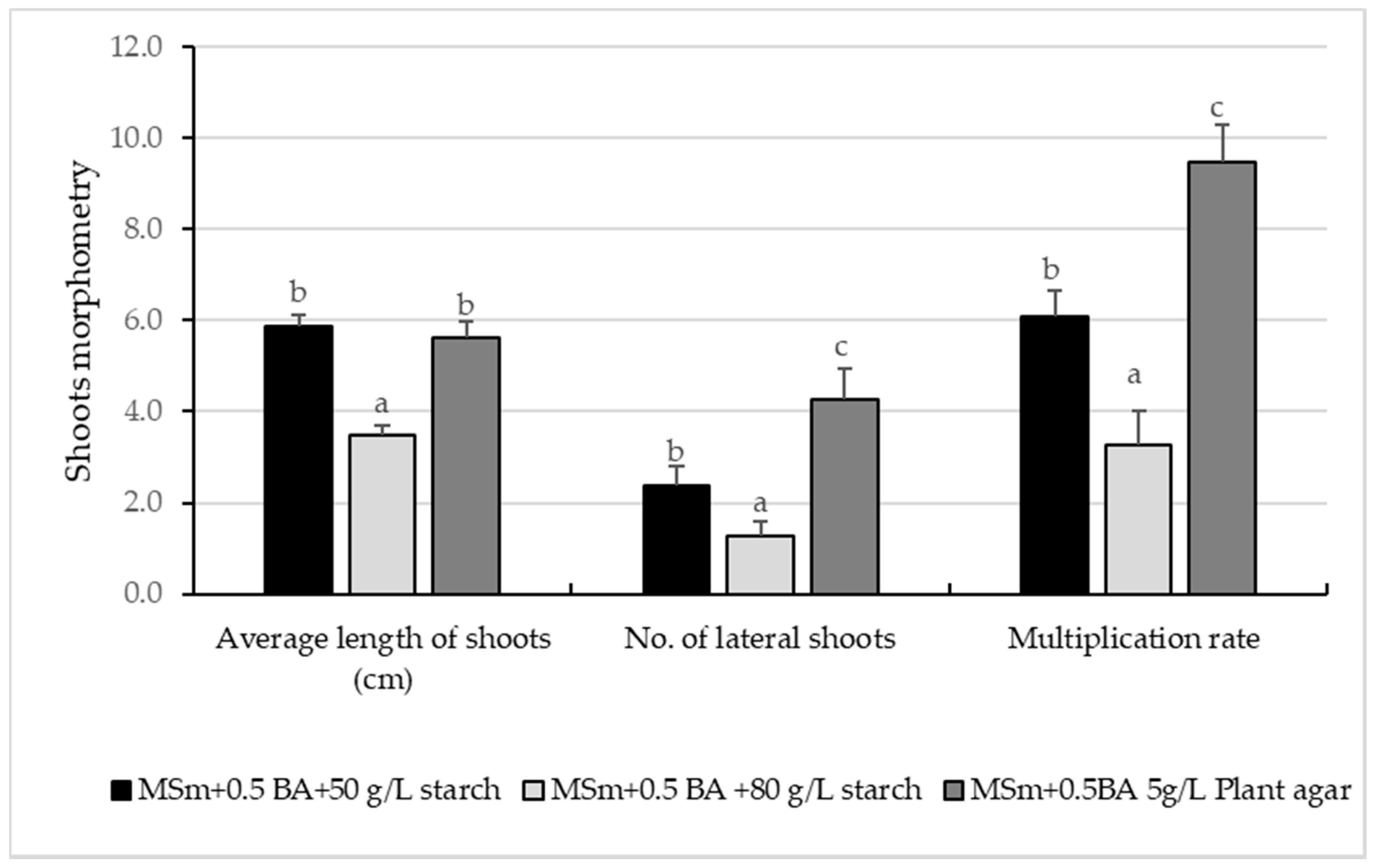
Thus, the mean value of the multiplication rate on the MSm medium supplemented with 0.5 mg L−1 BA was significantly higher (9.48 ± 0.78) compared to the values recorded (1.6 ± 0.19, 1.88 ± 0.13 and 3.2 ± 0.11) on MSm medium fortified with any of the other three types of cytokinins used, respectively, 2 mg L−1 KIN, 0.7 mg L−1 1 CPPU and 2 mg L−1 mT (Figure 1). However, there were no significant differences between the mean values of the multiplication rate in the MSm variants supplemented with KIN (1.6 ± 0.19) and 1-CPPU (1.88 ± 0.13) as shown in Figure 2.
Regarding the average length of the proliferated shoots, the shortest shoots were measured on MS medium supplemented with 2 mg L−1 KIN, with an average length of 3.01 ± 0.49 cm. The longest shoots (6.30 ± 0.34 cm) were measured on MS medium supplemented with 0.7 mg L−1 1 CPPU and 0.5 mg L−1 BA (5.25 ± 0.27 cm) without significant differences between the two mentioned variants of the culture media (Figure 1).
To test the effectiveness of gelling agents in hop Cascade cv. in vitro culture, three treatments were applied. The highest multiplication rate (9.76 ± 0.72) was recorded on MSm + 0.5 mg/L−1 BA gelled with Plant agar 5 g L−1, followed by the medium gelled with 50 g L−1 wheat starch, which provided a 6.09 ± 0.57 multiplication rate. The treatment with 80 g L−1 wheat starch reduced the multiplication rate to 3.27 ± 0.74. There were no statistically significant differences regarding shoot lengths between the treatment gelled with Plant agar and the one gelled with 50 g L−1 wheat starch (5.60 ± 0.34 cm and 5.88 ± 0.25 cm, respectively), but shoot length was significantly lower on the medium gelled with 80 g L−1 starch (3.48 ± 0.21 cm) (Figure 2).

3.2. Rooting and Acclimatization

In this study, on the MSm medium without PGRs, 86.66% of the shoots were in vitro rooted, while the average number of roots per plantlet was 6.95 ± 0.47, and the average root length was 1.13 ± 0.06 cm (Table 1).
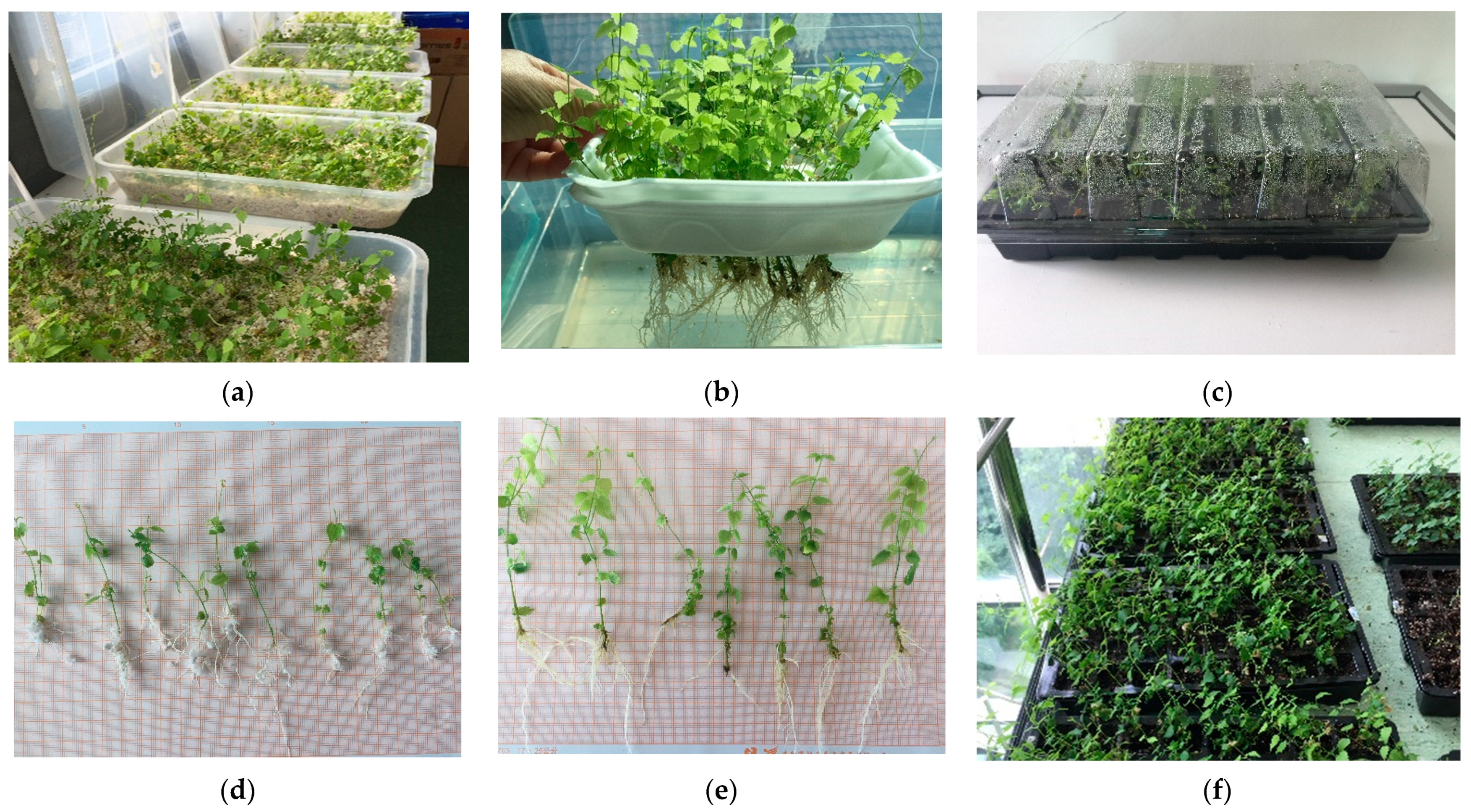
The explants cultured in vitro for rooting on MSm without PGRs generated an average number of 1.92 ± 0.08 shoots/explant and reached an average length of 5.65 ± 0.21 cm after 30 days of in vitro culture (Figure 3a). The plantlets rooted in vitro had a 98% acclimatization rate in floating hydroculture and 94% of the plants survived in greenhouse conditions (Table 1; Figure 3b,e).
The results of ex vitro rooting and acclimatization in perlite and also peat + perlite show that, although there were significant differences between the morphometric data of the shoots and roots, the highest rooting and acclimatization percentage was recorded in perlite (96.00 ± 0.60%) vs. peat + perlite (85.71 ± 0.27%), as shown in Table 1.
The shoots in vitro rooted and then acclimatized ex vitro exhibited a 94% survival rate, whereas the shoots rooted directly ex vitro and acclimatized in perlite and perlite with peat mix showed 100% survival rates in greenhouse conditions (Table 1).

3.3. Genetic Uniformity Assessment Based on RAPD and ScoT Markers

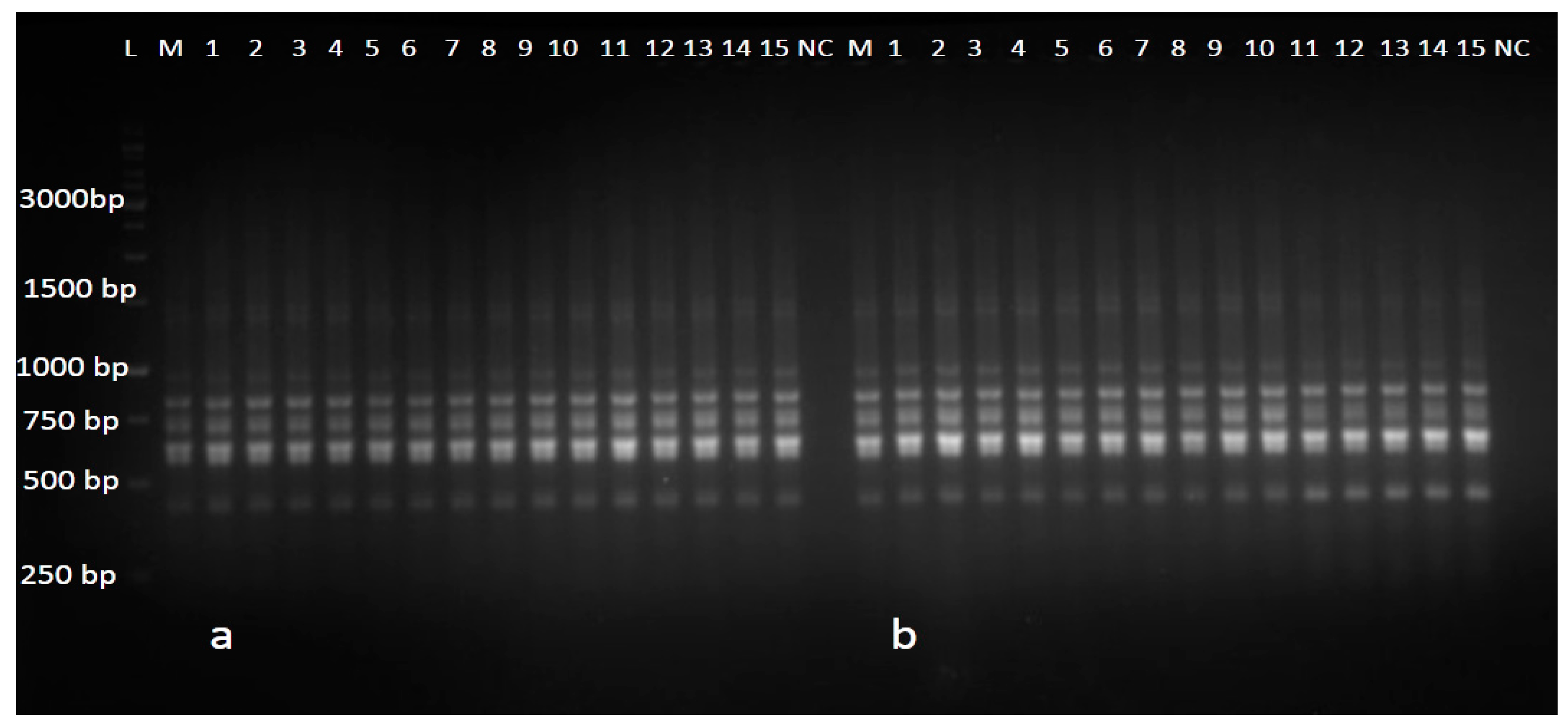
In the present study, RAPD and SCoT markers were used to assess the genetic uniformity between the selected in vitro regenerated hop plantlets and the mother plant. The 12 RAPD primers generated 66 distinct and scorable bands, with an average of 5.5 bands/primer. Each RAPD primer generated amplicons ranging in size from 305 bp (OPAB-18) to 1605 bp (OPD-16). The number of scorable monomorphic bands from each RAPD primer varied from three (OPA-04) to seven (OPX-03) (Table 2), and no polymorphism was observed between the in vitro raised plants and the mother plant (Figure 4).
SCoT primers showed amplification of 60 scorable PCR bands in the range of 354–2566 bp size (Table 2; Figure 5) with an average number of five monomorphic PCR bands per SCoT primer. The lowest number of bands (three) was recorded with primer SCoT2 and the highest number of bands (six) was obtained with the SCoT4, SCoT7, SCoT8 and SCoT25 primers, as presented in Table 2.

4. Discussion

Tissue culture techniques have been reported for a wide range of economically important species and have opened up the prospect of disease-free and genetically uniform plant micropropagation, key features for the large-scale production of industrial crops and by-products [35,36,37,38,39,40].
As in many crops, the availability and quality of hop planting material are considered major limiting factors for industrial exploitation [2]. In this context, rapid and efficient in vitro regeneration methods, which minimize somaclonal variation, are essential to ensure the mass propagation of commercial varieties.
Previous studies have shown that MS medium is favorable for hop cultivars in in vitro culture [26,27]. Based on the preliminary results from the scientific literature, for the elaboration of this micropropagation protocol (including all in vitro stages) the modified MS medium has been chosen for this study.

4.1. In Vitro Culture Initiation and Proliferation of Shoots

The major and most frequent problem of in vitro culture initiation is the increased risk of contamination of the explants, especially those harvested and prepared from underground organs [41,42]. Thus, the success of in vitro culture initiation largely depends on the use of non-contaminated juvenile plant material [43].
Previous findings have shown that the regeneration potential of hop cultivars in micropropagation systems has been achieved through the use of various explants, mainly shoots [27,44], as well as other plant organs [45,46,47,48].
Roy et al. [27] performed an efficient in vitro establishment stage for H138 hop variety (96.6% of viable explants) on modified MS medium supplemented with 0.57 µM indoleacetic acid (IAA) and 2.22 µM 6-benzylaminopurine (BA), but the efficiency of culture initiation decreased significantly (only 43.9% of the explants showed proliferation of axillary buds) when modified MS medium was supplemented with 0.54 µM naphthaleneacetic acid (NAA) and 2.22 µM BA. In this study, the single-node micro-cuttings inoculated in the initiation stage on MSm supplemented only with cytokinins, such as BA in a concentration of 0.5 mg L−1 (2.22 µM), generated 76% viable explants, exhibiting, thus, a high regenerative capacity.
One of the well-known effects of the high concentration of cytokinins in the medium is the reduction in the length of in vitro proliferated shoots. In contrast, low cytokinin concentrations have a stimulating effect on the growth of shoots individually or with a variable number of lateral shoots [49].
In this study, after analyzing the growth parameters, it was found that the type of cytokinin added in the medium and its concentration had a significant effect on Cascade cv. shoot proliferation.
Noticeably, KIN generated an unfavorable response on the average number of regenerated shoots/multinodal-explants and average shoot length in the proliferation stage of the Cascade cv. (see Figure 1). Our results are consistent with those reported by [44] in a wild variety of Iranian H. lupulus six-weeks proliferated on MS+ 2 mg L−1 KIN (1.5 ± 0.2 proliferated shoots/explant).
N-(2-Chloro-4-pyridyl)-N′-phenylurea (CPPU) is a highly active cytokinin-like plant growth regulator that promotes chlorophyll biosynthesis, cell division and cell expansion. Consequently, in addition to this study, CPPU were used in the proliferation stage of other micropropagated species [50,51,52] to regenerate shoots with a higher quality for acclimatization. For example, CPPU added in culture medium in the proliferation stage in Rubus fruticosus [53] and Lonicera kamtschatica [54] generated a higher number of shoots compared to BA. In contrast, BA added in the proliferation medium of H. lupulus Cascade cv. showed the best stimulatory effect on the number and length of shoots in comparison with CPPU.
Several species have been successfully propagated in vitro in the presence of various natural and synthetic cytokinins, derivatives of purine and urea. Meta-topoline (mT) was successfully used to replace 6-benzylaminopurine (BA) in the micropropagation of some species [55,56] but the results of this study showed that in the case of hop plants, mT generated a lower multiplication rate than BA (Figure 1). Following the testing of the four cytokinins it was highlighted that 6-benzylaminopurine, the growth regulator most commonly used in the propagation of plants, proved to be the most beneficial in the case of in vitro H. lupulus Cascade cv. culture. Moreover, on the MSm medium supplemented with 0.5 mg L−1 BA, no callus induction or morphological somaclonal variations were observed (supplementary Figure S1). Interestingly, BA-derived shoots were transferable to the rooting stage without the elongation stage requirement, as noted by [27] in a study of in vitro hop culture.

4.2. Rooting and Acclimatization

Gelling agents, especially Plant agar is a relatively expensive component of in vitro culture media, and some cheaper alternatives were found with beneficial effects in the micropropagation process of some species [57]. Wheat starch, for example, has been used in Goji berry and blackberry in vitro culture and provided not only higher multiplication rates as compared to media gelled with Plant agar but also a high quality of the regenerated shoots [58,59].
Even considering that the multiplication rate was slightly lower on the culture medium gelled with 50 g L−1 wheat starch, the length and quality of the shoots were similar to those obtained on the medium gelled with Plant agar, thus, the use of wheat starch as a gelling agent can be an alternative for the micropropagation of hop plants (Figure S1d). This finding is consistent with the results obtained by [60], who developed an in vitro micropropagation system for Czech hop mericlones (no. 521 6 of the cv. Osvald 72 and no. 6908, cv. Sladek.) using culture media gelled with 50 g L−1 starch + 0.5 g L−1 Gelrite. The use of these media resulted in the lower cost of micropropagation of healthy hop cultures without problems related to vitrification.
In this study, the MSm medium without PGRs was a good option for in vitro rooting of hop cv. Cascade. The plantlets rooted in vitro had 98% acclimatization rate in floating hydroculture and 94% of the plants survived in greenhouse conditions (Table 1). Contrary to our findings, [27] stated that on half-strength MS medium without PGRs, H138 cv. hop were not rooted, whereas in MS medium supplemented with 5.71 µM IAA + 2.46 µM IBA a high rooting percentage was obtained (86.5).
Similar results for the acclimatization of in vitro rooted shoots were reported for hop cultivars such as Tetnanger, Bor and Sladek transferred ex vitro into two substrates, Biona 112 and perlite. The acclimatization percentages were between 98% and 100%. The height of the plantlets was greater on substrate Biona 112, but the root systems were better developed in perlite [61].
From an economical point of view, in vitro rooting requires 35–75% of the total costs of micropropagation [62]. For this reason, commercial micropropagation labs often try to eliminate this stage from the production chain by combining ex vitro rooting and acclimatization in one stage [63,64,65]. This major change not only reduces plant production costs and time but also improves plant survival [66]. Moreover, the ex vitro rooted plants have well-developed root systems, which leads to better establishment and anchoring of plants when transferred in field conditions [67].
The acclimatization and concomitant rooting in one stage proposed in this study for hop plants does not require acclimatization spaces equipped with an artificial fog system because the acclimatization of the vitro plants was performed in trays covered with transparent plastic lids or in mini-greenhouses kept in rooms with a temperature of 23 ± 2 °C.
It is worth mentioning that previous micropropagation protocols for different hop varieties were established starting from different explants and based on the intermediate formation of the callus, which makes the regeneration prone to undesirable somaclonal variations [21,60], in contrast to the results of this research when plants were proliferated by the direct organogenesis of shoots.
To sum up, our findings indicate that the efficiency of this newly developed and low-cost micropropagation protocol for Humulus lupulus L. is provided by skipping a high-cost and time consuming step of the micropropagation process, namely, in vitro rooting, including, thus, only three major stages as follows: 1—the establishment of in vitro cultures on MSm medium with 0.5 mg L−1 BA gelled with Plant agar; 2—in vitro multiplication on MSm medium with 0.5 mg L−1 BA gelled with Plant agar or 50 g/L−1 wheat starch; 3—ex vitro rooting and acclimatization in perlite. This lack of the in vitro rooting stage replaced by ex vitro rooting frames a shorter and more efficient micropropagation system for Humulus lupulus L, which could easily be adapted for commercial, large-scale plant production purposes.

4.3. Genetic Uniformity Assessment Based on RAPD and ScoT Markers

Assessing the genetic uniformity of in vitro regenerants is very important if true-to-type plants are the desired end product for industrial crops. In this context, PCR-based molecular markers has been used successfully to evaluate the genetic stability of micropropagated plants with the mother plant [68,69,70,71,72,73].
Several research articles have already been published on the detection of genetic fidelity using RAPD markers in different crops such as Acacia mangium [74], Simmondsia chinensis [75], Ocimum gratissimum [76] Stevia rebaudiana [77], Origanum majorana [78] and Salvia hispanica [79]. It is worth mentioning that RAPD analysis is an easy-access technique for different laboratories but, usually, the reproducibility of the results are not satisfactory [80,81,82,83]. Therefore, SCoT markers, which are reproducible markers and are based on the short conserved region in plant genes surrounding the ATG translation start codon [84], have been used in this study to validate the RAPD results.
As far as we know, this study is the first report regarding the application of SCoT markers, which assesses the genetic uniformity of micropropagated plants of Humulus lupulus L. The advantage of using SCoT markers is that, unlike RAPD or ISSR which are based on non-coding regions of the genome, SCoT markers are correlated to functional genes as well as their corresponding traits [85]. In addition, being associated with the initiation codon, SCoT markers are abundant in the genome and can provide useful genetic information [86].
The results of this study confirm the true-to-type nature of the regenerated Humulus lupulus L. cv. Cascade plants with the mother plant and their lack of susceptibility to somaclonal variation. For practical uses, the techniques involved in the genetic comparison of micropropagated plantlets with mother plants can constitute a valuable informative tool to transfer genetically uniform plants to the field for their successful establishment [22].

5. Conclusions

The results of this research indicate that the micropropagated plants, according to the newly established protocol, were stable and genetically uniform as compared to their mother plant. Therefore, the production of hop planting material by using this micropropagation protocol has a great economic potential to obtain virus-free plants and to encourage the growth of Cascade or any other hop varieties on a large scale.

Supplementary Materials

The following are available online at https://www.mdpi.com/article/10.3390/agronomy11112268/s1, Figure S1: In vitro propagation of Humulus lupulus, cv. Cascade: (a) In vitro culture initiation on MSm + 0.5 mg L−1 BA; (b) In vitro shoot proliferation on medium MSm + 0.5 mg L−1 BA gelled with 5g L−1 Plant agar after 8 weeks of incubation; (c) In vitro shoot proliferation on MSm + 2 mg L−1 mT gelled with 5g L−1 Plant agar after 8 weeks of incubation; (d) In vitro shoot proliferation on MSm + 0.5 mg L−1 BA gelled with 50 g wheat starch after 8 weeks of incubation; (e) In vitro rooting of shoots cultured on hormone-free MSm; (f) Clusters of proliferated shoots.

Author Contributions

Conceptualization, D.C. and M.H.; methodology, D.C. (in vitro culture) and M.H. (genetic fidelity); investigation, D.C. and M.H.; resources, D.C.; data curation, D.C. and M.H.; writing—original draft preparation, D.C; writing—review and editing, M.H.; project administration, D.C.; funding acquisition, D.C. All authors have read and agreed to the published version of the manuscript.

Funding

This research was funded by a grant of the Romanian National Authority for Scientific Research and Innovation, CNCS/CCCDI—UEFISCDI, project number PN-III-P2-2.1-PTE-2019-0670, with-in PNCDI III.

Data Availability Statement

Not applicable.

Conflicts of Interest

The authors declare no conflict of interest.

References

  1. Small, E. A numerical and nomenclatural analysis of morpho-geographic taxa of Humulus. Syst. Bot. 1978, 3, 37–46. Available online: www.jstor.org/stable/2418532 (accessed on 23 August 2021). [CrossRef]
  2. Fortes, A.M.; Santos, F.; Pais, M.S. Organogenic nodule formation in hop: A tool to study morphogenesis in plants with biotechnological and medicinal applications. J. Biomed. Biotechnol. 2010, 13, 583691. [Google Scholar] [CrossRef]
  3. Rossini, F.; Virga, G.; Loreti, P.; Iacuzzi, N.; Ruggeri, R.; Provenzano, M.E. Hops (Humulus lupulus L.) as a novel multipurpose crop for the Mediterranean region of Europe: Challenges and opportunities of their cultivation. Agriculture 2021, 11, 484. [Google Scholar] [CrossRef]
  4. Magalhães, P.J.; Carvalho, D.O.; Cruz, J.M.; Guido, L.F.; Barros, A.A. Fundamentals and health benefits of xanthohumol, a natural product derived from hops and beer. Nat. Prod. Commun. 2009, 4, 591–610. [Google Scholar] [CrossRef] [PubMed] [Green Version]
  5. Carbone, K.; Macchioni, V.; Petrella, G.; Cicero, D.O. Exploring the potential of microwaves and ultrasounds in the green extraction of bioactive compounds from Humulus lupulus for the food and pharmaceutical industry. Ind. Crops Prod. 2020, 156, 112888. [Google Scholar] [CrossRef]
  6. Chadwick, L.R.; Pauli, G.F.; Farnsworth, N.R. The pharmacognosy of Humulus lupulus L. (hops) with an emphasis on estrogenic properties. Phytomedicine 2006, 13, 119–131. [Google Scholar] [CrossRef] [PubMed] [Green Version]
  7. Keiler, A.; Zierau, O.; Kretzschmar, G. Hop extracts and hop substances in treatment of menopausal complaints. Planta Med. 2013, 79, 576–579. [Google Scholar] [CrossRef] [PubMed] [Green Version]
  8. Milligan, S.; Kalita, J.C.; Pocock, V.; Van de Kauter, V.; Stevens, J.F.; Deinzer, M.L.; Rong, H.; De Keukeleire, D. The endocrine activities of 8-prenylnaringenin and related hop (Humulus lupulus L.) flavonoids. J. Clin. Endocrinol. Metab. 2000, 85, 4912–4915. [Google Scholar] [CrossRef] [PubMed]
  9. Tyrrell, E.; Archer, R.; Tucknott, M.; Colston, K.; Pirianov, G.; Ramanthan, D.; Dhillon, R.; Sinclair, A.; Skinner, G.A. The synthesis and anticancer effects of a range of natural and unnatural hop bacids on breast cancer cells. Phytochem. Lett. 2012, 5, 144–149. [Google Scholar] [CrossRef]
  10. Yong, W.K.; Ho, Y.F.; Abd Malek, S.N. Xanthohumol induces apoptosis and S phase cell cycle arrest in A549 non-small cell lung cancer cells. Pharmacog. Mag. 2015, 11, S275–S283. [Google Scholar] [CrossRef] [Green Version]
  11. Negrão, R.; Duarte, D.; Costa, R. Isoxanthohumol modulates angiogenesis and inflammation via vascular endothelial growth factor receptor, tumor necrosis factor alpha and nuclear factor kappa B pathways. BioFactors 2013, 39, 608–622. [Google Scholar] [CrossRef] [PubMed]
  12. Bland, J.S.; Minich, D.; Lerman, R.; Darland, G.; Lamb, J.; Tripp, M.; Grayson, N. Isohumulones from hops (Humulus lupulus) and their potential role in medical nutrition therapy. PharmaNutrition 2015, 3, 46–52. [Google Scholar] [CrossRef] [Green Version]
  13. Bocquet, L.; Sahpaz, S.; Rivière, C. An overview of the antimicrobial properties of hop. In Natural Antimicrobial Agents. Sustainable Development and Biodiversity; Mérillon, J.M., Riviere, C., Eds.; Springer: New York, NY, USA, 2018; Volume 19, pp. 31–54. [Google Scholar] [CrossRef]
  14. Hege, M.; Jung, F.; Sellmann, C.; Jin, C.; Ziegenhardt, D.; Hellerbrand, C.; Bergheim, I. An iso-α-acid-rich extract from hops (Humulus lupulus) attenuates acute alcohol-induced liver steatosis in mice. Nutrition 2018, 45, 68–75. [Google Scholar] [CrossRef] [PubMed]
  15. Ponticelli, M.; Russo, D.; Faraone, I.; Sinisgalli, C.; Labanca, F.; Lela, L.; Milella, L. The promising ability of Humulus lupulus L. Iso-α-acids vs. diabetes, inflammation, and metabolic syndrome: A systematic review. Molecules 2021, 26, 954. [Google Scholar] [CrossRef] [PubMed]
  16. Moir, M. Hops—A Millennium Review. J. Am. Soc. Brew. Chem. 2000, 58, 131–146. [Google Scholar] [CrossRef]
  17. DeLyser, D.Y.; Kasper, W.J. Hopped beer: The case for cultivation. Econ. Bot. 1994, 48, 166–170. [Google Scholar] [CrossRef]
  18. Faivre, C.; Ghedira, K.; Goetz, P.; Lejeune, R.; Staub, H. Humulus lupulus L. Phytothérapie 2007, 5, 86–89. [Google Scholar] [CrossRef]
  19. Almeida, A.D.R.; Maciel, M.V.D.O.B.; Machado, M.H.; Bazzo, G.C.; de Armas, R.D.; Vitorino, V.B.; Vitali, L.; Block, J.M.; Barreto, P.L.M. Bioactive compounds and antioxidant activities of Brazilian hop (Humulus lupulus L.) extracts. Int. J. Food Sci. Tech. 2020, 55, 340–347. [Google Scholar] [CrossRef]
  20. Mongelli, A.; Rodolfi, M.; Ganino, T.; Marieschi, M.; Caligiani, A.; Dall’Asta, C.; Bruni, R. Are Humulus lupulus L. ecotypes and cultivars suitable for the cultivation of aromatic hop in Italy? A phytochemical approach. Ind. Crops Prod. 2016, 83, 693–700. [Google Scholar] [CrossRef]
  21. Machado, M.P.; Gomes, E.N.; Francisco, F.; Bernert, A.F.; Bespalhok Filho, J.C.; Deschamps, C. Micropropagation and establishment of Humulus lupulus L. plantlets under field conditions at Southern Brazil. J. Agric. Sci. 2018, 10, 275. [Google Scholar] [CrossRef] [Green Version]
  22. Rajput, B.S.; Jani, M.; Ramesh, K.; Manokari, M.; Jogam, P.; Allini, V.R.; Kher, M.M.; Shekhawat, M.S. Large-scale clonal propagation of Bambusa balcooa Roxb.: An industrially important bamboo species. Ind. Crops Prod. 2020, 157, 112905. [Google Scholar] [CrossRef]
  23. Gurriarán, M.J.; Revilla, M.A.; Tamés, R.S. Adventitious shoot regeneration in cultures of Humulus lupulus L. (hop) cvs. Brewers gold and Nugget. Plant Cell Rep. 1999, 18, 1007–1011. [Google Scholar] [CrossRef]
  24. Reed, B.M.; Okut, N.; D’Achino, J.; Narver, L.; DeNoma, J. Cold storage and cryopreservation of hops (Humulus L.) shoot cultures through application of standard protocols. CryoL Lett. 2003, 24, 389–396. [Google Scholar]
  25. Postman, J.D.; DeNoma, J.S.; Reed, B.M. Detection and elimination of viruses in USDA hop (Humulus lupulus) germoplasm collection. Acta Hortic. 2005, 668, 143–148. [Google Scholar] [CrossRef]
  26. Fortes, A.M.; Pais, M.S. Organogenesis from internode-derived nodules of Humulus lupulus var. Nugget (Cannabinaceae): Histological and changes in the starch content. Am. J. Bot. 2000, 87, 971–979. [Google Scholar] [CrossRef] [PubMed]
  27. Roy, A.T.; Leggett, G.; Koutoulis, A. Development of a shoot multiplication system for hop (Humulus lupulus L.). Vitr. Cell Dev. Biol. Plant 2001, 37, 79–83. [Google Scholar] [CrossRef]
  28. Murashige, T.; Skoog, A. A revised medium for rapid growth and bioassay with tobacco tissue culture. Physiol. Plant 1962, 15, 473–497. [Google Scholar] [CrossRef]
  29. Clapa, D.; Fira, A.; Joshee, N. An efficient ex vitro rooting and acclimatization method for horticultural plants using float hydroculture. HortScience 2013, 48, 1159–1167. [Google Scholar] [CrossRef] [Green Version]
  30. Lodhi, M.A.; Guang-Ning, Z.; Weeden, F.N.F.; Reisch, B.I. A simple and efficient method for DNA extraction from grapevine cultivars and Vitis species. Plant Mol. Biol. Rep. 1994, 12, 6–13. [Google Scholar] [CrossRef]
  31. Pop, R.; Ardelean, M.; Pamfil, D.; Gaboreanu, I.M. The efficiency of different DNA isolation and purification in ten cultivars of Vitis vinifera. Bull. UASVM Anim. Sci. Biotechnol. 2003, 59, 259–261. [Google Scholar]
  32. Bodea, M.; Pamfil, D.; Pop, R.; Sisea, R.C. DNA isolation from desiccated leaf material from plum tree (Prunus domestica L.) molecular analysis. Bull. UASVM Hortic. 2016, 1, 1–2. [Google Scholar] [CrossRef] [Green Version]
  33. Williams, J.G.; Kubelik, A.R.; Livak, K.J.; Rafalski, J.A.; Tingey, S.V. DNA polymorphisms amplified by arbitrary primers are useful as genetic markers. Nucleic Acids Res. 1990, 18, 6531–6535. [Google Scholar] [CrossRef] [Green Version]
  34. Pop, R.; Has, I.; Pop, I.F.; Harta, M.; Pamfil, D. Genetic Diversity Analysis of Maize Inbred Lines from SCDA Turda-Romania Revealed by RAPD Molecular Markers. Bull. Univ. Agric. Sci. 2011, 68, 277–283. [Google Scholar]
  35. Grigoriadou, K.; Leventakis, N. Large scale commercial production of potato minitubers, using in vitro techniques. Potato Res. 1999, 42, 607–610. [Google Scholar] [CrossRef]
  36. Volk, T.A.; Verwijst, T.; Tharakan, P.J.; Abrahamson, L.P.; White, E.H. Growing Fuel: A Sustainability Assessment of Willow Biomass Crops. Front. Ecol. Environ. 2004, 2, 411. [Google Scholar] [CrossRef]
  37. Grudzinska, M.; Solarska, E.; Czubacka, A.; Przybys, M.; Fajbus, A. Elimination of Hop Latent Viroid from Hop plants by cold treatment and meristem tip culture. Phytopathol. Pol. 2006, 40, 21–30. [Google Scholar]
  38. Al-Taleb, M.M.; Hassawi, D.S.; Abu-Romman, S.M. Production of virus free potato plants using meristem culture from cultivars grown under Jordanian environment. J. Agric. Environ. Sci. 2011, 11, 467–472. [Google Scholar]
  39. Azeez, H.; Ibrahim, K.; Pop, R.; Pamfil, D.; Hârţa, M.; Bobiș, O. Changes induced by gamma ray irradiation on biomass production and secondary metabolites accumulation in Hypericum triquetrifolium Turra callus cultures. Ind. Crops Prod. 2017, 108, 183–189. [Google Scholar] [CrossRef]
  40. Adhikary, D.; Kulkarni, M.; El-Mezawy, A.; Mobini, S.; Elhiti, M.; Gjuric, R.; Ray, A.; Polowick, P.; Slaski, J.J.; Jones, M.P.; et al. Medical cannabis and industrial hemp tissue culture: Present status and future potential. Front. Plant Sci. 2021, 12, 275. [Google Scholar] [CrossRef]
  41. Singh, V.; Tyagi, A.; Chauhan, P.K.; Kumari, P.; Kaushal, S. Identification and prevention of bacterial contamination on explant used in plant tissue culture labs. Int. J. Pharm. Pharm. Sci. 2011, 3, 160–163. [Google Scholar]
  42. Obisesan, I.A.; Sakpere, A.M.; Amujoyegbe, B.J.; Akinropo, M.S. Explants selection for in vitro propagation of Pachyrhizus erosus L. Not. Sci. Biol. 2021, 13, 10844. [Google Scholar] [CrossRef]
  43. Singh, C.R. Review on problems and its remedy in plant tissue culture. Asian J. Biol. Sci. 2018, 11, 165–172. [Google Scholar] [CrossRef] [Green Version]
  44. Mafakheri, M.; Hamidoghli, Y. Micropropagation of hop (Humulus lupulus L.) via shoot tip and node culture. Acta Hortic. 2019, 1236, 31–36. [Google Scholar] [CrossRef]
  45. Batista, D.; Ascensão, L.; Sousa, M.J.; Pais, M.S. Adventitious shoot mass production of hop (Humulus lupulus L.) var. Eroica in liquid medium from organogenic nodule cultures. Plant Sci. 2000, 151, 47–57. [Google Scholar] [CrossRef]
  46. Horlemann, C.; Schwekendiek, A.; Höhnle, M.; Weber, G. Regeneration and Agrobacterium-mediated transformation of hop (Humulus lupulus L.). Plant Cell Rep. 2003, 22, 210–217. [Google Scholar] [CrossRef]
  47. Faragó, J.; Pšenáková, I.; Faragová, N. The use of biotechnology in hop (Humulus lupulus L.) improvement. Nova Biotechnol. 2009, 9, 279–293. [Google Scholar]
  48. Trojak-Goluch, A.; Czarnecka, D. The effects of explant source and hormone content on plant regeneration and induction of tetraploids in Humulus lupulus L. Vitr. Cell. Dev. Biol. Plant 2015, 51, 152–159. [Google Scholar] [CrossRef]
  49. Gaspar, T.; Kevers, C.; Faivre-Rampant, O.; Cre’vecoeur, M.; Penel, C.; Greppin, H.; Dommes, J. Changing concepts in plant hormone action. Vitr. Cell. Dev. Biol. Plant 2003, 39, 85–106. [Google Scholar] [CrossRef]
  50. Faisal, M.; Alatar, A.A.; Abdel-Salam, E.M.; Qahtan, A.A. Effects of 4-CPPU on in vitro multiplication and sustainable generation of Hibiscus rosa-sinensis L.‘White Butterfly’. Saudi J. Biol. Sci. 2020, 27, 412–416. [Google Scholar] [CrossRef]
  51. Li, Q.; Yu, P.; Lai, J.; Gu, M. Micropropagation of the potential blueberry rootstock—Vaccinium arboreum through axillary shoot proliferation. Sci. Hortic. 2021, 280, 109908. [Google Scholar] [CrossRef]
  52. San José, M.C.; Cernadas, M.J.; Janeiro, L.V. Optimization of Micropropagation Protocols in Some Woody Plants Using Meta-topolin. In Meta-Topolin: A Growth Regulator for Plant Biotechnology and Agriculture; Springer: Singapore, 2021; pp. 221–240. [Google Scholar] [CrossRef]
  53. Vescan, L.A.; Clapa, D.; Fira, A.; Pamfil, D. Micropropagation of cold resistant blackberry cultivar ‘Gazda’. Bull. USAMV Anim. Sci. Biotechnol. 2012, 69, 282–289. [Google Scholar]
  54. Fira, A.; Clapa, D.; Cristea, V.; Plopa, C. In vitro propagation of Lonicera kamtschatica. Agric. Sci. Pract. 2014, 1–2, 89–90. [Google Scholar]
  55. Gentile, A.; Gutiérrez, M.J.; Martinez, J.; Frattarelli, A.; Nota, P.; Caboni, E. Effect of meta-Topolin on micropropagation and adventitious shoot regeneration in Prunus rootstocks. Plant Cell Tiss. Organ. Cult. 2014, 118, 373–381. [Google Scholar] [CrossRef]
  56. Gentile, A.; Frattarelli, A.; Nota, P.; Condello, E.; Caboni, E. The aromatic cytokinin meta-topolin promotes in vitro propagation, shoot quality and micrografting in Corylus colurna L. Plant Cell Tiss. Organ. Cult. 2017, 128, 693–703. [Google Scholar] [CrossRef]
  57. Fira, A.; Magyar-Tábori, K.; Hudák, I.; Clapa, D.; Dobránszky, J. Effect of gelling agents on in vitro development of Amelanchier canadensis ‘Rainbow Pillar’. Int. J. Hortic. Sci. 2013, 19, 75–79. [Google Scholar] [CrossRef] [Green Version]
  58. Fira, A.; Clapa, D.; Simu, M. Studies regarding the micropropagation of some blackberry cultivars. Bull. UASVM Hortic. 2014, 71, 22–37. [Google Scholar] [CrossRef]
  59. Fira, A.; Joshee, N.; Cristea, V.; Simu, M.; Hârța, M.; Pamfil, D.; Clapa, D. Optimization of Micropropagation Protocol for Goji Berry (Lycium barbarum L). Bull. UASVM Hortic. 2016, 73, 141–150. [Google Scholar] [CrossRef] [Green Version]
  60. Smýkalová, I.; Ortová, M.; Lipavská, H.; Patzak, J. Efficient in vitro micropropagation and regeneration of Humulus lupulus on low sugar, starch-Gelrite media. Biol. Plant 2001, 44, 7–12. [Google Scholar] [CrossRef]
  61. Kastrytskaya, M.S.; Kukharchyk, N.V.; Hashenka, O.A. Acclimatization of hop cultivars after in vitro propagation. Soil Sci. Agrochem. 2014, 1, 370–379. [Google Scholar]
  62. Hussain, A.; Ahmed, I.; Nazir, H.; Ullah, I. Plant Tissue Culture: Current Status and Opportunities. In Recent Advances in Plant In Vitro Culture; Chapter 1; IntechOpen: London, UK, 2012; pp. 1–28. [Google Scholar] [CrossRef]
  63. Debergh, P.C.; Maene, L.J. A scheme for commercial propagation of ornamental plants by tissue culture. Sci. Hortic. 1981, 14, 335–345. [Google Scholar] [CrossRef]
  64. Hazarika, B.N. Acclimatization of tissue-cultured plants. Curr. Sci. 2003, 85, 1704–1712. Available online: http://www.jstor.org/stable/24109975 (accessed on 2 September 2021).
  65. Wozny, A.; Miler, N. LEDs application in ex vitro rooting and acclimatization of chrysanthemum (Chrysanthemum x grandiflorum/Ramat/Kitam). Electron. J. Pol. Agric. Univ. 2016, 19, 1–8. Available online: http://www.ejpau.media.pl/volume19/issue4/art-02.html (accessed on 4 September 2021).
  66. Singh, A.; Agarwal, P.K. Enhanced micropropagation protocol of ex vitro rooting of a commercially important crop plant Simmondsia chinensis (Link) Schneider. J. For. Sci. 2016, 62, 107–115. [Google Scholar] [CrossRef] [Green Version]
  67. Dinesh, R.M.; Patel, A.K.; Vibha, J.B.; Shekhawat, S.; Shekhawat, N.S. Cloning of mature pomegranate (Punica granatum) cv. Jalore seedless via in vitro shoot production and ex vitro rooting. Vegetos 2019, 32, 181–189. [Google Scholar] [CrossRef]
  68. Goto, S.; Thakur, R.C.; Ishii, K. Determination of genetic stability in long-term micropropagated shoots of Pinus thunbergii Parl. using RADP markers. Plant Cell Rep. 1998, 18, 193–197. [Google Scholar] [CrossRef] [PubMed]
  69. Tiwari, J.K.; Chandel, P.; Gupta, S.; Gopal, J.; Singh, B.P.; Bhardwaj, V. Analysis of genetic stability of in vitro propagated potato microtubers using DNA markers. Physiol. Mol. Biol. Plants 2013, 19, 587–595. [Google Scholar] [CrossRef] [Green Version]
  70. Saha, S.; Adhikari, S.; Dey, T.; Ghosh, P. RAPD and ISSR based evaluation of genetic stability of micropropagated plantlets of Morus alba L. variety S-1. Meta Gene 2016, 7, 7–15. [Google Scholar] [CrossRef]
  71. Thakur, J.; Dwivedi, M.D.; Sourabh, P.; Uniyal, P.L.; Pandey, A.K. Genetic homogeneity revealed using SCoT, ISSR and RAPD markers in micropropagated Pittosporum eriocarpum royle—An endemic and endangered medicinal plant. PLoS ONE 2016, 11, e0159050. [Google Scholar] [CrossRef] [PubMed] [Green Version]
  72. Sharma, U.; Rai, M.K.; Shekhawat, N.S.; Kataria, V. Genetic homogeneity revealed in micropropagated Bauhinia racemosa Lam. using gene targeted markers CBDP and SCoT. Physiol. Mol. Biol. Plants 2019, 25, 581–588. [Google Scholar] [CrossRef]
  73. Tikendra, L.; Koijam, A.S.; Nongdam, P. Molecular markers based genetic fidelity assessment of micropropagated Dendrobium chrysotoxum Lindl. Meta Gene 2019, 20, 100562. [Google Scholar] [CrossRef]
  74. Nanda, R.M.; Das, P.; Rout, G.R. In vitro clonal propagation of Acacia mangium Willd and its evaluation of genetic stability through RAPD marker. Ann. For. Sci. 2004, 61, 381–386. [Google Scholar] [CrossRef] [Green Version]
  75. Kumar, S.; Mangal, M.; Dhawan, A.K.; Singh, N. Assessment of genetic fidelity of micropropagated plants of Simmondsia chinensis (Link) Schneider using RAPD and ISSR markers. Acta Physiol. Planta 2011, 33, 2541–2545. [Google Scholar] [CrossRef]
  76. Saha, S.; Abdul, K.; Parthadeb, G. In vitro propagation of Ocimum gratissimum L. (Lamiaceae) and its evaluation of genetic fidelity using RAPD marker. Am. J. Plant Sci. 2012, 3, 64–74. [Google Scholar] [CrossRef] [Green Version]
  77. Modi, A.R.; Patil, G.; Kumar, N.; Singh, A.S.; Subhash, N. A Simple and efficient in vitro mass multiplication procedure for Stevia rebaudiana Bertoni and analysis of genetic fidelity of in vitro raised plants through RAPD. Sugar Tech. 2012, 14, 391–397. [Google Scholar] [CrossRef]
  78. Çetin, B. Evaluation of the genetic fidelity of in vitro raised plants of Origanum majorana L. using Random Amplified Polymorphic DNA. Celal Bayar Univ. J. Sci. 2018, 14, 237–239. [Google Scholar] [CrossRef]
  79. Yadav, A.; Kothari, S.L.; Kachhwaha, S.; Joshi, A. In vitro propagation of chia (Salvia hispanica L.) and assessment of genetic fidelity using random amplified polymorphic DNA and intersimple sequence repeat molecular markers. J. Appl. Biol. Biotechnol. 2019, 7, 42–47. [Google Scholar]
  80. Penner, G.A.; Bush, A.; Wise, R.; Kim, W.; Domier, L.; Kasha, K.; Laroche, A.; Scoles, G.; Molnar, S.J.; Fedak, G. Reproducibility of random amplified polymorphic DNA (RAPD) analysis among laboratories. Genome Res. 1993, 2, 341–345. [Google Scholar] [CrossRef] [Green Version]
  81. Micheli, M.R.; Bova, R.; Pascale, E.; D’Ambrosio, E. Reproducible DNA fingerprinting with the random amplified polymorphic DNA (RAPD) method. Nucleic Acids Res. 1994, 22, 1921. [Google Scholar] [CrossRef]
  82. Jones, C.J.; Edwards, K.J.; Castaglione, S.; Winfield, M.O.; Sala, F.; Van de Wiel, C.; Bredemeijer, G.; Vosman, B.; Matthes, M.; Daly, A.; et al. Reproducibility testing of RAPD, AFLP and SSR markers in plants by a network of European laboratories. Mol. Breed. 1997, 3, 381–390. [Google Scholar] [CrossRef]
  83. Ramos, J.R.; Telles, M.P.; Diniz-Filho, J.A.; Soares, T.N.; Melo, D.B.; Oliveira, G. Optimizing reproducibility evaluation for random amplified polymorphic DNA markers. Genet. Mol. Res. 2008, 7, 1384–1391. [Google Scholar] [CrossRef]
  84. Collard, B.C.; Mackill, D.J. Start codon targeted (SCoT) polymorphism: A simple, novel DNA marker technique for generating gene-targeted markers in plants. Plant Mol. Biol Rep. 2009, 27, 86–93. [Google Scholar] [CrossRef]
  85. Xiong, F.; Zhong, R.; Han, Z.; Jiang, J.; He, L.; Zhuang, W.; Tang, R. Start codon targeted polymorphism for evaluation of functional genetic variation and relationships in cultivated peanut (Arachis hypogaea L.) genotypes. Mol. Bio. Rep. 2011, 38, 3487–3494. [Google Scholar] [CrossRef] [PubMed]
  86. Chhajer, S.; Jukanti, A.K.; Kalia, R.K. Start codon targeted (SCoT) polymorphism-based genetic relationships and diversity among populations of Tecomella undulata (Sm.) Seem-an endangered timber tree of hot arid regions. Tree Genet. Genomes 2017, 13, 84. [Google Scholar] [CrossRef]
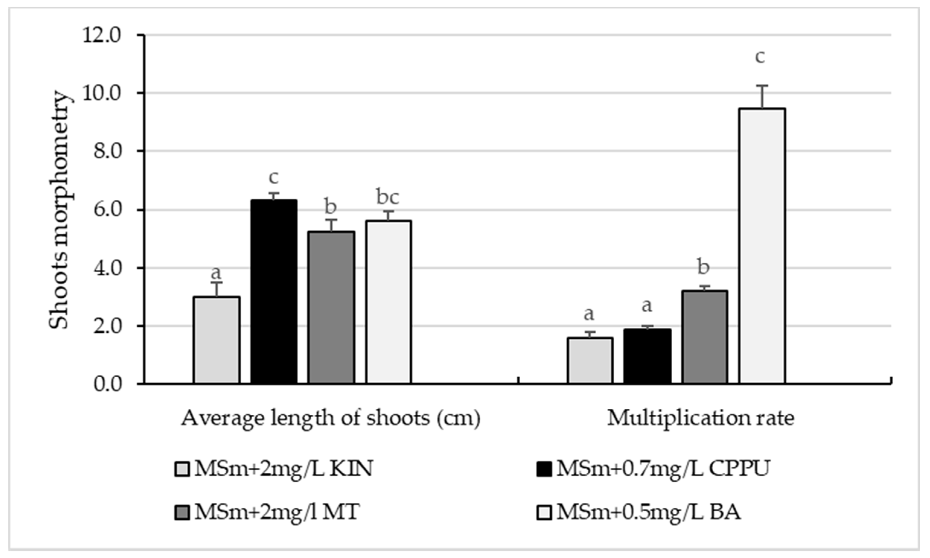
Figure 1. Effect of different types and concentrations of cytokinins on shoots’ proliferation in H. lupulus cv. Cascade after 8 weeks of in vitro culturing. Different lowercase letters above the bars indicate significant differences between the means of the same parameter according to Tukey’s HSD test (p < 0.05).
Agronomy 11 02268 g001
Figure 2. Influence of Plant agar and wheat starch on shoots’ proliferation in H. lupulus cv. Cascade. Different lowercase letters above the bars indicate significant differences between the means of the same parameter according to Tukey’s HSD test (p < 0.05).
Agronomy 11 02268 g002
Figure 3. Ex vitro acclimatization of Humulus lupulus, cv. Cascade: (a) Acclimatization in perlite. (b) Acclimatization in float hydroculture. (c) Acclimatization in covered cell trays in peat + perlite substrate (3:1, v/v). (d) Plants rooted ex vitro and acclimatized in perlite. (e) Plants rooted in vitro and subsequently acclimatized in float hydroculture. (f) Plants acclimatized in peat-based substrate.
Agronomy 11 02268 g003
Figure 4. Analysis of the genetic fidelity of micropropagated H. Lupulus cv Cascade plants with their mother plant. RAPD fingerprinting profile obtained with OPX-03 primer of in vitro (a) and ex vitro (b) rooted and raised plants. Lane L: 100 bp Ladder (Promega, USA); Lane M: DNA banding patterns of mother plant; Lane 1–15: DNA banding patterns of in vitro propagated plants.
Agronomy 11 02268 g004
Figure 5. The evaluation of genetic fidelity of micropropagated H. Lupulus cv. Cascade plants with their mother plant by SCoT markers. DNA fingerprinting profile obtained with SCoT-25 primer of in vitro (a) and ex vitro (b) rooted and raised plants. Lane L: 1 kb Ladder (Fermentas, Leon-Rot, Germany); Lane M: PCR banding patterns of mother plant DNA; Lane 1–15: PCR banding patterns of in vitro propagated plants DNA.
Agronomy 11 02268 g005
Table 1. Rooting and acclimatization data recorded in micropropagated plants of H. lupulus cv. Cascade.
Rooting MethodShoots’ and Roots’ MorphometryRooting (%)Survival under Greenhouse Conditions (%)
No.of ShootsLength of Shoots (cm)No. of RootsLength of Roots (cm)
In vitro rooting1.92 ± 0.08 c5.65 ± 0.21 b6.95 ± 0.47 b1.13 ± 0.06 b86.66 ± 0.39 a94 a
Ex vitro rooting on perlite1.55 ± 0.07 b4.89 ± 0.19 a6.56 ± 0.62 c1.29 ± 0.09 c96.00 ± 0.60 b100 b
Ex vitro rooting on peat + perlite1.35 ± 0.06 a 4.95 ± 0.15 a5.56 ± 0.52 a1.19 ± 0.19 a85.71 ± 0.27 a100 b
Values shown are means ± SE. Different lowercase letters in a column indicate significant differences between rooting methods (Tukey’s multiple comparison test, p < 0.05).
Table 2. Number and size range of amplified fragments generated by RAPD and SCoT markers in H. Lupulus cv. Cascade.
Primer CodePrimer Sequence (5′-3′)No. of Scorable Bands Size Range of Bands (bp)
OPA04AATCGCGCTG3525–1330
OPAB11GTGCGCAATG5325–1545
OPB08GTCCACACGG6220–1336
OPC08TGGACCGGTG6253–1408
OPD16AGGGCGTAAG5255–1605
OPF13GGCTGCAGAA6345–1218
OPF20GGTCTAGAGG6355–1115
OPAB18CTGGCGTGTC5305–1435
OPE14TGCGGCTGAG6410–1005
OPF16GGAGTACTGG5317–1214
OPX03TGGCGCAGTG7408–1415
OPP18GGCTTGGCCT6415–1138
Total bands -66-
SCOT1CAACAATGGCTACCACCA4554–1028
SCOT2CAACAATGGCTACCACCC3354–1464
SCOT3CAACAATGGCTACCACCG5528–2018
SCOT4CAACAATGGCTACCACCT6536–2520
SCOT5CAACAATGGCTACCACGA5514–2550
SCOT6CAACAATGGCTACCACGC5500–1578
SCOT7CAACAATGGCTACCACGG6564–2514
SCOT8CAACAATGGCTACCACGT6586–2058
SCOT9CAACAATGGCTACCAGCA5754–2566
SCoT10CAACAATGGCTACCAGCC4542–1564
SCOT16ACCATGGCTACCACCGAC5528–2012
SCOT25ACCATGGCTACCACCGGG6456–1036
Total bands-60-
Publisher’s Note: MDPI stays neutral with regard to jurisdictional claims in published maps and institutional affiliations.