Article

Potassium Positively Affects Skin Characteristics of Sweet Potato Storage Roots

1
Institute of Plant Sciences, Agricultural Research Organization, Gilat Research Center, Negev 85280, Israel
2
Institute of Plant Sciences, Agricultural Research Organization, Volcani Center, Rishon LeZion 7505101, Israel
3
Institute of Soil, Water and Environmental Sciences, Agricultural Research Organization, Gilat Research Center, Negev 85280, Israel
*
Author to whom correspondence should be addressed.
Agronomy 2020, 10(9), 1385; https://doi.org/10.3390/agronomy10091385
Submission received: 6 August 2020 / Revised: 25 August 2020 / Accepted: 10 September 2020 / Published: 14 September 2020
(This article belongs to the Section Soil and Plant Nutrition)

Abstract

:
Sweet potato (Ipomoea batatas) growth faces two critical problems: variability in storage root (SR) number and size among individual plants, and skinning injuries that render the SR susceptible to pathogen infections during storage. We hypothesized that application of potassium (K) fertilizer, an essential mineral for sweet potato, would contribute to increased yield, uniformity, and skin quality of SRs. Sweet potatoes were grown in sandy soil, which is poor in K, and in loess soil. The fertilizers potassium chloride (KCl) and polyhalite were applied before planting. Polyhalite is a hydrated sulfate of K, calcium, and magnesium that has been shown to improve potato skin appearance. Soil type was the major factor affecting SR yield—higher in sandy vs. loess soil. The K fertilizers did not affect yield in either soil type, or improve SR uniformity. However, the skin of the SRs from loess soil had more phellem layers and larger phellem cells following fertilization, mainly with KCl. Accordingly, the expression of suberin marker genes was significantly higher in mature vs. immature skin of SRs fertilized with KCl. Overall, soil type was the major factor affecting sweet potato yield, and addition of K positively affected skin morphology and related gene expression.

1. Introduction

Sweet potato (Ipomoea batatas L. Lam.) is a subtropical plant that is vegetatively propagated, with a short growth period and good production in various climates and farming systems. One of the problems in sweet potato growth is yield consistency among individual plants in the field, and uniformity of storage root (SR) size, ranging from a few huge SRs to many small ones that have no commercial value. The causes of this variability are not clear, but may involve unknown epigenetic and biochemical factors, or propagation methods that induce it [1,2,3]. Another major problem is postharvest skinning injuries of the SR periderm and exposure of the inner parenchyma tissue, leading to increased rates of moisture and weight loss, shriveling of the SR surface, increased susceptibility to pathogen attack, and unattractive appearance [4].
Potato tuber periderm provides a model system to study the sweet potato SR periderm. The periderm is a corky protective tissue of secondary origin that replaces the epidermis when the latter is damaged. The periderm is made up of three cell types: phellem, phellogen, and phelloderm [5]. The phellem (or cork) forms a series of layers at the outermost level of the periderm, and is derived from the meristematic phellogen layer (or cork cambium) below it. As phellem cells develop, they become suberized and then die, forming a protective layer termed ‘skin’. The phelloderm cells form the innermost layers of the periderm and are similarly derived from the phellogen layer, which is located directly above them. In sweet potato, the phelloderm resembles cortical cells and is usually distinguished by the radial position of the cells in reference to the phellogen [1]. The outer skin cells of sweet potato become partly lignified during growth and are progressively sloughed off. The phellogen layer remains active until harvest; its activity maintains skin thickness as the SR expands. However, phellogen activity is also responsible for the major problem of skinning injuries, as the superficial layers of the skin separate from the underlying parenchymatic tissue due to phellogen cell wall fracture [1,6].
Suberin, the corky material that accumulates in skin (phellem) cell walls, is a macromolecule containing both polyaromatic and polyaliphatic domains that is found between the primary cell wall and the plasma membrane. The aromatic domain is composed of monolignols and hydroxycinnamic (ferulic) acids and is covalently bound to the primary cell wall [7,8]. The aliphatic domain is a polyester that, upon transesterification, releases mainly C16–C28 α,ω-diacids and ω-hydroxyacids, with minor amounts of alkan-1-ols, alkanoic acids, and glycerol. The latter may be involved in crosslinking between the aromatic and aliphatic domains [9,10].
The suberin-biosynthesis pathway has not been completely resolved; it includes β-ketoacyl-coenzyme A (CoA) synthases (KCSs), fatty acyl-CoA reductases (FARs), long-chain acyl-CoA synthetases, cytochrome P450 monooxygenases, glycerol 3-phosphate acyltransferases (GPATs), and phenolic acyltransferases (reviewed in [11]). The most studied in potato are: StKCS6, which catalyzes suberin and wax compounds with chain lengths of C28 and longer [12], the cytochrome CYP86A33 which has been shown to promote the ω-hydroxylation step and its silencing, leading to a reduction in aliphatic suberin load and increased permeability of the periderm [13], and feruloyl transferase (FHT), suggested to ester link ferulic acid to ω-hydroxyacids and fatty alcohols into potato suberin [14]. Additional genes are associated with periderm initiation, development, and skin set [15,16]. Of these, ORGAN-SPECIFIC PROTEIN S2 (OSP) is a marker gene for skin maturation and skin set [15]. The sweet potato orthologs of these genes were identified and used in the present study.
There are few studies on the effect of ground fertilizers on sweet potato skin quality. The skin might be affected by better nutrition, supporting SR development, or by its direct interaction with the nutrient solution in the soil, which surrounds the SR. Our previous work showed that fertilization with minerals improves potato skin quality [17,18,19]. In light of those studies, we applied a similar approach to control the skinning problem in sweet potato, and further explored the effects of growth enhancers on SR yield and uniformity.
Two ground fertilizers were used in the present study, the mineral polyhalite and potassium chloride (KCl) salt. Polyhalite [K2Ca2Mg(SO4)4·2H2O] is a hydrated sulfate of potassium (K), calcium (Ca), and magnesium (Mg) in the relative proportions: 48% sulfur trioxide (SO3), 17% calcium oxide (CaO), 14% potassium oxide (K2O), and 6% magnesium oxide (MgO). The divalent ions, Ca and Mg, in the polyhalite, contribute to maintenance of the cell membrane, cell wall structure, and intracellular adhesion, by forming stable but reversible linkages between the pectin polar head groups in the cell wall [20,21,22,23]. Indeed, application of calcium chloride (CaCl2) has been shown to reduce the rate and severity of potato skin russeting [17]. The K in the polyhalite is considered a ‘macro’-nutrient. It is involved in several vital physiological processes, such as plant water management and osmoregulation, and is known to influence various aspects of product quality [24]. Plants with minor K deficiencies may suffer from reduced growth and productivity, while not always exhibiting visible symptoms [25]. Thus, the polyhalite provides Ca, Mg, and K as essential minerals for plant growth and skin quality. Moreover, the residual effect of the polyhalite as a fertilizer on crop growth is higher than that of the equivalent sulfate salts—K2SO4, CaSO4, and MgSO4 [26]. In accordance with the importance of K to plant growth, as already noted, observations in the field have suggested that increasing the concentration of K fertilization in potato may improve plant vigor and skin quality. Hence, KCl was the other ground fertilizer tested with sweet potato.
In the present work, we explored the effects of polyhalite and KCl fertilization on sweet potato SR yield, uniformity, and skin quality. The experiments were conducted in two soil types, sand and loess, where sweet potato is usually grown in Israel. Analyses included an anatomical study of SR skin and expression studies of skin-related genes.

2. Materials and Methods

2.1. Plant Material, Soil, Growth Management, and Fertilization Treatments

The experiments were carried out in the semiarid western Negev region of Israel, with an average annual rainfall of 200–300 mm during the winter (November–March), and dry summers. Experimental plots were located within two commercial fields with different soil types, light loess soil (loess plot) and sandy soil (sandy plot). Analysis of soil extracts before the growing season is reported in Table 1. Saturated paste soil solution extract was used for determination of electrical conductivity (EC) and nitrate (NO3), phosphorus (P), K, Ca, Mg, and Cl concentrations. Exchangeable K was determined with ammonium acetate extraction. K, Ca, and Mg were determined by an atomic absorption spectrophotometer (AAnalyst200 PerkinElmer, Waltham, MA). NO3 and P were determined using a discrete automatic chemical analyzer (Gallery Plus, Thermo Scientific, Vaata, Finland) and Cl using a chloridometer (Chloride 926, Sherwood Scientific, Cambridge, UK).
Rooted, virus-free cuttings of sweet potato cv. Georgia Jet were bought from Rahan Meristem Ltd., Israel (https://www.rahan.co.il) and were grown in a greenhouse under an Agril anti-insect cloth, from November 2017 to mid-June 2018. Planting in the commercial fields was on 24 May 2018 for the loess plot and 15 June 2018 for the sandy plot. For planting, 35 cm long sweet potato cuttings, exhibiting six large buds (primordia) were used. Four cuttings were planted per 1 m bed length; each bed consisted of two ridges with a total width of 1.93 m. Temperatures at both sites were 30–35 °C at the beginning of growth (mid-May to mid-July) and declined to 24 °C toward harvest (mid-November), 150 days after planting (DAP).
Daily irrigation was applied by sprinklers in the loess plot and by drip irrigation in the sandy plot, with water amounts that were 70% or 100% of the previous day’s evaporation rate calculated by Penman’s formula [27], respectively. During the first 100 days of growth, nitrogen (N) was applied at 100 kg ha−1 as ammonium nitrate by fertigation.
The two types of fertilizer, the polyhalite mineral and the KCl salt, were similarly applied before planting to the sandy and loess plots. Polyhalite was added in the form of Polysulphate® (http://www.polysulphate.com/fertilizing-potatoes-with-polysulphate/), a granular product (2–4 mm) produced by ICL at Boulby Mine in the UK, at 750 or 1500 kg ha−1 )100 or 200 kg ha−1 of K2O, respectively). KCl (Dead Sea Works, Tel Aviv, Israel) was applied at 350 kg ha−1 (200 kg ha−1 of K2O). Both fertilizers were incorporated into the soil manually.
The experimental plots were set up in a randomized block design, six replicate blocks for each treatment. There were four treatments: two K fertilizers, KCl and polyhalite, the latter at two concentrations, plus a control. Each block was 6 m wide and 10 m long, containing two ridges per bed. At the end of growth, around 150 days after planting (DAP), foliage was removed by tractor trimming, and the sweet potatoes were harvested manually by digging with a turning plow. To calculate the yield of each treatment, SRs were collected from 1 m of bed length, lengthwise, and average yield was calculated as kilogram per hectare, representing the six replicated blocks.
For each block and treatment, 10 representative SRs were taken for laboratory analysis.

2.2. Skin Anatomical Study

Samples of SR skin (blocks of 4 × 3 × 3 mm) were fixed in FAA (50% ethanol, 5% acetic acid, and 3.7% formaldehyde, v/v), dehydrated in an ethanol/Histo-Clear (Finkelman Chemicals, Petach Tikva, Israel) series and embedded in Paraplast (Paraplast Plus, McCormick Scientific, St. Louis, MO, USA) according to standard methods [28]. Tissue sections (15–20 µm) were stained with Safranin O/Fast Green (Sigma Chemicals, Rehovot, Israel) for examination of tissue morphology [29]. Sections were observed under a light microscope (Nikon Eclipse 80i, Nikon Instruments, Tokyo, Japan) and images were displayed on a monitor through a CMOS camera (Complementary Metal-Oxide Semiconductor, Nikon DS-Ri2) using the NIS elements imaging software. The same samples were viewed under UV light to detect autofluorescence of suberized cell walls in the skin. The Nikon Eclipse microscope was configured for epifluorescent illumination using the UV-2A ultraviolet fluorescence filter, which includes medium-width bandpass excitation filter EX330-380, a dichromatic mirror 400 nm (DM400), and barrier filter BA420.

2.3. RNA Extraction and Quantitative Real-Time PCR (qPCR)

Total RNA was extracted from SR peels according to Ginzberg et al. [30]. Peels were sampled from four SRs that were collected randomly from 1 m of bed length, from each block. The peels from each block were pooled and considered a biological replicate. Three blocks of each treatment were sampled similarly; in total, there were three biological replicates per treatment.
The cDNA was synthesized from total RNA using a QuantiTect Reverse Transcription Kit (Qiagen, Hilden, Germany), and ABsolute™ Blue QPCR SYBR® Green ROX Mix (Thermo Scientific) was used for qPCR according to the manufacturer’s protocol, with specific primers (Table 2). Primers were designed based on BLAST analysis of potato skin genes with RNA-seq results from sweet potato (Sweetpotato Genomics Resource database http://sweetpotato.plantbiology.msu.edu/index.shtml). Each qPCR was performed with three biological replicates, each of these with three technical replicates (i.e., the same sample was subjected to qPCR three times to avoid pipetting errors). Values in each sample were normalized to the levels of PHOSPHOLIPASE D1 ALPHA (PLD, JX177360.1) as the reference gene.

2.4. Data Analysis

Data were analyzed for statistical significance among means by Student’s t-tests (JMP software, http://www.jmp.com). Significant differences were determined at p < 0.05.

3. Results

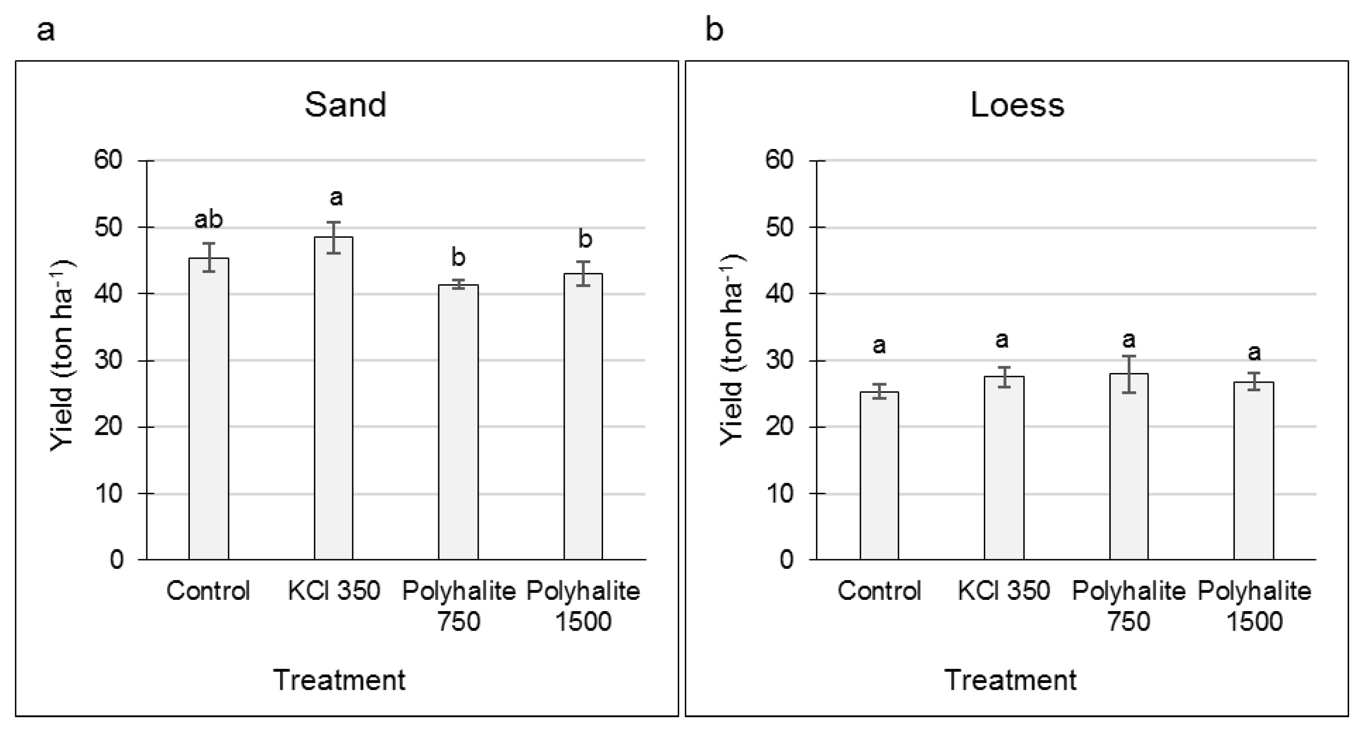
3.1. Effects of KCl and Polyhalite Fertilizers on SR Yield

The effects of KCl and polyhalite fertilizers on sweet potato SR yield and skin quality were tested in sandy and loess experimental plots within commercial fields. Treatments included fertilization with KCl at 350 kg ha−1, or polyhalite at 750 or 1500 kg ha−1. In both sandy and loess plots, high variability was obtained in the size and number of the SRs collected from each of the six blocks of each treatment (Figure 1 shows SRs from the loess plot experiment), irrespective of the type of fertilizer or control.
Total yield was calculated at the end of growth as average ton per hectare for each treatment. In sandy soil, KCl treatment resulted in a significant increase in yield compared to the polyhalite treatment at both concentrations; there was no significant difference between any of the treatments and the control (Figure 2a). In the loess soil, the total yield for each treatment was lower than that obtained in the sandy soil, and no differences were observed between the treatments (Figure 2b).

3.2. Effects of KCl and Polyhalite Fertilizers on SR Skin Anatomy

Histological analysis of SR skin was conducted to examine the effects of KCl and polyhalite treatments on skin morphology. Skin samples were taken at harvest, 150 DAP. Results showed differences in skin morphology between SRs that were grown in sandy soil compared to loess soil (Figure 3). In control plants, the skin of the SRs was thin, comprising 2–3 phellem cell layers. These phellem layers were uniform and well organized in the SRs from the loess plot (Figure 3e) compared to those from the sandy plot (Figure 3a). Application of the fertilizers modified the skin’s morphology. In loess soil, the KCl and polyhalite treatments increased its thickness by adding one phellem layer and expanding the size of the cells (Figure 3f–h); this was most prominent in the KCl treatment (Figure 3f). In the sandy soil, the KCl and polyhalite treatments contributed to better-organized skin compared to the control. However, the skin was thinner than that from the loess soil and consisted of only 1–2 phellem layers (Figure 3b–d). Interestingly, the KCl and 1500 kg ha−1 polyhalite treatments resulted in large, square-shaped phellem cells (Figure 3b,d,f,h) compared to the controls (Figure 3a,e). Overall, the morphology of the SR skin from loess soil was better organized than that from the sandy soil. Application of KCl to both soil types improved the organization of the phellem layers, whereas polyhalite modified skin anatomy to a lesser extent, with a greater impact in the loess soil; only its highest concentration improved phellem organization in the sandy soil.

3.3. Expression of Skin Molecular Markers and Effect of K Fertilizers

The recently published genome of sweet potato enabled the identification of SR skin-related genes based on their alignment to their well-identified potato orthologs (Table 2). Gene expression was analyzed in the skin of SRs from the sandy and loess plots at two developmental stages: mid-growth (around 100 DAP) when SR skin is in an immature stage of development, and at harvest (150 DAP), when the skin is in the maturation stage of development. Tested genes included the suberization-related genes KCS6, FHT, CYP86A33, GPAT3, and FAR3, and the OSP gene, which is related to the skin-setting process.
In general, the expression level of these genes in skin samples from sandy soil was higher than in samples from the loess soil (Figure 4). Nevertheless, in the loess soil, gene expression levels were higher in the mature vs. immature skin samples. This difference in gene expression was not significant for the control treatments, except for KCS6 and OSP genes (Figure 4b; compare gray and orange bars). Following the KCl treatment in the loess soil, the mature skin had significantly higher levels of gene expression than the immature skin for KCS6, CYP86A33, GPAT3, and FAR3 (Figure 4b; compare light blue and yellow bars). Following the polyhalite treatment in the loess soil, only CYP86A33 expression was significantly increased in the mature vs. immature periderm (Figure 4b, compare blue and red bars). Overall, in the loess soil, KCl treatment upregulated the expression of most of the suberin-related genes in the mature periderm compared to the immature periderm.
No such clear pattern was observed in samples from the sandy soil (Figure 4a). However, FHT, which did not show differential expression between immature and mature periderm in the loess soil, was strongly downregulated in mature periderm from the sandy soil, irrespective of the treatment. Interestingly, KCl upregulated OSP expression in both immature and mature periderm (Figure 4a), compared to the control and polyhalite treatment.

4. Discussion

The reported experiments were run under the assumption that supporting sweet potato plant growth and vigor by fertilizer application would contribute to better development of the SRs; as these are strong sinks, this would then contribute to increased SR yield, uniformity, and skin quality. In Israel, sweet potato is grown mostly in sandy soils that are poor in minerals compared to the local loess soil (Table 1). Growers in the region have indicated differences in growth pattern and yield between these two soil types, which initiated the present research. It has also been claimed that K fertilization should be increased, mainly in sandy soil, to support better growth of the crop. The fertilizers of choice were KCl and polyhalite, the latter consisting of the essential minerals Ca, Mg, and K. Polyhalite has also been shown to have some positive effects on potato skin appearance [18], and was thus considered a means of improving sweet potato skin quality, thus solving postharvest skinning problems.

4.1. Effects of K Fertilization on Yield

Our data indicated that the major factor affecting yield in the field was soil type, with higher yield obtained in sandy compared to loess soil (Figure 2). It is common knowledge that sweet potato prefers a fertile, well-drained, loose, fine sandy loam or very sandy soil. One explanation for this might be the relative deficiency of oxygen in loess soil compared to sandy soil, as a low oxygen level in the root zone has been found to reduce SR number and weight compared to an oxygen-enriched root zone [31].
Macroelements play a crucial role in the development of tuber roots in several plant species. For example, previous studies have demonstrated the potential for sweet potato yield to increase with K application [32]. Indeed, application of KCl in sandy soil contributed to a slight increase in yield (Figure 2a). However, polyhalite, a mineral that also provides a K source, had a negative effect on yield compared to KCl. Interestingly, polyhalite contains Mg and Ca in addition to K, and it has been shown that when polyhalite dissolves in the soil, Mg concentration increases first [26]. A high dose of Mg and Ca during SR establishment may compete with K and reduce the latter’s availability [33]. On the other hand, in sandy soils, Mg and Ca application may be necessary, in addition to K, to maintain a favorable balance [32].
The most important nutrient element for sweet potato, in terms of nutrient uptake per unit SR production, per unit area, is K. Since sweet potato is generally grown in highly weathered and leached soils where available K is low, the management of K assumes great significance [32]. Sweet potato-growing areas are generally acidic and have low extractable K concentrations. As K is also required for enhancement of the cambial activity that initiates SR development, it probably affects the activity of starch synthetase, which facilitates SR bulking [32].

4.2. SR Uniformity

A well-known phenomenon in sweet potato crops is the wide variation in the number and size of SRs produced by each individual plant, despite the same growth conditions, apparent homogeneity of the planted vegetative cuttings, and similar development of the aboveground foliage. Adventitious roots (ARs) that emerge from stem cuttings become fibrous roots and exhibit normal secondary growth and lignification of the stele, or develop into SRs that exhibit proliferation of the cambial cells that form starch-accumulating parenchyma cells. It has been shown that ARs in stem cuttings develop from dormant root primordia that are already present in the stem nodes of the mother plant [2]. Nodal position has a significant effect on the developmental status and number of root primordia inside the stem, determining the number and length of the ARs that have developed by 14 DAP. ARs originating from “older” nodes develop better than those originating from “younger” nodes. Moreover, early initiating ARs, 5–7 DAP, have greater potential to become SRs than their late-emerging counterparts [34]. In this context, the balance between development of the anomalous cambium, i.e., SR initiation, and lignification of the stele determines the final SR count [35,36,37]. Hence, sweet potato uniformity and yield are attributed to the maturation and physiological status of the cuttings; however, growth conditions of the transplanting that favor early development of ARs are important as well [34]. The first 50 days from planting was found to be the most significant period for irrigation in determining the amount of SRs [3], and elevated temperature affected SR initiation and bulking [38]. Initiation of SRs from ARs is characterized by an increase in lateral root development compared to their number on lignified roots [3]. Lateral root development is favored by soil moisture, N rate [3,39], and possibly K fertilization, as shown for the model plant Arabidopsis [40]. Recent work in sweet potato has demonstrated that K treatment in the form of K2SO4 significantly increases the number and weight of SRs per plant. This was explained by the increased activity of enzymes involved in sucrose degradation [41]. In our work, application of K in the form of polyhalite and KCl had no positive effect on SR uniformity with respect to SR number per plant or the weight of each SR (Figure 1). This was true for SRs from both soil types, sandy and loess, although the yield was higher in the former (Figure 2).

4.3. K Fertilization Affects Skin Characteristics

The sweet potato’s skin is the first line of defense, and its quality is critical in reducing or avoiding skinning injuries and wounding at harvest. Freshly harvested sweet potatoes have a thin skin that is very susceptible to breakage. The skinning process involves fracture of the longitudinal walls of skin cells as well as the radial walls of the phellogen cells, resulting in skin detachment from the SR surface [6,42]. Postharvest curing reinforces the skin tissue by enhancing suberization and lignification of the phellem cells, and improves skin adhesion to the parenchymatic layers below it, overall increasing SR protection against pathogen penetration [4,42]. High quality skin may prevent wounding and allow harvest mechanization, extended storage time, transport, and improve overall marketability of the sweet potato [43].
Our previous studies indicated a positive effect of mineral fertilization on potato skin [17,18,19]. The fertilizers, exploited by the roots, support plant growth; however, they may also interact directly with the skin of the potato tuber or of the sweet potato SR, both of which are buried in the soil and surrounded by the soil solution. The skin’s interaction with minerals in the soil was demonstrated in a series of independent experiments applying mineral fertilizers to potato in the form of CaCl2 or polyhalite. Tuber acquisition of Ca was suggested to result from direct uptake from the soil across the periderm [44,45]. This was supported by the finding of high Ca in the skin compared to tuber flesh [45,46]—the periderm contained 34% of the total tuber Ca concentration [45]. However, direct transfer across the periderm can only occur at early stages of tuber development while the skin is still a living tissue, before it becomes suberized [47,48]; in mature tubers, there is no significant transport of Ca from the soil across the periderm [47]. There are reports of higher Ca supply in the soil increasing the level of Ca in the tuber peel more than in its flesh [46], implying that Ca fertilization results in its accumulation in the peel. When polyhalite mineral is applied, an increase in periderm K level over that of Ca is observed [18]. It was suggested that in the maturing skin, which consists of heavily suberized dead cells, i.e., corky material, the polyhalite mineral elements Ca, Mg, and K interact with this inert material as ion exchangers, and K binding is favored over that of Ca and Mg [18,49]. Fertilization of potato with silica also resulted in a high level of the mineral in maturing periderm compared to tuber flesh [19]. As silicon accumulation by the plant requires specific transporters, this suggests passive absorption of the mineral from the soil solution to the periderm.
The above-mentioned minerals alter skin morphology and improve its quality. Fertilization with Ca reduced potato skin russeting [17]; silicon fertilization increased phellem cell area and the expression of suberin biosynthesis genes, and resulted in enrichment of the periderm cell walls with oxidized aromatic moieties—overall suggesting enhanced lignification and suberization [19]; and the polyhalite mineral—consisting of K, Ca, and Mg—resulted in increased phellem cell size, increased the expression of suberin biosynthesis genes, and improved potato skin appearance [18]. Divalent ions, such as Ca and Mg, contribute to maintenance of the cell membrane and cell wall structure by forming stable but reversible linkages between the pectin polar head groups in the cell wall [20,21]. K, as potassium phosphite, reinforced the cell wall of periderm cells, by increasing related enzymatic activity and the deposition of suberin and pectin [50].
The effect of the K fertilizers on sweet potato skin seemed to follow a similar pattern. Application of KCl improved the organization of skin cells, appeared to increase phellem cell volume, and as a result, increased skin thickness, whereas polyhalite modified skin anatomy to a lesser extent, with a greater impact in the loess soil (Figure 3).

4.4. Expression of Skin-Related Genes

In accordance with the anatomical data that showed differences in skin morphology between the sandy and loess soils, the expression profile of suberin-related genes also differed. The tested genes included the suberization-related genes KCS6 [12], FHT [14], CYP86A33 [13], GPAT3 [51], and FAR3 [52], and the OSP [15] gene that is related to the skin-setting process.
In the sandy soil, skin development seemed to be arrested, with only one or two phellem cell layers compared to the more developed skin from the loess soil (Figure 3). Accordingly, no differences were observed in gene expression between immature and mature skin from the sandy soil, compared to differential expression in skins from the loess soil. In the latter, the level of gene expression in the mature periderm was higher than in the immature one, albeit not significantly so for all genes (Figure 4). This greater gene expression indicated enhanced suberization in the maturing skin.
A detailed analysis of gene expression in skin from the loess soil indicated upregulation of KCS6, CYP86A33, GPAT3, and FAR3 in mature skin compared to immature skin following KCl fertilization (Figure 4b), suggesting an effect of KCl on skin development and suberization. This was supported by the anatomical study indicating a bigger phellem cell size compared to controls (Figure 3e,f). Overall, in the loess soil, KCl treatment upregulated most of the suberin-related genes in the mature vs. immature skin, which might contribute to both cell wall strength and resistance to water loss during postharvest treatments and storage [53]. The effect of polyhalite on gene expression was not as prominent as that of the KCl and only CYP86A33 showed significantly increased expression in the mature skin compared to the immature skin (Figure 4b). Application of polyhalite to potato plants (red-skinned cv. Rosanna) also upregulated the expression of CYP86A33 [18]. Interestingly, this gene is involved in ω-hydroxylation of suberin aliphatic monomers, which are necessary for the suberin’s typical lamellar organization and for periderm resistance to water loss [13].

5. Conclusions

Overall, sweet potato yield was higher in sandy soil than in loess soil; however, SR skin seemed to be more developed in the loess soil. Supplement of K in sandy soils improved sweet potato yield and contributed to better organization of the SR skin, especially in loess soil. The positive effect on skin anatomy was supported by upregulation of suberin-related genes in maturing skin compared to immature skin.

Author Contributions

Conceptualization, L.K., U.Y., and I.G.; Data curation, R.v.O. and A.K.-K.; Funding acquisition, L.K., U.Y., and I.G.; Methodology, L.K., U.Y., and I.G.; Supervision, I.G.; Writing—original draft, I.G.; Writing—review and editing, L.K., U.Y., and I.G. All authors have read and agreed to the published version of the manuscript.

Funding

This research was supported by the Center for Fertilization and Plant Nutrition (CFPN, https://www.cfpn.center/), and is a contribution of the ARO, Volcani Center.

Acknowledgments

The authors thank Tal Oged from Ego Growers Ltd. for providing the sweet potato experimental plots, and Gali Carmi and Lior Peled-Lichter from ICL, Israel, for providing the fertilization protocols and coordinating the experiments.

Conflicts of Interest

The authors declare no conflict of interest.

References

  1. Firon, N.; LaBonte, D.; Villordon, A.; McGregor, C.; Kfir, Y.; Pressman, E. Botany and physiology: Storage root formation and development. In The Sweetpotato; Loebenstein, G., Thottappilly, G., Eds.; Springer: Dordrecht, The Netherlands, 2009; pp. 13–26. [Google Scholar] [CrossRef]
  2. Ma, J.; Aloni, R.; Villordon, A.; Labonte, D.; Kfir, Y.; Zemach, H.; Schwartz, A.; Althan, L.; Firon, N. Adventitious root primordia formation and development in stem nodes of ‘Georgia Jet’ sweetpotato, Ipomoea batatas. Am. J. Bot. 2015, 102, 1040–1049. [Google Scholar] [CrossRef] [PubMed]
  3. Villordon, A.; LaBonte, D.; Solis, J.; Firon, N. Characterization of lateral root development at the onset of storage root initiation in ‘Beauregard’ sweetpotato adventitious roots. Hortscience 2012, 47, 961–968. [Google Scholar] [CrossRef] [Green Version]
  4. Afek, U.; Orenstein, J.; Nuriel, E. Increased quality and prolonged storage of sweet potatoes in Israel. Phytoparasitica 1998, 26, 307–312. [Google Scholar] [CrossRef]
  5. Reeve, R.M.; Hautala, E.; Weaver, M.L. Anatomy and compositional variation within potatoes 1. Developmental histology of the tuber. Am. Potato J. 1969, 46, 361–373. [Google Scholar] [CrossRef]
  6. Lulai, E.C.; Freeman, T.P. The importance of phellogen cells and their structural characteristics in susceptibility and resistance to excoriation in immature and mature potato tuber (Solanum tuberosum L.) periderm. Ann. Bot. 2001, 88, 555–561. [Google Scholar] [CrossRef] [Green Version]
  7. Bernards, M.A.; Lewis, N.G. The macromolecular aromatic domain in suberized tissue: A changing paradigm. Phytochemistry 1998, 47, 915–933. [Google Scholar] [CrossRef]
  8. Yan, B.; Stark, R.E. Biosynthesis, molecular structure, and domain architecture of potato suberin: A C-13 NMR study using isotopically labeled precursors. J. Agric. Food Chem. 2000, 48, 3298–3304. [Google Scholar] [CrossRef]
  9. Graça, J. Suberin: The biopolyester at the frontier of plants. Front. Chem. 2015, 3, 62. [Google Scholar] [CrossRef]
  10. Graça, J.; Pereira, H. Suberin structure in potato periderm: Glycerol, long-chain monomers, and glyceryl and feruloyl dimers. J. Agric. Food Chem. 2000, 48, 5476–5483. [Google Scholar] [CrossRef]
  11. Vishwanath, S.J.; Delude, C.; Domergue, F.; Rowland, O. Suberin: Biosynthesis, regulation, and polymer assembly of a protective extracellular barrier. Plant Cell Rep. 2015, 34, 573–586. [Google Scholar] [CrossRef]
  12. Serra, O.; Soler, M.; Hohn, C.; Franke, R.; Schreiber, L.; Prat, S.; Molinas, M.; Figueras, M. Silencing of StKCS6 in potato periderm leads to reduced chain lengths of suberin and wax compounds and increased peridermal transpiration. J. Exp. Bot. 2009, 60, 697–707. [Google Scholar] [CrossRef] [PubMed] [Green Version]
  13. Serra, O.; Soler, M.; Hohn, C.; Sauveplane, V.; Pinot, F.; Franke, R.; Schreiber, L.; Prat, S.; Molinas, M.; Figueras, M. CYP86A33-targeted gene silencing in potato tuber alters suberin composition, distorts suberin lamellae, and impairs the periderm’s water barrier function. Plant Physiol. 2009, 149, 1050–1060. [Google Scholar] [CrossRef] [PubMed] [Green Version]
  14. Serra, O.; Hohn, C.; Franke, R.; Prat, S.; Molinas, M.; Figueras, M. A feruloyl transferase involved in the biosynthesis of suberin and suberin-associated wax is required for maturation and sealing properties of potato periderm. Plant J. 2010, 62, 277–290. [Google Scholar] [CrossRef] [PubMed]
  15. Vulavala, V.K.; Fogelman, E.; Faigenboim, A.; Shoseyov, O.; Ginzberg, I. The transcriptome of potato tuber phellogen reveals cellular functions of cork cambium and genes involved in periderm formation and maturation. Sci. Rep. 2019, 9, 1–14. [Google Scholar] [CrossRef] [PubMed] [Green Version]
  16. Vulavala, V.K.; Fogelman, E.; Rozental, L.; Faigenboim, A.; Tanami, Z.; Shoseyov, O.; Ginzberg, I. Identification of genes related to skin development in potato. Plant Mol. Biol. 2017, 94, 481–494. [Google Scholar] [CrossRef] [PubMed]
  17. Ginzberg, I.; Minz, D.; Faingold, I.; Soriano, S.; Mints, M.; Fogelman, E.; Warshavsky, S.; Zig, U.; Yermiyahu, U. Calcium mitigated potato skin physiological disorder. Am. J. Potato Res. 2012, 89, 351–362. [Google Scholar] [CrossRef]
  18. Keren-Keiserman, A.; Baghel, R.S.; Fogelman, E.; Faingold, I.; Zig, U.; Yermiyahu, U.; Ginzberg, I. Effects of polyhalite fertilization on skin quality of potato tuber. Front. Plant Sci. 2019, 10. [Google Scholar] [CrossRef]
  19. Vulavala, V.K.; Elbaum, R.; Yermiyahu, U.; Fogelman, E.; Kumar, A.; Ginzberg, I. Silicon fertilization of potato: Expression of putative transporters and tuber skin quality. Planta 2016, 243, 217–229. [Google Scholar] [CrossRef]
  20. Olsson, K. Resistance to gangrene (Phoma exigua var.foveata) and dry rot (Fusarium solani var.coeruleum) in potato tubers. 1. The influence of pectin-bound magnesium and calcium. Potato Res. 1988, 31, 413–422. [Google Scholar] [CrossRef]
  21. Palta, J.P. Role of calcium in plant responses to stresses: Linking basic research to the solution of practical problems. HortScience 1996, 31, 51–57. [Google Scholar] [CrossRef] [Green Version]
  22. Jarvis, M.; Briggs, S.; Knox, J. Intercellular adhesion and cell separation in plants. Plantcell Environ. 2003, 26, 977–989. [Google Scholar] [CrossRef]
  23. Vincken, J.-P.; Borkhardt, B.; Bush, M.; Doeswijk-Voragen, C.; Dopico, B.; Labrador, E.; Lange, L.; McCann, M.; Morvan, C.; Muñoz, F. Remodelling pectin structure in potato. In Developments in Plant Genetics and Breeding; Elsevier: Amsterdam, The Netherlands, 2000; Volume 6, pp. 245–256. [Google Scholar]
  24. Zörb, C.; Senbayram, M.; Peiter, E. Potassium in agriculture—Status and perspectives. J. Plant Physiol. 2014, 171, 656–669. [Google Scholar] [CrossRef] [PubMed]
  25. Haberman, A.; Dag, A.; Shtern, N.; Zipori, I.; Erel, R.; Ben-Gal, A.; Yermiyahu, U. Long-term impact of potassium fertilization on soil and productivity in intensive olive cultivation. Agronomy 2019, 9, 525. [Google Scholar] [CrossRef] [Green Version]
  26. Yermiyahu, U.; Zipori, I.; Faingold, I.; Yusopov, L.; Faust, N.; Bar-Tal, A. Polyhalite as a multi nutrient fertilizer—Potassium, magnesium, calcium and sulfate. Isr. J. Plant Sci. 2017, 64, 145–157. [Google Scholar]
  27. Penman, H.L. Natural evaporation from open water, bare soil and grass. Proc. R. Soc. Lond. Ser. A Math. Phys. Sci. 1948, 193, 120–145. [Google Scholar]
  28. Ruzin, S.E. Plant Microtechnique and Microscopy; Oxford University Press: New York, NY, USA, 1999. [Google Scholar]
  29. Johansen, D.A. Plant Microtechniques; McGraw-Hill Book Company, Inc.: New York, NY, USA, 1940. [Google Scholar]
  30. Ginzberg, I.; Barel, G.; Ophir, R.; Tzin, E.; Tanami, Z.; Muddarangappa, T.; de Jong, W.; Fogelman, E. Transcriptomic profiling of heat-stress response in potato periderm. J. Exp. Bot. 2009, 60, 4411–4421. [Google Scholar] [CrossRef]
  31. Chua, K.; Kays, S.J. Effect of soil oxygen concentration on sweet potato storage root induction and/or development. HortScience 1981, 16, 71–73. [Google Scholar]
  32. Byju, G.; George, J. Potassium nutrition of sweet potato. Adv. Hortic. Sci. 2005, 19, 221–239. [Google Scholar]
  33. Fageria, N.K. Absorption of magnesium and its influence on the uptake of phosphorus, potassium, and calcium by intact groundnut plants. Plant Soil 1974, 40, 313–320. [Google Scholar] [CrossRef]
  34. Villordon, A.Q.; Bonte, D.R.L.; Firon, N.; Kfir, Y.; Pressman, E.; Schwartz, A. Characterization of Adventitious Root Development in Sweetpotato. HortScience 2009, 44, 651. [Google Scholar] [CrossRef]
  35. Firon, N.; LaBonte, D.; Villordon, A.; Kfir, Y.; Solis, J.; Lapis, E.; Perlman, T.S.; Doron-Faigenboim, A.; Hetzroni, A.; Althan, L. Transcriptional profiling of sweetpotato (Ipomoea batatas) roots indicates down-regulation of lignin biosynthesis and up-regulation of starch biosynthesis at an early stage of storage root formation. BMC Genom. 2013, 14, 460. [Google Scholar] [CrossRef] [PubMed] [Green Version]
  36. Wilson, L.; Lowe, S. The anatomy of the root system in West Indian sweet potato (Ipomoea batatas (L.) Lam.) cultivars. Ann. Bot. 1973, 37, 633–643. [Google Scholar] [CrossRef]
  37. Togari, Y. A study of tuberous root formation in sweet potato. Bull. Nat. Agric. Exp. Stn. 1950, 68, 1–96. [Google Scholar]
  38. Gajanayake, B.; Reddy, K.R.; Shankle, M.W.; Arancibia, R.A.; Villordon, A.O. Quantifying storage root initiation, growth, and developmental responses of sweetpotato to early season temperature. Agron. J. 2014, 106, 1795–1804. [Google Scholar] [CrossRef]
  39. Villordon, A.; LaBonte, D.; Firon, N.; Carey, E. Variation in nitrogen rate and local availability alter root architecture attributes at the onset of storage root initiation in ‘Beauregard’ sweetpotato. HortScience 2013, 48, 808–815. [Google Scholar] [CrossRef] [Green Version]
  40. Kellermeier, F.; Chardon, F.; Amtmann, A. Natural variation of arabidopsis root architecture reveals complementing adaptive strategies to potassium starvation. Plant Physiol. 2013, 161, 1421–1432. [Google Scholar] [CrossRef] [Green Version]
  41. Du, F.; Liu, H.; Yin, X.; Zhao, Q.; Shi, C. Potassium-mediated regulation of sucrose metabolism and storage root formation in sweet potato. Arch. Agron. Soil Sci. 2020. [Google Scholar] [CrossRef]
  42. Villavicencio, L.E.; Blankenship, S.M.; Yencho, G.C.; Thomas, J.F.; Raper, C.D. Temperature effect on skin adhesion, cell wall enzyme activity, lignin content, anthocyanins, growth parameters, and periderm histochemistry of sweetpotato. J. Am. Soc. Hortic. Sci. 2007, 132, 729–738. [Google Scholar] [CrossRef]
  43. Effendy, J.; La Bonte, D.R.; Baisakh, N. Identification and expression of skinning injury-responsive genes in sweetpotato. J. Am. Soc. Hortic. Sci. 2013, 138, 210–216. [Google Scholar] [CrossRef] [Green Version]
  44. Davies, H.; Millard, P. Fractionation and distribution of calcium in sprouting and non-sprouting potato tubers. Ann. Bot. 1985, 56, 745–754. [Google Scholar]
  45. Subramanian, N.K.; White, P.J.; Broadley, M.R.; Ramsay, G. The three-dimensional distribution of minerals in potato tubers. Ann. Bot. 2011, 107, 681–691. [Google Scholar] [CrossRef] [PubMed] [Green Version]
  46. Olsen, N.L.; Hiller, L.K.; Mikitzel, L.J. The dependence of internal brown spot development upon calcium fertility in potato tubers. Potato Res. 1996, 39, 165–178. [Google Scholar] [CrossRef]
  47. Busse, J.S.; Palta, J.P. Investigating the in vivo calcium transport path to developing potato tuber using 45Ca: A new concept in potato tuber calcium nutrition. Physiol. Plant. 2006, 128, 313–323. [Google Scholar] [CrossRef]
  48. Habib, A.; Donnelly, D.J. Calcium translocation and accumulation into potato tubers. Potato Res. 2002, 45, 17–24. [Google Scholar] [CrossRef]
  49. Shomer, I.; Novacky, A.J.; Pike, S.M.; Yermiyahu, U.; Kinraide, T.B. Electrical potentials of plant cell walls in response to the ionic environment. Plant Physiol. 2003, 133, 411–422. [Google Scholar] [CrossRef] [Green Version]
  50. Lobato, M.C.; Daleo, G.R.; Andreu, A.B.; Olivieri, F.P. Cell wall reinforcement in the potato tuber periderm after crop treatment with potassium phosphite. Potato Res. 2018, 61, 19–29. [Google Scholar] [CrossRef]
  51. Beisson, F.; Li-Beisson, Y.; Pollard, M. Solving the puzzles of cutin and suberin polymer biosynthesis. Curr. Opin. Plant Biol. 2012, 15, 329–337. [Google Scholar] [CrossRef]
  52. Domergue, F.; Vishwanath, S.J.; Joubes, J.; Ono, J.; Lee, J.A.; Bourdon, M.; Alhattab, R.; Lowe, C.; Pascal, S.; Lessire, R.; et al. Three Arabidopsis fatty acyl-coenzyme a reductases, FAR1, FAR4, and FAR5, generate primary fatty alcohols associated with suberin deposition. Plant Physiol. 2010, 153, 1539–1554. [Google Scholar] [CrossRef] [Green Version]
  53. Lulai, E.C.; Orr, P.H. Techniques for detecting and measuring developmental and maturational changes in tuber native periderm. Am. Potato J. 1994, 71, 489–505. [Google Scholar] [CrossRef]
Figure 1. Lack of uniformity in sweet potato. Storage roots (SRs) were collected at mid-growth—100 days after planting, following a preplanting application of fertilizers: KCl at 350 kg ha−1, or polyhalite at 750 or 1500 kg ha−1. Each treatment and the control were designed in six replicates and arranged in random blocks. Each frame represents a block, and demonstrates SRs collected from 1 m of the bed. The figure presents SRs collected from the plot with the loess soil and demonstrates the variability in SR number and size, irrespective of treatment. A similar phenomenon was obtained in the sandy plot.
Agronomy 10 01385 g001
Figure 2. Yields (ton ha−1) of sweet potato storage roots following application of the fertilizers KCl at 350 kg ha−1, or polyhalite at 750 or 1500 kg ha−1. Plants were grown in sandy (a) or loess soil (b). Values are average of six replicates with SE. Data were analyzed for statistical significance between means of the different treatments by Student’s t-test. Values followed by different letters are significantly different (p < 0.05).
Agronomy 10 01385 g002
Figure 3. Anatomical study of the skin of sweet potato storage roots (SRs). SRs were collected from plants grown in sandy (ad) and loess (eh) soils, and treated with a preplanting application of the fertilizers KCl at 350 kg ha−1 (b,f), or polyhalite at 750 (c,g) or 1500 (d,h) kg ha−1. Sections were taken from three independent SRs for each treatment, and one representative section is shown. Tissue morphology was observed under a light microscope. The morphology of the SR skin (located above the yellow line) was characterized by rectangular phellem cells arranged in columns (demonstrated by red arrows). Scale bar = 100 µm.
Agronomy 10 01385 g003
Figure 4. Expression of suberin-related genes KCS6, FHT, CYP86A33, GPAT3, and FAR3, and skin-set-related gene OSP, in skin of sweet potato storage roots (SRs) grown in sandy (a) and loess (b) soil types, and treated with a preplanting application of the fertilizers KCl at 350 kg ha−1, or polyhalite at 750 or 1500 kg ha−1. SRs were collected at around 100 days after planting (DAP) (blue–gray bars) and at harvest, 150 DAP (yellow–red bars, marked with inverted commas in the color key). Transcript levels were monitored by qPCR and expression levels were normalized relative to that of the reference gene PLD. Values are averages of three replicates with SE. Data were analyzed for statistical significance among means by Student’s t-test; different letters indicate significantly different values (p < 0.05).
Agronomy 10 01385 g004
Table 1. Soil composition and minerals before the growing season. Data are average of three replicates with SE.
Soil Type
SandLoess
Sand (%)83.2 ± 3.366.8 ± 3.5
Silt (%)2.3 ± 0.89.9 ± 1.4
Clay (%)14.5 ± 1.223.3 ± 2
Electrical conductivity (dS m−1) *1.20 ± 0.272.03 ± 0.13
pH *7.63 ± 0.047.63 ± 0.04
Minerals (mg kg−1 dry soil)
N-NO3 *8.62 ± 3.9135.32 ± 9.50
P *0.50 ± 0.090.02 ± 00
K *22.27 ± 4.1926.24 ± 2.76
Mg *7.45 ± 1.4016.43 ± 0.44
Ca *36.38 ± 8.8383.80 ± 4.19
Cl *16.94 ± 6.5364.05 ± 24.50
Exchangeable K * 217.2 ± 32.6514.1 ± 77.1
* Saturated paste solution.
Table 2. Sweet potato primers for periderm- and suberin-related genes based on their alignment to potato and Arabidopsis orthologs.
Gene CodeGenePotato IDTAIR IDSweet potato ID *Forward primer 5′→3′ Reverse primer 5′→3′ Length (bp)
CYP86A33FATTY ACID OMEGA-HYDROXYLASESotub06g032570AT5G58860itf04g28930.t1AAACGCATCGCTCTCAACTTGCTAATGCCGCCTTGAGATA227
FAR3FATTY ACYL-COA REDUCTASE 3Sotub06g031240 AT4G33790 itf14g11580.t1TTTGCAAGTATTTCCAGACAACACCCCCTTGAAGAATAAGTATGG101
FHTFERULOYL TRANSFERASESotub03g018220AT5G41040itf02g04740.t1AATACCAAGCCTGATCCTGTGATGCTTCTGTCCAGGAATG230
GPAT3GLYCEROL-3-PHOSPHATE 2-O-ACYLTRANSFERASE 4Sotub01g032090AT1G01610itf09g20300.t1CGATTGAAAAGCCGATTGATGTTGATTCCGAGCATGAGGT183
KSC63-KETOACYL-COA SYNTHASE 6Sotub02g029020AT1G68530 itf13g22850.t1CTCATCGTCAACTGCAGCATCAAATCTCGGGCCAAATCTA153
OSPORGAN-SPECIFIC PROTEIN S2-LIKESotub07g011080AT2G48030itf10g24760.t1GAGTATTGGGACGCGATGATCTGCTGAACATCCTTGGC300
PLDPHOSPHOLIPASE D1A itf10g15190.t1ATCGGAATCAGCAGTGATGGATGATGAGGCAAGCAGTGTG160
* Sweetpotato Genomics Resource database http://sweetpotato.plantbiology.msu.edu/index.shtml.

