Review

The Emerging Roles of Diacylglycerol Kinase (DGK) in Plant Stress Tolerance, Growth, and Development

1
College of Life Sciences, Engineering Research Center of the Chinese Ministry of Education for Bioreactor and Pharmaceutical Development, Jilin Agricultural University, Changchun 130118, China
2
College of Tropical Crops, Hainan University, Haikou 570228, China
*
Authors to whom correspondence should be addressed.
Agronomy 2020, 10(9), 1375; https://doi.org/10.3390/agronomy10091375
Submission received: 16 July 2020 / Revised: 1 September 2020 / Accepted: 3 September 2020 / Published: 12 September 2020
(This article belongs to the Special Issue Molecular Responses to Salt Stress in Crop Plants)

Abstract

:
Diacylglycerol kinase (DGK) is recognized as the key enzyme of the lipid signaling pathway, which involves the transduction of messages from hormones, neurotransmitters, and immunologic and growth factors. Regarding their essential role in animal physiology, many plant biologists have predicted a similar enzymatic influence in plants. However, a small number of recent studies have revealed the complexity of the involvement of DGK genes in the modulation of plant growth, development, and adaptation in both biotic and abiotic stress conditions. Here, we describe recent discoveries on the role of DGK genes in the plants’ responses to biotic or abiotic stressors. Moreover, we discuss how DGK enzymes regulate plant cellular activities during the adaptation of plants to a readily changing environment. DGK is an enzyme that plays a pivotal role in plant lipid signaling, by catalyzing the phosphorylation of the diacylglycerol (DAG) to phosphatidic acid (PA), which is a crucial molecule in a plant’s metabolic network, leading to its response to various external stresses. DGK enzymes are the principal moderators of PA generation in plant cells; this consequently affects its derived products—hence, enabling their activities in lipid signaling networks and cell homeostasis. Thus, understanding the DGK operational mode and interactions between the production and accumulation of PA would constitute a significant advancement in investigating the mechanism of stress adaptation in plants.

1. Introduction

Plants are continuously subjected to diverse environmental stresses and have developed efficient mechanisms to survive under such adverse conditions. Over the past decade, studies have been intensely focused on the molecular mechanisms that are involved in both biotic and abiotic stress responses and/or stress tolerance, with much priority on adaptive mechanisms relating to individual stresses [1,2].
Several studies have provided important avenues for the improvement of plant adaptation to external stress factors [3,4]. Studies on both biotic and abiotic stresses have independently revealed significant amounts of data for a plethora of genes and have successfully identified several stress-responsive genes [5,6,7,8,9]. These stress-responsive genes include the locus that encodes for the diacylglycerol kinase (DGK) enzyme, which stands out by its important roles in plant growth, development, and stress tolerance through phosphatidic acid (PA) signaling in plants. The enzymatic function of DGK coupled with PA production can moderate the activation of many phytohormones, controlling stomatal closure, regulating cell membrane permeability, and other cellular processes via altering its enzyme activity [10]. Lipids are fundamental metabolites in plant development and stress responses [11], and PA is the glycerophospholipid that plays a crucial role in the biosynthesis of several complex lipids, and it is also recognized as a key molecule in lipid signaling in plants [12,13]. The endoplasmic reticulum, the mitochondria, and the plastids are the main compartments of cells that generate PA, which is mostly derived from varied phospholipase networks in the plasma membrane and serves as a major precursor of phospholipids and glycolipids in plants [14]. PA is commonly known as one of the most important secondary messengers for signal transduction cascades—notably, in multiple plant abiotic stress responses such as salinity [15], abscisic acid [16], osmotic pressure [17,18], drought [19], wounding and nodulation factor [20] ethylene [20], cold [21] and other temperature changes [22], and pathogen attacks [23]. Overall, PA accumulation is closely linked to the mechanisms involved in plant cell adaptation to combat numerous stresses, and in some cases, positive-strand RNA viruses even use PA to activate their replication [24]. PA is mainly produced by the hydrolytic action of two important phospholipases—namely, phospholipase D (PLD) and phospholipase C (PLC)—coupled with the catalytic actions of diacylglycerol kinases, PLC, and DGK [25,26]. The PA generated from the PLC/DGK pathway has the activity with the largest scope during the molecular, cellular, and physiological responses. This explains its capacity to stimulate several reactions in plants via its activation of a large number of protein targets [14,27]. PA production from diacylglycerol (DAG) phosphorylation through the DGK enzyme activity is the main reaction of lipid signaling in plants. DGK enzymes can create local changes in cell membrane configurations through their generation of PA and via modifying its electrostatic charge, which leads to the enlistment and triggering of various proteins [28].
Diacylglycerol kinase is the fundamental membrane enzymatic protein that catalyzes the ATP-dependent phosphorylation of DAG to PA. DGK plays an important role in lipid homeostasis through the modulation of the abundance of PAs and diacylglycerols (DAG); those are among the pivotal molecules that hold essential roles in several plant signaling pathways, including the lipid signaling pathway [29,30], with the lipid signaling pathway known as a key network of plant growth and plant stress tolerances.
The efficiency of lipid signaling pathway depends on the actions of the secondary messengers, the signal transduction cascade events, and the nature or the composition of the engaged lipid. This pathway mainly recruits phosphoglycerolipids, such as diacylglycerol kinase (DGK), phosphatidylinositols (PI), phosphatidic acid (PA), nonspecific phospholipase C (NPC), PLC, and PLD [31], and the contribution of PLD to plant tip growth is also well-documented [26].
Amongst the phytohormones required for the modulation of plant growth and development, Brassinosteroids (BRs) are one of the eminent classes of steroid hormone involved in key processes. The linkage between BRs and PA activity was further exhibited, and the PA level appeared to be strongly increased in root meristem cells of Arabidopsis thaliana in response to BR treatment [32]. In 2016, Derevyanchuk and colleagues revealed that Epibrassinolide (EBL) plays an important role in the regulation of plant growth and PA production in Brassica napus under high salinity conditions [33]. The same study showed the existing interaction between DGKs and EBL, by displaying DGK involvement in the formation of PA molecules in response to BR treatment, using a specific DGK inhibitor, R59022 [34].
In addition, recent studies revealed attractive results, such as the inhibition of DGK function strongly affecting root elongation and the dgk4 knockout mutant developing defective pollen, as demonstrated via a transgenic knockdown approach of the Arabidopsis DGK2 gene. This DGK2 knockdown mutant had an alteration of gametophyte structure, which strongly disturbed the leaves and the growth of the roots [35]. In knockdown lines, a glycerolipid analysis displayed that, in floral buds and leaves of the Arabidopsis thaliana, both the metabolism of phosphatidylglycerol and phosphatidylinositol metabolism were affected differently by stress exposure. The expression of MpDGK2 from Malus prunifolia conferred drought stress tolerance in transgenic Arabidopsis through the modification of stomatal closure under water shortage conditions and the accumulation of hydrogen peroxide (H2O2). These outcomes suggest that DGKs are essential during gametogenesis, plant stress responses, and phospholipid metabolism involved in vegetative and reproductive growth [34,35,36,37].
Plants are generally confronted with variations of their environment, which, in some cases, can seriously affect their growth, development, and productivity. Besides its role as an effective gene family that is widely expressed in response to minerals like metals, metalloid ions, and other beneficial elements, DGK has emerged as being crucial in diverse plant stress responses [38]. In nonactivated cells, DGK activity lowly enables the DAG used for the biosynthesis of glycerol phospholipid, but this activity increases during the initiation of the phosphoinositide pathway [39]. PA and DAG are involved as different bioactive lipids in plant lipid signaling and have distinct cell targets. Recent studies positioned the diacylglycerol kinase as a key enzyme that regulates various cell reactions by controlling the parity between two important second messengers. Moreover, a critical reductive coenzyme (NADPH) is generated by DGK enzymes during the biosynthesis of flavonoids and lignin, while the activation of tobacco NADPH oxidase-dependent by DGK enzymes strongly increased during the cryptogenic stress treatment [40]. Additionally, during the apoplastic oxidative reaction in most plant-pathogen reactions, the production of reactive oxygen species is driven by NADPH oxidases [41].
This review provides a theoretical foundation for the research on plant stress resistance; it stands on research progresses on the PA derived from the catalytic activity of DGK enzymes, which is different from that derived from PLD. This difference is based on the fatty acid composition and differential 32Pi-labeling characteristics of PA [10]. In this section, we mostly highlight the recent research progress on the roles of diacylglycerol kinase in the plant lipid signaling pathways and investigate its corresponding mechanisms that allow a plant to cope with stress. More investigation on the biological knowledge of the DGK roles, both in biotic and abiotic stress, is still needed to provide supplementary information for further study advancement. Therefore, the understanding of novel DGK mechanisms in plant stress responses could provide a new avenue to enhance crop resistance and, consequently, boost crop yields to combat the food shortages that are currently being experienced in some parts of the world.

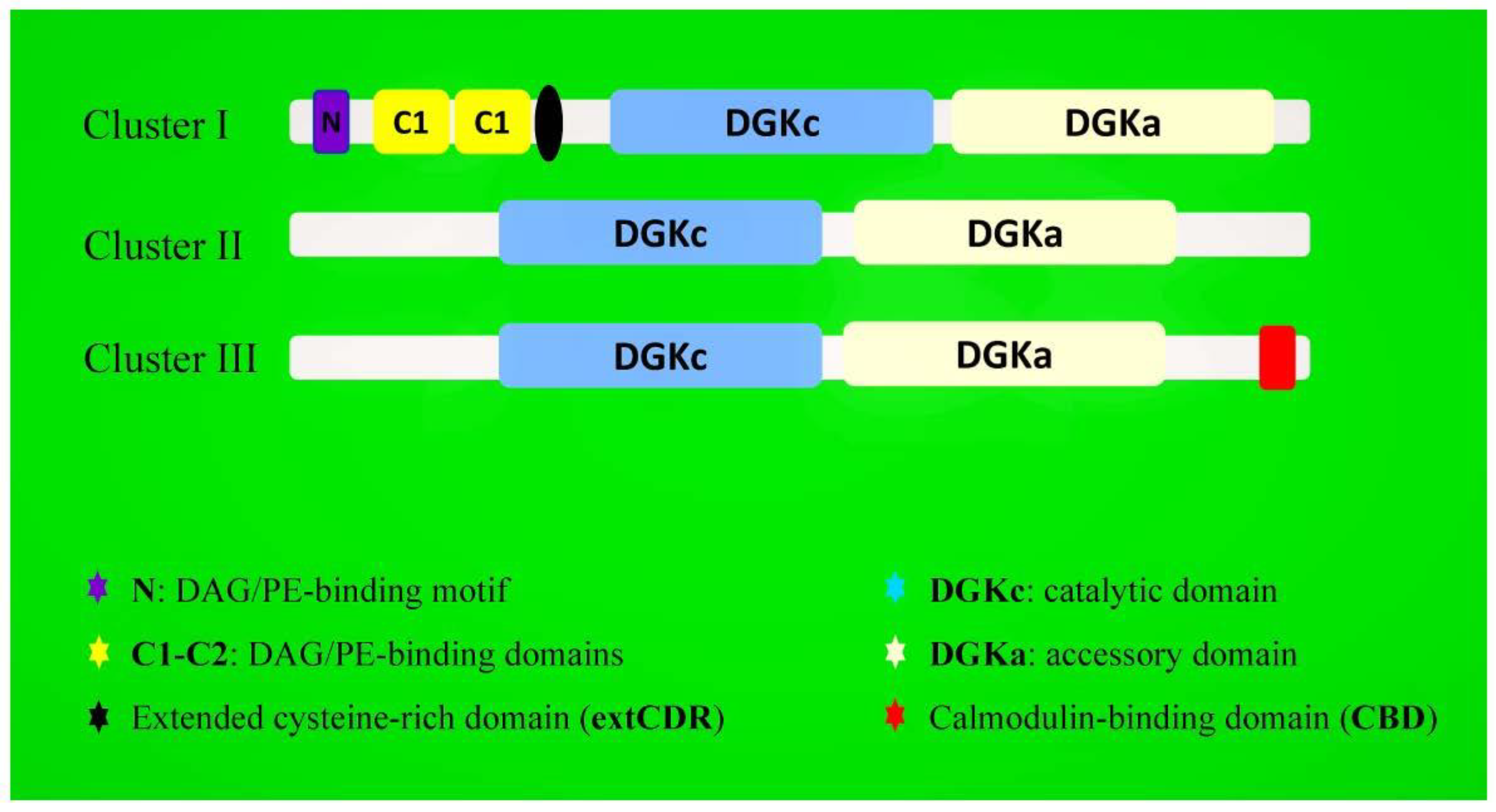
2. Structure and Isoforms of Plant DGK Genes

Many studies have been carried out on the expression of DGK genes in diverse organisms, with the highest diversity in mammals (10 isoforms), compared to only one DGK gene identified in Drosophila melanogaster [42]. In mammals, DGKs are classified into five subtypes, according to their conserved protein domains. Considering their catalytic and accessory domains, the catalytic domain is strongly conserved, with different characteristics within the subtypes [43,44]. The type I isoforms (DGKα, β, and γ) have two repeated calcium-binding EF hand motifs; type II DGK (δ, η, and κ) are characterized by a Pleckstrin homology domain; type III (DGKε) has a hydrophobic domain; type IV DGK (ζ and ι) show common myristoylated alanine-rich C kinase substrates (MARCKS) and ankyrin repeat (ANK) elements; and finally, the type V isoform (DGKθ) is characterized by three C1 domains. Multiple functional analyses on the DGK conserved domains either in animals, algae, or plants have also clearly highlighted the potential role of specific motifs and their regulations [45,46]. Meanwhile, many DGK isoforms were also described with higher diversity and to perform several pivotal roles in human cells [47].
In plants, DGKs also exist in various isoforms, with almost all containing similar conserved catalytic, accessory, and ATP-binding domains. The ATP-binding region is generally flanked by the consensus GXGXXG/A, which is essential for the kinase activity in controlling the balance between DAG and PA levels coordinately. Moreover, this domain directs the physiological functions in the appropriate cell types [34]. However, research on plant DGKs is only recent, and the majority of these studies have been mainly focused on the identification and the characterization of DGK isoforms by in-silico analyses in several plant models, with a well-known genome in which several isoforms were identified. The distribution of DGK isoforms in plants is widely spread, and the prominent research publications on plant DGKs have revealed 7 DGK isoforms in Arabidopsis thaliana (AtDGK) [34,48]; 8 isoforms in Oryza sativa (OsDGK) [49] and Malus domestica (MdDGK) [19]; 12 isoforms in Glycine max (GmDGK) [50]; and 7 isoforms in Zea mays (ZmDGK) [51,52], Solanum lycopersicum (SlDGK) [53], and Triticum aestivum (TaDGK) [54] (Figure 1).
The structure of plant DGK genes is less complex than that of animals [55]. In plants, following their protein-conserved domains, DGK genes are divided into three different clusters: clusters I, II, and III. Thus, in Cluster I, all plant DGKs hold a universal framework: (YT-upstream basic region-VP) -(3aa)-(DAG/PE-BD-1)-(12aa)-(DAG/PE-BD-2/extCRD-like)-(~130aa)-(DGKc/DGKa domain). While DGK genes that belong to clusters II and III have only conserved the catalytic (DGKc) and accessory (DGKa) domains (Figure 2). For example, cluster II (AtDGK3, AtDGK4, and AtDGK7) and III (AtDGK5 and AtDGK6) are deprived of a transmembrane helix but can still be engaged in the plasma membrane [40]. However, in Arabidopsis thaliana, it was previously proved that cluster I diacylglycerol kinases are sufficient to target the proteins of the cell membrane; this is because its conserved catalytic and C1-type domains are suggested as binding the substrate DAG [56].
The exon/intron organization of DGK genes in plants was found to be almost the same in individual clusters. For example, seven exons are found in cluster I DGKs, and 12 exons are harbored by cluster II and III DGKs, a finding that suggests that these genes belong to the same ancestors [57,58].

3. Subcellular Localization and Tissue Distribution of DGK Genes

A study from Massart and Zierath (2019) on the movement of diacylglycerol kinases during the metabolism of glucose and the energy consumed for cell homeostasis showed that DGK genes are present in various subcellular compartments and that their activities are closely dependent on extracellular stimuli, binding partners, and DAG availability [59]. Additionally, in quiescent cells, DGK activity remains very low but quickly increases upon stimuli; therefore, it can create different pools of DGK activities during eukaryotic cell metabolism [28]. The movement of DGK enzymes in different cell compartments mostly results from the intensity or the nature of the stimulation. For example, in mammalian cells, inactive DGKα switches into the plasma membrane in an active form during the calcium-dependent cell signaling pathway in response to the stimulation of extracellular receptors or cell-surface receptors [59]. In plants, a fusion recombinant protein method was used to characterize the subcellular localization of many DGK isoforms [53]. Nevertheless, further investigation on the intracellular localization and the characterization of the different plant DGK isoforms within different cell or tissue types is still necessary [60,61]. DGK subcellular localization in plants was mainly studied from 1989 to 1992; their activities were notably detected in plant key organelles, such as the nucleus [62,63], the cytoskeleton [64], chloroplasts [65], and the plasma membrane [64]. Additionally, it is possible to predict the subcellular localization of certain proteins through online bioinformatics tools [66,67]. This method predicted the endoplasmic reticulum and other key organelles as the main DGK destinations. In 2019, predictions for the subcellular localization of the whole DGK gene family were performed on two plant models: the Arabidopsis thaliana and Glycine max models [50]; the online investigation predicted the localization of DGKs in almost every key organelle of a plant cell (Table 1). However, in mammalian cells, DGKθ and DGKα were also found in the plasma membrane, indicating that they could transit from the cytosol to the plasma membrane [68,69].
The investigation on the subcellular localization of DGK genes reported their wide distribution in different cell compartments and revealed their movements across several organelle membranes. The presence of DGK genes was already reported in almost every plant tissue at a different stage of their development. In Arabidopsis, AtDGK1 and AtDGK2 protein encoding domain organizations are similar and belong to Cluster I of the plant DGKs. AtDGK1 cDNA was isolated and mainly expressed in roots, shoots, and leaves [38], while the AtDGK2 transcript was expressed in the whole plant, except in the stems [48]. The AtDGK7 transcript is prominent in flowers and young tissues; the expression of AtDGK2, AtDGK4, and AtDGK5b genes was found in stem cells; and further, AtDGK4 and GmDGK1 genes exhibited their highest levels of expression in the raceme. In the inflorescence and floral tissues, AtDGK3, AtDGK4, and AtDGK5 are expressed in petals, stamens, and pistils and highly expressed in the stamen [39,70]. Subsequently, six DGK genes (DGK1, DGK2, DGK4, DGK5, DGK7, and DGK8) were identified in apples with the high expression in the stems and flowers [71]. Another study on maize revealed DGKs in various plant tissues during the reproductive, vegetative, and developmental stages; this includes the stems, roots, seedlings, elongation stage, huge bellbottom stage, tasseling stage, the endosperm, and mature seeds. Findings on DGK genes from Arabidopsis, barley, soybean, rice, wheat, and tomato displayed their ubiquitous expression in most analyzed tissues of each of these plant species.

4. DGK in Plant Development

Diacylglycerol kinase is known as the integral membrane enzymatic protein that catalyzes the ATP-dependent phosphorylation of DAG to form PAs [72]. The coupled PLC/DGK pathway is the second-most prolific generator of PA in plants, and its production of PA decreases during the biosynthesis or the disruption of both phosphatidylinositol 4-phosphate (PtdIns(4)P) and phosphatidylinositol 4,5-bisphosphate (PtdIns(4,5)P2) [73].
The PLC/DGK network is involved in various forms of plant metabolism, especially during periods when the plant is experiencing external stress; its action requires the intervention of three principal metabolites: PA, PI-phospholipase C (PI-PLC), and DAG. During a plant’s immune response, PA pools that derive from the PLC/DGK signaling pathway are rapidly collected towards the stimuli, and only a little fraction of the collected PA belongs to the activity of PLD in the effector-triggered immunity (ETI) response [74,75]. PI-PLC generates DAG, which is mainly and promptly phosphorylated by DGK to produce a specific pool of PA different from that of PI-phospholipase D (PI-PLD) [76]. The catalytic action of the DGK enzymes combined with PIP2 hydrolysis can also generate a polyunsaturated PA from PLC during the signal transduction cascade in eukaryotic cells [77]. In 2008, Munnik and Testerink proposed that the regulation of root growth in response to any kind of stress was stimulated by polyunsaturated PA, and this was correlated to glyceraldehyde-3-phosphate dehydrogenase (GAPDH) degradation. Later, this hypothesis was confirmed when a recent study on the root tissues of Arabidopsis thaliana properly demonstrated that GAPDH/GAPC was targeted and modulated by PA from the PLC/DGK pathway in reaction to a salt stress treatment [78].
The underestimation of the DAG signaling pathway in plants is explained by the small affluence of DAG and its rapid transformation into PA by DGK enzymes, but PA obviously remains the principal regulatory element of plant lipid signaling. Therefore, direct actions of DGK enzymes in plant stress responses are emerging to be an important issue in plant stress resistance and demand more investigations [79].

4.1. DGK in Growth and Development

Genetic and biochemical studies have attributed a crucial role to DGK genes in plant abiotic stress responses in plants [80]. Meanwhile, some advances have been noted in the investigation on their roles in the growth and development of plants. In 2004, Zimmermann and colleagues showed that the expression of DGK4 was significant in flowers, juvenile leaves, and root apical meristems, with the highest expression in pollen tissues [81]. This finding urged more investigations on the possible role of DGK in plant metabolism during development. DGK plays a pivotal role in plant growth and development by regulating the transformation of the second messenger DAG to generate PA, which forms a central signaling molecule in plant cell metabolism. When investigating plant growth factors in Arabidopsis, the use of the DGK inhibitor R59022 differentially affected AtDGK2 and AtDGK7 activities in vitro and altered the plant growth and development, revealing a possible role for DGK genes in root growth [48]. A recent study from Yuan and colleagues revealed that OsDGK1-overexpressing seedlings supplemented with DAG restored the lateral root (LR) density and seminal root (SR) formation in mutant rice. Furthermore, the LR and SR densities were re-established in mutant dgk1 seedlings during PA treatment, compared to those of the wild type (WT). Furthermore, the authors demonstrated that the concentrations of DAG and PA, respectively, increased and decreased in response to the DGK inhibitor R59022 to restore the phenotypes of the roots in the OsDGK1-overexpressing seedlings. Together, these results highlighted the critical role of DGK as an associated lipid mediator during rice root development and the promotion of LR and SR formation. The protein-conserved domain analysis and the subcellular localization of DGK proteins in key plant organelles also suggest that DGK enzymes might strongly participate in various cellular metabolism processes during different plant developmental stages [82]. In addition, considering the important role of calcium in plant growth and development to regulate plant cell homeostasis [83], also revealed was the interaction between diacylglycerol kinase (LeCBDGK) and a CaM-calcium-modulated system that was first demonstrated in tomatoes by Snedden and Blumwald in 2000, where they suggested a functional role to LeDGK1 in the Ca2+/CaM-independent mechanism [53,84]. Thereby, during their different developmental stages, plants could balance PA formation using the CaM binding coupled with DGK enzymes, PLD, the calcium-transporting ATPase, and the plasma membrane-type (ACA8) (Figure 3).
Note that DAG is also generated upon the hydrolysis of PA by the CaM-binding phosphatase and that the reverse reaction of converting DAG to PA is also achieved by both the CaM-binding kinase and DGK [85]. During the process of stomatal closure, several other additional events in relation with the DGK enzyme activity may occur and require more attention [86,87]. Among these events, the association between PLDδ and free calcium from the cytoplasm ((Ca2+)cyt) was the most illustrative during the stomatal opening and closing [88].
Recent studies simultaneously conducted on seven DGK and eleven PA phosphatase genes showed specific expression patterns in distinct floral organs, displaying the role of the active reactions (phosphorylation/dephosphorylation) between DAG and PA in flowers [65]. PA from the DGK actions plays a significant role in flower development, and the level of the same PA was lower during the imbibition and higher in the seedling growth stage. However, the functional characterization and the regulatory mechanisms of DGK genes in plant physiology remains unclear and requires further research.

4.2. DGK in Nitric Oxide-Dependent Pollen Tube Guidance and Fertilization

Nitric oxide (NO) is a fundamental molecule that plays important roles in both animals and plant cell signaling. Nitric oxides are key elements for plant development, especially its well-known contribution during the generation of pollen tubes (PTs) and its mobilization in responses to both biotic and abiotic stresses. The intracellular reaction to NO is combined with certain important events, including the increase of the cytosolic Ca2+ level, actin organization, cell wall sedimentation, and vesicle trafficking [89,90,91]. In a recent study, Wong et al. (2019) demonstrated that the absence of pollen-specific DGK in the PTs of Arabidopsis slows down plant growth and increases the insensitivity to NO-dependent growth inhibition responses. Thus, the recombinant protein DGK (DGK4) improves the detection of stimuli and signaling in PTs. The faster diffusion of NO as a gas, along with other results, suggest that NO might serve in a long and rapid range of signaling networks during the programming phase of angiosperm reproduction; hence, combined with the DGK catalytic enzymes, this could form a fascinating avenue for studies of pollen tube guidance and fertilization [92].

5. DGK in Plant Stress Adaptation

Many studies on the roles of DGK genes in plant adaptation to environmental stresses have been performed in several plant species, such as Arabidopsis, soybeans, apples, and rice. However, the knowledge of the role of DGKs remains poorly described in higher plants. In mammalian cells, DGK is well-studied; in this case, DGK enzymes can transit from the cytosol to the plasma membrane and vice versa, and its ease of movement across the plasma membrane reveals the important transitioning role of DGK transmembrane proteins during the communication between the intracellular and extracellular cell mediums [93]. The same DGK transitioning study has not yet been carried out in plants but was linked in the proteomes of cultivated plants, and the physiological roles of DGK and the comprehensive phylogenetic analysis of DGK isoforms were identified by the Genevestigator platform [94].
Studies on plant DGK genes have demonstrated their important interactions with some known beneficial elements like sodium (Na) and aluminum (Al) with other metal and metalloid ions such as arsenic (As), silver (Ag), chromium (Cr), cadmium (Cd), and mercury (Hg) [95,96]. These beneficial elements are located in different cell compartments with distinct roles. This confirmed that DGK genes are widely spread in the plant cell while owing transitioning activities and suggested that DGK genes could modulate plant reactions to homeostasis-inducing elements.
PA production is strongly modulated by any mutation in an individual DGK gene in plants, indicating the functional redundancy for DGK genes [22]. This genetic redundancy gives more importance to the gene family in plants; thus, the kinase activity of recombinant DGK4 was determined by measuring the amount of the ATP energy used to phosphorylate DAG in plant cells [43,97]. The most efficient response in plants to biotic or abiotic stresses remains the prompt and brief generation of PA via the PLC/DGK pathway, and this process is implicated in the osmotic stress response caused by dehydration [98], salt [17,99], and temperature stress [21,22]. Today, an increasing indicator underlines the emerging role of DGK enzymes in plant key processes, such as plants’ growth, development, and responses to environmental stimuli and stresses.

5.1. Biotic Stress

The plant biotic stresses are external stress factors that are caused by other living organisms, such as bacteria, viruses, fungi, arachnids, nematodes, weeds, and insects. In contrast to the abiotic stress, which is linked to environmental factors, biotic stress is the main cause of harvest losses in agriculture, since it can directly rob the nutrients of the host plant, reducing its vigor or even leading to death [100].
Plants are sessile organisms that, therefore, cannot avoid various external stress factors. Besides their immobility and the complexity of their adaptive response system, plants have nonetheless developed many sophisticated mechanisms to neutralize biotic stress agents. A plethora of biotic stress resistance genes encode for many signaling pathways, involving several fundamental metabolites that are stimulated by kinase cascade activities. This kinase activity is mainly associated with varied vital metabolites such as reactive oxygen species (ROS), cell calcium (Ca2+), abscisic acid (ABA), jasmonate acid (JA), salicylic acid (SA), and ethylene, which are important second messengers involved in plant signal transduction during the stress response. Discoveries on the DGK activities in diverse metabolisms have been displayed in several plants, and the notifications of their potential roles in the modulation of the PA pathway that derives from the transcription of plant PLC, DGK, or PLD genes remain unclear. However, the induced enzymatic activities of DGK proteins were increased upon pathogen infection or elicitor treatment in tomato [101], rice [102,103,104], tobacco [105], and Arabidopsis plants [106]. The DGK enzyme activity is the main moderator of lipid signaling in plants; it modifies the membrane configuration and its electrostatic charge, leading to the enlistment and triggering of various proteins [28].
The role of DGK5 in the production of ROS was revealed by using the FLS2-BAK1 receptor complex in a signaling cascade of flagellin recognition (downstream) and the NADPH oxidase RbohD during ROS formation (upstream). This study also displayed DGK activity as the principal origin of the detected plant PA and suggested that it can step into the basal transcriptome regulation, the stimulation of callose accumulation in the apoplast, and the tolerance to the pathogen Pseudomonas syringae pv in tomato variety DC3000 [107]. The PA generated from DGK activity plays an important role in the activation of NADPH oxidase and, consequently, stimulated ROS signaling in the response of a plant to abiotic stresses. In addition, genetic studies generated more evidence to supply the different roles of DGK in the modulation of plant reactions to biotic stress. For example, during the AvrRpt2-induced disease resistance responses in tomatoes, PA accumulation via the PLC-DGK pathway was prompted (rapid response in minutes), while the PLD pathway was involved in the second phase (hours) [74,75]. Additionally, in peas (Pisum sativum), the inhibition of DGK activity using the complex R59022 slowed down the conversion of DAG to PA and upgraded the elicitor-mediated accumulation of the phytoalexin pisatin, inducing phenylalanine ammonia-lyase (PAL) expression [108]. The overexpression of the rice DGK gene (OsBIDK1) in tobacco improved its tolerance to Tobacco mosaic virus (TMV) and Phytophthora parasitica var. nicotianae infections [54]. In another case, the nitric oxide response was regulated by the guanylyl cyclase domain of DGK4, providing the relation between two important second messengers, Ca2+ signaling and nitric oxide (NOX), in the tube guidance [109]. These messengers (NOX and Ca2+) are both engaged in the functional role of DGK4; therefore, the authors suggested a bifunctional method that might operate as a link between signaling pathways [110]. Additionally, two DGK homologs, AtDGK5 in Arabidopsis and tomato SlDGK1, have the CBD domains in one of their two splice variants, suggesting a physiological function of DGK genes in Ca2 signaling, since we know that cytosolic Ca2+ is a common element in biotic and abiotic stress signaling pathways.
In addition, phospholipases A2 (PLA2) represents a family of many unrelated proteins with similar enzymatic activities, which classifies them in two groups: the Ca2+-independent PLA2 and the lipoprotein-associated PLA2s (lp-PLA2), also known as platelet-activating factor acetylhydrolase (PAF-AH). Several members of the PLA2 family, such as PLP2a and pPLA-IIα, combined with DGKs are among proteins that increase in concentration during plant immune responses to biotic stresses [111], inducing a fast PA accumulation upon exposure to pathogenic infections by oomycetes, fungi, or bacteria [112].
In 2003, Den Hartog et al. demonstrated that, during plant stress responses, the origin of PA production is closely linked to its predicted role and the involved signal pathway. The authors used the Nod factors, or xylanase, to stimulate PA production in a cell-suspension culture of Medicago sativa. This result revealed that PA produced by the stimulation of the PI-PLC/DGK network was trigged by Nod factor, Chito-tetraose, or xylanase treatments, while both PI-PLC and PLD were activated by the Nod factor treatments in the same cell suspension [113,114]. This study displayed the importance of DGK in plant immune responses, the crosstalk between the PI-PLC/DGK and PLD networks, and their distinct reactions in plant symbiosis.
In summary, several functions are attributed to DGK during immune reactions in animals, but much remains to be proven in the context of similar actions in plants. Although there is little evidence that shows their fundamental roles in plant immune responses against biotic aggressors, however, the existing relationships between DGK and the key metabolites of immune responses in plants under biotic stresses remain to be investigated.

5.2. Abiotic Stress

During their adaptive mechanisms in response to abiotic stresses like salinity, drought, and cold, plants accumulate many biomolecules that cannot have a functional role in plant metabolism. These molecules that accumulate in response to changes in environmental parameters can be osmolytes like proline [115], heat shock proteins (HSPs) [116,117], dehydrins, late embryogenesis abundant (LEA) proteins [118], betaine and glycine [119,120], inositol, abscisic acid ethylene [115,121], and jasmonate [122]. At the cellular level, plant cell integrity is conserved by the controlling mechanism that maintains the protein composition and the membrane fluidity of cells [123].
Sudden environmental changes trigger different adaptive mechanisms in plants. These mechanisms are all efficient but differ from each other according to the responsive metabolic pathways that are activated in plants. It is important to note that plants are differently activated; it depends on the intensity and the kind of stimuli they are experiencing. In plants, the most frequent environmental or abiotic stresses are heavy-metal, drought, salt, and cold, among others. All these abiotic stress factors have, as a consequence, the activation of several molecular signaling pathways, characterized by a specific accumulation of the biomolecules listed above [124]. Among these pathways, the lipid signaling pathway, which has PA as a driving force, stands out from the others as being the most promising pathway in the phenomenon of plant resistance to environmental stresses. The omnipotence of the PA molecule in fighting against abiotic stress in plants is no longer to be discussed. However, its close relationship with the catalyzing DGK enzymes is not deeply studied. In addition, PA production by the DGK pathway has been shown to be the fastest and most efficient generator pathway during the responsive reaction to abiotic stress in plants. Thus, recent studies are mainly interested in the role of these DGK genes, as well as their mechanisms, in plant abiotic stress responses.

5.2.1. Drought Stress

Drought is a major stress factor that affects plant growth. The response to drought stress is mainly regulated by important signal transduction pathways, including the lipid signaling pathway. The latter is the most important molecular pathway, because it drives the production of vital molecules like PA [125,126], PIs [127], oxylipins [128], and sphingolipids [40,129]. Many adaptive systems are activated in plants when they are exposed to water stress, giving rise to some critical changes at the biomolecular, physiological, and phenotypical levels. Stomatal closure, ROS clean-up, ABA-mediated responses, and photosynthesis systems are the main responses during drought stress.
DAG is known as an important second messenger in plants; it is involved in the fastest plant reaction to principal stress factors such as drought, cold, salinity, or certain stress-respondent hormones like ABA and ethylene [97,130], and DAG also initiates the synthesis of PA through DGK activities [131]. The coupled PLC/DGK through its derived PA is widely involved in ABA-mediated responses to drought stress, with the stomata closure as the physiological reaction, which is characterized by the production of phytosphingosine phosphates (PSPs) [132,133,134]. The sphingosine phosphates (SPHP) production is currently meditated by activated phytosphingosine kinases (SPHKs) [135].
The mechanism of transcriptional regulation of a plant in response to drought stress by the action of DGK genes remains unclear and demands more investigation. Previous studies in maize plants revealed that the PI-PLCs (ZmPLC) and PLDs (ZmPLDα1) and ZmDGKs (ZmDGK1 and ZmDGK3) were also involved in ABA biosynthesis, which improved a plant’s response to drought stress [136]. Recent studies on genes expression profile analyses have suggested an important role for DGK genes in drought and salt tolerance [94]. Thus, in the last decade, many in-silico analyses have presented more hypotheses on the importance of DGK genes in plant drought stress resistance. For example, in maize under drought, three genes: ZmDGK1, ZmDGK3, and ZmDGK4 were upregulated at all time points of the treatment [51]. MpDGK2 from Malus prunifolia confers drought stress tolerance in transgenic Arabidopsis by influencing the mechanism of stomatal closure and by strongly regulating hydrogen peroxide (H2O2) accumulation under conditions of water shortage. This double action changes the antioxidant enzyme activity, which is the major reaction in a plant’s adaptive response mechanisms. These results exhibit fundamental roles of DGKs in plant growth and its tolerance under drought stress [34,35,36,37]. The presence of cis-regulatory element ABRE (drought stress-responsive) in many promoters of the soybean DGK gene family, and the characterization of their relative expression by qRT-PCR, exhibited the upregulation of almost all GmDGKs during drought stress [50]. Additionally, some apple DGK genes (MdDGK4 and MdDGK8) have been reported to be significantly induced under drought stress, suggesting their involvement in plant drought stress defense. DGK gene transcripts were also found to be highly expressed in apples in response to drought stress [19]. Meanwhile, studies have proved that DREB2 gene expression (dehydration responsive element binding protein) could be equally affected by PI-PLC inhibitors and DGK inhibitors. These pieces of evidence postulate that DGK genes play a crucial role in the activation of the dehydration responsive element binding protein (DREB2) during plants’ water shortage stress responses [137].

5.2.2. Cold Stress

To cope with environmental changes, plants engage various signaling pathways that are involved in metabolic, molecular, and physiological adaptations [138,139]. Numerous studies have tried explaining the mechanism of plant adaptive responses to low temperatures, and the most studied model in plant cold stress responses remains Arabidopsis thaliana [140]. Usually, the induced reactions to plant cold stress acclimations are associated with the production of several plant regulatory factors like NO, ROS [141], and mitogen-activated protein kinases (MPKs) [142]. However, the expression of cold-responsive (COR) genes is immediately stimulated, initiating plant physiological changes in response to coldness, nonfreezing temperatures, and other environmental signals (Figure 4).
The fastest generation of PA derives from the phosphorylation of PtdInsP to PtdIns(4,5)P2; two second messengers Ins(1,4,5)P3 and DAG are also generated from the same process. Therefore, in response to cold stress, DGK genes use ATP to generate PA from the phosphorylation of DAG [143].
The PLC/DGK pathway was found to be activated by cold shock stimuli in Arabidopsis cell suspension, and AtDGK1 and AtDGK2 were listed among a number of genes that were upregulated in response to cold stress [144,145,146]. In another study, expressions of DGK genes were also upregulated within 30 min in maize roots and leaves in response to cold stress [52]. Besides ROS, NO, and MPK element mobilizations, plant cold acclimation can also be triggered by ABA accumulation. In this case, deficient or impaired ABA biosynthesis could decrease the efficiency of a plant’s cold stress response [147,148]. However, the accumulation of ABA in plant cells can enhance the adjustment in Ca2+ flow, which, consequently, affects its specific mechanisms during abscisic acid signal transduction in guard cells [149]. In maize, the interactions between DGK genes (ZmDGK1 and ZmDGK3) and ABA biosynthesis were previously demonstrated under abiotic stress treatments [136]. This interaction shows ABA as a potential regulator element involved in plant cold stress response mechanisms [150]. However, it remains evident that the cold stress tolerance response copes with the induction of diverse molecules, such as H2O2 [151], calmodulin (CaM) [152], NO [153], MPK [154,155], and CPK [156,157,158]. It is important to note here that the induced membrane rigidification can activate Ca2+ channels in the plasma membrane and increase the production of PA through the PLC/DGK pathway [159].
Recent studies on the cold stress response of Arabidopsis clearly showed that DGK genes are involved in plant-chilling response mechanisms, which include regulating the homeostasis of triacylglycerol (TAG), DAG, and PA [160,161]. Additionally, some DGK genes (AtDGK2, AtDGK3, and AtDGK5) conferred an improved freezing tolerance and slowed down the production of PA in plants under cold stress treatments [65]. Several DGK isoforms are likely to be programmed to convert the DAG into PA in plant responses to low temperatures. In another study, the PA generation was not seriously affected in various DGK single mutants; yet, no genetic evidence has been reported for their role in cold responses [10,22]. The cold stress-induced stimulation of both PLD and PLC/DGK in Arabidopsis cell suspensions, and the expression of separate DGK gene clusters, revealed that DGK genes are upstream of different lipid signaling pathways in plant cold stress responses [162]. While the DGK network operates either simultaneously or as a supplementary mechanism to that of PLDs to trigger PA generation in response to cold stress [65,163].

5.2.3. Saline and Osmotic Stresses

Soil salinity can trigger different reactions in plants. According to their biomass production under salt stress, plants are grouped into four subgroups: Eu-halophytes, activated by a moderate degree of salinity (Suaeda maritima and Salicornea europaea), Facultative halophytes, with growth faculties at a low salinity (Aster tripolium and Plantago maritima), Non-halophytes, which have a low salt tolerance (Gossypium sp. and Hordeum sp.), and Halophobic plants, which cannot tolerate salts (Glycine max and Phaseolus vulgaris) [164].
In general, halophytes represent a group of plants that have developed some elegant mechanisms to adapt their complete life cycles to the circumstances of high-salinity stress. Salinity is a severe source of environmental stress that considerably reduces crop yield. Among transient methods that plants use in response to saline and osmotic stresses, proline accumulation appears to be the best indicator. It has been suggested that leaves accumulate more proline in order to maintain their chlorophyll levels and cell turgor and protect photosynthetic activities under salt stress [165]. The proline accumulation is greatest in plant leaves during the salt stress response, decreasing the GA3 and ABA levels and increasing the accumulation of endogenous jasmonate (JA) and salicylic acid (SA) [166]. In response to salinity, plants can also increase their PA accumulation via PLD and PLC trigging PA generation through the catalytic activity of DGK enzymes. The latest process is a pivotal process in the mechanism of plant adaptation to saline and osmotic stresses [167,168].
During saline and osmotic stress states, PA is generated by the combined action of PLD and DGK activity, which could also play a fundamental role in proline accumulation [169]. Studies in both DGK and PLD inhibition, using DGK-inhibitors and 1-butanol, suggested that DGK and PLD inhibition could adjust the proline levels in plants during saline and osmotic stress treatments. At the same time, the PA pool generated through DGK activity might be assigned to proline synthesis and accumulation, since DGK inhibitors promptly stop proline accumulation [25,169]. The authors of these studies clearly demonstrated the direct interactions existing between DGK activity and proline accumulation in plants’ salt stress responses. Furthermore, a study from Darwish and colleagues showed that the fastest and most transient accumulation of PA was generated through DGK activity during the salt stress treatment in rice leaves, underlining the capital role of the PLC/DGK pathway in earlier plant stress responses. The role of DGK genes is well-elucidated in plant root development, and its catalytic role in PA generation leads to the activation of the serine/threonine kinases (SNRK2); the latter is also implicated in plant responses to abiotic stresses and the abscisic acid (ABA)-dependent response. SNRK2 remains one of the most critical active elements in the preservation of plant root architecture under saline conditions [78,170].
In reaction to salinity, the plant response mechanism is usually coupled with oxidative stress because of the spontaneous generation of ROS [171,172,173]. Plant responses to saline and osmotic stress are well-understood. Several studies are being conducted on important mechanisms that regulate the salt uptake, plant-functioning tissues, and lipid signaling during salt stress [174,175,176,177,178]. During salt stress reactions, the ion homeostasis of cells becomes imbalanced, disrupting the control of ion transit (ion influx and efflux) across the plasma membrane and vacuolar ion sequestration. This ionic imbalance also affects the redox homeostasis and cellular energy storage, which are two important factors in plant cellular architecture [179,180,181,182].
In rice leaves, PA generation during salt treatment was suggested to be mainly triggered by DGK activities under the experimental conditions [183]. The silencing of the DGK gene expressions in rice under salt stress treatments revealed the significant repression of some key transcriptional factors, such as the nonexpresser of pathogenesis gene 1 (OsNPR1) and calcineurin B-like protein-interacting protein kinases (OsCIPK15). While the transient RNA interference highly repressed the expression of multiple OsDGK genes in rice protoplasts, the knockdown of OsDGKs caused the increase in OsCIPK15 in rice under the salt stress treatments. These results propose that DGK enzymes can regulate both the abiotic and the biotic stresses through diverse signaling pathways [49].
PA is rapidly generated within seconds to minutes in response to salt stress treatments in several plant systems, as demonstrated with the suspension-cultured cells of tomatoes, tobacco, Arabidopsis, tobacco pollen tubes, seedlings, and the leaves of Arabidopsis [184]. A study on Chlamydomonas, which is a genus of unicellular green algae, in response to a salt stress treatment shows that a rapid generation of PA from DGK activity could lead to lysophosphatidic acid (LPA) formation via the activity of PLA2 [185]. This can be justified by the fact that the rapid reactions of PA and PLA were similar to that of PPI and correlated with their generation through the DGK-PLA2 pathway. Thus, PA can also be generated by DGK-PLA2 activity, leading to the formation of important metabolites such as monogalactosyldiacylglycerol (MGDG), cytidine monophosphate-phosphatidic acid (CMP-PA), CMP-PA synthase (CDS), and diacylglycerol pyrophosphate (DGPP). Some of these metabolites can be virtually absent in nonstimulated cells but rapidly increase during salt stress treatments. In this case, the prompt accumulation of DGPP can be considered as a typical example (Figure 5) [186]. DGKs represent a fundamental group of enzymes that are actively involved in the lipid signaling pathway and the generation of the key lipid messengers. However, the role and the mechanism of DGKs in PA generation during hormonal signaling remains unclear. Various DGK genes were revealed to be upregulated by BR treatment, which is a famous class of steroid phytohormones. BRs are reputed for their numerous roles in the regulation of plant growth and development [32,187,188,189,190]; they are also reputed to promote key reactions during a plant’s adaptation to abiotic stresses [191,192,193].
A recent study has revealed epibrassinolide (EBL) as a key regulator factor of plant growth and development, which can be an alternative regulatory factor in plants under salt stress [33]. In Arabidopsis, the suppression of BR accumulation strongly damages the BR-dependent processes in response to salt stress, and this reaction was more severe for DGK mutants than for WT plants. This suggests an important role for DGK genes in the BR-dependent response reaction against salt stress and proves that the inhibition of EBL accumulation confers as highly sensitive to salinity to DGK mutants [194].

5.2.4. Heavy Metal Stress

Over the past decade, admirable progress in several research studies in the area of plant growth and adaptation has been noticed, especially those conducted on plant stress induced by heavy metals. The transcriptomic analysis of several plants like Arabidopsis, Brassica, and Lycopersicum has assigned important roles to various transcription factors (TFs) such as bHLH, bZIP, AP2, ERF, and DREB in the mechanism of plants facing stress induced by heavy metals. That also includes the involvement of some special genes like the DGK genes [195,196,197]. Beneficial elements constitute a group of elements, including heavy metals, that can stimulate hormesis, which is presented by many toxicologists as a biphasic dose-response. Low-dose stimulation by environmental agents reveals the beneficial effects of heavy metals in plants, while high-dose stimulation exhibits their inhibitory or toxic effects [198]. In principle, these beneficial elements do not constitute a vital component in the metabolism of every plant, but under diverse conditions, they can become very necessary in certain taxa, according to their concentrations, the environmental conditions, and, especially, to plant species. During the low concentration stages of these elements, positive responses can be triggered in terms of growth, yield, and responses to environmental stresses [95,199]. Due to the wide distribution of DGKs in plants, certain studies have been carried out in order to determine their expressions in several varieties of plants in their responses to different types of stress. Thus, among these studies, it was shown that the DGK genes of Arabidopsis (AtDGK), rice (OsDGK), tomatoes (SlDGK), soybeans (GmDGK), wheat (TaDGK), and barley (HvDGK) were differentially regulated in response to Ag, Al, As, Cd, Cr, Hg, and Na in different plant tissues. For example, when barley was exposed to mercury (Hg), the HvDGK3a, HvDGK3b, and HvDGK3c genes were strongly expressed in the roots. The same effect was observed for AtDGK5b and AtDGK7 in the roots of Arabidopsis when the latter was exposed to silver (Ag), but when exposed to cadmium (Cd), the same genes exhibited a very low expression in Arabidopsis shoots [80]. In 2005, Gómez-Merino proved that the AtDGK2 gene was significantly expressed in Arabidopsis in response to aluminum (Al)-induced stress [200].
During their treatment with sodium or sodium arsenate, the OsDGK2 in rice and SiDGK1 tomatoes were both strongly expressed in leaves. These results expose the role of DGK genes in plants’ defenses against heavy metals stress, suggesting the existence of an interaction between these heavy metal beneficial elements and the network of PLC/DGK in plants [197].

6. Conclusions and Perspectives

The role of lipids as modulators and signaling molecules in plants is now widely appreciated and demands further investigation due to their prominent activity in every area of cell biology [201]. Over the past twenty years, several studies have been conducted to further understand the PLC/DGK pathway, which is mainly activated in response to both biotic and abiotic stresses in plants. The PLC/DGK pathway results from a rapid accumulation of PA in response to cell exposure to various environmental stimulators; it can also be stimulated by pathogenic infection, such as bacteria, fungi, or oomycete infections of plants. The available data on DGK gene families for many plant species, such as Arabidopsis, tomatoes, soybeans, rice, apples, barley, maize, and others, clearly demonstrated a novel and important PA signaling pathway. The novel PA signaling pathway is mainly engaged in plant responses to both biotic and abiotic stresses. However, the role of DGK in plants remains not well-understood; plant investigators have managed to clone and characterize many DGK genes in order to lay a good foundation for further researches, including the functional analysis of diacylglycerol kinases in plant growth, development, and stress resistance. Many DGK isoforms have been identified in several plant species by in-silico analyses, and significant hypotheses on their pivotal roles in lipid signaling were noted. Nevertheless, the involvement of DGK genes in lipid signaling molecules in response to environmental stress still needs to be further studied in plants under stress conditions such as water shortage, salinity, freezing, and pathogen interactions, among others. DGK gene expression patterns vary in mammals with the development stage and environmental conditions [202]; great progress in the clarification of their roles and modes of action in mammalian cells is notable. It is possible to test the interactions between the DGK and their protein targets, but the elucidation of protein-protein interactions for the plant DGKs is still puzzling. Thus, to better understand the mechanism of plant adaptation to environmental stress, it will be a good asset to combine metabolic, molecular, and genetic approaches to further clarify the protein-protein interactions around DGK enzyme reactions in plant cells.

Author Contributions

Conceptualization, H.L. and I.C.K.F. wrote this manuscript. X.-W.L., F.-W.W., T.K., and Y.Z. participated in the writing and modification of this manuscript. Validation, H.L. and F.-W.W. All authors have read and agreed to the published version of the manuscript.

Funding

This research was supported by the National Key Research and Development Program (2016YFD0101005), the Natural Science Foundation of Science Technology Department of Jilin Province (20180101028JC and 20190201259JC), and the National Natural Science Foundation of China (31401144 and 31601323).

Conflicts of Interest

The authors declare no conflict of interest.

References

  1. Ramegowda, V.; Senthil-Kumar, M. The interactive effects of simultaneous biotic and abiotic stresses on plants: Mechanistic understanding from drought and pathogen combination. J. Plant Physiol. 2015, 176, 47–54. [Google Scholar] [CrossRef] [PubMed] [Green Version]
  2. AbuQamar, S.; Luo, H.; Laluk, K.; Mickelbart, M.V.; Mengiste, T. Crosstalk between biotic and abiotic stress responses in tomato is mediated by the AIM1 transcription factor. Plant J. 2009, 58, 347–360. [Google Scholar] [CrossRef]
  3. Mao, X.; Zhang, H.; Tian, S.; Chang, X.; Jing, R. TaSnRK2. 4, an SNF1-type serine/threonine protein kinase of wheat (Triticum aestivum L.), confers enhanced multistress tolerance in Arabidopsis. J. Exp. Bot. 2009, 61, 683–696. [Google Scholar] [CrossRef] [PubMed] [Green Version]
  4. Sreenivasulu, N.; Sopory, S.; Kishor, P.K. Deciphering the regulatory mechanisms of abiotic stress tolerance in plants by genomic approaches. Gene 2007, 388, 1–13. [Google Scholar] [CrossRef] [PubMed]
  5. Narsai, R.; Wang, C.; Chen, J.; Wu, J.; Shou, H.; Whelan, J. Antagonistic, overlapping and distinct responses to biotic stress in rice (Oryza sativa) and interactions with abiotic stress. BMC Genom. 2013, 14, 93. [Google Scholar] [CrossRef] [PubMed] [Green Version]
  6. Luo, M.; Liang, X.Q.; Dang, P.; Holbrook, C.C.; Bausher, M.G.; Lee, R.D.; Guo, B.Z. Microarray-based screening of differentially expressed genes in peanut in response to Aspergillus parasiticus infection and drought stress. Plant Sci. 2005, 169, 695–703. [Google Scholar] [CrossRef]
  7. Shaik, R.; Ramakrishna, W. Genes and co-expression modules common to drought and bacterial stress responses in Arabidopsis and rice. PLoS ONE 2013, 8, e77261. [Google Scholar] [CrossRef] [Green Version]
  8. Sharma, R.; De Vleesschauwer, D.; Sharma, M.K.; Ronald, P.C. Recent advances in dissecting stress-regulatory crosstalk in rice. Mol. Plant 2013, 6, 250–260. [Google Scholar] [CrossRef] [Green Version]
  9. Tippmann, H.F.; Schlüter, U.; Collinge, D.B. Common themes in biotic and abiotic stress signalling in plants. In Floriculture, Ornamental and Plant Biotechnology; Global Science Books: London, UK, 2006; pp. 52–67. [Google Scholar]
  10. Arisz, S.A.; Testerink, C.; Munnik, T. Plant PA signaling via diacylglycerol kinase. Biochim. Biophys. Acta (BBA) Mol. Cell Biol. Lipids 2009, 1791, 869–875. [Google Scholar] [CrossRef]
  11. Barbaglia, A.M.; Hoffmann-Benning, S. Long-distance lipid signaling and its role in plant development and stress response. In Lipids in Plant and Algae Development; Springer: Berlin/Heidelberg, Germany, 2016; pp. 339–361. [Google Scholar]
  12. Ohlrogge, J.; Browse, J. Lipid biosynthesis. Plant Cell 1995, 7, 957. [Google Scholar]
  13. Testerink, C.; Munnik, T. Phosphatidic acid: A multifunctional stress signaling lipid in plants. Trends Plant Sci. 2005, 10, 368–375. [Google Scholar] [CrossRef] [PubMed]
  14. Testerink, C.; Munnik, T. Molecular, cellular, and physiological responses to phosphatidic acid formation in plants. J. Exp. Bot. 2011, 62, 2349–2361. [Google Scholar] [CrossRef] [PubMed] [Green Version]
  15. Zhang, Q.; Lin, F.; Mao, T.; Nie, J.; Yan, M.; Yuan, M.; Zhang, W. Phosphatidic acid regulates microtubule organization by interacting with MAP65-1 in response to salt stress in Arabidopsis. Plant Cell 2012, 24, 4555–4576. [Google Scholar] [CrossRef] [Green Version]
  16. Zhang, W.; Qin, C.; Zhao, J.; Wang, X. Phospholipase Dα1-derived phosphatidic acid interacts with ABI1 phosphatase 2C and regulates abscisic acid signaling. Proc. Natl. Acad. Sci. USA 2004, 101, 9508–9513. [Google Scholar] [CrossRef] [PubMed] [Green Version]
  17. Munnik, T.; Meijer, H.J.; Ter Riet, B.; Hirt, H.; Frank, W.; Bartels, D.; Musgrave, A. Hyperosmotic stress stimulates phospholipase D activity and elevates the levels of phosphatidic acid and diacylglycerol pyrophosphate. Plant J. 2000, 22, 147–154. [Google Scholar] [CrossRef]
  18. Testerink, C.; Dekker, H.L.; Lim, Z.Y.; Johns, M.K.; Holmes, A.B.; de Koster, C.G.; Ktistakis, N.T.; Munnik, T. Isolation and identification of phosphatidic acid targets from plants. Plant J. 2004, 39, 527–536. [Google Scholar] [CrossRef]
  19. Li, Y.; Tan, Y.; Shao, Y.; Li, M.; Ma, F. Comprehensive genomic analysis and expression profiling of diacylglycerol kinase gene family in Malus prunifolia (Willd.) Borkh. Gene 2015, 561, 225–234. [Google Scholar] [CrossRef]
  20. Munnik, T. Phosphatidic acid: An emerging plant lipid second messenger. Trends Plant Sci. 2001, 6, 227–233. [Google Scholar] [CrossRef]
  21. Ruelland, E.; Cantrel, C.; Gawer, M.; Kader, J.C.; Zachowski, A. Activation of phospholipases C and D is an early response to a cold exposure in Arabidopsis suspension cells. Plant Physiol. 2002, 130, 999–1007. [Google Scholar] [CrossRef] [Green Version]
  22. Arisz, S.A.; van Wijk, R.V.; Roels, W.; Zhu, J.K.; Haring, M.A.; Munnik, T. Rapid phosphatidic acid accumulation in response to low temperature stress in Arabidopsis is generated through diacylglycerol kinase. Front. Plant Sci. 2013, 4, 1. [Google Scholar] [CrossRef] [Green Version]
  23. Zhang, Q.; Xiao, S. Lipids in salicylic acid-mediated defense in plants: Focusing on the roles of phosphatidic acid and phosphatidylinositol 4-phosphate. Front. Plant Sci. 2015, 6, 387. [Google Scholar] [CrossRef] [PubMed] [Green Version]
  24. Hyodo, K.; Taniguchi, T.; Manabe, Y.; Kaido, M.; Mise, K.; Sugawara, T.; Taniguchi, H.; Okuno, T. Phosphatidic acid produced by phospholipase D promotes RNA replication of a plant RNA virus. PLoS Pathog. 2015, 11, e1004909. [Google Scholar] [CrossRef] [PubMed]
  25. Villasuso, A.L.; Di Palma, M.A.; Aveldaño, M.; Pasquaré, S.J.; Racagni, G.; Giusto, N.M.; Machado, E.E. Differences in phosphatidic acid signalling and metabolism between ABA and GA treatments of barley aleurone cells. Plant Physiol. Biochem. 2013, 65, 1–8. [Google Scholar] [CrossRef] [PubMed]
  26. Pleskot, R.; Li, J.; Žárský, V.; Potocký, M.; Staiger, C.J. Regulation of cytoskeletal dynamics by phospholipase D and phosphatidic acid. Trends Plant Sci. 2013, 18, 496–504. [Google Scholar] [CrossRef] [PubMed]
  27. Pleskot, R.; Pejchar, P.; Bezvoda, R.; Lichtscheidl, I.K.; Wolters-Arts, M.; Marc, J.; Žárský, V.; Potocký, M. Turnover of phosphatidic acid through distinct signaling pathways affects multiple aspects of pollen tube growth in tobacco. Front. Plant Sci. 2012, 3, 54. [Google Scholar] [CrossRef] [Green Version]
  28. Baldanzi, G.; Bettio, V.; Malacarne, V.; Graziani, A. Diacylglycerol kinases: Shaping diacylglycerol and phosphatidic acid gradients to control cell polarity. Front. Cell Dev. Biol. 2016, 4, 140. [Google Scholar] [CrossRef] [Green Version]
  29. Eichmann, T.O.; Lass, A. DAG tales: The multiple faces of diacylglycerol—Stereochemistry, metabolism, and signaling. Cell. Mol. Life Sci. 2015, 72, 3931–3952. [Google Scholar] [CrossRef] [Green Version]
  30. Liu, Y.; Su, Y.; Wang, X. Phosphatidic acid-mediated signaling. In Lipid-Mediated Protein Signaling; Springer: Berlin/Heidelberg, Germany, 2013; pp. 159–176. [Google Scholar]
  31. Wang, X.; Chapman, K.D. Lipid signaling in plants. Front. Plant Sci. 2013, 4, 216. [Google Scholar] [CrossRef] [Green Version]
  32. Yamagami, A.; Chieko, S.; Sakuta, M.; Shinozaki, K.; Osada, H.; Nakano, A.; Asami, T.; Nakano, T. Brassinosteroids regulate vacuolar morphology in root meristem cells of Arabidopsis thaliana. Plant Signal. Behav. 2018, 13, e1417722. [Google Scholar] [CrossRef] [Green Version]
  33. Derevyanchuk, M.; Kretynin, S.; Iakovenko, O.; Litvinovskaya, R.; Zhabinskii, V.; Martinec, J.; Blume, Y.; Khripach, V.; Kravets, V. Effect of 24-epibrassinolide on Brassica napus alternative respiratory pathway, guard cells movements and phospholipid signaling under salt stress. Steroids 2017, 117, 16–24. [Google Scholar] [CrossRef]
  34. Gómez-Merino, F.C.; Brearley, C.A.; Ornatowska, M.; Abdel-Haliem, M.E.; Zanor, M.I.; Mueller-Roeber, B. AtDGK2, a novel diacylglycerol kinase from Arabidopsis thaliana, phosphorylates 1-stearoyl-2-arachidonoyl-sn-glycerol and 1, 2-dioleoyl-sn-glycerol and exhibits cold-inducible gene expression. J. Biol. Chem. 2004, 279, 8230–8241. [Google Scholar] [CrossRef] [PubMed] [Green Version]
  35. Vaz Dias, F.; Serrazina, S.; Vitorino, M.; Marchese, D.; Heilmann, I.; Godinho, M.; Rodrigues, M.; Malhó, R. A role for diacylglycerol kinase 4 in signalling crosstalk during Arabidopsis pollen tube growth. New Phytol. 2019, 222, 1434–1446. [Google Scholar] [CrossRef] [PubMed]
  36. Angkawijaya, A.E.; Nguyen, V.C.; Gunawan, F.; Nakamura, Y. A Pair of Arabidopsis Diacylglycerol Kinases Essential for Gametogenesis and ER Phospholipid Metabolism in Leaves and Flowers. Plant Cell 2020. [Google Scholar] [CrossRef] [PubMed]
  37. Tan, Y.; Wang, L. MpDGK2, a Novel Diacylglycerol Kinase from Malus prunifolia, Confers Drought Stress Tolerance in Transgenic Arabidopsis. Plant Mol. Biol. Rep. 2020, 1–9. [Google Scholar] [CrossRef]
  38. Katagiri, T.; Mizoguchi, T.; Shinozaki, K. Molecular cloning of a cDNA encoding diacylglycerol kinase (DGK) in Arabidopsis thaliana. Plant Mol. Biol. 1996, 30, 647–653. [Google Scholar] [CrossRef]
  39. Arana-Ceballos, F.A. Biochemical and Physiological Studies of Arabidopsis Thaliana Diacylglycerol Kinase 7 (AtDGK7). Ph.D. Thesis, University of Potsdam, Potsdam, Germany, 2006. [Google Scholar]
  40. Cacas, J.L.; Gerbeau-Pissot, P.; Fromentin, J.; Cantrel, C.; Thomas, D.; Jeannette, E.; Kalachova, T.; Mongrand, S.; Simon-Plas, F.; Ruelland, E. Diacylglycerol kinases activate tobacco NADPH oxidase-dependent oxidative burst in response to cryptogein. Plant Cell Environ. 2017, 40, 585–598. [Google Scholar] [CrossRef]
  41. Sagi, M.; Fluhr, R. Production of reactive oxygen species by plant NADPH oxidases. Plant Physiol. 2006, 141, 336–340. [Google Scholar] [CrossRef] [Green Version]
  42. Masai, I.; Hosoya, T.; Kojima, S.I.; Hotta, Y. Molecular cloning of a Drosophila diacylglycerol kinase gene that is expressed in the nervous system and muscle. Proc. Natl. Acad. Sci. USA 1992, 89, 6030–6034. [Google Scholar] [CrossRef] [Green Version]
  43. Gupta, R.S.; Epand, R.M. Phylogenetic analysis of the diacylglycerol kinase family of proteins and identification of multiple highly-specific conserved inserts and deletions within the catalytic domain that are distinctive characteristics of different classes of DGK homologs. PLoS ONE 2017, 12, e0182758. [Google Scholar] [CrossRef]
  44. Mérida, I.; Avila-Flores, A.; Merino, E. Diacylglycerol kinases: At the hub of cell signalling. Biochem. J. 2007, 409, 1–18. [Google Scholar] [CrossRef] [Green Version]
  45. Sakane, F.; Imai, S.I.; Yamada, K.; Murakami, T.; Tsushima, S.; Kanoh, H. Alternative splicing of the human diacylglycerol kinase δ gene generates two isoforms differing in their expression patterns and in regulatory functions. J. Biol. Chem. 2002, 277, 43519–43526. [Google Scholar] [CrossRef] [PubMed] [Green Version]
  46. Sakane, F.; Imai, S.I.; Kai, M.; Yasuda, S.; Kanoh, H. Diacylglycerol kinases: Why so many of them? Biochim. Biophys. Acta (BBA) Mol. Cell Biol. Lipids 2007, 1771, 793–806. [Google Scholar] [CrossRef]
  47. Sasaki, H.; Hozumi, Y.; Hasegawa, H.; Ito, T.; Takagi, M.; Ogino, T.; Watanabe, M.; Goto, K. Gene expression and localization of diacylglycerol kinase isozymes in the rat spinal cord and dorsal root ganglia. Cell Tissue Res. 2006, 326, 35–42. [Google Scholar] [CrossRef] [PubMed]
  48. Gómez-Merino, F.C.; Arana-Ceballos, F.A.; Trejo-Téllez, L.I.; Skirycz, A.; Brearley, C.A.; Dörmann, P.; Mueller-Roeber, B. Arabidopsis AtDGK7, the Smallest Member of Plant Diacylglycerol Kinases (DGKs), Displays Unique Biochemical Features and Saturates at Low Substrate Concentration the DGK Inhibitor R59022 Differentially Affects ATDGK2 and AtDGK7 Activity in vitro and Alters Plant Growth and Development. J. Biol. Chem. 2005, 280, 34888–34899. [Google Scholar] [PubMed] [Green Version]
  49. Ge, H.; Chen, C.; Jing, W.; Zhang, Q.; Wang, H.; Wang, R.; Zhang, W. The rice diacylglycerol kinase family: Functional analysis using transient RNA interference. Front. Plant Sci. 2012, 3, 60. [Google Scholar] [CrossRef] [PubMed] [Green Version]
  50. Carther, K.F.; Ketehouli, T.; Ye, N.; Yang, Y.H.; Wang, N.; Dong, Y.Y.; Yao, N.; Liu, X.M.; Liu, W.C.; Li, X.W.; et al. Comprehensive Genomic Analysis and Expression Profiling of Diacylglycerol Kinase (DGK) Gene Family in Soybean (Glycine max) under Abiotic Stresses. Int. J. Mol. Sci. 2019, 20, 1361. [Google Scholar] [CrossRef] [Green Version]
  51. Gu, Y.; Zhao, C.; He, L.; Yan, B.; Dong, J.; Li, Z.; Yang, K.; Xu, J. Genome-wide identification and abiotic stress responses of DGK gene family in maize. J. Plant Biochem. Biotechnol. 2018, 27, 156–166. [Google Scholar] [CrossRef]
  52. Sui, Z.; Niu, L.; Yue, G.; Yang, A.; Zhang, J. Cloning and expression analysis of some genes involved in the phosphoinositide and phospholipid signaling pathways from maize (Zea mays L.). Gene 2008, 426, 47–56. [Google Scholar] [CrossRef]
  53. Snedden, W.A.; Blumwald, E. Alternative splicing of a novel diacylglycerol kinase in tomato leads to a calmodulin-binding isoform. Plant J. 2000, 24, 317–326. [Google Scholar] [CrossRef]
  54. Zhang, W.; Chen, J.; Zhang, H.; Song, F. Overexpression of a rice diacylglycerol kinase gene OsBIDK1 enhances disease resistance in transgenic tobacco. Mol. Cells Springer Sci. Bus. Media BV 2008, 26, 258–264. [Google Scholar]
  55. Liu, X.; Zhai, S.; Zhao, Y.; Sun, B.; Liu, C.; Yang, A.; Zhang, J. Overexpression of the phosphatidylinositol synthase gene (ZmPIS) conferring drought stress tolerance by altering membrane lipid composition and increasing ABA synthesis in maize. Plant Cell Environ. 2013, 36, 1037–1055. [Google Scholar] [CrossRef] [PubMed]
  56. Vaultier, M.N.; Cantrel, C.; Guerbette, F.; Boutté, Y.; Vergnolle, C.; Çiçek, D.; Bolte, S.; Zachowski, A.; Ruelland, E. The hydrophobic segment of Arabidopsis thaliana cluster I diacylglycerol kinases is sufficient to target the proteins to cell membranes. FEBS Lett. 2008, 582, 1743–1748. [Google Scholar] [CrossRef] [PubMed] [Green Version]
  57. McGrath, C.L.; Gout, J.F.; Johri, P.; Doak, T.G.; Lynch, M. Differential retention and divergent resolution of duplicate genes following whole-genome duplication. Genome Res. 2014, 24, 1665–1675. [Google Scholar] [CrossRef] [PubMed] [Green Version]
  58. Lynch, M.; O’Hely, M.; Walsh, B.; Force, A. The probability of preservation of a newly arisen gene duplicate. Genetics 2001, 159, 1789–1804. [Google Scholar] [PubMed]
  59. Massart, J.; Zierath, J.R. Role of diacylglycerol kinases in glucose and energy homeostasis. Trends Endocrinol. Metab. 2019, 30, 603–617. [Google Scholar] [CrossRef]
  60. Kobayashi, N.; Hozumi, Y.; Ito, T.; Hosoya, T.; Kondo, H.; Goto, K. Differential subcellular targeting and activity-dependent subcellular localization of diacylglycerol kinase isozymes in transfected cells. Eur. J. Cell Biol. 2007, 86, 433–444. [Google Scholar] [CrossRef]
  61. Vermeer, J.E.; van Wijk, R.; Goedhart, J.; Geldner, N.; Chory, J.; Gadella, T.W., Jr.; Munnik, T. In vivo imaging of diacylglycerol at the cytoplasmic leaflet of plant membranes. Plant Cell Physiol. 2017, 58, 1196–1207. [Google Scholar] [CrossRef] [Green Version]
  62. Hendrix, K.W.; Assefa, H.; Boss, W.F. The polyphosphoinositides, phosphatidylinositol monophosphate and phosphatidylinositol bisphosphate, are present in nuclei isolated from carrot protoplast. Protoplasma 1989, 151, 62–72. [Google Scholar] [CrossRef]
  63. Xie, S.; Naslavsky, N.; Caplan, S. Diacylglycerol kinases in membrane trafficking. Cell. Logist. 2015, 5, e1078431. [Google Scholar] [CrossRef] [Green Version]
  64. Lundberg, G.A.; Sommarin, M. Diacylglycerol kinase in plasma membranes from wheat. Biochim. Biophys. Acta (BBA) Lipids Lipid Metab. 1992, 1123, 177–183. [Google Scholar] [CrossRef]
  65. Tan, W.J.; Yang, Y.C.; Zhou, Y.; Huang, L.P.; Xu, L.; Chen, Q.F.; Yu, L.J.; Xiao, S. Diacylglycerol Acyltransferase and Diacylglycerol KINASE modulate triacylglycerol and phosphatidic acid production in the plant response to freezing stress. Plant Physiol. 2018, 177, 1303–1318. [Google Scholar] [CrossRef] [PubMed] [Green Version]
  66. Savojardo, C.; Martelli, P.L.; Fariselli, P.; Profiti, G.; Casadio, R. BUSCA. An integrative web server to predict subcellular localization of proteins. Nucleic Acids Res. 2018, 46, W459–W466. [Google Scholar] [CrossRef] [PubMed]
  67. Sperschneider, J.; Catanzariti, A.M.; DeBoer, K.; Petre, B.; Gardiner, D.M.; Singh, K.B.; Dodds, P.N.; Taylor, J.M. LOCALIZER: Subcellular localization prediction of both plant and effector proteins in the plant cell. Sci. Rep. 2017, 7, 1–14. [Google Scholar] [CrossRef] [PubMed] [Green Version]
  68. Shirai, Y.; Segawa, S.; Kuriyama, M.; Goto, K.; Sakai, N.; Saito, N. Subtype-specific translocation of diacylglycerol kinase α and γ and its correlation with protein kinase C. J. Biol. Chem. 2000, 275, 24760–24766. [Google Scholar] [CrossRef] [Green Version]
  69. Wissing, J.B.; Wagner, K.G. Diacylglycerol kinase from suspension cultured plant cells: Characterization and subcellular localization. Plant Physiol. 1992, 98, 1148–1153. [Google Scholar] [CrossRef] [Green Version]
  70. Yunus, I.S.; Cazenave-Gassiot, A.; Liu, Y.C.; Lin, Y.C.; Wenk, M.R.; Nakamura, Y. Phosphatidic acid is a major phospholipid class in reproductive organs of Arabidopsis thaliana. Plant Signal. Behav. 2015, 10, e1049790. [Google Scholar] [CrossRef] [Green Version]
  71. Li, Y.C.; Meng, F.R.; Zhang, C.Y.; Zhang, N.; Sun, M.S.; Ren, J.P.; Niu, H.B.; Wang, X.; Yin, J. Comparative analysis of water stress-responsive transcriptomes in drought-susceptible and-tolerant wheat (Triticum aestivum L.). J. Plant Biol. 2012, 55, 349–360. [Google Scholar] [CrossRef]
  72. Möbius, K.; Kazemi, S.; Güntert, P.; Jakob, A.; Heckel, A.; Becker-Baldus, J.; Glaubitz, C. Global response of diacylglycerol kinase towards substrate binding observed by 2D and 3D MAS NMR. Sci. Rep. 2019, 9, 1–17. [Google Scholar] [CrossRef] [Green Version]
  73. Delage, E.; Ruelland, E.; Guillas, I.; Zachowski, A.; Puyaubert, J. Arabidopsis type-III phosphatidylinositol 4-kinases β1 and β2 are upstream of the phospholipase C pathway triggered by cold exposure. Plant Cell Physiol. 2012, 53, 565–576. [Google Scholar] [CrossRef]
  74. De Jong, C.F.; Laxalt, A.M.; Bargmann, B.O.; De Wit, P.J.; Joosten, M.H.; Munnik, T. Phosphatidic acid accumulation is an early response in the Cf-4/Avr4 interaction. Plant J. 2004, 39, 1–12. [Google Scholar] [CrossRef]
  75. Andersson, M.X.; Kourtchenko, O.; Dangl, J.L.; Mackey, D.; Ellerström, M. Phospholipase-dependent signalling during the AvrRpm1-and AvrRpt2-induced disease resistance responses in Arabidopsis thaliana. Plant J. 2006, 47, 947–959. [Google Scholar] [CrossRef] [PubMed]
  76. Haucke, V.; di Paolo, G. Lipids and lipid modifications in the regulation of membrane traffic. Curr. Opin. Cell Biol. 2007, 19, 426–435. [Google Scholar] [CrossRef] [PubMed] [Green Version]
  77. Hodgkin, M.N.; Pettitt, T.R.; Martin, A.; Michell, R.H.; Pemberton, A.J.; Wakelam, M.J. Diacylglycerols and phosphatidates: Which molecular species are intracellular messengers? Trends Biochem. Sci. 1998, 23, 200–204. [Google Scholar] [CrossRef]
  78. McLoughlin, F.; Arisz, S.A.; Dekker, H.L.; Kramer, G.; de Koster, C.G.; Haring, M.A.; Munnik, T.; Testerink, C. Identification of novel candidate phosphatidic acid-binding proteins involved in the salt-stress response of Arabidopsis thaliana roots. Biochem. J. 2013, 450, 573–581. [Google Scholar] [CrossRef] [Green Version]
  79. Muthan, B.; Roston, R.L.; Froehlich, J.E.; Benning, C. Arabidopsis chloroplast diacylglycerol pools by selectively targeting bacterial diacylglycerol kinase to suborganellar membranes. Plant Physiol. 2013, 163, 61–74. [Google Scholar] [CrossRef] [Green Version]
  80. Escobar-Sepúlveda, H.F.; Trejo-Téllez, L.I.; Pérez-Rodríguez, P.; Hidalgo-Contreras, J.V.; Gómez-Merino, F.C. Diacylglycerol kinases are widespread in higher plants and display inducible gene expression in response to beneficial elements, metal, and metalloid ions. Front. Plant Sci. 2017, 8, 129. [Google Scholar] [CrossRef] [Green Version]
  81. Zimmermann, P.; Hirsch-Hoffmann, M.; Hennig, L.; Gruissem, W. GENEVESTIGATOR. Arabidopsis microarray database and analysis toolbox. Plant Physiol. 2004, 136, 2621–2632. [Google Scholar] [CrossRef] [Green Version]
  82. Yuan, S.; Kim, S.C.; Deng, X.; Hong, Y.; Wang, X. Diacylglycerol kinase and associated lipid mediators modulate rice root architecture. New Phytol. 2019, 223, 261–276. [Google Scholar] [CrossRef]
  83. Hepler, P.K. Calcium: A central regulator of plant growth and development. Plant Cell 2005, 17, 2142–2155. [Google Scholar] [CrossRef]
  84. Wood, N.T. Production of a novel calmodulin-binding DGK by alternative splicing. Trends Plant Sci. 2001, 6, 50. [Google Scholar] [CrossRef]
  85. Gaur, R.K.; Sharma, P. Approaches to Plant Stress and Their Management; Springer: Berlin/Heidelberg, Germany, 2014. [Google Scholar]
  86. Suhita, D.; Raghavendra, A.S.; Kwak, J.M.; Vavasseur, A. Cytoplasmic alkalization precedes reactive oxygen species production during methyl jasmonate-and abscisic acid-induced stomatal closure. Plant Physiol. 2004, 134, 1536–1545. [Google Scholar] [CrossRef] [PubMed] [Green Version]
  87. Islam, M.M.; Munemasa, S.; Hossain, M.A.; Nakamura, Y.; Mori, I.C.; Murata, Y. Roles of AtTPC1, vacuolar two pore channel 1, in Arabidopsis stomatal closure. Plant Cell Physiol. 2010, 51, 302–311. [Google Scholar] [CrossRef] [PubMed]
  88. Li, M.; Hong, Y.; Wang, X. Phospholipase D-and phosphatidic acid-mediated signaling in plants. Biochim. Biophys. Acta (BBA) Mol. Cell Biol. Lipids 2009, 1791, 927–935. [Google Scholar] [CrossRef] [PubMed]
  89. Prado, A.M.; Colaço, R.; Moreno, N.; Silva, A.C.; Feijó, J.A. Targeting of pollen tubes to ovules is dependent on nitric oxide (NO) signaling. Mol. Plant 2008, 1, 703–714. [Google Scholar] [CrossRef]
  90. Prado, A.M.; Porterfield, D.M.; Feijó, J.A. Nitric oxide is involved in growth regulation and re-orientation of pollen tubes. Development 2004, 131, 2707–2714. [Google Scholar] [CrossRef] [Green Version]
  91. Wang, Y.; Chen, T.; Zhang, C.; Hao, H.; Liu, P.; Zheng, M.; Baluška, F.; Šamaj, J.; Lin, J. Nitric oxide modulates the influx of extracellular Ca2+ and actin filament organization during cell wall construction in Pinus bungeana pollen tubes. New Phytol. 2009, 182, 851–862. [Google Scholar] [CrossRef]
  92. Wong, A.; Donaldson, L.; Portes, M.T.; Eppinger, J.; Feijó, J.A.; Gehring, C. The Arabidopsis Diacylglycerol Kinase 4 is involved in nitric oxide-dependent pollen tube guidance and fertilization. Development 2020. [Google Scholar] [CrossRef]
  93. Alonso, R.; Rodríguez, M.C.; Pindado, J.; Merino, E.; Mérida, I.; Izquierdo, M. Diacylglycerol kinase α regulates the secretion of lethal exosomes bearing Fas ligand during activation-induced cell death of T lymphocytes. J. Biol. Chem. 2005, 280, 28439–28450. [Google Scholar] [CrossRef] [Green Version]
  94. Zimmermann, P.; Bleuler, S.; Laule, O.; Martin, F.; Ivanov, N.V.; Campanoni, P.; Oishi, K.; Lugon-Moulin, N.; Wyss, M.; Hruz, T.; et al. ExpressionData-A public resource of high quality curated datasets representing gene expression across anatomy, development and experimental conditions. BioData Min. 2014, 7, 1–11. [Google Scholar] [CrossRef] [Green Version]
  95. Pilon-Smits, E.A.; Quinn, C.F.; Tapken, W.; Malagoli, M.; Schiavon, M. Physiological functions of beneficial elements. Curr. Opin. Plant Biol. 2009, 12, 267–274. [Google Scholar] [CrossRef]
  96. Poschenrieder, C.; Cabot, C.; Martos, S.; Gallego, B.; Barceló, J. Do toxic ions induce hormesis in plants? Plant Sci. 2013, 212, 15–25. [Google Scholar] [CrossRef] [PubMed]
  97. Topham, M.K.; Prescott, S.M. Diacylglycerol kinases. Diacylglycerol kinases. In Handbook of Cell Signaling; Elsevier: Amsterdam, The Netherlands, 2010; pp. 1177–1182. [Google Scholar]
  98. Hong, S.; Kim, S.A.; Guerinot, M.L.; McClung, C.R. Reciprocal interaction of the circadian clock with the iron homeostasis network in Arabidopsis. Plant Physiol. 2013, 161, 893–903. [Google Scholar] [CrossRef] [PubMed] [Green Version]
  99. Yu, L.; Nie, J.; Cao, C.; Jin, Y.; Yan, M.; Wang, F.; Liu, J.; Xiao, Y.; Liang, Y.; Zhang, W. Phosphatidic acid mediates salt stress response by regulation of MPK6 in Arabidopsis thaliana. New Phytol. 2010, 188, 762–773. [Google Scholar] [CrossRef] [PubMed]
  100. Newton, A.C.; Johnson, S.N.; Gregory, P.J. Implications of climate change for diseases, crop yields and food security. Euphytica 2011, 179, 3–18. [Google Scholar] [CrossRef]
  101. van der Luit, A.H.; Piatti, T.; van Doorn, A.; Musgrave, A.; Felix, G.; Boller, T.; Munnik, T. Elicitation of suspension-cultured tomato cells triggers the formation of phosphatidic acid and diacylglycerol pyrophosphate. Plant Physiol. 2000, 123, 1507–1516. [Google Scholar] [CrossRef] [PubMed] [Green Version]
  102. Young, S.A.; Wang, X.; Leach, J.E. Changes in the plasma membrane distribution of rice phospholipase D during resistant interactions with Xanthomonas oryzae pv oryzae. Plant Cell 1996, 8, 1079–1090. [Google Scholar] [CrossRef]
  103. Yamaguchi, T.; Minami, E.; Shibuya, N. Activation of phospholipases by N-acetylchitooligosaccharide elicitor in suspension-cultured rice cells mediates reactive oxygen generation. Physiol. Plant. 2003, 118, 361–370. [Google Scholar] [CrossRef]
  104. Yamaguchi, T.; Minami, E.; Ueki, J.; Shibuya, N. Elicitor-induced activation of phospholipases plays an important role for the induction of defense responses in suspension-cultured rice cells. Plant Cell Physiol. 2005, 46, 579–587. [Google Scholar] [CrossRef]
  105. Suzuki, K.; Yano, A.; Nishiuchi, T.; Nakano, T.; Kodama, H.; Yamaguchi, K.; Shinshi, H. Comprehensive analysis of early response genes to two different microbial elicitors in tobacco cells. Plant Sci. 2007, 173, 291–301. [Google Scholar] [CrossRef]
  106. De Torres Zabela, M.; Fernandez-Delmond, I.; Niittyla, T.; Sanchez, P.; Grant, M. Differential expression of genes encoding Arabidopsis phospholipases after challenge with virulent or avirulent Pseudomonas isolates. Mol. Plant-Microbe Interact. 2002, 15, 808–816. [Google Scholar] [CrossRef] [Green Version]
  107. Kalachova, T.; Puga-Freitas, R.; Kravets, V.; Soubigou-Taconnat, L.; Repellin, A.; Balzergue, S.; Zachowski, A.; Ruelland, E. The inhibition of basal phosphoinositide-dependent phospholipase C activity in Arabidopsis suspension cells by abscisic or salicylic acid acts as a signalling hub accounting for an important overlap in transcriptome remodelling induced by these hormones. Environ. Exp. Bot. 2016, 123, 37–49. [Google Scholar] [CrossRef]
  108. Toyoda, K.; Kawahara, T.; Ichinose, Y.; Yamada, T.; Shiraishi, T. Potentiation of phytoalexin accumulation in elicitor-treated epicotyls of pea (Pisum sativum) by a diacylglycerol kinase inhibitor. J. Phytopathol. 2000, 148, 633–636. [Google Scholar]
  109. Domingos, P.; Prado, A.M.; Wong, A.; Gehring, C.; Feijo, J.A. Nitric oxide: A multitasked signaling gas in plants. Mol. Plant 2015, 8, 506–520. [Google Scholar] [CrossRef] [PubMed] [Green Version]
  110. Sousa, E.; Kost, B.; Malhó, R. Arabidopsis phosphatidylinositol-4-monophosphate 5-kinase 4 regulates pollen tube growth and polarity by modulating membrane recycling. Plant Cell 2008, 20, 3050–3064. [Google Scholar] [CrossRef] [Green Version]
  111. Elmore, J.M.; Liu, J.; Smith, B.; Phinney, B.; Coaker, G. Quantitative proteomics reveals dynamic changes in the plasma membrane during Arabidopsis immune signaling. Mol. Cell. Proteom. 2012, 11. [Google Scholar] [CrossRef] [Green Version]
  112. Laxalt, A.M.; Munnik, T. Phospholipid signalling in plant defence. Curr. Opin. Plant Biol. 2002, 5, 332–338. [Google Scholar] [CrossRef]
  113. Den Hartog, M.; Musgrave, A.; Munnik, T. Nod factor-induced phosphatidic acid and diacylglycerol pyrophosphate formation: A role for phospholipase C and D in root hair deformation. Plant J. 2001, 25, 55–65. [Google Scholar] [CrossRef]
  114. Den Hartog, M.; Verhoef, N.; Munnik, T. Nod factor and elicitors activate different phospholipid signaling pathways in suspension-cultured alfalfa cells. Plant Physiol. 2003, 132, 311–317. [Google Scholar] [CrossRef] [Green Version]
  115. Xiong, J.; Zhang, L.; Fu, G.; Yang, Y.; Zhu, C.; Tao, L. Drought-induced proline accumulation is uninvolved with increased nitric oxide, which alleviates drought stress by decreasing transpiration in rice. J. Plant Res. 2012, 125, 155–164. [Google Scholar] [CrossRef]
  116. Vierling, E. The roles of heat shock proteins in plants. Annu. Rev. Plant Biol. 1991, 42, 579–620. [Google Scholar] [CrossRef]
  117. Lipiec, J.; Doussan, C.; Nosalewicz, A.; Kondracka, K. Effect of drought and heat stresses on plant growth and yield: A review. Int. Agrophys. 2013, 27, 463–477. [Google Scholar] [CrossRef]
  118. Banerjee, A.; Roychoudhury, A. Group II late embryogenesis abundant (LEA) proteins: Structural and functional aspects in plant abiotic stress. Plant Growth Regul. 2016, 79, 1–17. [Google Scholar] [CrossRef]
  119. Sakamoto, A.; Murata, N. The role of glycine betaine in the protection of plants from stress: Clues from transgenic plants. Plant Cell Environ. 2002, 25, 163–171. [Google Scholar] [CrossRef] [PubMed]
  120. Chen, T.H.; Murata, N. Glycinebetaine protects plants against abiotic stress: Mechanisms and biotechnological applications. Plant Cell Environ. 2011, 34, 1–20. [Google Scholar] [CrossRef]
  121. Finkelstein, R. Abscisic acid synthesis and response. Arab. Book Am. Soc. Plant Biol. 2013, 11. [Google Scholar] [CrossRef] [Green Version]
  122. Zhao, Y.; Dong, W.; Zhang, N.; Ai, X.; Wang, M.; Huang, Z.; Xiao, L.; Xia, G. A wheat allene oxide cyclase gene enhances salinity tolerance via jasmonate signaling. Plant Physiol. 2014, 164, 1068–1076. [Google Scholar] [CrossRef] [Green Version]
  123. Bohnert, H.J.; Nelson, D.E.; Jensen, R.G. Adaptations to environmental stresses. Plant Cell 1995, 7, 1099. [Google Scholar] [CrossRef]
  124. Juszczak, I.; Bartels, D. LEA gene expression, RNA stability and pigment accumulation in three closely related Linderniaceae species differing in desiccation tolerance. Plant Sci. 2017, 255, 59–71. [Google Scholar] [CrossRef]
  125. Bargmann, B.O.; Laxalt, A.M.; Riet, B.T.; Van Schooten, B.; Merquiol, E.; Testerink, C.; Haring, M.A.; Bartels, D.; Munnik, T. Multiple PLDs required for high salinity and water deficit tolerance in plants. Plant Cell Physiol. 2008, 50, 78–89. [Google Scholar] [CrossRef] [Green Version]
  126. Gasulla, F.; vom Dorp, K.; Dombrink, I.; Zähringer, U.; Gisch, N.; Dörmann, P.; Bartels, D. The role of lipid metabolism in the acquisition of desiccation tolerance in C raterostigma plantagineum: A comparative approach. Plant J. 2013, 75, 726–741. [Google Scholar] [CrossRef]
  127. Drøbak, B.K.; Watkins, P.A. Inositol (1, 4, 5) trisphosphate production in plant cells: An early response to salinity and hyperosmotic stress. FEBS Lett. 2000, 481, 240–244. [Google Scholar] [CrossRef]
  128. Savchenko, T.; Kolla, V.A.; Wang, C.Q.; Nasafi, Z.; Hicks, D.R.; Phadungchob, B.; Chehab, W.E.; Brandizzi, F.; Froehlich, J.; Dehesh, K. Functional convergence of oxylipin and abscisic acid pathways controls stomatal closure in response to drought. Plant Physiol. 2014, 164, 1151–1160. [Google Scholar] [CrossRef] [PubMed] [Green Version]
  129. Ng, C.K.; Carr, K.; McAinsh, M.R.; Powell, B.; Hetherington, A.M. Drought-induced guard cell signal transduction involves sphingosine-1-phosphate. Nature 2001, 410, 596–599. [Google Scholar] [CrossRef] [PubMed]
  130. Topham, M.K. Signaling roles of diacylglycerol kinases. J. Cell. Biochem. 2006, 97, 474–484. [Google Scholar] [CrossRef] [PubMed]
  131. Chakradhar, T.; Reddy, R.A.; Chandrasekhar, T. Protein Kinases and Phosphatases in Stress Transduction: Role in Crop Improvement. In Plant Signaling Molecules; Elsevier: Amsterdam, The Netherlands, 2019; pp. 533–547. [Google Scholar]
  132. Distéfano, A.M.; Scuffi, D.; García-Mata, C.; Lamattina, L.; Laxalt, A.M. Phospholipase Dδ is involved in nitric oxide-induced stomatal closure. Planta 2012, 236, 1899–1907. [Google Scholar] [CrossRef]
  133. Distefano, A.M.; GARCÍA-MATACA, C.; Lamattina, L.; Laxalt, A.M. Nitric oxide-induced phosphatidic acid accumulation: A role for phospholipases C and D in stomatal closure. Plant Cell Environ. 2008, 31, 187–194. [Google Scholar] [CrossRef]
  134. Guo, L.; Devaiah, S.P.; Narasimhan, R.; Pan, X.; Zhang, Y.; Zhang, W.; Wang, X. Cytosolic glyceraldehyde-3-phosphate dehydrogenases interact with phospholipase Dδ to transduce hydrogen peroxide signals in the Arabidopsis response to stress. Plant Cell 2012, 24, 2200–2212. [Google Scholar] [CrossRef] [Green Version]
  135. Villasuso, A.L.; Molas, M.L.; Racagni, G.; Abdala, G.; Machado-Domenech, E. Gibberellin signal in barley aleurone: Early activation of PLC by G protein mediates amylase secretion. Plant Growth Regul. 2003, 41, 197–205. [Google Scholar] [CrossRef]
  136. Daszkowska-Golec, A. The role of abscisic acid in drought stress: How aba helps plants to cope with drought stress. In Drought Stress Tolerance in Plants; Springer: Berlin/Heidelberg, Germany, 2016; Volume 2, pp. 123–151. [Google Scholar]
  137. Djafi, N.; Vergnolle, C.; Cantrel, C.; Wietrzyński, W.; Delage, E.; Cochet, F.; Puyaubert, J.; Soubigou-Taconnat, L.; Gey, D.; Collin, S.; et al. The Arabidopsis DREB2 genetic pathway is constitutively repressed by basal phosphoinositide-dependent phospholipase C coupled to diacylglycerol kinase. Front. Plant Sci. 2013, 4, 307. [Google Scholar] [CrossRef] [Green Version]
  138. Fowler, S.; Thomashow, M.F. Arabidopsis transcriptome profiling indicates that multiple regulatory pathways are activated during cold acclimation in addition to the CBF cold response pathway. Plant Cell 2002, 14, 1675–1690. [Google Scholar] [CrossRef] [Green Version]
  139. Thomashow, M.F. Plant cold acclimation: Freezing tolerance genes and regulatory mechanisms. Annu. Rev. Plant Biol. 1999, 50, 571–599. [Google Scholar] [CrossRef] [PubMed] [Green Version]
  140. Van Buskirk, H.A.; Thomashow, M.F. Arabidopsis transcription factors regulating cold acclimation. Physiol. Plant. 2006, 126, 72–80. [Google Scholar] [CrossRef]
  141. Amir, R.; Hussain, S.; Noor-ul-Ain, H.; Hussain, A.; Yun, B.W. ROS Mediated Plant Defense against Abiotic Stresses. In Plant Biotechnology: Progress in Genomic Era; Springer: Berlin/Heidelberg, Germany, 2019; pp. 481–515. [Google Scholar]
  142. Lv, X.; Li, H.; Chen, X.; Xiang, X.; Guo, Z.; Yu, J.; Zhou, Y. The role of calcium-dependent protein kinase in hydrogen peroxide, nitric oxide and ABA-dependent cold acclimation. J. Exp. Bot. 2018, 69, 4127–4139. [Google Scholar] [CrossRef] [PubMed]
  143. Xiong, L.; Schumaker, K.S.; Zhu, J.-K. Cell signaling during cold, drought, and salt stress. Plant Cell 2002, 14 (Suppl. 1), S165–S183. [Google Scholar] [CrossRef] [PubMed] [Green Version]
  144. Hirayama, T.; Ohto, C.; Mizoguchi, T.; Shinozaki, K. A gene encoding a phosphatidylinositol-specific phospholipase C is induced by dehydration and salt stress in Arabidopsis thaliana. Proc. Natl. Acad. Sci. USA 1995, 92, 3903–3907. [Google Scholar] [CrossRef] [Green Version]
  145. Li, W.; Li, M.; Zhang, W.; Welti, R.; Wang, X. The plasma membrane–bound phospholipase Dδ enhances freezing tolerance in Arabidopsis thaliana. Nat. Biotechnol. 2004, 22, 427–433. [Google Scholar] [CrossRef]
  146. Lee, B.-H.; Henderson, D.A.; Zhu, J.-K. The Arabidopsis cold-responsive transcriptome and its regulation by ICE1. Plant Cell 2005, 17, 3155–3175. [Google Scholar] [CrossRef] [Green Version]
  147. Gilmour, S.J.; Thomashow, M.F. Cold acclimation and cold-regulated gene expression in ABA mutants of Arabidopsis thaliana. Plant Mol. Biol. 1991, 17, 1233–1240. [Google Scholar] [CrossRef]
  148. Mantyla, E.; Lang, V.; Palva, E.T. Role of abscisic acid in drought-induced freezing tolerance, cold acclimation, and accumulation of LT178 and RAB18 proteins in Arabidopsis thaliana. Plant Physiol. 1995, 107, 141–148. [Google Scholar] [CrossRef] [Green Version]
  149. Brandt, B.; Munemasa, S.; Wang, C.; Nguyen, D.; Yong, T.; Yang, P.G.; Poretsky, E.; Belknap, T.F.; Waadt, R.; Alemán, F.; et al. Calcium specificity signaling mechanisms in abscisic acid signal transduction in Arabidopsis guard cells. Elife 2015, 4, e03599. [Google Scholar] [CrossRef]
  150. Huang, X.; Shi, H.; Hu, Z.; Liu, A.; Amombo, E.; Chen, L.; Fu, J. ABA is involved in regulation of cold stress response in bermudagrass. Front. Plant Sci. 2017, 8, 1613. [Google Scholar] [CrossRef] [PubMed] [Green Version]
  151. Jiang, M.; Zhang, J. Water stress-induced abscisic acid accumulation triggers the increased generation of reactive oxygen species and up-regulates the activities of antioxidant enzymes in maize leaves. J. Exp. Bot. 2002, 53, 2401–2410. [Google Scholar] [CrossRef] [PubMed] [Green Version]
  152. Hu, X.; Jiang, M.; Zhang, J.; Zhang, A.; Lin, F.; Tan, M. Calcium–calmodulin is required for abscisic acid-induced antioxidant defense and functions both upstream and downstream of H2O2 production in leaves of maize (Zea mays) plants. New Phytol. 2007, 173, 27–38. [Google Scholar] [CrossRef] [PubMed]
  153. Neill, S.; Barros, R.; Bright, J.; Desikan, R.; Hancock, J.; Harrison, J.; Morris, P.; Ribeiro, D.; Wilson, I. Nitric oxide, stomatal closure, and abiotic stress. J. Exp. Bot. 2008, 59, 165–176. [Google Scholar] [CrossRef] [PubMed] [Green Version]
  154. Zhang, A.; Jiang, M.; Zhang, J.; Tan, M.; Hu, X. Mitogen-activated protein kinase is involved in abscisic acid-induced antioxidant defense and acts downstream of reactive oxygen species production in leaves of maize plants. Plant Physiol. 2006, 141, 475–487. [Google Scholar] [CrossRef] [Green Version]
  155. Xing, Y.; Jia, W.; Zhang, J. AtMKK1 mediates ABA-induced CAT1 expression and H2O2 production via AtMPK6-coupled signaling in Arabidopsis. Plant J. 2008, 54, 440–451. [Google Scholar] [CrossRef]
  156. Zhu, S.Y.; Yu, X.C.; Wang, X.J.; Zhao, R.; Li, Y.; Fan, R.C.; Shang, Y.; Du, S.Y.; Wang, X.F.; Wu, F.Q.; et al. Two calcium-dependent protein kinases, CPK4 and CPK11, regulate abscisic acid signal transduction in Arabidopsis. Plant Cell 2007, 19, 3019–3036. [Google Scholar] [CrossRef] [PubMed] [Green Version]
  157. Ding, Y.; Cao, J.; Ni, L.; Zhu, Y.; Zhang, A.; Tan, M.; Jiang, M. ZmCPK11 is involved in abscisic acid-induced antioxidant defence and functions upstream of ZmMPK5 in abscisic acid signalling in maize. J. Exp. Bot. 2012, 64, 871–884. [Google Scholar] [CrossRef] [Green Version]
  158. Zou, J.J.; Li, X.D.; Ratnasekera, D.; Wang, C.; Liu, W.X.; Song, L.F.; Zhang, W.Z.; Wu, W.H. Arabidopsis CALCIUM-DEPENDENT PROTEIN KINASE8 and CATALASE3 function in abscisic acid-mediated signaling and H2O2 homeostasis in stomatal guard cells under drought stress. Plant Cell 2015, 27, 1445–1460. [Google Scholar] [CrossRef] [Green Version]
  159. Vaultier, M.N.; Cantrel, C.; Vergnolle, C.; Justin, A.M.; Demandre, C.; Benhassaine-Kesri, G.; Çiçek, D.; Zachowski, A.; Ruelland, E. Desaturase mutants reveal that membrane rigidification acts as a cold perception mechanism upstream of the diacylglycerol kinase pathway in Arabidopsis cells. FEBS Lett. 2006, 580, 4218–4223. [Google Scholar] [CrossRef] [Green Version]
  160. Xu, J.; Carlsson, A.S.; Francis, T.; Zhang, M.; Hoffman, T.; Giblin, M.E.; Taylor, D.C. Triacylglycerol synthesis by PDAT1 in the absence of DGAT1 activity is dependent on re-acylation of LPC by LPCAT2. BMC Plant Biol. 2012, 12, 4. [Google Scholar] [CrossRef] [PubMed] [Green Version]
  161. Chen, Q.F.; Xu, L.; Tan, W.J.; Chen, L.; Qi, H.; Xie, L.J.; Chen, M.X.; Liu, B.Y.; Yu, L.J.; Yao, N.; et al. Disruption of the Arabidopsis defense regulator genes SAG101, EDS1, and PAD4 confers enhanced freezing tolerance. Mol. Plant 2015, 8, 1536–1549. [Google Scholar] [CrossRef] [PubMed] [Green Version]
  162. Vergnolle, C.; Vaultier, M.N.; Taconnat, L.; Renou, J.P.; Kader, J.C.; Zachowski, A.; Ruelland, E. The cold-induced early activation of phospholipase C and D pathways determines the response of two distinct clusters of genes in Arabidopsis cell suspensions. Plant Physiol. 2005, 139, 1217–1233. [Google Scholar] [CrossRef] [PubMed] [Green Version]
  163. Tan, W.-J.; Xiao, S.; Chen, Q.-F. Potential role of salicylic acid in modulating diacylglycerol homeostasis in response to freezing temperatures in Arabidopsis. Plant Signal. Behav. 2015, 10, e1082698. [Google Scholar] [CrossRef] [Green Version]
  164. Rozema, J.; Schat, H. Salt tolerance of halophytes, research questions reviewed in the perspective of saline agriculture. Environ. Exp. Bot. 2013, 92, 83–95. [Google Scholar] [CrossRef] [Green Version]
  165. Reddy, P.S.; Jogeswar, G.; Rasineni, G.K.; Maheswari, M.; Reddy, A.R.; Varshney, R.K.; Kishor, P.K. Proline over-accumulation alleviates salt stress and protects photosynthetic and antioxidant enzyme activities in transgenic sorghum [Sorghum bicolor (L.) Moench]. Plant Physiol. Biochem. 2015, 94, 104–113. [Google Scholar] [CrossRef] [Green Version]
  166. Meringer, M.V.; Villasuso, A.L.; Margutti, M.P.; Usorach, J.; Pasquaré, S.J.; Giusto, N.M.; Machado, E.E.; Racagni, G.E. Saline and osmotic stresses stimulate PLD/diacylglycerol kinase activities and increase the level of phosphatidic acid and proline in barley roots. Environ. Exp. Bot. 2016, 128, 69–78. [Google Scholar] [CrossRef]
  167. Hou, Q.; Ufer, G.; Bartels, D. Lipid signalling in plant responses to abiotic stress. Plant Cell Environ. 2016, 39, 1029–1048. [Google Scholar] [CrossRef]
  168. Julkowska, M.M.; Testerink, C. Tuning plant signaling and growth to survive salt. Trends Plant Sci. 2015, 20, 586–594. [Google Scholar] [CrossRef]
  169. Meringer, M.V.; Villasuso, A.L.; Pasquaré, S.J.; Giusto, N.M.; Machado, E.E.; Racagni, G.E. Comparative phytohormone profiles, lipid kinase and lipid phosphatase activities in barley aleurone, coleoptile, and root tissues. Plant Physiol. Biochem. 2012, 58, 83–88. [Google Scholar] [CrossRef]
  170. McLoughlin, F.; Galvan-Ampudia, C.S.; Julkowska, M.M.; Caarls, L.; van der Does, D.; Laurière, C.; Munnik, T.; Haring, M.A.; Testerink, C. The Snf1-related protein kinases SnRK2. 4 and SnRK2. 10 are involved in maintenance of root system architecture during salt stress. Plant J. 2012, 72, 436–449. [Google Scholar] [CrossRef] [PubMed] [Green Version]
  171. Tsugane, K.; Kobayashi, K.; Niwa, Y.; Ohba, Y.; Wada, K.; Kobayashi, H. A recessive Arabidopsis mutant that grows photoautotrophically under salt stress shows enhanced active oxygen detoxification. Plant Cell 1999, 11, 1195–1206. [Google Scholar] [CrossRef] [PubMed] [Green Version]
  172. Hernández, J.A.; Ferrer, M.A.; Jiménez, A.; Barceló, A.R.; Sevilla, F. Antioxidant systems and O2.−/H2O2 production in the apoplast of pea leaves. Its relation with salt-induced necrotic lesions in minor veins. Plant Physiol. 2001, 127, 817–831. [Google Scholar] [CrossRef]
  173. Isayenkov, S. Physiological and molecular aspects of salt stress in plants. Cytol. Genet. 2012, 46, 302–318. [Google Scholar] [CrossRef] [Green Version]
  174. Hasegawa, P.M.; Bressan, R.A.; Zhu, J.K.; Bohnert, H.J. Plant cellular and molecular responses to high salinity. Annu. Rev. Plant Biol. 2000, 51, 463–499. [Google Scholar] [CrossRef] [Green Version]
  175. Flowers, T. Improving crop salt tolerance. J. Exp. Bot. 2004, 55, 307–319. [Google Scholar] [CrossRef]
  176. Munns, R. Genes and salt tolerance: Bringing them together. New Phytol. 2005, 167, 645–663. [Google Scholar] [CrossRef]
  177. Ashraf, M.; Akram, N.A. Improving salinity tolerance of plants through conventional breeding and genetic engineering: An analytical comparison. Biotechnol. Adv. 2009, 27, 744–752. [Google Scholar] [CrossRef]
  178. Agarwal, P.K.; Shukla, P.S.; Gupta, K.; Jha, B. Bioengineering for salinity tolerance in plants: State of the art. Mol. Biotechnol. 2013, 54, 102–123. [Google Scholar] [CrossRef]
  179. Baena-González, E.; Rolland, F.; Thevelein, J.M.; Sheen, J. A central integrator of transcription networks in plant stress and energy signalling. Nature 2007, 448, 938–942. [Google Scholar] [CrossRef]
  180. Jaspers, P.; Kangasjärvi, J. Reactive oxygen species in abiotic stress signaling. Physiol. Plant. 2010, 138, 405–413. [Google Scholar] [CrossRef] [PubMed]
  181. Miller, G.A.; Suzuki, N.; Ciftci-Yilmaz, S.U.; Mittler, R.O. Reactive oxygen species homeostasis and signalling during drought and salinity stresses. Plant Cell Environ. 2010, 33, 453–467. [Google Scholar] [CrossRef] [PubMed]
  182. Zhu, J.; Lee, B.H.; Dellinger, M.; Cui, X.; Zhang, C.; Wu, S.; Nothnagel, E.A.; Zhu, J.K. A cellulose synthase-like protein is required for osmotic stress tolerance in Arabidopsis. Plant J. 2010, 63, 128–140. [Google Scholar] [CrossRef] [PubMed] [Green Version]
  183. Darwish, E.; Testerink, C.; Khalil, M.; El-Shihy, O.; Munnik, T. Phospholipid signaling responses in salt-stressed rice leaves. Plant Cell Physiol. 2009, 50, 986–997. [Google Scholar] [CrossRef] [PubMed] [Green Version]
  184. Sanchez, J.-P.; Chua, N.-H. Arabidopsis PLC1 is required for secondary responses to abscisic acid signals. Plant Cell 2001, 13, 1143–1154. [Google Scholar] [CrossRef] [PubMed] [Green Version]
  185. Arisz, S.A.; Munnik, T. The salt stress-induced LPA response in Chlamydomonas is produced via PLA2 hydrolysis of DGK-generated phosphatidic acid. J. Lipid Res. 2011, 52, 2012–2020. [Google Scholar] [CrossRef] [Green Version]
  186. König, S.; Mosblech, A.; Heilmann, I. Stress-inducible and constitutive phosphoinositide pools have distinctive fatty acid patterns in Arabidopsis thaliana. FASEB J. 2007, 21, 1958–1967. [Google Scholar] [CrossRef] [Green Version]
  187. Gendron, J.M.; Liu, J.S.; Fan, M.; Bai, M.Y.; Wenkel, S.; Springer, P.S.; Barton, M.K.; Wang, Z.Y. Brassinosteroids regulate organ boundary formation in the shoot apical meristem of Arabidopsis. Proc. Natl. Acad. Sci. USA 2012, 109, 21152–21157. [Google Scholar] [CrossRef] [Green Version]
  188. González-García, M.P.; Vilarrasa-Blasi, J.; Zhiponova, M.; Divol, F.; Mora-García, S.; Russinova, E.; Caño-Delgado, A.I. Brassinosteroids control meristem size by promoting cell cycle progression in Arabidopsis roots. Development 2011, 138, 849–859. [Google Scholar] [CrossRef] [Green Version]
  189. Lucini, L.; Rouphael, Y.; Cardarelli, M.; Bonini, P.; Baffi, C.; Colla, G. A vegetal biopolymer-based biostimulant promoted root growth in melon while triggering brassinosteroids and stress-related compounds. Front. Plant Sci. 2018, 9, 472. [Google Scholar] [CrossRef] [Green Version]
  190. Liu, X.; Yang, Q.; Wang, Y.; Wang, L.; Fu, Y.; Wang, X. Brassinosteroids regulate pavement cell growth by mediating BIN2-induced microtubule stabilization. J. Exp. Bot. 2018, 69, 1037–1049. [Google Scholar] [CrossRef] [PubMed]
  191. Wu, P.; Gao, H.B.; Zhang, L.L.; Xue, H.W.; Lin, W.H. Phosphatidic acid regulates BZR1 activity and brassinosteroid signal of Arabidopsis. Mol. Plant 2014, 7, 445–447. [Google Scholar] [CrossRef] [PubMed] [Green Version]
  192. Kagale, S.; Divi, U.K.; Krochko, J.E.; Keller, W.A.; Krishna, P. Brassinosteroid confers tolerance in Arabidopsis thaliana and Brassica napus to a range of abiotic stresses. Planta 2007, 225, 353–364. [Google Scholar] [CrossRef] [PubMed]
  193. Sharma, I.; Kaur, N.; Pati, P.K. Brassinosteroids: A promising option in deciphering remedial strategies for abiotic stress tolerance in rice. Front. Plant Sci. 2017, 8, 2151. [Google Scholar] [CrossRef] [PubMed]
  194. Derevyanchuk, M.; Kretynin, S.; Kolesnikov, Y.; Litvinovskaya, R.; Martinec, J.; Khripach, V.; Kravets, V. Seed germination, respiratory processes and phosphatidic acid accumulation in Arabidopsis diacylglycerol kinase knockouts–The effect of brassinosteroid, brassinazole and salinity. Steroids 2019, 147, 28–36. [Google Scholar] [CrossRef] [PubMed]
  195. Shameer, K.; Ambika, S.; Varghese, S.M.; Karaba, N.; Udayakumar, M.; Sowdhamini, R. STIFDB—Arabidopsis stress responsive transcription factor dataBase. Int. J. Plant Genom. 2009, 2009. [Google Scholar] [CrossRef] [Green Version]
  196. LeDuc, D.L.; AbdelSamie, M.; Móntes-Bayon, M.; Wu, C.P.; Reisinger, S.J.; Terry, N. Overexpressing both ATP sulfurylase and selenocysteine methyltransferase enhances selenium phytoremediation traits in Indian mustard. Environ. Pollut. 2006, 144, 70–76. [Google Scholar] [CrossRef]
  197. Singh, S.; Parihar, P.; Singh, R.; Singh, V.P.; Prasad, S.M. Heavy metal tolerance in plants: Role of transcriptomics, proteomics, metabolomics, and ionomics. Front. Plant Sci. 2016, 6, 1143. [Google Scholar] [CrossRef] [Green Version]
  198. Calabrese, E.J. Hormesis: A fundamental concept in biology. Microb. Cell 2014, 1, 145. [Google Scholar] [CrossRef] [Green Version]
  199. Carvalho, M.E.; Castro, P.R.; Azevedo, R.A. Hormesis in plants under Cd exposure: From toxic to beneficial element? J. Hazard. Mater. 2020, 384, 121–434. [Google Scholar] [CrossRef]
  200. Barker, A.V.; Pilbeam, D.J. Handbook of Plant Nutrition; CRC Press: Boca Raton, FL, USA, 2015. [Google Scholar]
  201. Hannun, Y.A.; Obeid, L.M. Principles of bioactive lipid signalling: Lessons from sphingolipids. Nat. Rev. Mol. Cell Biol. 2008, 9, 139–150. [Google Scholar] [CrossRef] [PubMed]
  202. Van Blitterswijk, W.J.; Houssa, B. Properties and functions of diacylglycerol kinases. Cell. Signal. 2000, 12, 595–605. [Google Scholar] [CrossRef]
Figure 1. Phylogenetic analyses of diacylglycerol kinase (DGK) genes in four plant models: Glycine max (Gm), Arabidopsis thaliana (At), Oryza sativa (Os), and Malus domestica.
Agronomy 10 01375 g001
Figure 2. A generalized representation of plant DGK protein-conserved domains in a different cluster. The purple box represents an upstream basic region, the yellow boxes, two DAG-binding domains (C1 and C2), black box, an extended cysteine-rich domain (extCRD), the diacylglycerol kinase accessory domain (DGKa) by the cream colored boxes, the diacylglycerol kinase catalytic domain (DGKc) by a blue box, and calmodulin-binding domain (CBD) by the red box.
Agronomy 10 01375 g002
Figure 3. The role of DGKs in plant development; maintaining of the balance between the phosphatidic acids (PA) generation and the diacylglycerol (DAG) production by the coupled actions of the phospholipase D (PLD) and diacylglycerol kinase (DGK), which are both activated by the CaM-binding enzymes. phosphatidylcholine (PC), phosphatidylethanolamine (PE), and phosphatidylglycerol (PG) also activate PLD.
Agronomy 10 01375 g003
Figure 4. Role of DGKs in different pathways involved in the mechanisms of plant stress responses. The membrane-bound phospholipases are stimulated by the external stress and generated lipid signaling molecules in response to the stress. In this response mechanism, DGK (diacylglycerol kinase) promotes a chain of reactions that includes DAG (diacylglycerol) IP3 (inositol trisphosphate), PA (phosphatidic acid), MPK (mitogen-activated-protein kinase), the production of nitric oxide (NO), the Atypical Protein Phosphatase 2A (PP2A), and reactive oxygen species (ROS) in the cold stress response. PLC (phospholipase C), PLD (phospholipase D), SPHK (sphingolipid kinase), and SNRK2 (serine/threonine kinases) generate, and phytosphingosine phosphate (PSP) influences the ABA-dependent regulation of the plant abiotic stress responses. Finally, cytidine monophosphate-phosphatidic acid (CMP-PA), lysophosphatidic acid (LPA) and diacylglycerol pyrophosphate (DGPP). can also stimulate many cold-responsive (COR) genes.
Agronomy 10 01375 g004
Figure 5. An overview of the general function of DGKs in the eukaryotic cells. After the phosphatidylinositol-phospholipase C (PI-PLC) actions that produce DAG (diacylglycerol), the DGK (diacylglycerol kinase) catalyzes the formation of PA (phosphatidic acid), which is the precursor of cytidine monophosphate-phosphatidic acid (CMP-PA) by CMP-PA synthase (CDS), diacylglycerol pyrophosphate (DGPP), and Lysophosphatidic acid (1-acyl-2-lyso-sn-glycero-3-phosphatidic acid; LPA) formation via the phospholipases A2 (PLA2) in eukaryote cells during the salt stress treatment.
Agronomy 10 01375 g005
Table 1. Subcellular localization data found on DeepLoc-1.0; the uncolored cases represent the highest values for each gene in different parts of the cell. From top to bottom, the values in the table are grouped according to clusters I, II, and III (from Carther et al.) [50].
GenesNucleusCytoplasmPeroxisomeCell MembraneMitochondrionLysosomeGolgi ApparatusEndoplasmic ReticulumExtra CellularPlastid
GmDGK20.000200.00010.78580.00080.01520.09350.104400
GmDGK110.00460.00040.01120.47330.02780.07280.15360.25360.0010.0017
GmDGK120.00130.00030.00130.60590.00410.03950.17340.17360.00010.0006
AtDGK10.000300.00020.80150.00040.00440.11110.08210.00010
ATDGK20.0001000.78340.00010.00070.17330.042200
GmDGK50.07810.52420.19850.07340.05890.01870.00950.02660.00550.0066
GmDGK60.22880.60910.03390.00660.04140.01330.01050.01240.00880.0351
GmDGK70.24190.48860.0340.05860.0920.02730.01960.02540.00530.0072
GmDGK100.4630.33720.01230.04820.06880.01250.00870.00460.00670.0379
AtDGK30.44140.27740.10850.06660.03770.01740.01640.01510.01050.009
AtDGK40.38480.35910.01540.13090.0080.0430.01720.01170.02790.0021
AtDGK70.50910.35670.00710.05670.00480.01750.01390.01120.02220.0001
GmDGK10.2270.40390.11290.05270.10440.01450.04080.02410.00710.0126
GmDGK30.21220.36070.20870.06770.07820.00970.01870.01740.00910.0187
GmDGK40.2120.490.1120.06630.05530.01730.0140.01140.01090.0108
GmDGK80.25630.59640.01520.05690.0210.01690.01170.00890.0130.0036
GmDGK90.20120.54830.07580.09890.01420.02020.01540.00960.01410.0022
AtDGK50.16920.61450.08360.04340.03130.01860.01540.01120.00870.004
AtDGK60.2870.60740.00770.05580.00590.01430.00760.00430.00810.00019

