Article

Isolate-Dependent Inheritance of Resistance Against Pseudoperonospora cubensis in Cucumber

Faculty of Life Sciences, Bar Ilan University, Ramat-Gan 529000, Israel
*
Author to whom correspondence should be addressed.
Agronomy 2020, 10(8), 1086; https://doi.org/10.3390/agronomy10081086
Submission received: 9 July 2020 / Revised: 19 July 2020 / Accepted: 23 July 2020 / Published: 27 July 2020
(This article belongs to the Special Issue Analysis of Crop Genetic and Germplasm Diversity)

Abstract

:
Six wild accessions of Cucumis sativum were evaluated for resistance against each of the 23 isolates of the downy mildew oomycete Pseudoperonospora cubensis. The isolates originated from Israel, Europe, USA, and Asia. C. sativum PI 197088 (India) and PI 330628 (Pakistan) exhibited the highest level of resistance against multiple isolates of P. cubensis. Resistance was manifested as reduced lesion number, lesion size, sporangiophores and sporangia per lesion and enhanced encasement of haustoria with callose and intensive accumulation of lignin in lesions of both Plant Introductions (PIs) compared to the susceptible C. sativum SMR-18. In the field, much smaller AUDPC (Area Under Disease Progress Curve) values were recorded in PI 197088 or PI 330628 as compared to SMR-18. Each PI was crossed with SMR-18 and offspring progeny plants were exposed to inoculation with each of several isolates of P. cubensis in growth chambers and the field during six growing seasons. F1 plants showed partial resistance. F2 plants showed multiple phenotypes ranging from highly susceptible (S) to highly resistant (R, no symptoms) including moderately resistant (MR) phenotypes. The segregation ratio between phenotypes in growth chambers ranged from 3:1 to 1:15, depending on the isolate used for inoculation, suggesting that the number of genes, dominant, partially dominant, or recessive are responsible for resistance. In the field, the segregation ratio of 1:15, 1:14:1, or 1:9:6 was observed. F2 progeny plants of the cross between the two resistant PI’s were resistant, except for a few plants that were partially susceptible, suggesting that some of the resistance genes in PI 197088 and PI 330328 are not allelic.

1. Introduction

Downy mildew (DM) is a devastating foliar disease of cucurbits with a global distribution. The causal agent, Pseudoperonospora cubensis (Berk. and Curt.) Rost. (Oomycota, Peronosporaceae), is an obligate biotrophic oomycete pathogen that attacks over 40 host plant species belonging to 20 genera of the Cucurbitaceae [1,2]. Typical symptoms in cucumber consist of chlorotic irregular lesions with sporulation on the lower leaf surface. Several review articles provide basic information on the biology, epidemiology, and control of the disease [1,2,3,4,5]. Infection may take place if free leaf moisture is available for ≥2 h at an appropriate temperature [6]. The sporangia release biflagellate zoospores that swim towards the stomata where they encyst, germinate, and penetrate. Hyphae grow into the intercellular space, colonize the mesophyll tissue, and establish intracellular haustoria for nutrient uptake. Haustoria also deliver effector proteins to facilitate the establishment and/or combat the host plant’s defense response system [3,7]. At ≥4 days post-inoculation, hyaline sporangiophores emerge from stomata bearing dark sporangia at their tip. Sporangia are dispersed by wind or rain and continue the asexual disease cycle. Lesion development is strongly affected by temperature and light. Sporulation occurred in darkness at a moisture-saturated atmosphere. Sporangial yield is strongly influenced by the availability of photosynthate. Labeled C14-CO2 it was supplied to infected cucumber plants during the day and found in carbohydrates in the sporangia that were produced the following night [8].
Current control measures of downy mildew in cucurbits rely on fungicide applications. However, the frequent appearance of insensitive isolates to the current chemistries rendered them ineffective. The novel fungicide oxathiapiprolin that targets oxysterol binding proteins provides a relief due to its excellent systemic activity against DM in cucumber [9,10].
In the past two decades, major changes in the population structure of P. cubensis occurred: new genotypes, races, pathotypes, and mating types were reported from around the globe [4,5,11,12,13]. Possible mechanisms may involve the cultivation of large acreages of monocultures, the introduction of new cultivars, changes in climatic conditions (e.g., global warming), and the migration, mutation, and sexual recombination of the pathogen. By using ISSR (Inter-Simple Sequence Repeats) and SRAP (Sequence Related Amplified Polymorphism) markers, Polat et al. [14] discovered remarkable genetic diversity within and among isolates of P. cubensis in Europe and Asia. While isolates from Turkey and the Czech Republic exhibited uniform genetic background, the isolates from Israel were clearly distinguished from the others, probably due to migration and/or frequent sexual reproduction of the pathogen in Israel. Wallace et al. [15] used 10 SSR (Single Sequence Repeats) markers to show that in the USA, P. cubensis has two distinct, host-adapted clades at the cucurbit species level. Clade 1 isolates preferentially infect Cucurbita pepo, Cucurbita maxima, Cucurbita moschata, Citrullus lanatus, and wild hosts Momordica charantia, and Momordica balsamina, while clade 2 isolates preferentially infect Cucumis sativus, Cucumis melo, and the wild host Lagenaria siceraria. Clade 1 showed random mating and evidence of recombination and clade 2 non-random mating and no evidence of recombination. In Israel, the A1 isolates preferably infect Cucumis sativus, Cucumis melo, and Lagenaria sp. while the A2 isolates preferably infect Cucurbita pepo, Cucurbita maxima, and Cucurbita moschata [16].
Several introductions of wild cucumber were reported to carry resistance genes/QTLs (Quantitative Trait Loci) against P. cubensis, including PI 197085, PI 197087, PI 197088, PI 330628, Chinese Long, TH118FLM, and Cucumis hystrix [17]. Overall, many QTLs associated with resistance to DM have been identified across seven chromosomes [18]. However, researchers in different countries reported on a different number of genes or QTLs that confer resistance against the disease, probably because they worked in different environments, used different isolates of the pathogen, and/or used different evaluation methods. PI 197087 was reported to carry one, two, or three recessive genes, or two or three partially dominant genes for resistance. PI 197088 was reported to have 3–14 QTLs residing on chromosomes 1–7. Table 1 summarizes the genetic data available in the literature on genes/QTLs conferring resistance of cucumber against DM caused by P. cubensis.
Despite the extensive screening and breeding efforts that were done to identify sources of resistance and to incorporate them into commercial cultivars [9], no cultivars currently offer a high level of resistance to the populations of P. cubensis that occur in different parts of the world. The reasons for the lack of resistant cultivars may derive from the heterozygosity of the resistant sources used for breeding, the continuous changes in the population structure of the pathogen, and the difficulty to pyramid the number of genes/QTLs in one cultivar.
The most promising current sources of resistance are PI 197088 and PI 330628 [30]. However, no data are available on the magnitude of their resistance against different isolates of P. cubensis from different parts of the world.
The objectives of this study were to: (i) stabilize PI 197088 and PI 330628 for resistance against multiple isolates of P. cubensis from different parts of the world. (ii) study the mechanism of resistance of PI 197088 and PI 330628 against P. cubensis. (iii) determine the mode of inheritance of resistance in PI 197088 and PI 330628 against multiple isolates of P. cubensis from different parts of the world.

2. Methods and Materials

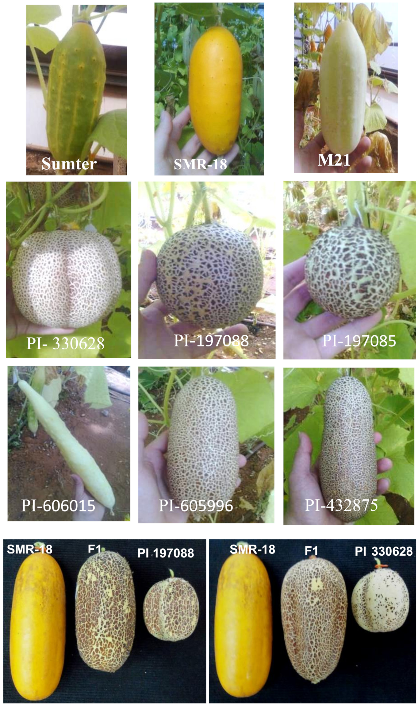
2.1. Cucumber Accessions

Nine accessions of cucumber (Cucumis sativus L) were obtained from Todd Whener, NCSU Charleston, NC, USA. Three accessions M-21, SMR-18, and Sumter are commercial cultivars whereas six Plant Introduction (PI) accessions PI 197085, PI 197087, PI 197088, PI 606015, PI 605996 and PI 432875 are wild C. sativum.

2.2. Pathogen

Forty-four field isolates of P. cubensis that were collected during 1980–2018 from 13 countries were available to perform the different experiments in the present study (Table 2), including two F1 hybrid isolates that were produced in our laboratory by crossing A1 and A2 field isolates from different hosts as described before [2]. A subset of 23 isolates was used to screen the resistance of PI 197088 and PI 330628 while other subsets of isolates were used to determine the resistance of F2 and F3 populations. The isolates were maintained by repeated inoculation of detached cucumber leaves of the universal susceptible cucumber line Nadiojny (own bred). Long-term maintenance of the isolates was done by storing freshly-sporulating leaves in dry paper bags at −80 °C.

2.3. Crosses

The susceptible SMR-18 and the resistant PI 197088 and PI 330628 were self-pollinated for three generations to ensure homozygosity (see below). Crosses were made between the susceptible SMR-18 and each of the resistant PI 197088 or PI 330628. Another cross was done between these two resistant accessions. F1 plants were grown in a net-house (insect-proof) and self-pollinated to obtain F2 populations. A single fruit was harvested from each F1 plant and the F2 seeds were grown the following season in net houses and self-pollinated to obtain F3 plants.

2.4. Inoculation of Detached Leaves

Parents, F1, F2, and F3 plants were grown erect in net-houses during 2014–2019. The third leaf from the top of 15–20-leaf plants were excised, placed on a wet filter paper in flat plastic trays (60 × 40 × 5 cm), lower surface uppermost, and spray-inoculated with a sporangial suspension of P. cubensis (2000 sporangia per mL). Trays were covered with transparent plastic bags and kept for 16 h in a dew chamber at 18 °C in the dark and thereafter in a growth chamber at 20 °C (14 h light/day, 100 µmole·s−1·m2) for 7 days.

2.5. Disease Assessment in Detached Leaves

Two readings were taken from each detached leaf at 7 days post-inoculation (dpi): the proportion of leaf area (0–100%) occupied with downy mildew lesions and the intensity of sporulation (0–3 scale) as visualized with a ×10 magnifying lens. The two values were multiplied to obtain a disease scoring scale of 0–300 (Figure 1). Leaves showing a score of 0–20; 21–200; 201–300 were considered resistant (R); moderately resistant (MR) and susceptible (S), respectively.

2.6. Inheritance of Resistance in Detached Leaves Taken from Adult Plants

Leaves (3rd leaf from the top) were detached daily from adult F2 plants growing in the field (summer 2014), placed on moistened filter paper inside sealed transparent trays and spray-inoculated with sporangia of each of the 22 isolates (PI 197088 × SMR-18) or 14 isolates (PI 330628 × SMR-18) of P. cubensis. The percentage of leaf area infected and sporulation intensity at ×10 magnification on a scale of 0–3 were recorded at 7 dpi.

2.7. Inheritance of Resistance in Intact Field-Grown Plants

Two-leaf plants of parents of F1 and F2 families were transplanted to the field in mid-March. Plants were grown erect in net houses (50 × 6 m) during the spring seasons of 2013, 2014, 2016, 2017, and 2019. In 2016, plants were also grown in the autumn season. F3 plants were similarly grown in the field in 2014. Natural infection with downy mildew initiated at 2–3 weeks after planting. Percentage of leaf area occupied by downy mildew lesions was visually assessed in each plant once or twice a week for 5–6 weeks after planting (unless stated otherwise). AUDPC (area under disease progress curve) was used to categorize plants as resistant, moderately resistant, or susceptible to the disease. A plant showing an AUDPC value of ≤5% of the maximal AUDPC value in that season was considered resistant whereas a plant showing an AUDPC value of ≥80% of that maximal value was considered susceptible. All other plants were considered moderately resistant.

2.8. Microscopy

The methods used by Cohen et al. [33] were used. Briefly, healthy and infected leaf discs were clarified in boiling ethanol, placed in aniline blue solution (0.05% aniline blue in 0.05 M K2HPO4, pH 8.9) at 4 °C for 24 h, stained with 0.01% calcofluor (Sigma) and examined with an Olympus A70 epi-fluorescent microscope. Sporangiophores on leaf surface fluoresced blue and sporangia looked dark. Fungal structures inside the leaf showed green-yellow fluorescence. Callose-encased haustoria were seen yellow. Staining for lignin was done with ethanol-clarified leaf discs. They were placed on microscope slides, treated with 2% phloroglucinol in methanol, and then with 0.25% HCl. A red color was visible in the lignified mesophyll cell.

3. Results

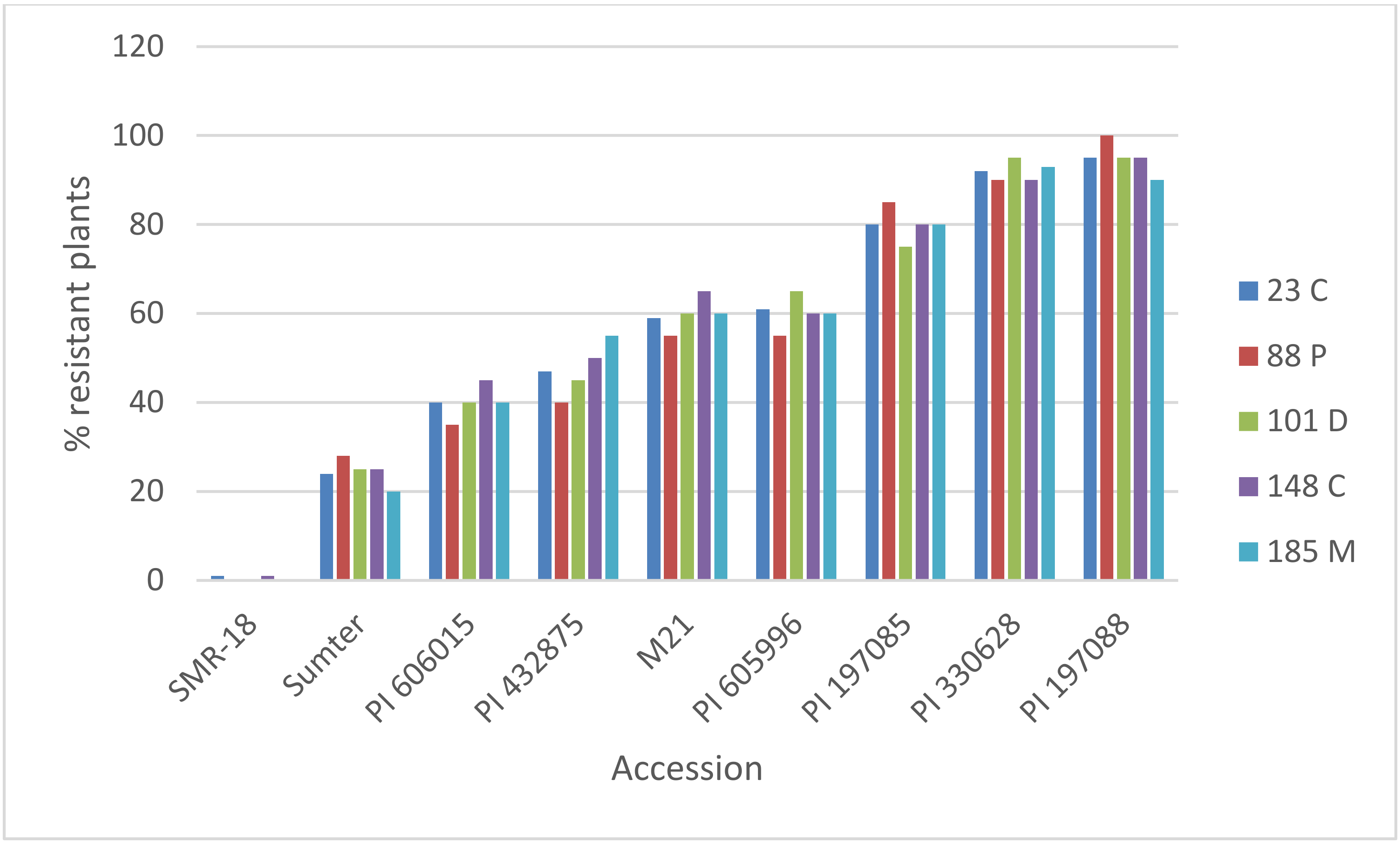
3.1. Resistance of C. sativus to Multiple Isolates of P. cubensis

The fruits of nine accessions of cucumber used in this study: SMR-18, M-21, Sumter, PI 606015, PI 432875, PI 605996, PI 197085, PI 197088, and PI 330628 are shown in Figure 2. Also shown are the F1 fruits of PI 197088 × SMR-18 and PI 330628 × SMR-18.
The resistance of nine accessions of Cucumis sativus (15–28 plants per accession, detached leaf bioassay) against each of five Israeli isolates of P. cubensis is shown in Figure 3. Most resistant accessions were PI 197088 and PI 330628. To test the homozygosity of the two most resistant accessions, detached leaves taken from 19 individual plants of PI 197088 and 19 individual plants of PI 330628 were each inoculated with each of the 23 isolates of P. cubensis from different parts of the world. The resistance profile of each plant is shown in Table 3. Nine PI 197088 plants (Figure 4A) and three PI 330628 plants (Figure 4B) were resistant to all 23 isolates of the pathogen, suggesting heterozygosity of the original accessions. One resistant plant of each PI was self-pollinated for two more generations and its offspring plants were all found resistant to these isolates of the pathogen. These two plants were used to study the resistance mechanisms and the mode of inheritance of resistance.

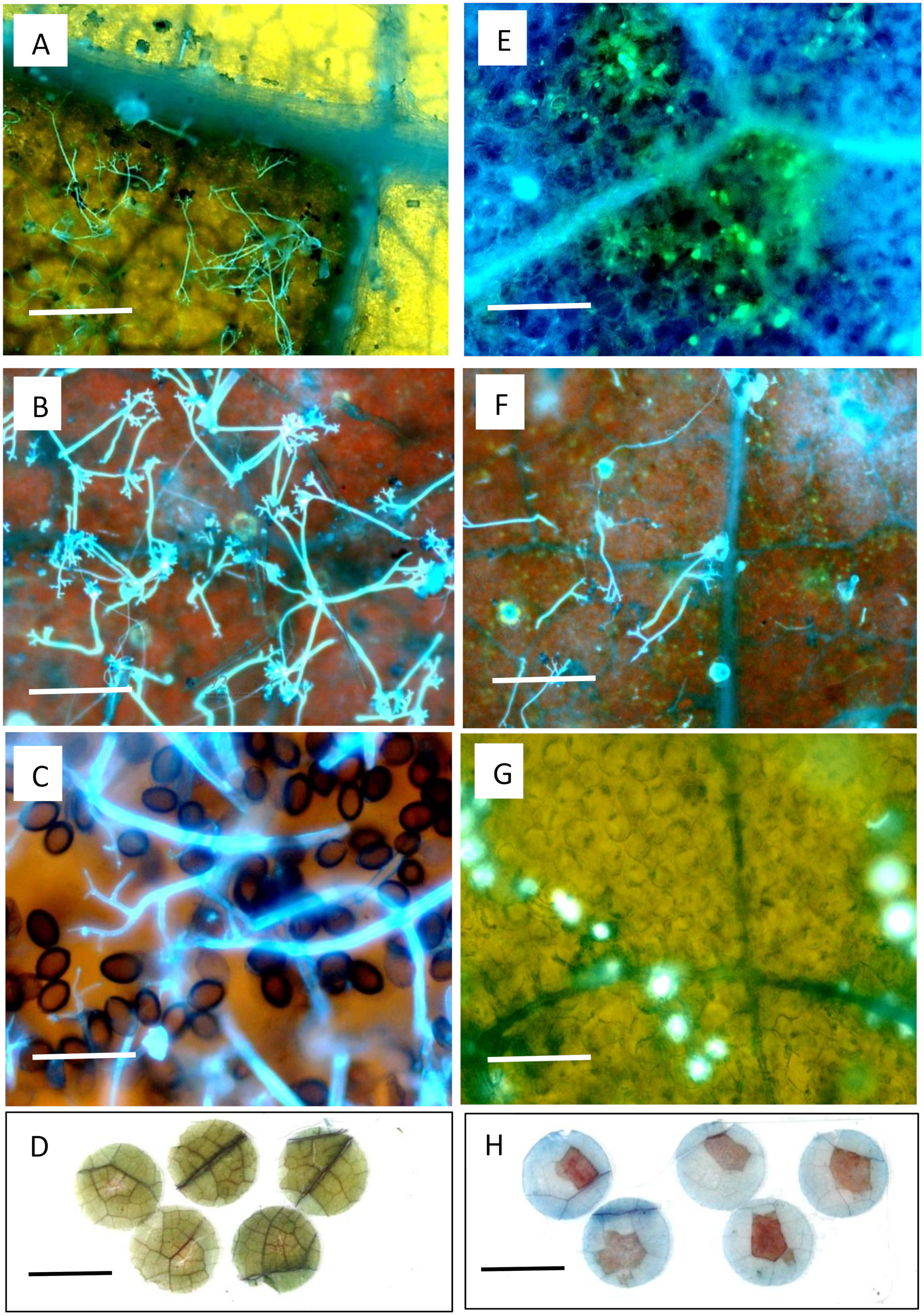
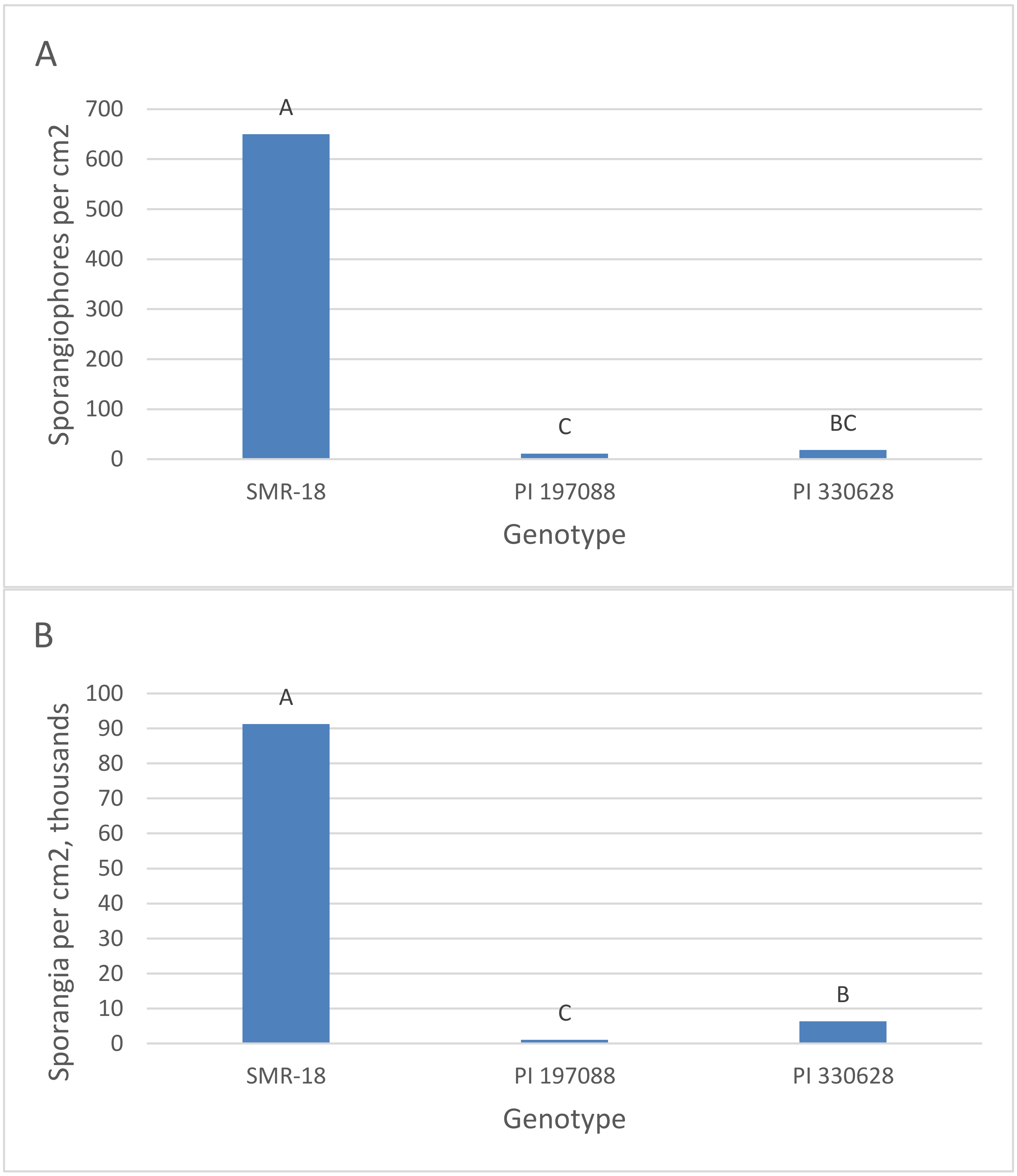
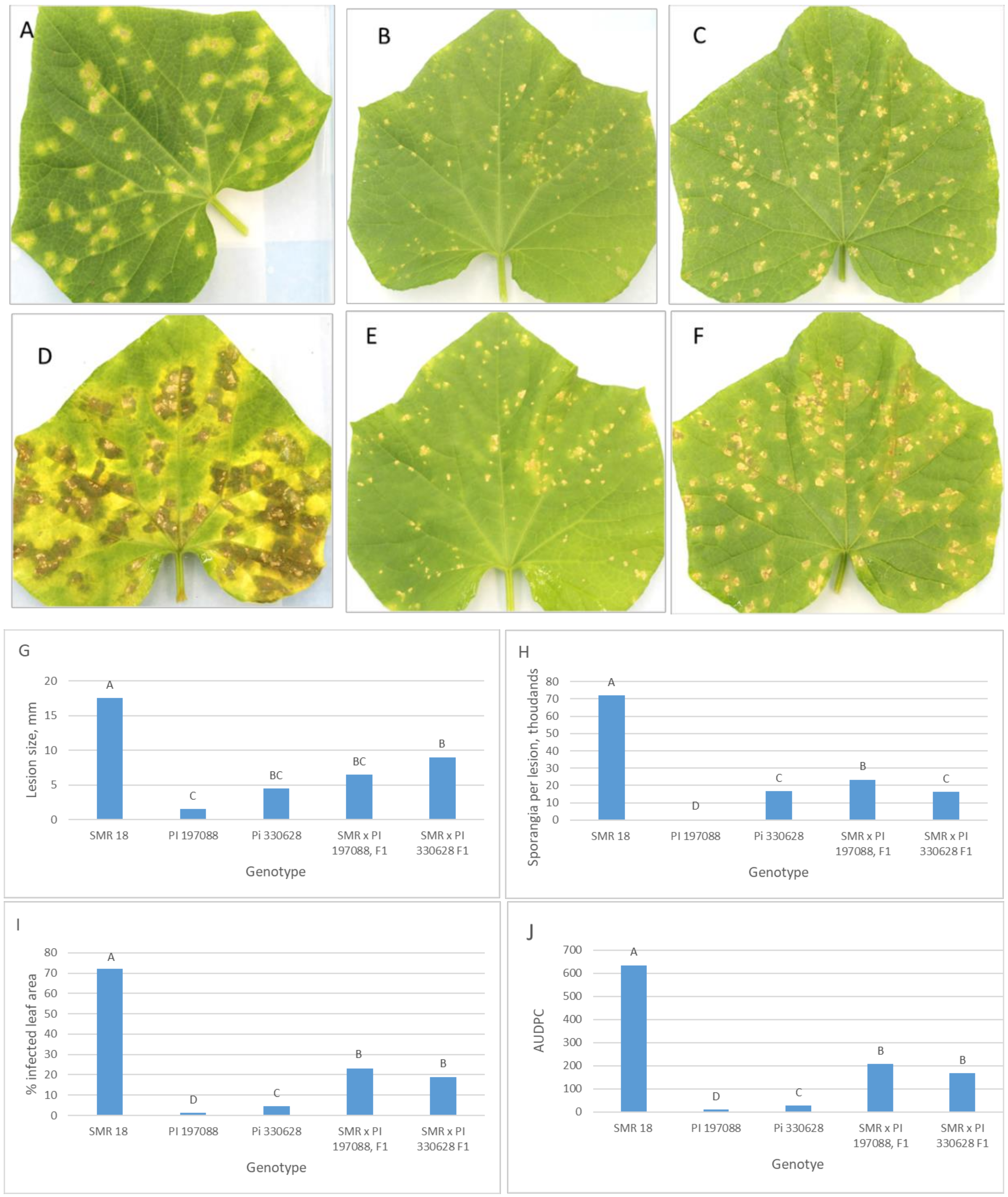
3.2. Microscopy of Resistance in PI 197088 and PI 330628

Leaf discs (15 mm diameter) were removed at 6 dpi from detached infected leaves (isolate 83C) of SMR-18, PI 197088, and PI 330628 and examined under UV illumination. Abundant sporangiophores with sporangia were seen in the susceptible SMR-18 (Figure 5A–C). In contrast, a few mycelium runners bearing callose-encased haustoria were seen in PI 197088 (Figure 5E,G). Similar callose depositions were seen in PI 330628 (not shown). Sporangiophores were partially branched with no sporangia seen in the resistant plants (Figure 5F). Bright-field microscopy of phloroglucinol-stained infected leaf discs showed no lignin staining in SMR-18 (Figure 5D) but heavy lignin accumulation in the resistant PI 197088 (Figure 5H). Similar lignin accumulation was seen in the resistant PI 330628 (not shown). Resistance to downy mildew in PI 197088 and PI 330628 was stable at a colonization temperature of 14 °C (not shown).

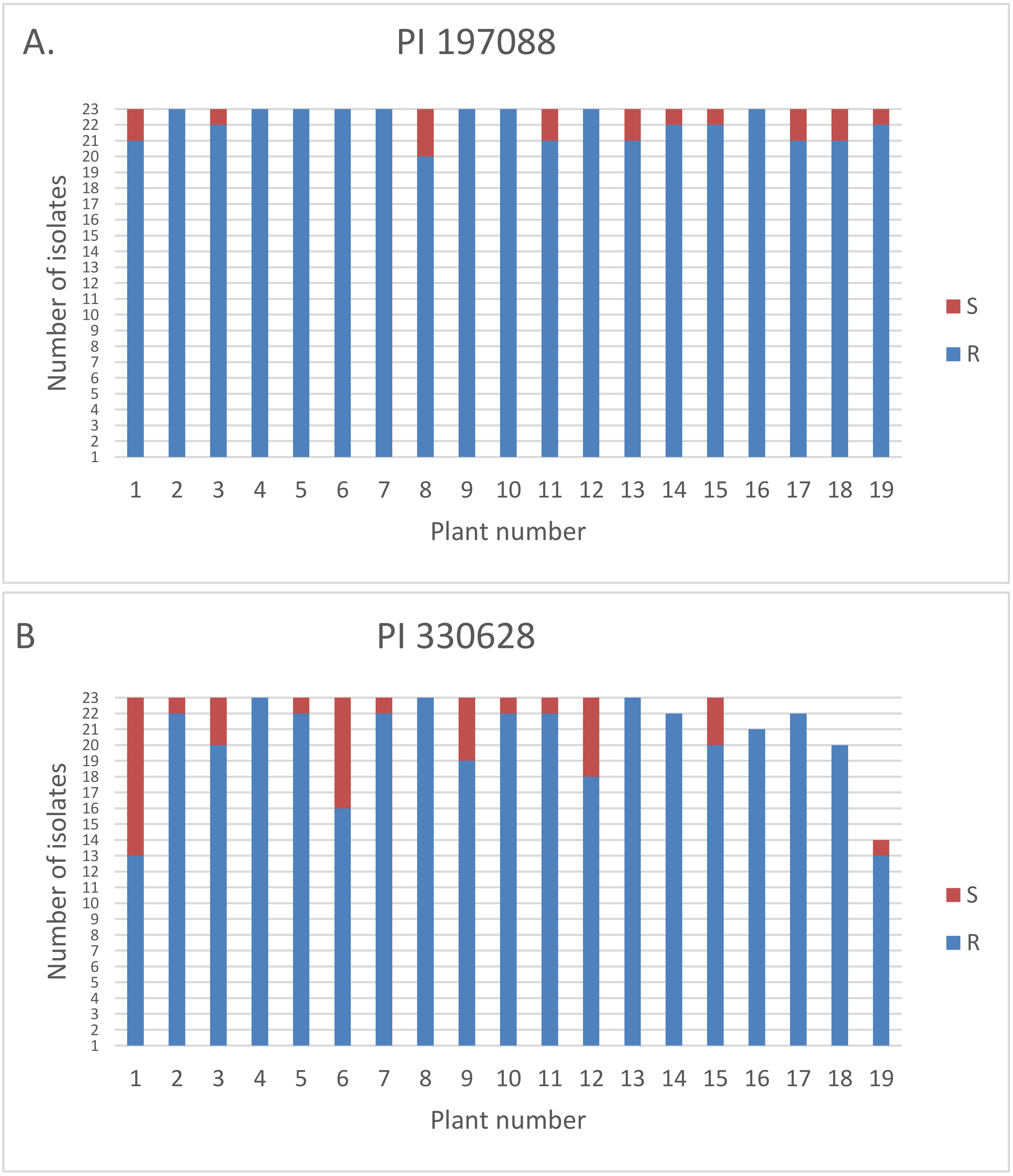
3.3. Quantification of Resistance in Adult Plants

The third leaf from the top of adult SMR-18, PI 197088, and PI 330628 plants (n = 10) grown in a net house was detached and drop-inoculated on the lower leaf surface with isolate 260. Fifteen mm leaf discs were sampled at 7 dpi for microscopic examination of sporulation. UV-epifluorescence microscopy of calcofluor-stained leaf discs revealed 650 ± 123 sporangiophores/cm2 in SMR-18 as against 11 ± 8 and 18 ± 12 sporangiophores/cm2 in PI 197088 and PI 330628, respectively (Figure 6A). The sporangiophores in SMR-18 were branched dichotomously three times, twice in PI 330628 and only once in PI 197088. The number of sporangia produced in SMR-18, PI 197088, and PI 330628 was 91 ± 5, 1 ± 0.3, and 6 ± 1 thousands of sporangia per cm2, respectively (Figure 6B). Similar results were obtained with other isolates of the pathogen (not shown). The data indicated that PI 197088 is slightly more resistant to downy mildew than PI 330628.
Artificial inoculation of intact plants (10-leaf stage) in the field with isolate 260 resulted at 9 dpi with the production of chlorotic lesions in SMR-18 and minute necrotic lesions in PI 197088 and PI 330628 (Figure 7A–C). Some infected leaves were detached, placed on wet filter paper and incubated in a growth chamber at 20 °C for three days (14 h light a day, 100 µmole·s−1·m2). At 13 dpi, SMR-18 produced the largest lesions (17.5 ± 3.5 mm) and the highest number of sporangia 71.9 ± 6.2 × 10³ per lesion whereas PI 197088 produced the smallest lesions (1.5 ± 0.7 mm) with the lowest number of sporangia per lesion (0.4 ± 0.1 × 10³) (Figure 7G,H). The F1 hybrid plants produced intermediate-size lesions with a moderate number of sporangia per lesion (Figure 7G,H). Percent leaf area infected at 27 dpi in fully-grown, blooming plants in the field is shown in Figure 7I. While SMR-18 exhibited 72% infected leaf area, PI197088 and PI330628 showed 1.2 and 4.4% infected leaf area, respectively. Their F1 plants were moderately infected (Figure 7I). AUDPC values at the end of the season are shown in Figure 7J. These results indicated that resistance of 197088 and 330628 is controlled by partially dominant gene(s).
When SMR-18, PI 197088, and PI 330628 (n = 10) were planted in a net house and thus exposed to natural infection, severe downy mildew developed in SMR-18 within four weeks after planting whereas no disease symptoms were seen in PI 197088 or PI 330628 (Figure 8). The disease was not visible in these two PI’s even at four months after planting when they carried mature fruits (produced by hand pollination), suggesting that homozygosity may avoid the appearance of the susceptible reaction reported to occur in these accessions at an advanced stage of growth in the field [28].
Resistance to downy mildew in melon Cucumis melo PI 124111F was shown to be active at colonization temperatures of >15 °C [34] due to the expression of eR genes [35]. Here, we observed that both PI 197088 and PI 330628 sustained full resistance to multiple isolates of P. cubensis when incubated after inoculation at either 14 °C or 20 °C (detached leaf bioassay), suggesting that unlike melon, expression of resistance against downy mildew in cucumber occurs at a low temperature of 14 °C.

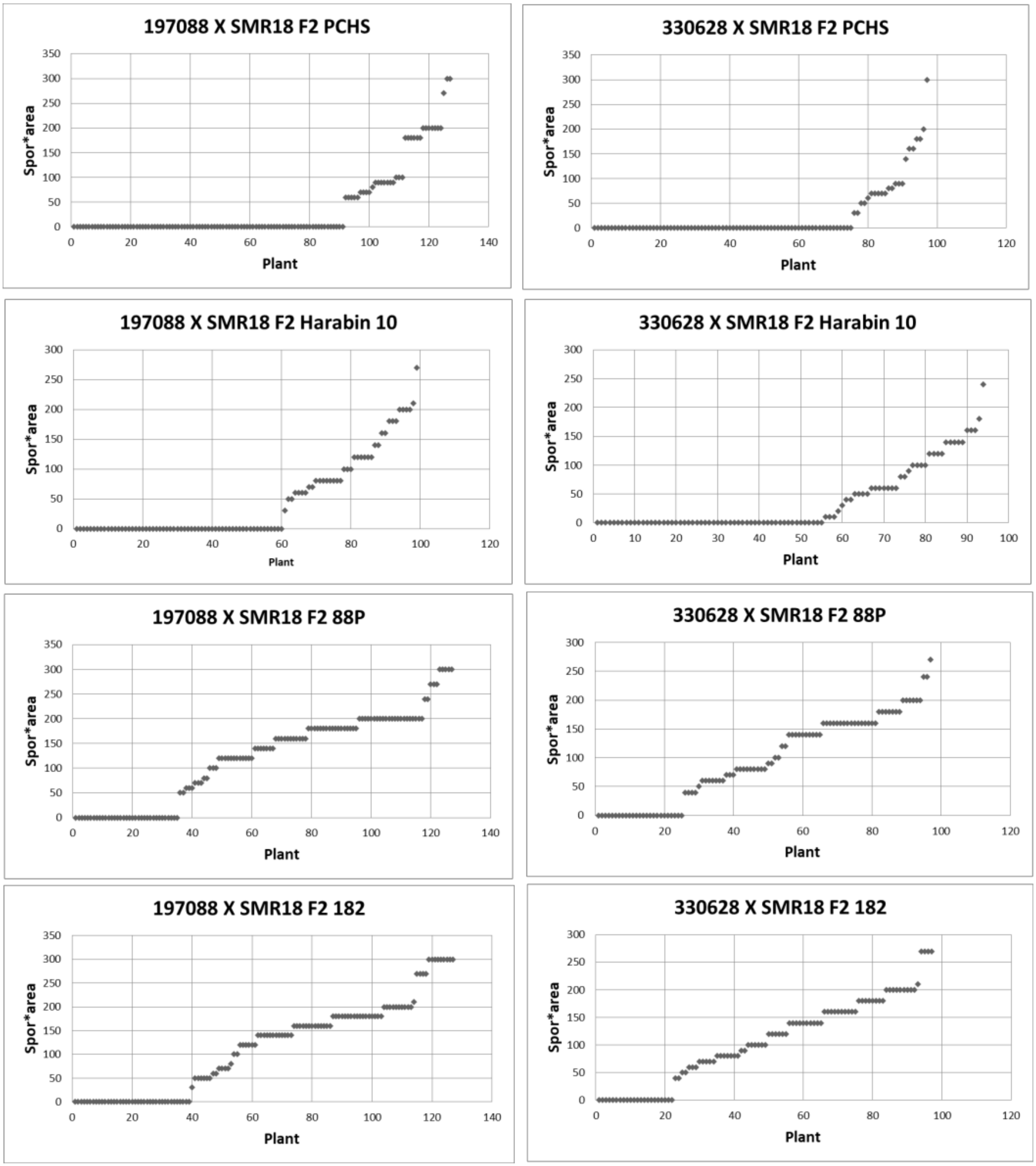
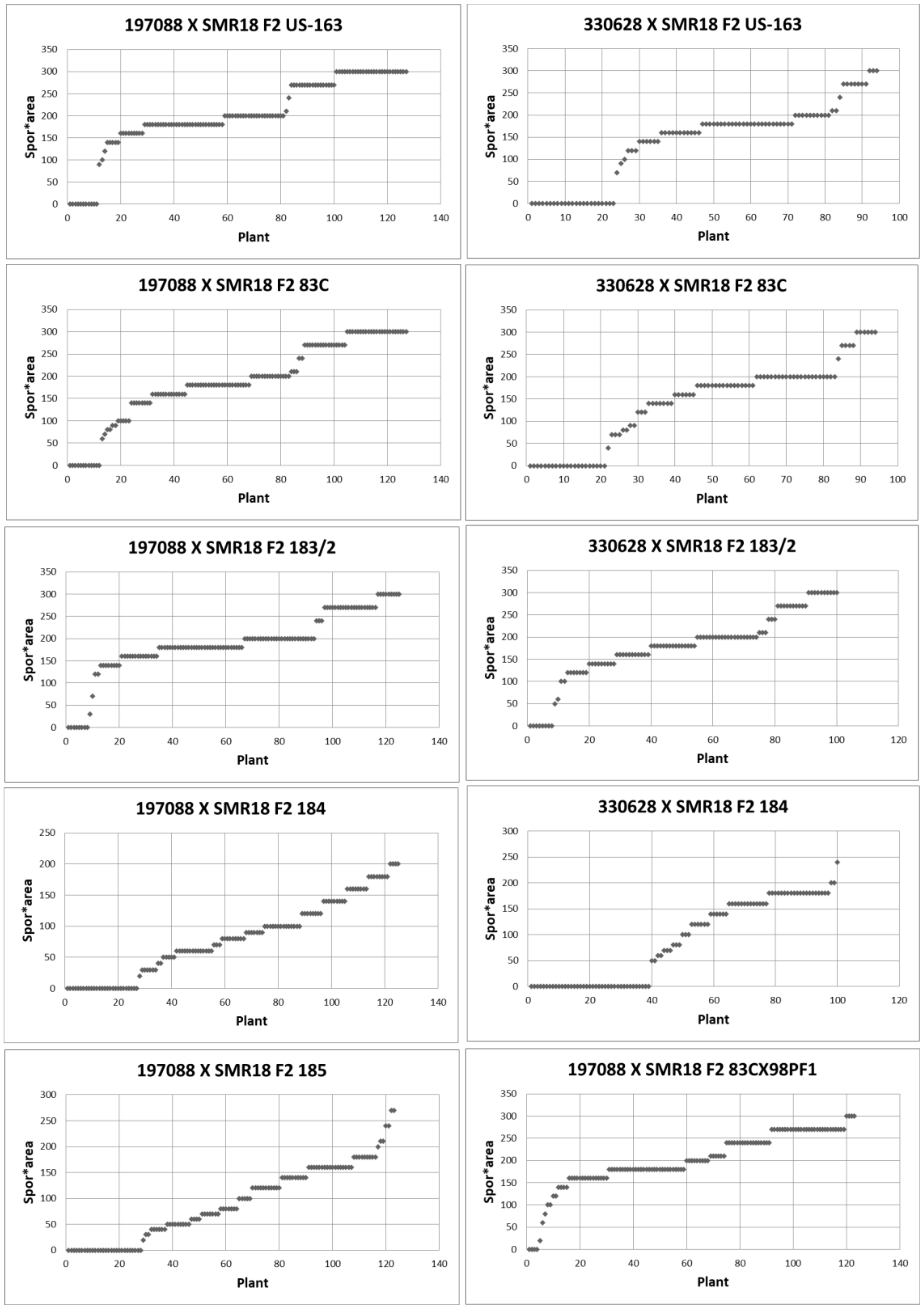
3.4. Inheritance of Resistance in Detached Leaves of Adult Plants

A set of 22 isolates was used to inoculate F2 plants of the cross PI 197088 × SMR 18 and a set of 14 isolates (all included in the former set) was used to inoculate F2 plants of the cross PI 330628 × SMR 18. Graphical illustrations of the disease scores are given for only 10 and 8 isolates of the above respective crosses (Figure 9 and Figure 10). Full numerical scores for all 22 and 14 isolates are given in Table 4. Data in Figure 9 and Figure 10 show a unique segregation pattern for each isolate. With some isolates, a similar pattern was seen for F2 of both crosses PI 197088 × SMR 18 and PI 330628 × SMR 18. Large differences in the response to inoculation were observed between plants, depending on the isolate used for inoculation. A single plant could react with different scores when inoculated with different isolates. The Mendelian analyses of the data are presented in Table 4. When plants’ responses were classified into two categories, F2 plants of the cross PI 197088 × SMR-18 showed five segregation ratios, depending on the isolate used for inoculation: 3:1 (2 isolates), 1:3 (11 isolates), 1:15 (6 isolates), 9:7 (2 isolates), and 13:3 (one isolate) (Table 4A). When three categories were used for classification, only 2 isolates out of 22, obeyed the Mendelian segregation of 9:6:1 (Table 4A). No inheritance model could be assigned to the results obtained with the other 19 isolates. Five segregation ratios were observed when two categories were applied for classification of the F2 plants of the cross PI 330628 × SMR-18: 3:1 (1 isolate), 1:3 (8 isolates), 1: 15 (2 isolates), 7:9 (2 isolates), and 9:7 (1 isolate) (Table 4B). When three categories were used for classification, only one isolate out of 14 obeyed a segregation ratio of 9:7. No inheritance model could be assigned to the results obtained for the other isolates (Table 4B).

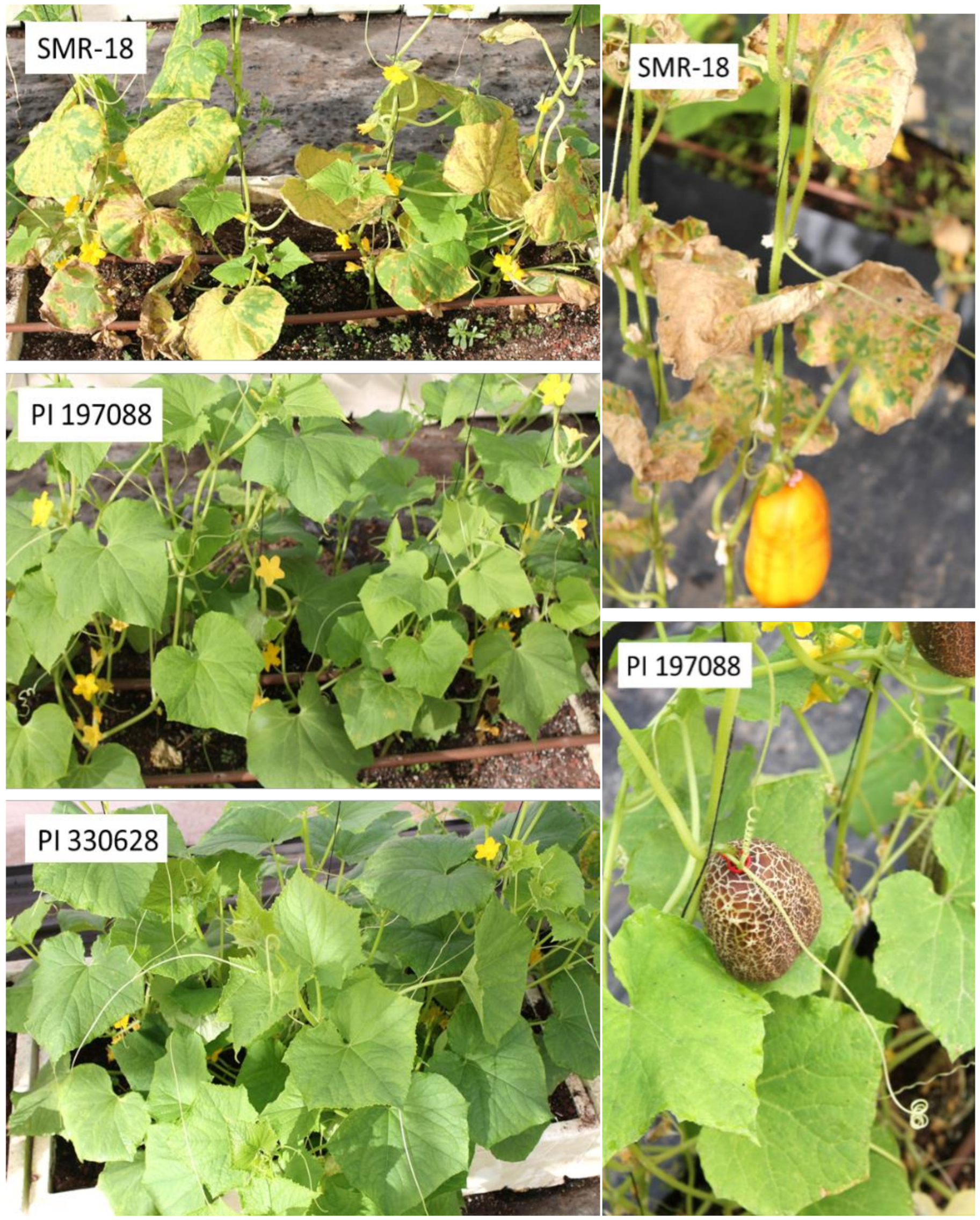
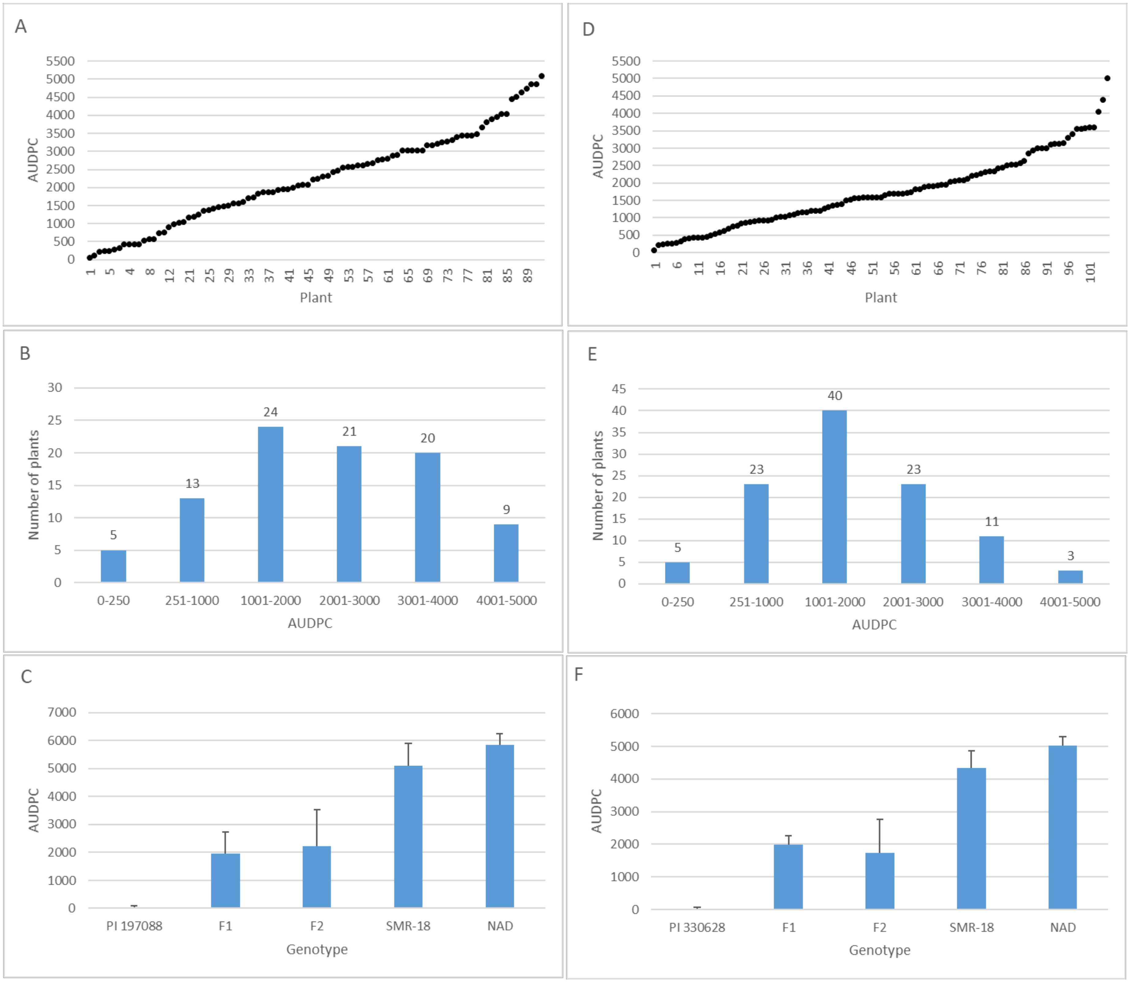
3.5. Inheritance of Resistance in Intact Field-Grown Plants

The response of the parents F1 and F2 plants to downy mildew in the field in 2019 is shown in Figure 11. A continuous response pattern to the disease was observed in F2 plants of PI 197088 × SMR-18 (Figure 11A,B) and of PI 330628 × SMR-18 (Figure 11D,E). Both resistant parents were completely resistant all along the season (until fruit maturity) whereas F1 plants were moderately resistant (Figure 11C,F).
Data in Table 5 summarize the segregation for the resistance of F2 plants in the field during 2013–2019. F2 plants of the cross PI 197088 × SMR-18 were tested in six seasons whereas F2 plants of the cross PI 330628 × SMR-18 were tested in two seasons. When two categories were used to classify the response of the plants to the disease (R and S),, two segregation ratios were observed, 1:15 or 1:63. When three categories were used for classification (R, MR, and S), two segregation ratios were observed, 1:14:1 or 1:9:6. The data suggest that genetic control of resistance in F2 plants varies between seasons, probably depending on the isolate prevailing in the field at each season.

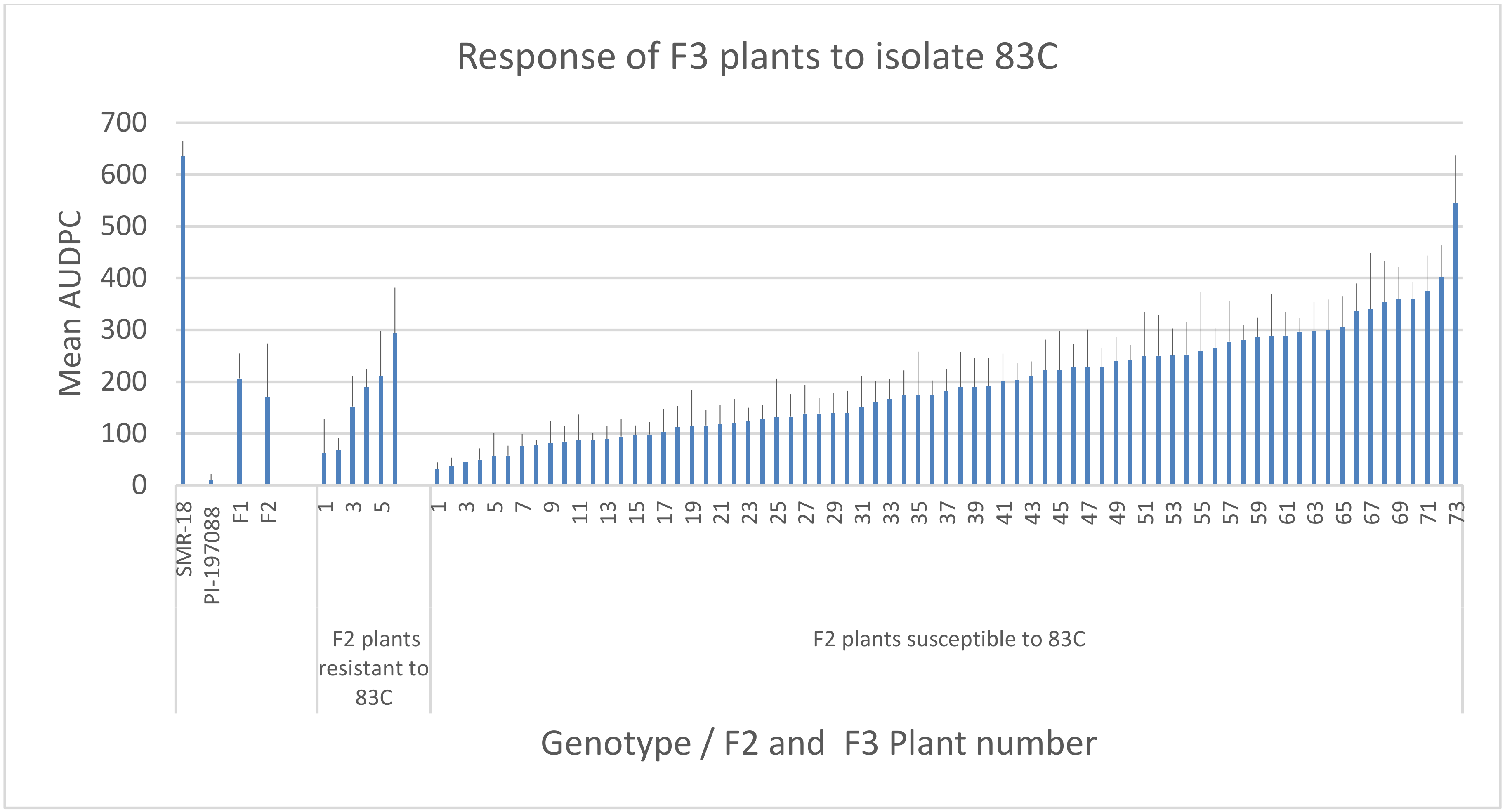
3.6. Resistance in F2: F3 Plants

Two field-grown resistant F2 plants (107 and 111) of the cross PI 197088 × SMR-18, were tested (detached leaf bioassay) for resistance against 14 isolates of P. cubensis. Plant 107 was resistant to all isolates, except to the hybrid isolate 83C × 98P, while plant 111 was resistant to all isolates except to the hybrid isolate 172 B × 183 C (Table 6A), suggesting enhanced virulence of hybrid isolates. Each plant was self-pollinated and detached leaves from the F3 plants growing in the field were inoculated with each of the four isolates of the pathogen. The segregation data are shown in Table 6B. F3 plants segregated into R: S at a ratio of 3:1, 1:3, 1:15, or 7:9, depending on the isolate used for inoculation. No genetic model fits the segregation data when three response categories were used because no MR-scored plants were detected in the progenies (Table 6B).

3.7. Resistance of F3 Plants in the Field

Seventy-nine F2 plants of the cross PI 197088 × SMR-18, six resistant (score 0), and 73 susceptible (score ≥ 200) (Figure 10) were self-pollinated to produce F3 seeds. Ten plants of each F3 entry were transplanted to the field and exposed to natural infection. Disease records were taken at weekly intervals for 22 days after the onset of the disease. Mean AUDPC and SD values for each F3 entry (n = 10) are shown in Figure 12. Mean AUDPC of SMR-18, PI 197088, and their F1 were 635, 10, and 206, respectively. F3 entries derived from resistant F2 plants showed AUDPC values ranging from 62 to 293 and those derived from susceptible F2 plants showed AUDPC values ranging from 31–545 (Figure 12). The results indicate that F3 plants are heterozygous for resistance, regardless of whether they were derived from a resistant or a susceptible F2 plant.

4. Discussion

The population of P. cubensis in the field may consist of many isolates, pathotypes, or races with varying degrees of pathogenicity or virulence thus rendering host resistance ineffective. Indeed, combating downy mildew (DM) in cucumber through host plant resistance or fungicide applications has become more complex in the past two decades due to the emergence of new pathotypes, races, and mating types of the causal agent P. cubensis. Old cucumber cultivars resistant to DM succumbed to the new pathotypes, and the old fungicidal chemistries lost activity due to the prevalence of resistant isolates of the pathogen [4,13]. Breeding cucumber for DM resistance is a long and laborious task due to the lack of stable, multi-race resistant sources and the complex mode of resistance inheritance.
Here we identified two sources of wild cucumber with multi-race/pathotype resistance. We characterized the mechanism of their resistance and determined the way they inherit resistance to their progeny plants. Because the resistance of accession to a local isolate of P. cubensis does not necessarily mean that it will be resistant to isolates that prevail in other locations, we used a large collection of isolates from different parts of the world to screen resistance. We developed a detached leaf bioassay in which we could determine the resistance of a single plant to multiple isolates of P. cubensis. We thus were able, for the first time, to study the mode of inheritance of resistance to multiple isolates and predict the performance of the resistant pedigrees in other countries.
Of the six Cucumis sativum genotypes known to exhibit resistance against P. cubensis [4], only PI 197088 and PI 330628 [28,30] exhibited multiple-isolate resistance. They were self-pollinated for three generations to bring their multiple-isolate resistance to homozygosity. The stabilized lines were used for the inheritance studies reported here.
When grown in the field under natural epiphytotic conditions, no disease was observed on the leaves of PI 197088 or PI 330628. However, when artificially inoculated in the field or in growth chambers, a few necrotic lesions did appear. Microscopic observations revealed that PI 197088 and PI 330628 exhibit similar responses to artificial inoculation with P. cubensis. The pathogen ceased developing at a relatively late stage after penetration and developed some initial hyphae and haustoria. The haustoria formed were encased with callose, which probably inhibits the intake of nutrients into the mycelium, while the infected cells accumulated lignin-like, phloroglucinol-positive materials. A similar structural mode of resistance was observed in melons resistant to P. cubensis [33,34]. These defense compounds still allowed some deteriorated sporangiophores to emerge from the stoma but almost totally prevented sporangial production. We show here that unlike the resistance in melon which breaks down at 14 °C [34], the resistance in PI 197088 and PI 330628 remained effective at a low colonization temperature of 14 °C.
We used a double visual scoring system (percent infected leaf area and sporulation intensity) to determine the level of resistance to DM in detached leaves (Figure 1). We observed that leaves taken from F2 plants of the cross PI 197088 × SMR-18 or 330628 × SMR-18 segregated in their phenotypic responses to infection with P. cubensis ranging from complete resistance to high susceptibility. The pattern of segregation depended on the isolate used for inoculation. The differential pattern of response to different isolates indicated that a number of genes might be involved in resistance. The Mendelian analysis was employed to the segregated populations after categorical classification into S: R or R: MR: S. The analysis of two categories R and S indicated that resistance in PI 197088 or PI 330628 is controlled by either 1 dominant, 1 recessive, or 2 recessive genes, depending on the isolate used for inoculation. Analysis with three categories of S, MR, and R did not fit, in most cases, any Mendelian model of segregation.
F3 plants, derived from either susceptible or resistant F2 plants of the cross PI 197088 × SMR-18, showed a continuous pattern of variable resistance to DM in the field. When detached leaves were inoculated with different isolates, F3 plants segregated R:S 3:1 or 1:3, depending on the isolate used for inoculation, reaffirming that inheritance of resistance to DM is isolate-dependent.
Interestingly, field-grown F2 plants of the cross between the two resistant genotypes PI 197088 × PI 330628 were all fully resistant at the end of the season (no DM symptoms). However, two out of 75 plants in 2017 and one out of 130 plants in 2019 showed a few DM lesions, consisting of about 5% infected leaf area. Neighboring SMR-18 plants showed about 90% leaf area infected. This suggests that PI 197088 and PI 330628 differ in at least one gene for resistance. On the other hand, they share one QTL dm4.1 as suggested by Wang et al. [28].
Isolate-dependent inheritance of disease resistance is a rarely reported phenomenon [36]. Lapin et al. [37] showed that unlike most natural Arabidopsis thaliana accessions that are susceptible to one or more isolates of the downy mildew pathogen Hyaloperonospora arabidopsidis, accession C24 is resistant to all isolates tested. The resistance of C24 was found to be a multigenic trait with complex inheritance. Many identified resistance loci were isolate-specific and located on different chromosomes. Among the C24 resistance QTLs, there were dominant, codominant, and recessive loci. Interestingly, none of the identified loci significantly contributed to resistance against all three tested isolates.
Unlike wild cucumbers, resistance of the wild melon (Cucumis melo L) PI 124111F against P. cubensis is broad-spectrum but not isolate-specific [38]. That resistance was controlled genetically by two partially dominant, complementary loci [39]. Unlike other plant disease resistance genes, which confer an ability to resist infection by pathogens expressing corresponding avirulence genes, the resistance of PI 124111F to P. cubensis is controlled by enhanced expression of the enzymatic resistance (eR) genes At1 and At2. These constitutively expressed genes encode the photorespiratory peroxisomal enzyme proteins glyoxylate aminotransferases. The low expression of At1 and At2 in susceptible melon lines is regulated mainly at the transcriptional level. This regulation is independent of infection with the pathogen. Transgenic melon plants overexpressing either of these eR genes displayed the enhanced activity of glyoxylate aminotransferases and remarkable resistance against P. cubensis [35,40]. Our attempts to transfer At1 and At2 to cucumber did not succeed (Cohen, unpublished data).
The results presented here corroborate with other studies in which multiple QTLs for resistance against P. cubensis were identified in PI 197088 and PI 330628. (Table 1). Wang et al. [30] reported QTL mapping results for DM resistance with F2:3 families from the cross between DM-resistant inbred line PI 330628 (WI7120) and susceptible ‘9930’. Four QTLs, dm2.1, dm4.1, dm5.1, and dm6.1 were consistently and reliably detected across at least three of the four environments which together could explain 62–76% phenotypic variations. Among them, dm4.1 and dm5.1 were major effect QTL and dm2.1 and dm6.1 had moderate and minor effects, respectively.
Wang et al. [28] used recombinant inbred lines from a cross between PI 197088 and the susceptible line ‘Coolgreen’. Phenotypic data on responses to natural DM infection were collected in three years and five locations from replicated field trials in North Carolina. The observed ratings followed a normal distribution that covered a large range of ratings at each environment and date. The interaction effects of genotype-by-location and genotype-by-year were significant at all ratings. QTL analysis identified 11 QTL for DM resistance harbored on chromosomes 1–6, accounting for more than 73.5% total phenotypic variance. Among the 11 DM resistance QTLs, dm5.1, dm5.2, and dm5.3 were major effect contributing QTL whereas dm1.1, dm2.1, and dm6.2 conferred susceptibility. The QTL dm4.1 which had a moderate effect was likely the same as the major-effect QTL dm4.1 detected in PI 330628 [30]. Three DM QTLs dm2.1, dm5.2, and dm6.1, were co-localized with powdery mildew (PM) QTLs, pm2.1, pm5.1, and pm6.1, respectively, which was consistent with the observed linkage of PM and DM resistances in PI 197088.
Katz et al. [29] reported on nine QTLs associated with resistance of PI 197088 against each of the seven isolates of P. cubensis. They examined for two years the response of a segregating F2 family (PI-197088 × SMR-18, n = 170) to seven isolates in growth chambers and the field. NGS (Next-Generation Sequencing) was performed for genotyping, and polymorphic SNPs were obtained from the same populations in both years. QTLs obtained for isolate 23C- resided on chromosomes 4 and 5; for isolate Pol.1- on chromosomes 1, 4, and 5; for isolate Pol.4- on chromosome 7; for isolate US-506- on chromosomes 1 and 2; for isolate 81C- on chromosomes 4 and 5; for isolate 88C- on chromosomes 3 and 6; for isolate 90C- on chromosomes 1, 4, and 6; for field isolate 2016, on chromosomes 3 and 5, and for field isolate 2017- on chromosomes 4 and 5. These authors concluded that the inheritance of resistance against DM in PI 197088 was isolate-dependent.
Tian et al. [41] sequenced 14% of the genome of one isolate of P. cubensis and identified 32 putative RXLR effector proteins and 29 secreted peptides with high similarity to RXLR effectors. They suggested that these effectors might play pivotal roles in pathogen fitness and pathogenicity. Sexual reproduction of the pathogen [12,42] may result in recombinant isolates which carry various combinations of effector proteins. It might, therefore, occur that isolates evolved in different parts of the world and therefore belong to different races, pathotypes, and mating types, each carries a unique set of effectors. Of this set of effectors, some might be secreted while others may not. Of the secreted effectors, some may recognize certain R genes in the host while others will not. This will make some host genotypes resistant to some genotypes of the pathogen.
The isolate-dependent inheritance of resistance of cucumber against P. cubensis may indicate that each isolate secretes a different battery of effectors that ignite a unique set of R genes in the host, thus making the inheritance of resistance isolate-dependent.

Author Contributions

T.C., D.K., Y.B.N., R.H., and B.H.B.D. did the investigations. A.E.R.—curation. Y.C. supervision and writing of the manuscript. All authors have read and agreed to the published version of the manuscript.

Funding

This research received no external funding

Conflicts of Interest

The authors declare no conflict of interest.

References

  1. Palti, J.; Cohen, Y. Downy mildew of cucurbits (Pseudoperonospora cubensis)—The fungus and its hosts, distribution, epidemiology and control. Phytoparasitica 1980, 8, 109–147. [Google Scholar] [CrossRef]
  2. Lebeda, A.; Cohen, Y. Cucurbit downy mildew (Pseudoperonospora cubensis)—Biology, ecology, epidemiology, host-pathogen interaction and control. Eur. J. Plant Pathol. 2011, 129, 157–192. [Google Scholar] [CrossRef]
  3. Savory, E.; Granke, L.L.; Quesada-Ocampo, L.M.; Varbanova, M.; Hausbeck, M.K.; Day, B. The cucurbit downy mildew pathogen Pseudoperonospora cubensis. Mol. Plant Pathol. 2011, 12, 217–226. [Google Scholar] [CrossRef]
  4. Cohen, Y.; Langenberg, K.M.V.D.; Wehner, T.C.; Ojiambo, P.S.; Hausbeck, M.; Quesada-Ocampo, L.M.; Lebeda, A.; Sierotzki, H.; Gisi, U.; Whener, T.C. Resurgence of Pseudoperonospora cubensis—The causal agent of cucurbit downy mildew. Phytopathology 2015, 105, 1–15. [Google Scholar] [CrossRef] [Green Version]
  5. Ojiambo, P.; Gent, D.H.; Quesada-Ocampo, L.M.; Hausbeck, M.K.; Holmes, G.J. Epidemiology and population biology of Pseudoperonospora cubensis: A model system for management of downy mildews. Annu. Rev. Phytopathol. 2015, 53, 223–246. [Google Scholar] [CrossRef] [Green Version]
  6. Cohen, Y. The combined effects of temperature, leaf wetness and inoculum concentration on infection of cucumbers with Pseudoperonospora cubensis. Can. J. Bot. 1977, 55, 1478–1487. [Google Scholar] [CrossRef]
  7. Whisson, S.C.; Boevink, P.C.; Moleleki, L.N.; Avrova, A.O.; Morales, J.G.; Gilroy, E.M.; Armstrong, M.R.; Grouffaud, S.; Van West, P.; Chapman, S.; et al. A translocation signal for delivery of oomycete effector proteins into host plant cells. Nature 2007, 450, 115–118. [Google Scholar] [CrossRef]
  8. Perl, M.; Cohen, Y.; Rotem, J. The effect of humidity during darkness on the transfer of assimilates from cucumber leaves to sporangia of Pseudoperonospora cubensis. Physiol. Plant Pathol. 1972, 2, 113–122. [Google Scholar] [CrossRef]
  9. Cohen, Y. The novel oomycide oxathiapiprolin inhibits all stages in the asexual life cycle of Pseudoperonospora cubensis—Causal agent of cucurbit downy mildew. PLoS ONE 2015, 10, e0140015. [Google Scholar] [CrossRef] [Green Version]
  10. Cohen, Y.; Rubin, A.E.; Galperin, M. Control of cucumber downy mildew with novel fungicidal mixtures of oxathiapiprolin. Phytoparasitica 2018, 46, 689–704. [Google Scholar] [CrossRef]
  11. Call, A.D.; Criswell, A.D.; Wehner, T.C.; Ando, K.; Grumet, R. Resistance of cucumber cultivars to a new strain of cucurbit downy mildew. HortScience 2012, 47, 171–178. [Google Scholar] [CrossRef]
  12. Cohen, Y.; Rubin, A.E. Mating type and sexual reproduction of Pseudoperonospora cubensis, the downy mildew agent of cucurbits. Eur. J. Plant Pathol. 2012, 132, 577–592. [Google Scholar] [CrossRef]
  13. Thomas, A.; Carbone, I.; Lebeda, A.; Ojiambo, P. Virulence structure within populations of Pseudoperonospora cubensis in the United States. Phytopathology 2017, 107, 777–785. [Google Scholar] [CrossRef] [Green Version]
  14. Polat, I.; Baysal, O.; Mercati, F.; Kitner, M.; Cohen, Y.; Lebeda, A.; Carimi, F. Characterization of Pseudoperonospora cubensis isolates from Europe and Asia using ISSR and SRAP molecular markers. Eur. J. Plant Pathol. 2014, 139, 641–653. [Google Scholar] [CrossRef]
  15. Wallace, E.C.; D’Arcangelo, K.N.; Quesada-Ocampo, L.M. Population analyses reveal two host-adapted clades of Pseudoperonospora cubensis, the causal agent of cucurbit downy mildew, on commercial and wild cucurbits. Phytopathology 2020. [Google Scholar] [CrossRef]
  16. Cohen, Y.; Rubin, A.E.; Galperin, M. Host preference of mating type in Pseudoperonospora cubensis, the downy mildew causal agent of cucurbits. Plant Dis. 2013, 97, 292. [Google Scholar] [CrossRef]
  17. Pang, X.; Zhou, X.; Wan, H.; Chen, J. QTL mapping of downy mildew resistance in an introgression line derived from interspecific hybridization between cucumber and Cucumis hystrix. J. Phytopathol. 2013, 161, 536–543. [Google Scholar] [CrossRef]
  18. Li, L.; He, H.; Zou, Z.; Li, Y. QTL analysis for downy mildew resistance in cucumber inbred line PI 197088. Plant Dis. 2018, 102, 1240–1245. [Google Scholar] [CrossRef] [Green Version]
  19. Szczechura, W.; Staniaszek, M.; Kłosińska, U.; Kozik, E.U. Molecular Analysis of New Sources of Resistance to Pseudoperonospora cubensis (Berk. et Curt.) Rostovzev in Cucumber. Russ. J. Genet. 2015, 51, 974–979. [Google Scholar] [CrossRef]
  20. Van Vliet, G.J.A.; Meysing, W.D. Inheritance of resistance to Pseudoperonospora cubensis Rost. in cucumber (Cucumis sativus L.). Euphytica 1974, 23, 251–255. [Google Scholar] [CrossRef]
  21. McFerson, J.R. A Screening Procedure for Determining Levels of Resistance in Cucumber. Master’s Thesis, Texas A&M University, College Station, TX, USA, 1978. [Google Scholar]
  22. Pershin, A.F.; Medvedeva, N.I.; Medvedev, A.V. Quantitative approach to studying the genetics of disease resistance. IV. Interaction of the genetic systems for resistance to powdery and downy mildews in cucumber. Genet. USSR. 1988, 24, 484–493. [Google Scholar]
  23. Doruchowski, R.W.E.; Lakowska-Ryk, E. Inheritance of Resistance to Downy Mildew (Pseudoperonospora cubensis Berk & Curt) in Cucumis sativus. In Proceedings of the 5th EUCARPIA Symposium, Warsaw, Poland, 27–31 July 1992; Doruchowski, R.W., Kozik, E., Niemirowicz Szczytt, K., Eds.; Research Institute of Vegetable Crops and Warsaw Agricultural University: Warsaw, Poland, 1992; pp. 132–138. [Google Scholar]
  24. Petrov, L.; Boogert, K.; Sheck, L.; Baider, A.; Rubin, E.; Cohen, Y. Resistance to downy mildew, Pseudoperonospora cubensis, in cucumbers. Acta Hortic. 2000, 510, 203–209. [Google Scholar] [CrossRef]
  25. Angelov, D. Inheritance of resistance to downy mildew, Pseudoperonospora cubensis (Berk. & Curt.) Rostow. In Proceedings of the Report of the 2nd National Symposium of Plant Immunity, Plovdiv, Bulgaria, 1994; Volume 3, pp. 99–105. [Google Scholar]
  26. Caldwell, D.; Chan, E.; de Vries, J.; Joobeur, T.; King, J.; Reina, A.; Shetty, N. Methods and Compositions for Identifying Downy Mildew Resistant Cucumber Plants. U.S. Patent Application No. 2011/0126309 A1, April 2011. [Google Scholar]
  27. Yoshioka, Y.; Sakata, Y.; Sugiyama, M.; Fukino, N. Identification of quantitative trait loci for downy mildew resistance in cucumber (Cucumis sativus L.). Euphytica 2014, 198, 265–276. [Google Scholar] [CrossRef]
  28. Wang, Y.; Vandenlangenberg, K.; Wen, C.; Wehner, T.C.; Weng, Y. QTL mapping of downy and powdery mildew resistances in PI 197088 cucumber with genotyping-by-sequencing in RIL population. Theor. Appl. Genet. 2018, 131, 597–611. [Google Scholar] [CrossRef]
  29. Katz, D.; Hammer, R.; Ben Naim, Y.; Cohen, Y. Resistance to downy mildew in cucumber breeding lines. Phytoparasitica 2018, 46, 320. [Google Scholar]
  30. Wang, Y.; Vandenlangenberg, K.; Wehner, T.C.; Kraan, P.A.G.; Suelmann, J.; Zheng, X.; Owens, K.; Weng, Y. QTL mapping for downy mildew resistance in cucumber inbred line WI7120 (PI 330628). Theor. Appl. Genet. 2016, 1298, 1493–1505. [Google Scholar] [CrossRef]
  31. Zhang, S.P.; Liu, M.M.; Miao, H.; Zhang, S.Q.; Yang, Y.H.; Xie, B.Y.; Wehner, T.C.; Gu, X.F. Chromosomal mapping and QTL analysis of resistance to downy mildew in Cucumis sativus. Plant Dis. 2013, 97, 245–251. [Google Scholar] [CrossRef] [Green Version]
  32. Win, K.T.; Vegas, J.; Zhang, C.; Song, K.; Lee, S. QTL mapping for downy mildew resistance in cucumber via bulked segregant analysis using next-generation sequencing and conventional methods. Theor. Appl. Genet. 2017, 130, 199–211. [Google Scholar] [CrossRef]
  33. Cohen, Y.; Eyal, H.; Hanania, J.; Malik, Z. Ultrastructure of Pseudoperonospora cubensis in muskmelon genotype susceptible and resistant to downy mildew. Physiol. Mol. Plant Pathol. 1989, 34, 27–40. [Google Scholar] [CrossRef]
  34. Balass, M.; Cohen, Y.; Bar-Joseph, M. Temperature-dependent resistance to downy mildew in muskmelon: Structural responses. Physiol. Mol. Plant Pathol. 1993, 43, 11–20. [Google Scholar] [CrossRef]
  35. Taler, D.; Galperin, M.; Benjamin, I.; Cohen, Y.; Kenigsbuch, D. Plant eR genes encoding for photorespiratory enzymes confer resistance against disease. Plant Cell 2004, 16, 172–184. [Google Scholar] [CrossRef] [Green Version]
  36. Chen, T.; Cohen, Y. Isolate-dependent inheritance of resistance to downy mildew in cucumber. Phytoparasitica 2013, 41, 462. [Google Scholar]
  37. Lapin, D.; Meyer, R.C.; Takahashi, H.; Bechtold, U.; Ackerveken, G.V.D. Broad-spectrum resistance of Arabidopsis C24 to downy mildew is mediated by different combinations of isolate-specific loci. New Phytol. 2012, 196, 1171–1181. [Google Scholar] [CrossRef]
  38. Cohen, Y.; Eyal, H. Downy mildew-, powdery mildew- and fusarium wilt-resistant muskmelon breeding line PI-124111F. Phytoparasitica 1987, 15, 187–195. [Google Scholar] [CrossRef]
  39. Kenigsbuch, D.; Cohen, Y. Inheritance of resistance to downy mildew in Cucumis melo PI 124112 and commonalty of resistance genes with C. melo PI 124111F. Plant Dis. 1992, 76, 615–617. [Google Scholar] [CrossRef]
  40. Benjamin, I.; Kenigsbuch, D.; Galperin, M.; Abrameto, J.A.; Cohen, Y. Cisgenic melons over expressing glyoxylate-aminotransferase are resistant to downy mildew. Eur. J. Plant Pathol. 2009, 125, 355–365. [Google Scholar] [CrossRef]
  41. Tian, M.; Win, J.; Savory, E.; Burkhardt, A.K.; Held, M.A.; Brandizzi, F.; Day, B. 454 Genome sequencing of Pseudoperonospora cubensis reveals effector proteins with a QXLR translocation motif. Mol. Plant Microbe Interact. 2011, 24, 543–553. [Google Scholar] [CrossRef] [Green Version]
  42. Thomas, A.; Carbon, I.; Cohen, Y.; Ojiambo, P. Occurrence and distribution of mating types of Pseudoperonospora cubensis in the USA. Phytopathology 2017, 107, 313–321. [Google Scholar] [CrossRef] [Green Version]
Figure 1. Phenotypic score panel of downy mildew in detached leaves of cucumber infected with P. cubensis. A score composes of percent leaf area infected multiplied by sporulation intensity (at ×10 magnification on a scale of 0–3). The scores given to the leaves in the upper row are: 0, 0.5, 20, and 80, and in the lower row are: 150, 180, 237.5, and 285. Plants showing a score of 0–20, 21–100, and 101–300 were considered resistant, moderately resistant, and susceptible, respectively.
Agronomy 10 01086 g001
Figure 2. Fruits of three cultivated and six wild Cucumis sativus L. and F1s of two wild types with SMR-18.
Agronomy 10 01086 g002
Figure 3. Resistance of nine accessions of Cucumis sativus (15–28 plants per accession, detached leaf bioassay) against each of five Israeli isolates of P. cubensis. The origin of isolates is shown in Table 2.
Agronomy 10 01086 g003
Figure 4. Response of 19 individual plants of Cucumis sativus PI 197088 (A) and 19 plants of PI 330628 (B) to inoculation with 23 isolates of P. cubensis. A detached leaf bioassay. S = susceptible. R = resistant.
Agronomy 10 01086 g004
Figure 5. Development of P. cubensis (isolate 83C) in detached leaves of cucumber at 6 dpi in a growth chamber at 20 °C. (AD), SMR-18, susceptible. Note in (A) that the lesion is confined between the veins. Note in (B) the dichotomously branched sporangiophore in blue. Note in (C) the dark oval sporangia. (EH), PI 197088 or PI 330628, resistant. Note in (E) and (G) the yellow dotes which are callose-encased haustoria. Note in (F) that sporangiophores are branched only once. Scale bar: (A) and (E) = 200 µm. (B) and (F) = 100 µm. (C) and (G) = 50 µm. (DH), 15 mm. (AC) and (EG) are UV micrographs after calcofluor staining; (DH), bright field micrographs, phloroglucinol staining of ethanol-clarified leaf discs.
Agronomy 10 01086 g005
Figure 6. Quantification of resistance against isolate 260 of P. cubensis in detached leaves of PI 197088 and PI 330628 (F3) in growth chambers. (A) Formation of sporangiophores at 7 dpi. (B) Sporangial production at 7 dpi. Different letters on bars indicate a significant difference at α = 0.05 (t-test).
Agronomy 10 01086 g006
Figure 7. Phenology of downy mildew symptoms in leaves of field-grown intact cucumber plants (n = 10) after artificial spray inoculation with sporangia of isolate 260 of P. cubensis. (AC), symptoms at 9 dpi. (D,E) symptoms after an additional 4 days of incubation in a growth chamber at 20 °C (13 dpi). (A,D)—SMR-18. (B,E)—PI 197088. (C,F)—PI 330628. (G,H), lesion size, and sporulation at 13 dpi of parental genotypes and their F1 progeny plants. (I)—Percentage infected leaf area in the field at 27 dpi. (J) AUDPC in the field at 56 dpi. Different letters on bars indicate a significant difference at α = 0.05 (t-test).
Agronomy 10 01086 g007
Figure 8. The appearance of downy mildew caused by P. cubensis in field-grown cucumber plants at 4 weeks (left) and 12 weeks (right) after plating. Note extensive disease symptoms in the susceptible SMR-18 but no disease symptoms in the resistant PI 197088 nor in the resistant PI 330628. BIU Farm, summer 2019 (left), spring 2020 (right).
Agronomy 10 01086 g008
Figure 9. Scores of the response of F2 plants derived from the cross PI 197088 × SMR-18 or the cross PI 330628 × SMR-18 to inoculation with five isolates of P. cubensis. A detached leaf bioassay. Leaves were taken from field-grown plants.
Agronomy 10 01086 g009
Figure 10. Scores of the response of F2 plants derived from the cross PI 197088 × SMR-18 or the cross PI 330628 × SMR-18 to inoculation with six (or five) isolates of P. cubensis. A detached leaf bioassay. Leaves were taken from field-grown plants.
Agronomy 10 01086 g010
Figure 11. Development of downy mildew in cucumber plants during 52 days under field conditions in autumn 2019. (A) The area under disease progress curve (AUDPC) of 92 F2 plants of the cross PI 197088 × SMR-18. (B) Categorical distribution of the data presented in (A). (C) Mean AUDPC of the resistant parent PI 197088, the susceptible parent SMR-18, their F1 and F2 progeny plants. NAD is another susceptible line. (D) The area under disease progress curve (AUDPC) of 105 F2 plants of the cross PI 330628 × SMR-18. (E) Categorical distribution of the data presented in (D). (F) Mean AUDPC of the resistant parent PI 330628, the susceptible parent SMR-18, their F1 and F2 progeny plants. NAD is another susceptible line.
Agronomy 10 01086 g011
Figure 12. The response of F3 plants (derived from 6 F2 resistant plants and 73 F2 susceptible plants) to isolate 83C of P. cubensis in the field. The F3 data represent the mean and SD of 10 plants per entry.
Agronomy 10 01086 g012
Table 1. A literature survey showing the reported genes and QTLs responsible for the resistance of cucumber genotypes against downy mildew caused by Pseudoperonospora cubensis. (Abbreviations: RAPD—Random Amplification of Polymorphic DNA; SCAR—Sequence Characterized Amplified Regions; SNP—Single nucleotide polymorphism; RIL—Recombinant Inbred Line; SSR—Single Sequence Repeats).
GenotypeCountryMethodGenes/QTLsChromosomeReference
PI 197085EUF2, RAPD, SCAR35[19]
PI 197087EU 1 recessive [20]
PI 197087USA 3 [21]
PI 197087Russia 3 partially dominant [22]
PI 197087EU 3 recessive [23]
PI 197087Israel 2 partially dominant [24]
PI 197088EU 2 recessive [25]
PI 197088USASNP32, 4, 5[26]
PI 197088JapanRIL141, 3, 5, 6, 7[27]
PI 197088USARIL/SNP111, 2, 3, 4, 5, 6[28]
PI 197088ChinaF2:F3/SSR51, 3, 4, 5[18]
PI 197088IsraelF2/SNP91, 2, 3, 4, 5, 6, 7[29]
PI 330628USA, EUF2:F3/SNP52, 4, 5, 6[30]
Chinese LongChinaF2:F3/SSR51, 5, 6[31]
TH118FLMKoreaF2:F3/SNP52, 4, 5, 6[32]
Table 2. Isolates of Pseudoperonospora cubensis used in this study. (For pathotype description see Table 1 in [4].
IsolateYearCountry HostMating TypePathotype
1PCHS1980JapanUnknownA13
2C11982South CarolinaC. meloA13
3621995Czech RepublicC. sativumA13
4662000FranceC. sativumA13
523 C2008IsraelC. sativumA24
6US-2992008MichiganC. sativumA13
7US-1632008FloridaC. lanatusA15
852010BulgariaC. sativumA13
972010BulgariaC. sativumA13
10172010TurkeyC. sativumA13
11212010TurkeyC. sativumA13
12562010TurkeyC. sativumA13
1381 C2011SpainC. sativumA02
1483 C2011SpainC. sativumA13
1584 C2011SpainC. sativumA13
1688 P2011IsraelC. maximaA26
1790 p2012IsraelC. maximaA27
1898 P2011IsraelC. maximaA16
19101 D2011IsraelC. maximaA26
201092011UkraineC. sativumA13
211482011IsraelC. sativumA13
22US-5042011New YorkC. sativum
23US-5062011OhioC. sativumA13
24Noam 19P2011KenyaC. maximaA26
25Noam C2011IsraelC. sativumA1 3
26TW-012011TaiwanC. sativumA13
27151/17 2012IsraelC. sativumA13
281852012IsraelC. meloA13
291712012IsraelC. sativumA13
30182 D2012IsraelC. sativumA13
31183/2 2012IsraelC. sativumA13
321842012IsraelC. moschataA26
33C-292012ChinaC. sativumA13
34Harbin 102012ChinaC. sativumA13
35SG-112012ChinaC. sativumA13
36Petiole 12012IsraelC. moschataA26
37172 B × 183 C, F12012IsraelC. meloHybrid
3883 C x 98 P, F12012IsraelC. meloHybrid
39197 C2013IsraelC. sativumA13
40198 B2013IsraelC. pepoA26
412452015IsraelC. sativumA13
42Pol 12016PolandC. sativumA13
43Pol 42016PolandC. sativumA13
442602018IsraelC. sativumA13
Table 3. Response of detached leaves of 19 individual plants of PI 197088 and 19 individual plants of PI 330628 to inoculation with each of the 23 isolates of P. cubensis. Isolates originated from Israel, Europe, USA, and Asia. White R = resistant (score 0–20). Blue S = susceptible (score 21–300). Green * = not determined. Disease records were taken at 7 dpi (20 °C, 14 h light/day).
Isolate
Plant Number8898101D148151/17171Noam C1721566684C7510962C1US-163US-504US-506US-299TW-01PCHS
PI 197088 (1)SRRRRRRRRRRRRSRRRRRRRRR
PI 197088 (2)RRRRRRRRRRRRRRRRRRRRRRR
PI 197088 (3)SRRRRRRRRRSRRRRSSRRRRRR
PI 197088 (4)RRRRRRRRRRRRRRRRRRRRRRR
PI 197088 (5)RRRRRRRRRRRRRRRRRRRRRRR
PI 197088 (6)RRRRRRRRRRRRRRRRRRRRRRR
PI 197088 (7)RRRRRRRRRRRRRRRRRRRRRRR
PI 197088 (8)RRRRSSRRRSRRRRRRRSRRRRR
PI 197088 (9)RRRRRRRRRRRRRRRRRRRRRRR
PI 197088 (10)RRRRRRRRRRRRRRRRRRRRRRR
PI 197088 (11)RRRRRRRRRRRRRRRRSRRRRRS
PI 197088 (12)RRRRRRRRRRRRRRRRRRRRRRR
PI 197088 (13)RRRRSSRSRSSRRRRRSRRRRRR
PI 197088 (14)RRRRSSRRRRRRRRRRRRRRRRR
PI 197088 (15)RRRRRRRRRRRRRRRRRSRRRRS
PI 197088 (16)RRRRRRRRRRRRRRRRRRRRRRR
PI 197088 (17)SRRRRRRRRRRRRRRRRRRRRRS
PI 197088 (18)RRRRRSRRRRRRRRSRRRRRRRR
PI 197088 (19)RRRRRSRRRRRRRRRRRRRRRRR
PI 330628 (1)SRRRRSRRSSSRRSSSSRRRRRS
PI 330628 (2)RRRRRRRRRRRRRRSRRRRRRRR
PI 330628 (3)RRRRRRRRRSSRRRRRSRRRRRR
PI 330628 (4)RRRRRRRRRRRRRRRRRRRRRRR
PI 330628 (5)RRRRRRRRRSRRRRRRRRRRRRR
PI 330628 (6)RRRRSSRSRSSRSRSRRRRRRRR
PI 330628 (7)RRRRRRRRRRRRRSRRRRRRRRR
PI 330628 (8)RRRRRRRRRRRRRRRRRRRRRRR
PI 330628 (9)SRRRRRRRSRSRRRRRRRRRRRS
PI 330628 (10)RRRRRRRRRRRRRRRRRRRRRRS
PI 330628 (11)RRRRRRRRRRRRRSRRRRRRRRR
PI 330628 (12)SRRRRRRSRSSRRRRSRRRRRRR
PI 330628 (13)RRRRRRRRRRRRRRRRRRRRRRR
PI 330628 (14)RRRRRRRRRRR*RRRRRRRRRRR
PI 330628 (15)RRRRRSRRRSRRRRRRRRRRRRS
PI 330628 (16)RRRRRRRRRRR*RRRR*RRRRRR
PI 330628 (17)RRRRRRRRRRRRRRRR*RRRRRR
PI 330628 (18)RRR*RRRRRRRRRRRR*RRRR*R
PI 330628 (19)RRR*RSR*R****R*R*RRRR*R
Table 4. Inheritance of resistance against multiple isolates of P. cubensis in detached leaves of cucumber. R = resistant (score 0–20). MR—moderately resistant (score 21–200). S = susceptible (score 201–300). * = model not accepted. ** = no Mendelian model.
A. 197088 × SMR-18, F2
Two categoriesThree categories
Number of plantsNumber of plants
IsolateTotalRSRatiopX2TotalRMRSRatiopX2
PCHS12791363:10.3840.759127913339:6:1**
Harbin 1010061399:70.3380.917100613369:6:10.6220.951
88P12735921:30.5050.444127358481:2:1**
182D12739881:30.1372.2071273975131:2:1**
US-163127111161:150.2621.260127117244******
83C127121151:150.1362.218127127639******
183/212581171:150.9450.00512588829******
18412325981:30.2311.43412325980******
18512329941:30.7160.13312329922******
83CX98P, F112341191:150.1701.88712348732******
172BX183C, F111824941:30.2421.36711824913******
US-50412370539:70.8830.022123704769:6:10.8200.397
SG-111047971:150.8390.04110473265******
Noam 19P11841771:3**11841752******
Petiole 11045991:150.5430.36910453861******
23 C155361191:30.6100.260155361118******
Pol 1155471081:30.1262.3421554789191:2:1**
Pol 41581342413:30.2521.315158134240******
US-506155114413:10.6760.174155114410******
81 C151301211:30.1452.121151301210******
88 C148351131:30.7040.144148351130******
90 P156461101:30.1961.675156461100******
B. 330628 × SMR-18, F2
Number of plantsNumber of plants
IsolateTotalRSRatiopX2TotalRMRSRatioPX2
PCHS9775223:10.5980.278977521112:3:1**
Harbin 109455399:70.6590.19594553909:70.6590.195
88P9725721:30.8600.0319725693******
182D9722751:30.5980.2789722714******
US-1639423711:30.9050.014942358131:2:1**
83C9320731:30.4360.60693206211******
183/2997921:150.7360.1149977220******
1849938611:3**9938601******
1858528571:30.0912.859852843141:2:1**
83CX98P, F1867791:150.4690.5248675425******
172BX183C, F16812561:3**681245111:2:1**
US-50412950797:90.2531.305129505821******
101D12555707:90.9550.00312555655******
Noam 19P6716511:30.8320.04567164471:2:1**
Table 5. Inheritance of resistance against P. cubensis in intact cucumber plants in multiple years under field conditions at BIU Farm, Israel. R = resistant (AUDPC—≤5% of maximal value). MR—moderately resistant (AUDPC—6–79% of maximal value). S = susceptible (AUDPC—≥80% of maximal value).
A. 197088 × SMR-18, F2
Number of plants Number of plants
YearIsolateTotalRSRatiopX2TotalRMRSRatiopX2
2013natural755701:150.8810.0227556821:14:10.4391.648
2014artificial, 83C180111691:150.9390.00618011158111:14:10.9940.013
2016artificial, 245C 953861:150.2121.5599538931:14:10.1903.322
2016natural15631531:630.7170.132156395581:9:6**
2017natural 1035981:150.5580.342103557411:9:6**
2019artificial, 260925871:150.7470.1049257891:14:10.3662.012
B. 330628 × SMR-18, F2
2013natural886821:150.8260.0488867571:14:10.7760.506
2019artificial, 26010551001:150.5290.39710559731:14:10.2742.592
* = model not accepted.
Table 6. Segregation of resistance to downy mildew in F2 and F3 plants derived from the cross PI 197088 × SMR-18. A—Response of two F2 plants (107 and 111) to inoculation with 14 isolates of P. cubensis. R = resistant (score 0–20). S = susceptible (score 21–300). B—Response and segregation for the resistance of F3 plants derived from plants 107 and 111 after inoculation with four isolates of P. cubensis. R = resistant (score 0–20). MR- moderately resistant (score 21–200). S = susceptible (score 201–300). Plants were grown in the field and used for detached leaf bioassays.
A. 197088 × SMR-18, F2
IsolatePlant 107Plant 111
PCHSRR
Harbin 10RR
88PRR
182DRR
US-163RR
83CRR
183/2RR
184RR
185RR
83C × 98P, F1SR
172B × 183C, F1RS
US-504RR
SG-11RR
Petiole 1RR
B. 197088 × SMR-18, F3
Number of plants
Plant/IsolateTotalRSRatioX2p
Plant 107
182 D4030103:110
83C × 98P, F14214281:30.2121.556
83C415361:150.1162.473
US-5044011291:30.7150.133
Plant 111
182 D3411231:30.3220.980
83C × 98P, F1368281:30.7000.148
83C357281:30.4950.467
US-5043513227:90.0972.752
Number of plants
Plant/IsolateTotalRMRSRatioX2p
Plant 107
182 D40301003:1:0****
83C × 98P, F142142801:3:0****
83 C4153601:3:0****
US-50440112901:3:0****
Plant 111
182 D34112301:2:0****
83C × 98P, F13682801:3:0****
83 C3572801:3:0****
US-5043513211*****
* = model not accepted. ** = no Mendelian models.

