Article

Influence of Exogenous Hydrogen Peroxide on Plant Physiology, Leaf Anatomy and Rubisco Gene Expression of the Ficus deltoidea Jack var. Deltoidea

by
Rosnah Jamaludin
1,
Nashriyah Mat
1,
Khamsah Suryati Mohd
1,
Noor Afiza Badaluddin
1,
Khairil Mahmud
2,
Mohammad Hailmi Sajili
1 and
Mohammad Moneruzzaman Khandaker
1,*
1
School of Agriculture Science & Biotechnology, Faculty of Bioresources and Food Industry, Universiti Sultan Zainal Abidin, Besut Campus, Besut 22200, Terengganu, Malaysia
2
Department of Crop Science, Faculty of Agriculture, Universiti Putra Malaysia, Seri Kembangan 43400, Selangor, Malaysia
*
Author to whom correspondence should be addressed.
Agronomy 2020, 10(4), 497; https://doi.org/10.3390/agronomy10040497
Submission received: 21 February 2020 / Revised: 26 March 2020 / Accepted: 27 March 2020 / Published: 1 April 2020
(This article belongs to the Section Horticultural and Floricultural Crops)

Abstract

:
This study was carried out to investigate the regulatory effects of hydrogen peroxide (H2O2) on the growth, photosynthesis, biochemical properties, leaf anatomy and Rubisco gene expression in Ficus deltoidea var. deltoidea, a slow-growing medicinal herb. Results showed that 20-mM H2O2 treatment increased plant height, net photosynthetic rate, stomatal conductance and chlorophyll content of the plants by 10%, 20%, 127% and 57%, respectively, than a control plant. In addition, 20 mM H2O2 treatment significantly increased the carotene, total phenolic, total flavonoid and total sugar content than the control plant. The applications of H2O2 did not produce any negative effects on the leaf area, chlorophyll fluorescence, quantum yield or antioxidant activity of F. deltoidea plants. In regard to leaf anatomy, it was observed that the applications of H2O2 at 15 mM significantly improved cellular structure, leaf veins and promoted cell proliferation. Treated leaves developed a palisade layer, thickened leaf surface, the widest stomatal openings and a well-developed vascular bundle when compared to the control plant. Employing reverse transcription polymerase chain reaction (RT-PCR), the study showed that the Rubisco gene was expressed at a higher level in 15 mM H2O2 treatments than in 20 mM H2O2 treatments. The results indicate that H2O2 increased the Rubisco expression ratio up to 16-fold when compared to the untreated plants. It was conclusive that spraying 15 mM and 20 mM H2O2 twice a week enhanced growth, photosynthesis, the stomatal aperture, improved leaf anatomy and helped to regulate the expression of the Rubisco gene.

1. Introduction

Ficus deltoidea Jack, commonly known as Mas Cotek derived from the Moraceae family, has received much attention in Malaysia and has become an international favorite due to its therapeutic properties and nutritional benefits. The species can be found in Malaysia, Africa, Thailand, Hawaii, Indonesia and Southern Philippines [1]. F. deltoidea can be used to treat postpartum depression, assists in the contraction of muscles of the uterus and also helps heal the uterus and vaginal canal, particularly after birth [2]. This study further describes that F. deltoidea is also widely used in controlling blood pressure, sexual desire and assisting womb recovery after delivery. It is also used to regulate cholesterol levels in obese patients and to suppress blood sugar levels in hypertension patients. Other common uses are as a migraine reliever, for toxin removal, for deferring menopause in women, reducing nausea, for joint pain and for enhancing blood circulation [3]. As a result of its usefulness, F. deltoidea could be more viable in the commercial market except that it is a slow-growing plant. This study aims to pinpoint the best way to enhance regulatory processes within the F. deltoidea by using hydrogen peroxide.
Based on Cheseman [4], H2O2 plays dual roles in plants at low and normal concentrations (one to five mmol/g FW) since it acts as a messenger molecule involved in adaptive signaling and in triggering tolerance against various abiotic stresses; and, at high concentrations, (H2O2 above seven mmol/g FW) orchestrates the programmed cell death. H2O2 also takes part in the ABA-induced stomatal opening and closing. Kim and Portis [5], reported that H2O2 is made as a side reaction of oxygenation of Rubisco and that this side reaction occurs more frequently at higher temperatures. Rubisco is the most abundant protein on earth that catalyzes both photosynthetic carbon fixation and photorespiratory oxygen incorporation in plants. During photorespiration, Rubisco react with oxygen (O2) to produce fixed CO2, NH3 and energy. H2O2 regulates the expression of various genes through protein oxidation, activation and regulation of kinase transduction cascades [6]. F. deltoidea is a slow-growing herb which attributes its slow growth to low photosynthetic capacity and its inability to be grown under direct sunlight. This slow growth leads to low biomass accumulation which is very important if it is to be used for commercialization. The plant also cannot tolerate water stress, thus sufficient water (optimum soil moisture content of 6% to 8%) needs to be supplied for optimal growth. F. deltoidea is prone to a sudden loss of leaves even with slight to moderate stress applied to it, such as an increase in temperature or arid conditions. To overcome these problems, this study proposes to use a chemical growth treatment to increase the growth rate, improve the leaf histology and increase the plant’s biomass yield.
Hydrogen peroxide (H2O2) exists naturally in plants and it has been reported that H2O2 acts as a regulator, terminator and also growth stimulator of several types of plants. Due to H2O2′s positive regulating effects on plants, this study uses H2O2 as a chemical stimulant to overcome F. deltoidea’s early phase slow-growing problem. In this study the regulatory effects of H2O2 on F. deltoidea var. deltoidea were investigated on growth, photosynthesis, biochemical properties, leaf histology, primary or secondary metabolites accumulation and the Rubisco (rbcL) gene expression. The study proposes that exogenous H2O2 can regulate plant physiology, primary or secondary metabolites accumulation, improve leaf anatomy and increase expression of the rubisco gene of F. deltoidea.

2. Materials and Methods

2.1. Experimental Site and Plant Propagation

All of the experiments were conducted from December 2014 to February 2017 at the research farm of Universiti Sultan Zainal Abidin, Besut Campus, Terengganu, Malaysia. F. deltoidea var. deltoidea (FD 156) was collected from Kampung Sungai Nibong, Batu Pahat, Johor, Malaysia and were cut, then propagated, to be used in the experiments for treatment application. Twenty-five (25) stem cuttings of similar size and with a similar number of branches were selected and planted into a prepared media which consisted of cocoa peat, paddy husk and perlite in the ratio of 2:1:1, respectively. Approximately 2.5 g of NPK fertilizer in the ratio of 10:10:20 was applied once a month. The selected plants were arranged under sunlight-proof shades and watered everyday with sprinkler irrigation for about 15 min per session. The temperature under the shades was about 21–30 °C, with maximum PAR of 500–1000 µEm−2s−1 and relative humidity of 60% to 90%. Soil moisture content was maintained at 80%. Weeding was done manually using hand tools once a month to keep the plot area free of weeds.

2.2. Treatment Application

The leaves and shoots were sprayed manually with approximately 10 mL of H2O2 of 5, 10, 15, 20 mM and water (control) twice a week throughout the vegetative growth period of one and a half months. A total number of 15 foliar sprays were carried out with 50 mL of H2O2 for each treatment on 5 plants in three-day intervals from the vegetative stage up to the flowering stage. Upper and lower surface of all the leaves of plants were treated with H2O2 and H2O (control). Treatment applications were carried out several times during the experimental period for repeated measurements of photosynthesis, determinations of biochemical parameters and gene expression studies.

2.3. Measurements of Growth and Physiological Parameters

In this study, growth parameters such as leaf number, plant height and leaf area were measured to determine the effects of H2O2 on the growth of the F. deltoidea plants. The number of leaves were counted manually, and the leaf area was measured using a Leaf Area Meter. The plant height was measured using a measuring tape from the base of the plant on the soil surface to the top of the youngest shoot.

2.4. Measurement of Chlorophyll Content, Chlorophyll Fluorescence and Photosynthetic Parameters

The data for chlorophyll content was taken with the SPAD 502 Plus Meter (Konica Minolta; Opti-Science, Hudson, NH, USA). The data for chlorophyll fluorescence was taken with a Handy PEA Meter (Hansatech Instruments, King’s Lynn, UK). The data were presented in three levels; lower fluorescence (F₀), variable fluorescence (Fv) and maximum fluorescence (Fm). Quantum yield (Fv/Fm) was measured at 28 °C temperature and time range = 10 μs−3 [7]. The net photosynthetic rate (Pn), transpiration rate (tr), internal CO2 and stomatal conductance (gsw) were measured using the CI-340 Handheld Photosynthesis System (Bio-Science, Camas, WA USA). Photosynthetic characteristics measurements were made one day after treatments of H2O2 from 9.00 am to 1.00 pm, according to the procedure as described by Khandaker et al. [8]. In the measurement process, the light intensity and leaf chamber temperature were 1000 molm−2s−1 and 24 °C. Humidity levels of the reference and sample chambers were set a 30 g kg−1. Stomatal ratio was set at 0.5 and measurements made on a leaf of treated and untreated plants. Five leaves per sample plants were analyzed for measurements of photosynthetic characteristics.

2.5. Determination of Total Sugar, Total Phenolic Content, Flavonoid Content, Antioxidant Activity and Pigment Content

A crude methanolic extract was prepared from the treated and untreated F. deltoidea leaves for biochemical analysis, according to the procedure as described by Haq et al. [9] with some modifications. Three days after treatment application, the green mature leaves were harvested and kept in a refrigerator at 4 °C prior to biochemical analysis. Harvested leaves were weighed, washed and then dried in a dryer at 45 °C for three consecutive days until the leaves were totally dry. The dry weight of each sample was recorded before and after grinding when 10 g of the ground leaves were then soaked in 70% methanol for three consecutive days in an orbital shaker to provide continuous shaking. The filtrate was obtained after filtration using Whatman filter paper number one. The filtrate was then put into a rotary evaporator to produce the crude methanolic extract.
The sugar content in the crude methanolic extract was determined according to Dubois et al. [10]. The reaction of sugar content determination was initiated by mixing 200 µL crude extract with 200 µL of 5% phenol into test tubes, followed by addition of 2 mL of concentrated sulfuric acid instantaneously. The sample tube was immersed in a 90 °C water bath for 5 min. The absorbance of the mixture was recorded at 490 nm, with glucose solution as the standard. The total phenolic content of the extract was determined by using the method developed by Singleton and Rossi [11]. A total of 10 µL of crude extract was mixed with 450 µL of distilled water and 2.5 mL of 0.2 N Folin–Ciocalteu reagent. After 5 min, 2 mL of 10% sodium carbonate was added. The mixture was incubated for 30 min at 37 °C. The absorbance of blue-colored solution was recorded at 765 nm. Gallic acid was used as a standard when the phenolic content was expressed as milligram gallic acid equivalents (GAE) per gram of dry weight. The total flavonoid content was measured according to Chang et al. [12] using the aluminum chloride colorimetric analysis. A total of 10 µL of 1 mg/mL crude extract was mixed with 60 µL of methanol, 10 µL of 10% AlCl2, 10 µL of 1M potassium acetate and 120 µL of distilled water. The mixture was let at room temperature for 30 min. The absorbance was recorded at 415 nm. Quercetin was used as a standard for the measurement of the flavonoid content and the results were expressed at milligram quercetin equivalents (QE) per gram dry weight. An ABTS assay was performed to check the antioxidant activity of treated and untreated fresh leaves of plants according to the ABTS assay kit’s manual (Sigma-Aldrich, St. Louis, MO, USA). A total of 10 µL Trolox or sample extract and 150 µL of ABTS substrate working solution were added with 20 µL of Myoglobin working solution. The mixture was let to incubate for 5 min at room temperature. A total of 100 µL of stop solution were added to each well. Absorbance was recorded at 405 nm by using multi plate reader. Chlorophyll a, b and carotenoid contents of treated and untreated F. deltoidea leaves were analyzed. The concentrations of chlorophyll a, b and carotenoid were counted based on the formula of Arnon [13].

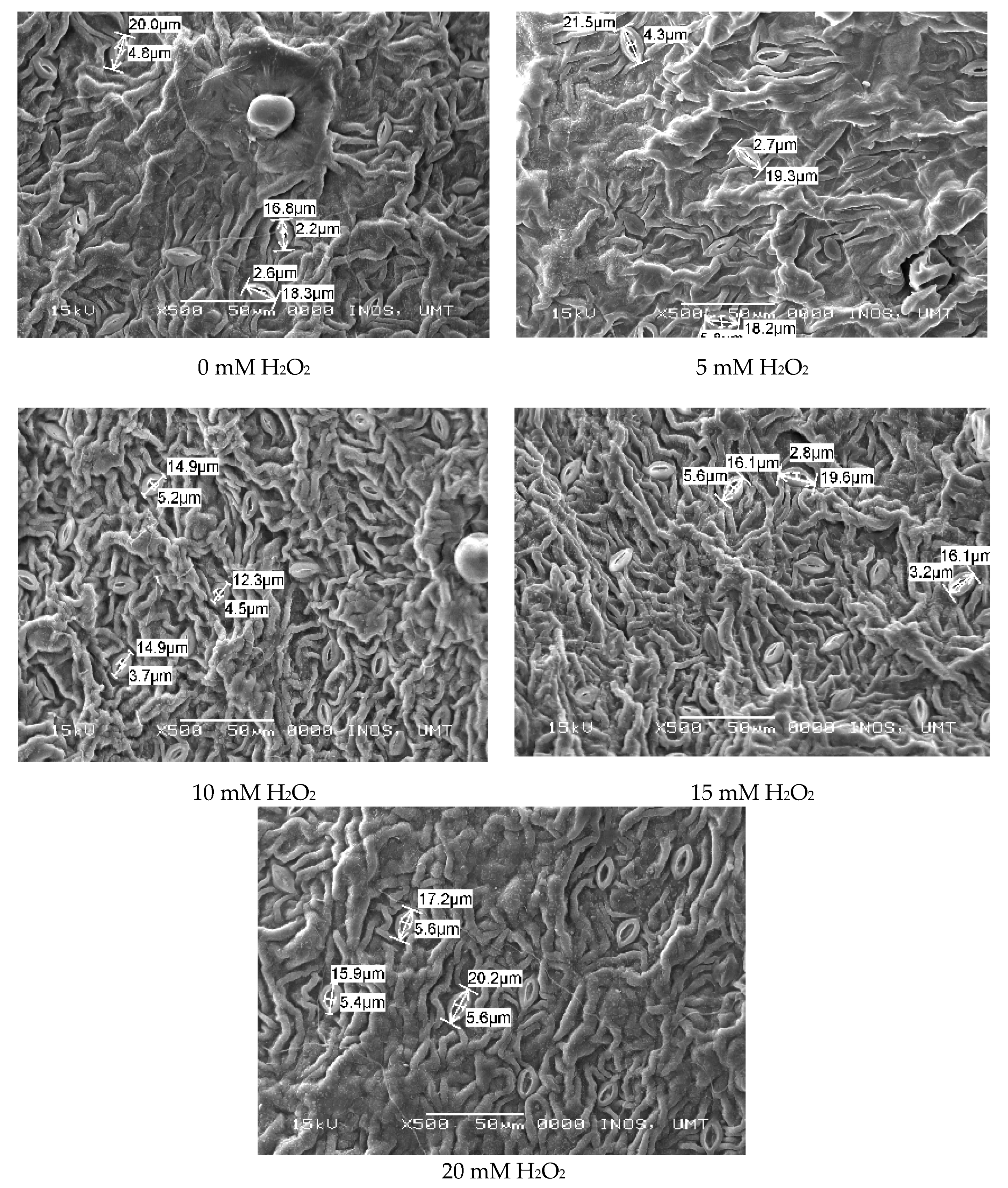
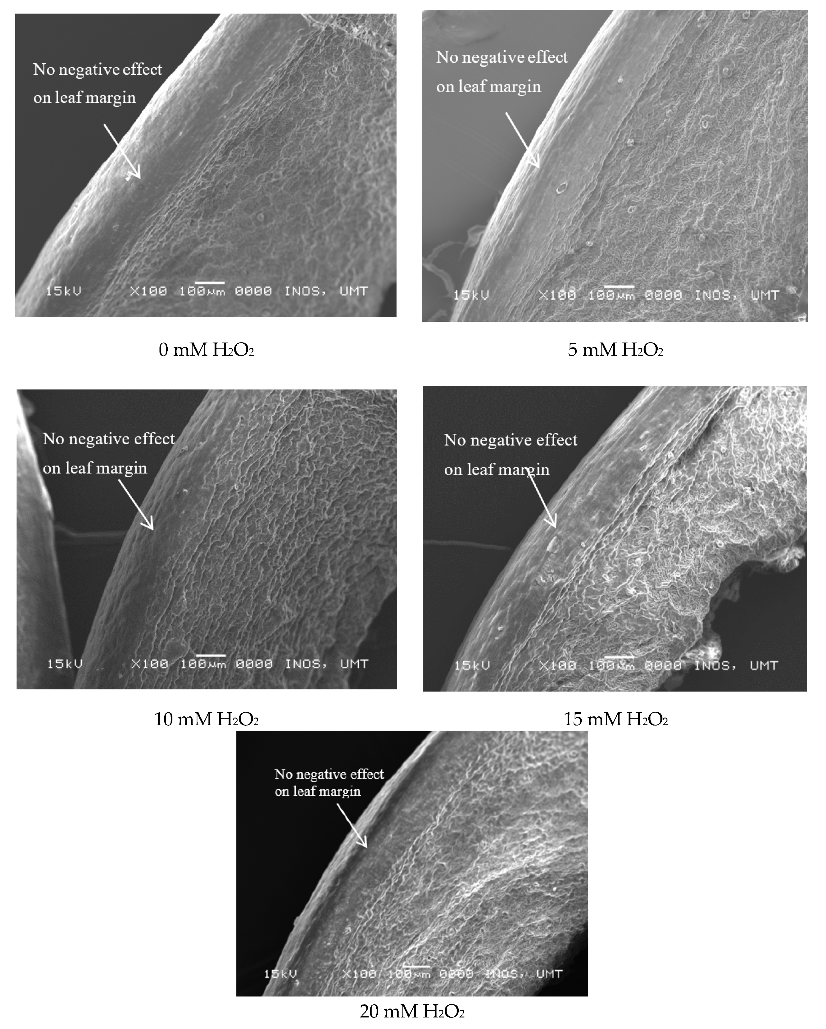
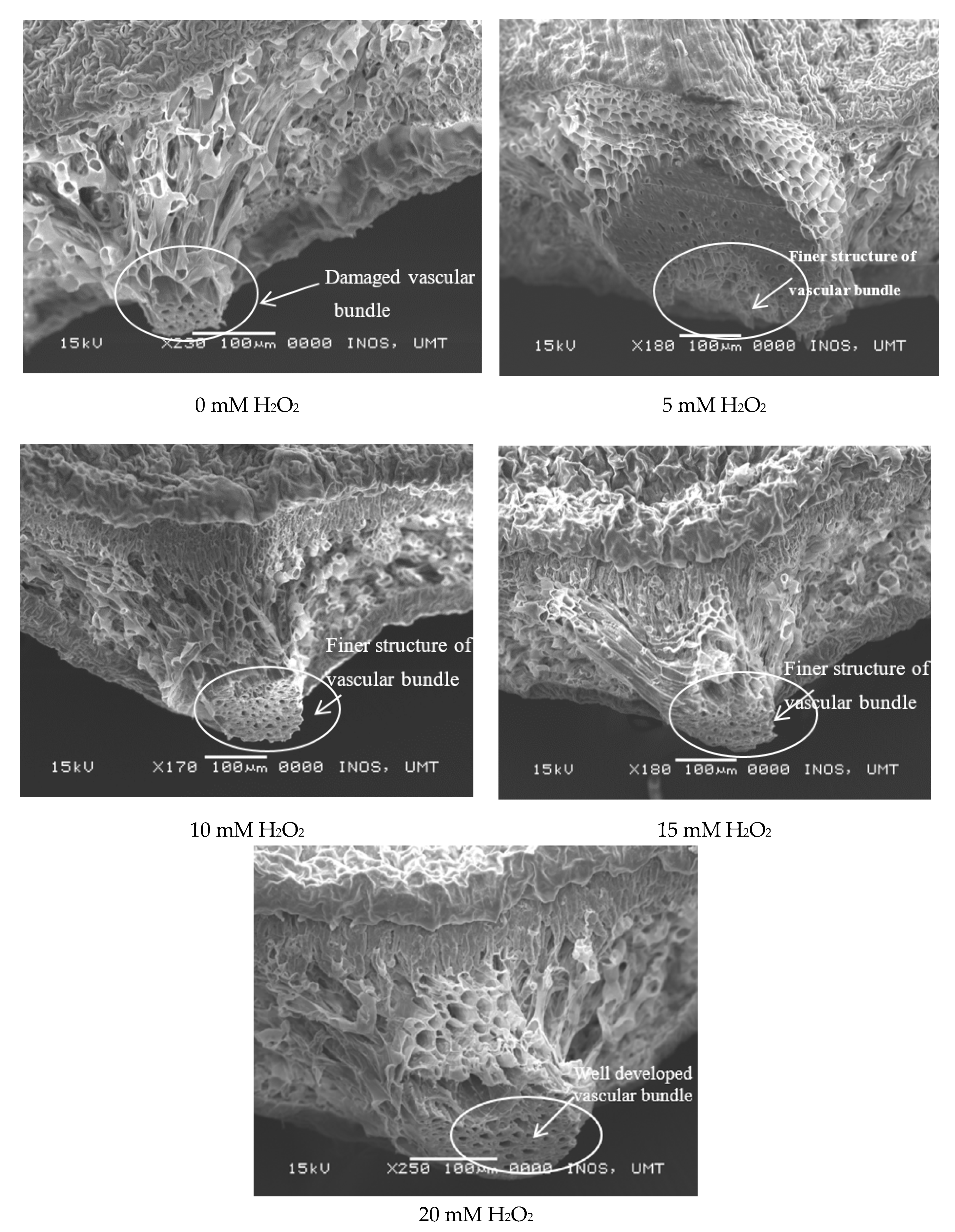
2.6. Leaf Histological Study (SEM)

A leaf histological study was done at the Electron Microscope Scanning laboratory, Institute of Oceanography and Environment (INOS), Universiti Malaysia Terengganu (UMT), Terengganu using the Scanning Electron Microscopy (SEM) (Model JEOL JSm-636OLA, JEOL, Tokyo, Japan) and following the standard procedure by Rohini et al. [14]. Treated and untreated leaves were collected and preserves in 80% ethanol for 12 h prior to study. After 12 h, the leaves were cut into standard size (1 cm × 1 cm) and were fixed with 2.5 g glutaraldehyde in 0.1 M Sodium cacodylate for 3 h. Then, the sample were rinse in 0.1 M sodium cacodylate buffer (pH 7.2) for 3 times with 15 min interval for each rinse. Next, the samples were let to dehydrate in series of ethanol (35%, 50%, 60%, 70%, 80%, 90%, 95% and 100%). Lastly, hexamethyldisilazane (HMDS) were used for further dehydration. The prepared samples were coated with gold and observed under SEM.

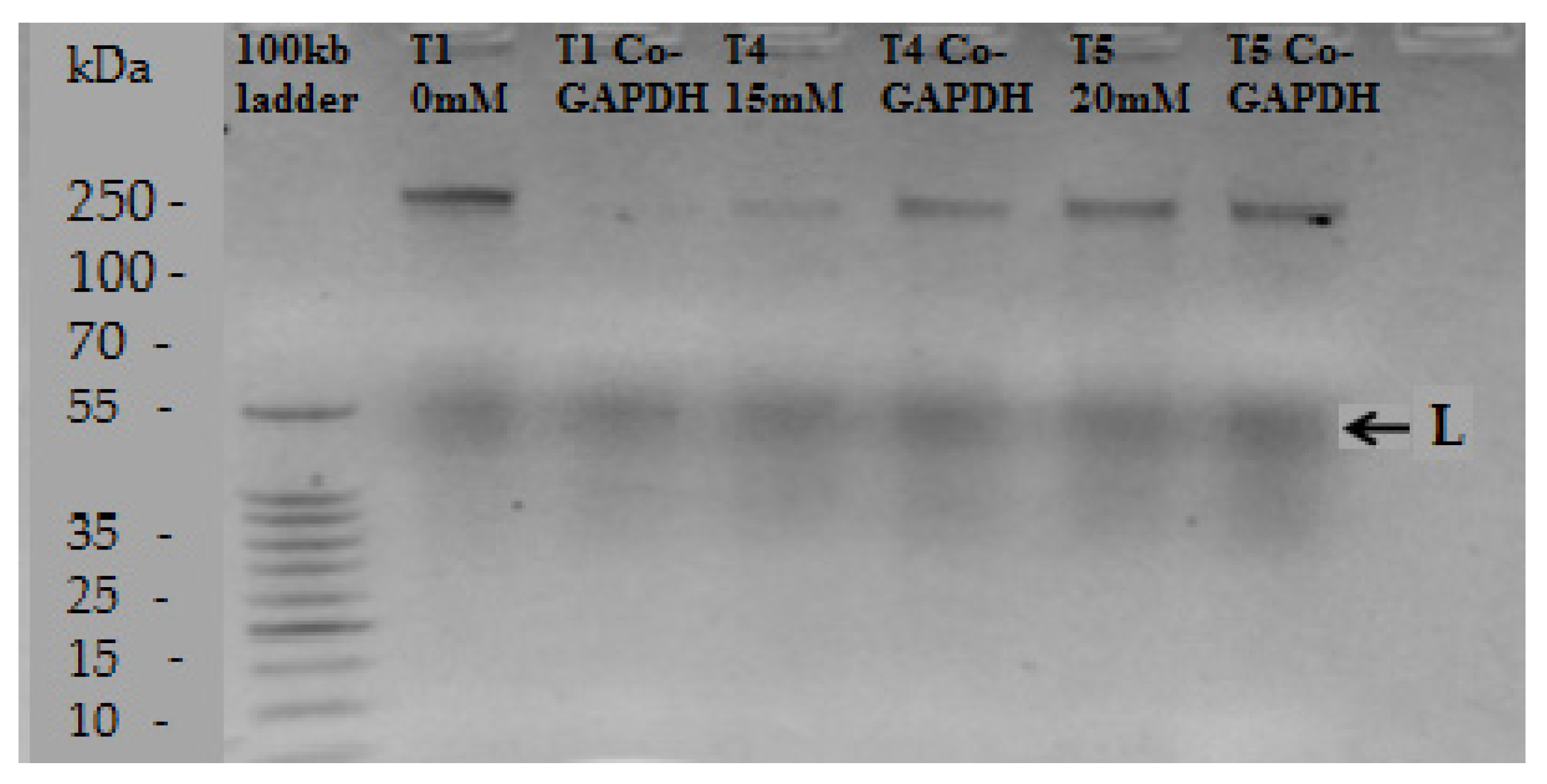
2.7. RNA Extraction and Purification, Synthesis of cDNA and RT-PCR

The RNA extraction was completed by following the manual of the SV RNA Isolation System kit (Promega, Madison, WI, USA). To prepare the total Ribonucleic acid (RNA), a fresh leaf was harvested at three days after treatment application. The leaf was later frozen and ground together with liquid nitrogen and only 30 mg of leaf tissue was used for total RNA extraction. RNA purification was carried out using a centrifuge at 12000–14000 rpm. The detailed procedure was carried out according to the method described by Wang et al. [15]. Synthesis of the cDNA was done by following the manual of Takara Prime Script 1st strand cDNA synthesis kit, Takara Bio, USA. Prior to proceeding with PCR and amplification, reverse transcriptase was inactivated by incubating the reaction tubes in a heat-block at 70 °C for 15 min. In this work rbcL primers 5′-TGTCACCAAAAACAGAGACT-3′ and 5′-TTCCATACTTCACAAGCAGC-3′, were respectively used as templates for quantitative RT-PCR analysis. The expression of the Co-GAPDH (glyceraldehydes-3-phosphate-dehydrogenase) gene was used as an internal control. The reaction volumes of 20 µL contained 10 µL of SYBR Premix Ex Taq (2×), 0.8 µL of forward primer (TGTACTACAGTTCGGCGGAG) and 0.8 µL reverse primer (TCCATACCTCACAAGCAGCA), 0.4 µL of reference dye, 2.0 µL of template and additional distilled water to the total volume of 20 µL. The expression data were analyzed using the Rotor-Gene 6000 Series Software 1.7 (Build 65, Germantown, USA) data analysis software and the 2−ΔΔCT, the method described by Livak and Schmittgen [16]. This study used relative quantification expression to compare with the untreated control sample as reference.

2.8. Statistical Analysis

The experimental design for all experiments were Completely Randomized Design (CRD) with six replications. The data were analyzed using the SPSS-17 statistical software, IBM Rochester, Canada. The one-way ANOVA was applied to evaluate the significant difference of the parameters studied in this experiment. Tukey’s test (HSD) was used to compare different concentrations of H2O2 whenever ANOVA showed significant differences among the means.

3. Results

3.1. The Effects of Hydrogen Peroxide (H2O2) on Growth and Photosynthesis

This study shows that H2O2 treatments promote growth in the number of leaves of Ficus deltoidea var. deltoidea. Untreated plants produced the lowest number of leaves, which was shown to be 16% lower than H2O2-treated plants which had more than 600 leaves on average (Table 1). For the plant height, the same pattern was observed. The untreated plant had the lowest plant height which was 10% lower than the 20 mM treated plant. Plants treated with 5 and 10 mM of H2O2 had medium height which was 5% higher than the control group. The plants with 20 mM H2O2 treatments had the highest height, which was 10% higher than the control plant (Table 1). Leaf area did not differ significantly between the treated and untreated plants, which was recorded as around 7–9 cm2 on average (Table 1).
The photosynthetic rate was significantly different between the untreated and H2O2-treated plants. It was observed that untreated plants had the lowest photosynthetic rate in comparison to the treated plants, which was 2.38 μmol m−2/s. The net photosynthetic rate showed incremental increases with the application of H2O2 directly on plant leaves. With 5, 10 and 15 mM H2O2 treatments, plants had a medium photosynthetic rate which was 6%, 11% and 12% higher than the untreated plant. The net photosynthetic activity was 1.2-fold higher than the control in treated plants with 20-mM H2O2 (Table 1). On the other hand, the transpiration rate was not significantly different among treated plants and the control, as seen in Table 1. For stomatal conductance, H2O2-treated plants were significantly different from the untreated plant (Table 1). The leaves stomatal conductance were 1.31, 1.77, 2.08 and 2.27-fold higher than the control with the 5, 10, 15 and 20 mM treatments, respectively (Table 1).
Internal CO2 was significantly different from each other among the treated and untreated plants. The lowest internal CO2 was determined in the untreated plant. The internal CO2 increased with the continued application of H2O2. The highest internal CO2 concentration was 10% higher in the 20 mM H2O2-treated plants compared to the control plant (Table 2). From this study, we show the chlorophyll content (SPAD value) of F. deltoidea leaves was not significantly different among treated and untreated plants as seen in Table 2. The same trend was observed in lower (F0) and higher (Fm) chlorophyll fluorescence. However, relative variable (Fv) fluorescence was significantly different from the treated and untreated plants (Table 2). Overall, quantum or photosynthetic yield was not significantly different when comparing the treated plants with the control, as shown in Table 2.

3.2. Regulatory Effects of Hydrogen Peroxide (H2O2) on Biochemical Properties

The chlorophyll content of H2O2-treated leaves was determined to compare it with the control. In this study, two types of chlorophyll were studied; chlorophyll a and chlorophyll b. It was observed that H2O2-treated leaves were significantly different from the control in both chlorophyll a and chlorophyll b. The H2O2 at 20 mM had the highest chlorophyll a content which was 57% higher than the control; meanwhile the highest chlorophyll b content was 56% and 67% higher than the control in 15 mM and 20 mM H2O2 treatments respectively (Table 3). The lowest chlorophyll value was recorded in the control plants.
The exogenous H2O2 treatments produced significant effects on the carotenoid content of F. deltoidea plants. Results showed that carotenoid content increased with H2O2 concentration (Table 3). Maximum values of carotenoid were recorded in 15 and 20 mM H2O2-treated plants which were 22% and 26% higher than the control plant respectively (Table 3).
For total phenolic and flavonoid contents, the same pattern of significant increments was observed as in the carotenoid content. For total phenolic content (TPC), the control plant had the lowest TPC value which was 27% less than the 20 mM treated plants which recorded the highest TPC content at 168 mg GAE/g DL extract. The lowest total flavonoid content (TFC) was found in the control plant and TFC increased with treatment concentration. Plants treated with 10 and 15 mM H2O2 increased 42% and 78% higher, respectively, in flavonoid content than the control plants (Table 3). In this study, a total soluble sugar content (TSC) test was carried out to measure carbohydrates or the natural sugar content of treated F. deltoidea leaves due to its sensitivity and simplicity. The results of the tests showed that control plants had the lowest sugar content which was 1.62 µmol/g of leaves with dried extract. As the concentration of applied H2O2 increased (5, 10, 15 and 20 mM), the results for the total sugar content also increased significantly. H2O2 at 20 mM had the highest total sugar content which was 1.89-fold higher than the control as shown in Table 3. This may be due to increases in available photosynthates under this optimum growth condition. In addition, antioxidant activity determined by the ABTS method was 1.28-fold greater in the 20 mM treated F. deltoidea plants compared to the control plant even though their differences were not statistically significant at 5% (Table 3).

3.3. Regulatory Effects of Hydrogen Peroxide on Leaf Anatomy

The histological studies were done to study the effects of H2O2 treatment on plant leaves, focusing on stomatal aperture, leaf margin and leaf cross sections, to determine the direct effects of H2O2 on plant cells. It was observed that H2O2 applications do promote enlargement of the stomatal aperture. From Figure 1, it can be seen that single stomatal openings increased significantly with the application of exogenous H2O2. The H2O2 at 0 mM (untreated) had the smallest stomatal opening which was 3.2 μm although it had the longest stomatal length (17.47 μm). On the other hand, the 20 mM H2O2-treated leaves had the widest stomatal opening which was 5.88 μm with a stomatal length of 16.63 μm (Figure 1).
Figure 2 shows the direct effects of exogenous applications of H2O2 towards stomatal size on the upper (adaxial) leaf surface. On average, there was less stomata presence on the upper surface of the leaf. It was observed that H2O2 treatment increased the stomatal opening with moderate doses. However, untreated leaves still had the smallest openings which were, on average, 2.5 to 3.9 μm when viewed under SEM. The largest opening was observed in 20 mM H2O2-treated leaves with ranges from 2.3 to 6.6 μm.
Figure 3 shows the effects of exogenous applications of H2O2 on the stomatal closure at the lower (abaxial) surface of F. deltoidea leaves. It can be observed that more significant amounts of stomata were present under the leaf surface. It was observed that H2O2 treatment increased the stomatal openings at moderate doses.
Control plants had the smallest stomatal openings which were between 2.2 µm to 4.8 µm when viewed under SEM. This was followed by 5 mM H2O2 treatments with 2.7 µm to 4.3 µm stomatal openings and 10 mM H2O2 treatments with 3.7 µm to 5.2 µm stomatal openings. The 15 mM H2O2 treatment showed 2.8 µm to 5.6 µm stomatal openings, which was similar to the stomatal opening in the 20 mM H2O2 treatment, at 5.4 µm to 5.6 µm (Figure 3). Figure 4 displays the effects of H2O2 treatments on leaf margins of Ficus deltoidea leaves. Generally, no negative effect of H2O2 treatment was detected on the leaves’ leaf margin on treated and untreated plants. It can be said that no damage was caused by H2O2 direct application via spraying on the leaves’ surfaces.
Figure 5 exhibits the F. deltoidea leaf cross section when viewed under SEM. Obviously, it can be seen that H2O2-treated leaves had better leaf structure compared to untreated leaves. Damaged and thinner palisade and spongy mesophyll cells were found in untreated leaves than H2O2-treated leaves. Results showed that H2O2-treated leaves had more of an arranged palisade mesophyll layer and had thicker leaf structure than untreated leaves (Figure 5).
Figure 6 exhibits the effects of H2O2 treatments on leaves’ vascular bundles of F. deltoidea var. deltoidea. Our results showed that H2O2 treatments improved the leaf structure especially the xylem and phloem tissues in vascular bundles when compared to untreated leaves. It was clearly shown that all the treated leaves had a well-developed vascular bundle, with less damage, as compared to untreated leaves (Figure 6). It can be said that H2O2 application improves the structure of the leaf vein and promotes cell proliferation.

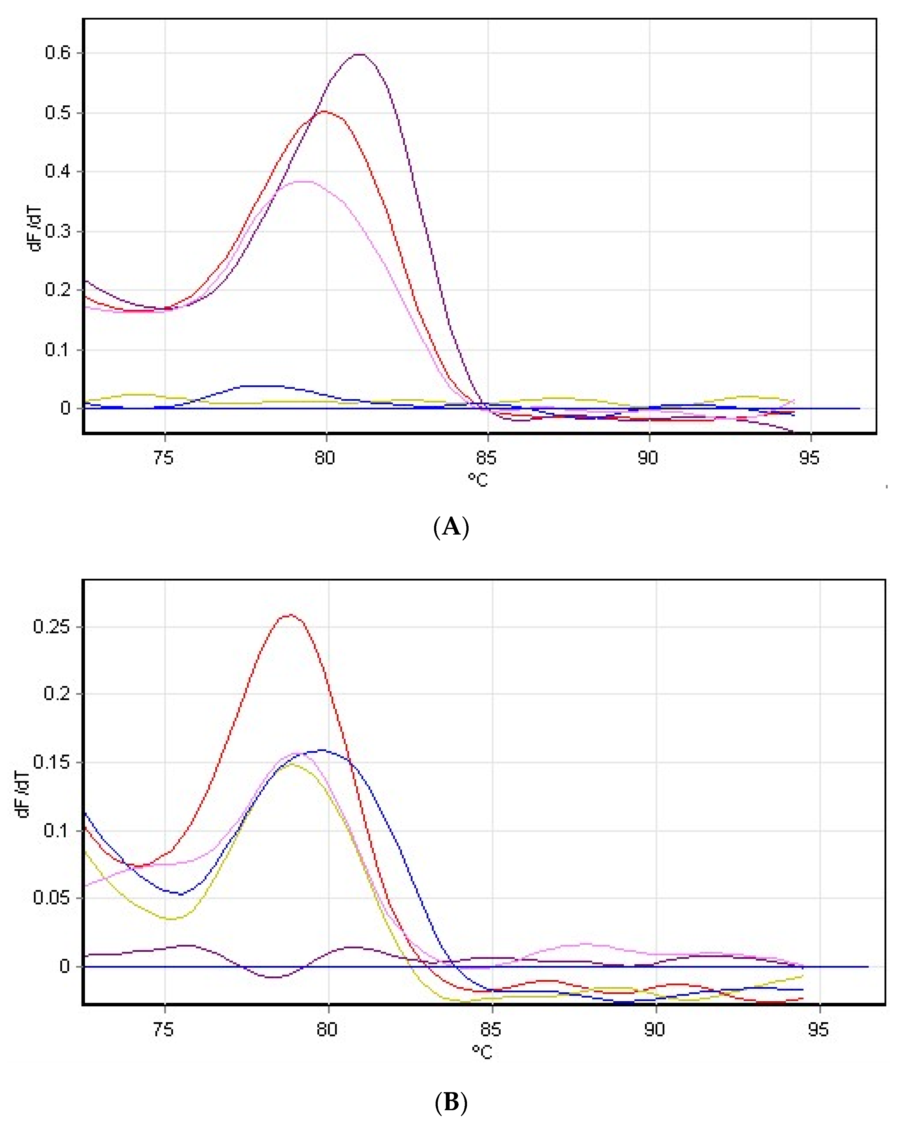
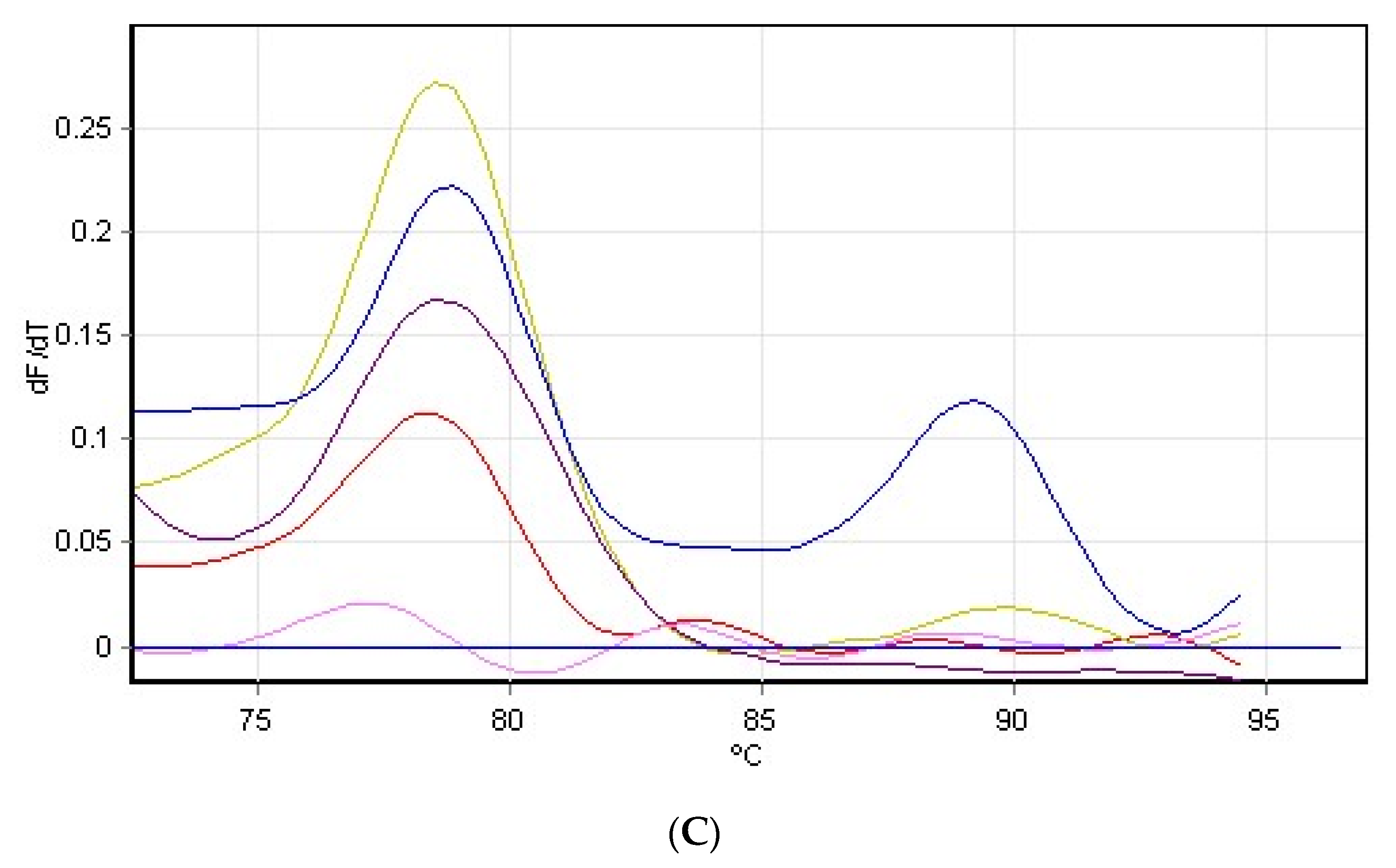
3.4. Hydrogen Peroxide on Rubisco Gene Expression

In this study, Rubisco gene expression was used to determine the regulatory effects of H2O2 on the Ficus deltoidea plant, specifically, whether it regulated rbcL gene expression or not, with a comparison to the housekeeping gene Co-GAPDH as a mean. Only three treatments preceded the gene expression study, to determine which treatment was the best when compared to untreated plants. From the study, it is concluded that 15 and 20 mM H2O2 treatments had the highest stimulating effects on plant growth, photosynthesis, histological study, minerals accumulation and biochemical properties. As a result of this, Real-Time PCR (RT-PCR) was done to conclude this experiment.
Figure 7 shows the qualitative analysis of rbcL gene expression when viewed under gel electrophoresis. In general, it was determined that rbcL gene was up-regulated in comparison to Co-GAPDH. We can see the presence of rbcL gene in both 15 and 20 mM H2O2-treated leaves and also in the untreated plant when compared to the housekeeping gene (Co-GAPDH). Subsequent details about the Rubisco gene expression were described via results obtained from the RT-PCR analysis.
The results were obtained from RT-PCR of the ∆∆CT method. Absolute quantification was done to compare the 15 and 20 mM H2O2-treated plants, with Co-GAPDH as housekeeping gene and 0 mM H2O2 as a reference gene following the Pfaffl method. From the RT-PCR study, it was determined that the rbcL gene expression ratio of the 15 mM H2O2-treated plant was the highest with a 4.796 times expression ratio than the control. The expression ratio showed that rbcL gene expression was a much higher ratio in the 15 mM H2O2-treated plant than in the 20 mM H2O2-treated plant (Table 4).
Other than that, the fold change was also calculated following the 2−∆∆CT method in comparison to 0 mM as the control gene. It was determined that 15 mM H2O2-treated plants had a 0.11582-fold change than the control gene (0 mM H2O2). Treated plants with 20 mM H2O2 had a 0.534-fold change of the rbcL gene than the control gene (0 mM H2O2). It was obvious that 15 mM increased the expression of rbcL gene at a much better rate and at a higher fold rate than the 20 mM H2O2 treatment. The details of expression ratios and fold changes of the rbcL gene are tabulated in Table 4.
Figure 8 shows the melting curve of the rbcL gene of F. deltoidea var. deltoidea plant. Since SYBR green binds to double stranded DNA, there will be a drop in fluorescence as strands separate. For any given piece of DNA, there is a very small temperature range at which the strands will separate which depends on G/C content, length and other factors. At temperatures between 60–90 °C, there will be a sharp drop in fluorescence. It was clear that the RT-PCR melting reaction occurred at temperatures between 78.3–81 °C. The good primers, 0.8 µL of forward primer (TGTACTACAGTTCGGCGGAG) and 0.8 µL reverse primer (TCCATACCTCACAAGCAGCA), produced only one major drop in fluorescence. This drop strongly suggested that there was only one product in the reaction; namely Rubisco (rbcL) which is the protein/enzyme responsible for CO2 fixation during photosynthesis.

4. Discussion

In this study, four concentrations of hydrogen peroxide (H2O2) were used through foliar spraying, to study the effects of exogenous applications of H2O2 on plant physiology, leaf anatomy and Rubisco gene expression of Ficus deltoidea var. deltoidea. Hydrogen peroxide was sprayed onto F. deltoidea leaves, instead of other parts of the plant, because the leaf is a major route for nanoparticle entry into the plant system [17]. Our results on plant growth and development show that H2O2 treatment on F. deltoidea plants enhanced their growth and development. This is because of hydrogen peroxide’s role in protein initiation in plant signaling and development during the early growth of pea seedlings [18]. This study added that the induction of these polypeptides may correlate the hydrogen peroxide to the greater seedling growth when pea seeds were pretreated with 20 mM H2O2.
The application of exogenous 15 and 20 mM H2O2 treatments provided the best stimulating effects on the plants’ height and number of leaves. These results were supported by Neill et al. [19], who reported that the H2O2 concentration at an appropriate level can promote plant growth and development. The exogenous H2O2 may impact metabolic and antioxidant enzyme activity in favor of plant growth and development. Average leaf area was recorded in this study. When treated plants were compared with untreated plants, leaf area was found to not be affected. The main factor that leads to this result may be due to a leaf trait that was genetically inherited and commonly unchanged by external factors in these plants. However, another study showed that the application of H2O2 promoted a higher growth rate in lettuce leaves [20].
During this experiment, it was determined that the photosynthesis rate was significantly and incrementally different from untreated plants of F. deltoidea. Mittler et al. [21] also reported that H2O2 stimulated the photosynthesis process in plants. H2O2 acts as signal molecule which mediates photosynthesis, stomatal movement and biochemical reactions during plant growth and development. H2O2 application induces the activity of sucrose phosphate synthase (SPS), an enzyme important in the formation of sucrose from triose phosphates during and after photosynthesis in plants [22]. In this study, the non-significant effects of H2O2 on the transpiration rate of F. deltoidea plants was recorded. Gil et al. [23] also stated that H2O2 injected into heavy clay loam soil had no significant effect on avocado transpiration rate. This study indicated that the spraying of H2O2 significantly improved the stomatal conductance of the F. deltoidea var. deltoidea plants. Contradictory results reported by Konstantinos et al. [24], stated that amine oxidase was produced by H2O2 to stimulate the stomatal closure in Vitis vinifera plants. In their study, the concentration amount which created the negative impact on the stomatal conductance of plants was not clear. An increase stomatal conductance is dependent on the density, size and degree of stomatal opening. This study also indicated that exogenous H2O2 increase the stomatal size at both surfaces of the leaves. May be the exogenous H2O2 is associated with improved gas exchange or stomatal conductance rather than with non-enzymatic antioxidant system [25]. These results state that the improved stomatal conductance may increase net photosynthetic rates and cause higher accumulations of internal CO2. Our results indicate that chlorophyll contents were higher in 15 and 20 mM H2O2-treated plants. Butcher et al. [26] demonstrated that the application of hydrogen peroxide on Pelargonium tomentosum yielded the highest production of chlorophyll within all the replicates and differed significantly from the control.
This study indicates that the application of 20 mM H2O2 significantly improved the chlorophyll fluorescence of F. deltoidea var. deltoidea plants, with a much higher result than plants not treated with H2O2. It has been reported that reduced chlorophyll fluorescence of stress plan can be reversed by the application exogenous H2O2 [27]. May be the exogenous H2O2 caused up-regulation of plant antioxidant system which closely related to the efficiency of photosystem II. This study demonstrates that the application of different concentrations of H2O2 did not significantly affect the quantum yield of the F. deltoidea var. deltoidea plants. Kautsky et al. [28] explained that chlorophyll fluorescence yield changes as a consequence of the reduction of electron acceptors in the photosynthetic pathway, downstream of PSII, notably plastoquinone and in particular, QA.
This study indicates that application of exogenous H2O2 significantly improved both chlorophyll a and chlorophyll b content in F. deltoidea plants. In addition, carotenoid content was significantly higher in treated plants. Jeong et al. [29] also reported that β-carotene biosynthesis is stimulated by H2O2 treatment in Blakeslea trispora. However, this result was contradictory to a study conducted by Kim et al. [30], who demonstrated that direct application of H2O2 at higher doses (100 mM to 400 mM) onto a fresh cut tomato caused it to undergo color changes due to reduced carotenoid content. These results indicate that the application of H2O2 significantly improved total phenolic content of treated F. deltoidea var. deltoidea plants. The same pattern was observed in total flavonoid content, where H2O2-treated plants had significantly higher flavonoid content. The possible reason for the increase of secondary metabolites could be due to H2O2′s involvement as signal molecules in phenolic synthesis. Nyathi and Baker, [31] also stated that H2O2 may regulate the PAL, CHS and stilbene synthase gene expression, which are related to the synthesis of phenols and flavonoids in plants
This study shows that the application of H2O2 increased the total sugar content in F. deltoidea plants. Ozaki et al. [22] also reported similar positive effects of H2O2 on total sugar accumulation in melons. This may be due to the effects of exogenous H2O2 as it induces the activity of the sucrose phosphate synthase (SPS) enzyme, which regulates the formation of sucrose from triose phosphates during and after photosynthesis in rice seedlings at the transcriptional level [32]. In this study, ABTS antioxidant tests were not significantly different from each other in the treated and untreated plants. However, Zhang et al. [33] mentioned that pretreatment with H2O2 promoted antioxidant activities of plants, as well as hindered damages caused by chilling, salt, heat and osmotic exogenous stress. Hydrogen peroxide may be responsible for the improvement of the antioxidant status within plants by activating gene expression of PAL, CHS and stilbene synthase [31].
This study shows that direct application of H2O2 on plants’ leaves provoke a positive stimulatory effect on the plants’ stomatal aperture, as viewed in a single stomatal opening in the result. Previous studies have suggested that oxidative stress, resulting from exposure to H2O2, has had a remarkable effect on the stomatal aperture [34]. The study of stomata was extended to the stomatal size at the upper and lower surfaces of the leaves with both surfaces reportedly having a different stomatal density. It was determined that the lower surface of the leaves had higher stomatal density than the upper surface of the leaves. This study showed that the application of H2O2 created a moderate stomatal opening at the upper surface of the leaves, while the smallest stomatal openings were observed in the untreated leaves. At both surfaces of the leaves, 20 mM H2O2 treatment was reported to produce the largest stomatal size. Ishibashi et al. [35] reported that spraying H2O2 on soybean alleviates stress through the maintenance of leaf water content, and that this water retention was caused by the promotion of oligosaccharide biosynthesis rather than by rapid stomatal closure.
In addition, H2O2 application was determined to have no negative effect on F. deltoidea var. deltoidea leaf margin. Doke et al. [36] suggested the usage of H2O2 as an antimicrobial agent for preventing microbial infestation in plants when the suitable dosage of usage could be selected from 5 mM H2O2 to 20 mM H2O2 as these dosages were proven to not caused any physical damage to the plant leaf.
This study on the effects of H2O2 on leaf cross-sections demonstrated that the usage of H2O2 improves the structure of palisade and spongy mesophyll cells when viewed under a SEM. Moreover, less damage was observed in cell structures from the 15 and 20 mM H2O2-treated leaves than untreated leaves. The improved structure of H2O2-treated leaves may be due to the exogenous application of H2O2, which improved the cell structure by lignification and the cross-linking of cell wall structural proteins. Also, H2O2 takes part in a resistance mechanism for reinforcement of plant cell wall lignification, cross-linking of cell wall structural proteins, phytoalexin production and resistance enhancement [37]. Furthermore, Bradley et al. [38] suggested that the oxidative burst of reactive oxygen species (ROS) such as H2O2 drives crosslinking of the cell wall.
Other than that, the regulatory effects of H2O2 on leaves’ vascular bundles of the F. deltoidea var deltoidea plants were realized. Orozco-Cardenas and Ryan [39] stated that hydrogen peroxide was generated systematically in wounded leaves and was localized in the vascular tissues. This study shows that H2O2-treated leaves had a thicker structure of leaf veins, including both xylem and phloem. It can be said that H2O2 application improved the structure of leaf veins and promoted cell proliferation. It was reported earlier that the thickening of minor veins is the result of cell proliferation. The result was in parallel to a study conducted by Jafariyan and Zarea [40], who stated that soaking seeds of Azospirillum in 50% H2O2 solution resulted in a stimulant effect by enhancing areas of the vascular bundle and upper epidermis. In addition, Thordal-Christensen [41] reported that H2O2 was localized within the cell walls of xylem vessels with secondary thickening and within walls of surrounding cells.
Finally, after previous results, 15 and 20 mM H2O2 treatments were used in the gene expression study to determine which treatment was the best concentration to enhance plant growth and development. Rubisco is the most abundant protein in leaves and there is evidence from studies on the altering expression of Rubisco. Kasai et al. [42] stated that Rubisco activates changes in the plant’s activity and the amount of Rubisco in a leaf significantly affects the photosynthetic rate of plants. Our results indicate that 15 mM H2O2 had a more significant rbcL gene expression ratio than the 20 mM H2O2-treated F. deltoidea var. deltoidea plants. This shows 15 mM H2O2-treated plants had slightly higher fold changes within rbcL gene expression than 20 mM H2O2 and the untreated plants. In general, 15 mM H2O2 had better Rubisco gene expression compared to control and this is because of the role of H2O2 in triggering plant growth and development. Cui et al. [43] advocates that the exogenous application of H2O2 onto plants induced gene expression at mRNA levels, during somatic embryogenesis of Lycium barbarum. Other than this, a study conducted by Chen et al. [44], showed that chondrocytes cultured with 100 μm of H2O2 had an increase in ROS amounts in intercellular and intracellular cells, 15-fold higher than the control which lead to oxidative stress and thus spikes in expression of MMP-1 and MMP-13 genes. Significantly, H2O2 has been recognized as the ROS that induced the largest changes in the levels of gene expression in plants and this was probably due to its relative stability [45].

5. Conclusions

It can be concluded that spraying 15 and 20 mM of H2O2 significantly enhances plant growth and development of F. deltoidea. Net photosynthetic rate, chlorophyll content and stomatal conductance significantly increased in all the H2O2 treatments. In addition, 15 and 20 mM of H2O2 significantly improved the carotenoids, phenols, flavonoids and total sugar content in plants. Also, H2O2 treatment as a foilar spray did not cause any harmful effects on the plants’ cell, tissue and, as a bonus, improved leaf vain structure. It was also found that rubisco (rbcL) gene expression in 15 mM H2O2-treated plants was higher than those of the 20 mM H2O2-treated plants. As a result, it is concluded that applications of 15–20 mM H2O2 could enhanced plant growth, development, biochemical properties, cellular structure and Rubisco gene expression.

Author Contributions

Data curation, R.J. and M.M.K.; investigation, K.M.; methodology, M.M.K. and M.H.S.; supervision, M.M.K. and N.M.; validation, K.S.M.; writing—original draft, M.M.K.; writing—review & editing, N.A.B. and M.M.K. All authors have read and agreed to the published version of the manuscript.

Funding

This research was supported by the Ministry of Education Malaysia from the Fundamental Research Grant Scheme, FRGS Grant No. FRGS/2/2014/SG03/UNISZA/02/01.

Acknowledgments

We are grateful to Anuar McAfee for his English editing and proof reading of this paper.

Conflicts of Interest

The authors declare no conflict of interest.

References

  1. Hasham, R.; Choi, H.K.; Sarmidi, M.R.; Park, C.S. Protective effects of a Ficus deltoidea extract against UVB-induced photoageing in skin cells. Biotechnol. Bioprocess Eng. 2013, 18, 185–193. [Google Scholar] [CrossRef]
  2. Fasihuddin, B.A.; Din, L.B. Medicinal Plants Used by Various Ethnic Groups in Sabah; Paper presented at the french Malaysian-symposium on natural products; Dept. of Chemistry, University of Malaya: Kuala Lumpur, Malaysia, 2002; p. 85. [Google Scholar]
  3. Misbah, H.; Abdul Aziz, A.; Aminudin, N. Antidiabetic and antioxidant properties of F. deltoidea fruit extracts and fractions. BMC complement. Alt. Med. 2013, 13, 118. [Google Scholar] [CrossRef] [PubMed] [Green Version]
  4. Cheeseman, J.M. Hydrogen Peroxide concentrations in leaves under natural conditions. J. Exp. Bot. 2006, 57, 2435–2444. [Google Scholar] [CrossRef] [Green Version]
  5. Kim, K.; Portis, A.R. Oxygen-dependent H2O2 production by Rubisco. FEBS Lett. 2004, 571, 124–128. [Google Scholar] [CrossRef] [PubMed] [Green Version]
  6. Lariguet, P.; Ranocha, P.; De Meyer, M.; Barbier, O.; Penel, C.; Dunand, C. Identification of a H2O2 signalling pathway in the control of light-dependent germination in Arabidopsis. Planta 2013, 238, 381–395. [Google Scholar] [CrossRef] [PubMed]
  7. Khandaker, M.M.; Hossain, A.S.; Osman, N.; Boyce, A.N. Application of girdling for improved fruit retention, yield and fruit quality in Syzygium samarangense under field conditions. Int. J. Agric. Biol. 2011, 13, 18–24. [Google Scholar]
  8. Khandaker, M.M.; Boyce, A.N.; Osman, N. The influence of hydrogen peroxide on the growth, development and quality of wax apple (Syzygium samarangense, [Blume] Merrill & L.M. Perry var. jambu madu) fruits. Plant Physiol. Biochem. 2012, 53, 101–110. [Google Scholar] [CrossRef]
  9. Haq, M.; Sani, W.; Hossain, A.B.M.S.; Taha, R.M.; Monneruzzaman, K.M. Total phenolic contents, antioxidant and antimicrobial activities of Bruguiera gymnorrhiza. J. Med Plants Res. 2011, 5, 4112–4118. [Google Scholar]
  10. Dubois, M.; Gilles, K.; Hamilton, J.K.; Robers, P.A.; Smith, F. A colorimetric method for the determination of sugar and related substances. Anal. Chem. 1956, 28, 350–356. [Google Scholar] [CrossRef]
  11. Singleton, V.; Rossi, J.A.J. Colorimetry of total phenolic with phosphomolybdic-phosphotungstic acid reagents. Am. J. Enol. Viticult. 1965, 16, 144–158. [Google Scholar]
  12. Chang, C.; Yang, M.; Wen, H.; Chern, J. Estimation of total flavonoid content in propolis by two complementary colorimetric methods. J. Food Drug Anal. 2002, 10, 178–182. [Google Scholar]
  13. Arnon, D.I. Copper enzymes in isolated chloroplasts, polyphenoxidase in beta vulgaris. Plant Physiol. 1949, 24, 1–15. [Google Scholar] [CrossRef] [PubMed] [Green Version]
  14. Gowtham, H.G.; Hariprasad, P.; Brijesh, S.S.; Niranjana, S.R. Biological control of Phomopsis leaf blight of brinjal (Solanum melongena L.) with combining phylloplane and rhizosphere colonizing beneficial bacteria. Biol. Cont. 2016, 101, 123–129. [Google Scholar]
  15. Wang, B.; Chen, J.; Chen, L.; Wang, X.; Wang, R.; Ma, L.; Peng, S.; Luo, J.; Chen, Y. Combined drought and heat stress in Camellia oleifera cultivars: Leaf characteristics, soluble sugar and protein contents, and Rubisco gene expression. Trees 2015, 29, 1483–1492. [Google Scholar] [CrossRef]
  16. Livak, K.J.; Schmittgen, T.D. Analysis of relative gene expression data using real-time quantitative PCR and the 2ΔΔCT Method. Methods 2001, 25, 402–408. [Google Scholar] [CrossRef]
  17. Anjum, S.A.; Xiao-yu, X.; Long-chang, W.; Muhammad, F.S.; Chen, M.; Wang, L. Morphological, physiological and biochemical responses of plants to drought stress. Afr. J. Agril. Res. 2011, 6, 2026–2032. [Google Scholar] [CrossRef]
  18. Barba-Espín, G.; Diaz-Vivancos, P.; Job, D.; Belghazi, M.; Job, C.; Hernández, J.A. Understanding the role of H2O2 during pea seed germination: A combined proteomic and hormone profiling approach. Plant Cell Environ. 2011, 34, 1907–1919. [Google Scholar] [CrossRef]
  19. Neill, S.; Desikan, R.; Hancock, J. Hydrogen peroxide signalling. Current Opinion in Plant Biol. 2002, 5, 388–395. [Google Scholar] [CrossRef]
  20. Watanabe, K.; Yachi, C.; Song, X.J.; Kakuyama, S.; Nishibe, M.; Michigami, S. Measurements of atmospheric hydroperoxides at a rural site in central Japan. J. Atmos. Chem. 2018, 75, 71–84. [Google Scholar] [CrossRef]
  21. Mittler, R.; Vanderauwera, S.; Gollery, M.; Breusegem, F. Reactive oxygen gene network of plants. Trends Plant Sci. 2004, 9, 490–498. [Google Scholar] [CrossRef]
  22. Ozaki, K.A.U.; Tomoko, T.; Shinagawaa, F.; Yoshito, T.; Takabeb, T.; Takahisa, H.; Tasuku, H.; Ashwani, K.R. Enrichment of sugar content in melon fruits by hydrogen peroxide treatment. J. Plant Physiol. 2009, 166, 569–578. [Google Scholar] [CrossRef]
  23. Gil, P.M.; Ferreyra, R.E.; Barrera, C.M.; Zuniga, C.E.; Gurovich, L.R. Effects of injecting hydrogen peroxide into heavy clay loam soil on plant water status, net CO2 assimilation, biomass, and vascular anatomy of avocado trees. Chilean J. Agril. Res. 2009, 69, 97–106. [Google Scholar] [CrossRef]
  24. Konstantinos, P.A.; Imene, T.; Panagiotis, M.N.; Roubelakis-Angelakis, K.A. ABA-dependent amine oxidases-derived H2O2 affects stomata conductance. Plant Signal. Behavior. 2010, 5, 1153–1156. [Google Scholar] [CrossRef] [PubMed] [Green Version]
  25. Gondim, F.A.; Miranda, R.S.; Filho, E.G.; Prisco, J.T. Enhanced salt tolerance in maize plants induced by H2O2 leaf spraying is associated with improved gas exchange rather than with non-enzymatic antioxidant system. Theoret. Exp. Plant Physiol. 2013, 25, 251–260. [Google Scholar] [CrossRef]
  26. Butcher, J.D.; Charles, P.L.; Johannes, C.C. A study of oxygenation techniques and the chlorophyll responses of pelargonium tomentosum grown in deep water culture hydroponics. Hort. Sci. 2017, 52, 952–957. [Google Scholar] [CrossRef] [Green Version]
  27. Khan, M.I.R.; Khan, N.A.; Masood, A.; Per, T.S.; Asgher, M. Hydrogen Peroxide Alleviates Nickel-Inhibited Photosynthetic Responses through Increase in Use-Efficiency of Nitrogen and Sulfur, and Glutathione Production in Mustard. Front. Plant Sci. 2016, 7, 44. [Google Scholar] [CrossRef] [PubMed] [Green Version]
  28. Kautsky, H.; Appel, W.; Amann, H. Chlorophyll fluorescence and carbon assimilation. Biochem. Zeit. 1960, 322, 277–292. [Google Scholar]
  29. Jeong, J.; Lee, I.; Kim, S.W.; Park, Y.H. Stimulation of β-carotene synthesis by hydrogen peroxide in Blakeslea trispora. Biotechnol. Lett. 1999, 21, 683–686. [Google Scholar] [CrossRef]
  30. Kim, H.J.; Fonseca, J.M.; Kubota, C.; Choi, J.H. Effect of hydrogen peroxide on quality of fresh-cut tomato. J. Food Sci. 2007, 72, S463–S467. [Google Scholar] [CrossRef]
  31. Nyathi, Y.; Baker, A. Plant peroxisomes as a source of signalling molecules. Biochim. Biophys. Acta. 2006, 1763, 1478–1495. [Google Scholar] [CrossRef] [Green Version]
  32. Uchida, A.; Jagendorf, A.T.; Hibino, T.; Takabe, T.; Takabe, T. Effects of hydrogen peroxide and nitric oxide on both salt and heat stress tolerance in rice. Plant Sci. 2002, 163, 515–523. [Google Scholar] [CrossRef]
  33. Zhang, X.L.; Jia, X.F.; Yu, B.; Gao, Y.; Bai, J.G. Exogenous hydrogen peroxide influences antioxidant enzyme activity and lipid peroxidation in cucumber leaves at low light. Sci. Hort. 2011, 129, 656–662. [Google Scholar] [CrossRef]
  34. Pei, Z.M.; Murata, Y.; Benning, G.; Thomine, S.; Klusener, B.; Allen, G.J.; Grill, E.; Schroeder, J.I. Calcium channels activated by hydrogen peroxide mediate abscisic acid signalling in guard cells. Nature 2000, 406, 731–734. [Google Scholar] [CrossRef] [PubMed]
  35. Ishibashi, Y.; Yamaguchi, H.; Yuasa, T.; Iwaya-Inoue, M.; Arima, S.; Zheng, S.H. Hydrogen peroxide spraying alleviates drought stress in soybean plants. J. Plant Physiol. 2011, 168, 1562–1567. [Google Scholar] [CrossRef] [PubMed]
  36. Doke, N.; Miura, Y.; Sanchez, L.M.; Park, H.J.; Noritake, T.; Yoshioka, H.; Kawakita, K. The oxidative burst protects plants against pathogen attack: Mechanism and role as an emergency signal for plant bio-defence. Rev. Gene 1996, 179, 45–51. [Google Scholar] [CrossRef]
  37. Dempsey, D.A.; Klessig, D.F. Signals in plant disease resistance. Bull. de l’Inst. Pasteur 1995, 93, 167–186. [Google Scholar] [CrossRef]
  38. Bradley, D.J.; Kjellbom, P.; Lamb, C.J. Elicitor-and wound-induced oxidative cross-linking of a proline-rich plant cell wall protein: A novel, rapid defense response. Cell 1992, 70, 21–30. [Google Scholar] [CrossRef]
  39. Orozco-Cárdenas, M.L.; Ryan, C. Hydrogen peroxide is generated systemically in plant leaves by wounding and systemin via the octadecanoid pathway. Proc. Nat. Acad. Sci. USA 1999, 96, 6553–6557. [Google Scholar] [CrossRef] [Green Version]
  40. Jafariyan, T.; Mohammad, J.Z. Hydrogen peroxide affects plant growth promoting effects of Azospirillum. J. Crop Sci. Biotechnol. 2016, 19, 167–175. [Google Scholar] [CrossRef]
  41. Thordal-Christensen, H.; Shang, Z.; Wei, Y.; Collinge, D.B. Subcellular localization of H2O2 in plants. H2O2 accumulation in papillae and hypersensitive response during the barley powdery mildew interaction. Plant J. 1997, 11, 1187–1194. [Google Scholar] [CrossRef]
  42. Kasai, M.; Koide, K.; Ichikawa, Y. Effect of Pot Size on Various Characteristics Related to Photosynthetic Matter Production in Soybean. Plants. Int. J. Agron 2012, 2012, 1–7. [Google Scholar] [CrossRef] [Green Version]
  43. Cui, K.; Xing, G.; Liu, X.; Wang, Y.; Kairong, C.; Gengsheng, X.; Yafu, W. Effect of hydrogen peroxide on somatic embryogenesis of Lycium barbarum L. Plant Sci. 1999, 146, 9–16. [Google Scholar] [CrossRef]
  44. Chen, M.P.; Yang, S.H.; Chou, C.H.; Yang, K.C.; Wu, C.C.; Cheng, Y.H.; Lin, F.H. The chondroprotective effects of ferulic acid on hydrogen peroxide-stimulated chondrocytes: Inhibition of hydrogen peroxide-induced pro-inflammatory cytokines and metalloproteinase gene expression at the mRNA level. Inflam. Res. Off. J. Eur. His. Res. Soc. 2010, 59, 587–595. [Google Scholar] [CrossRef] [PubMed]
  45. Li, Z.; Wakao, S.; Fischer, B.B.; Niyogi, K.K. Sensing and responding to excess light. Ann. Rev. Plant Biol. 2009, 60, 239–260. [Google Scholar] [CrossRef]
Figure 1. Scanning Electron Microscopy (SEM) photographs show single stomatal size of the H2O2-treated and untreated leaf of Ficus deltoidea var. deltoidea plant.
Agronomy 10 00497 g001
Figure 2. SEM photographs showing the stomatal closure at upper surface of the treated and untreated leaves Ficus deltoidea var. deltoidea plant.
Agronomy 10 00497 g002
Figure 3. SEM photographs showing the stomatal closure at lower surface of the treated and untreated leaves Ficus deltoidea var. deltoidea plant.
Agronomy 10 00497 g003
Figure 4. SEM photographs show that no damage was caused by H2O2 direct application via spraying on the leaves of Ficus deltoidea var. deltoidea plant.
Agronomy 10 00497 g004
Figure 5. SEM photographs (cross section) showing the spongy mesophyll cells, upper and lower epidermis of the treated and untreated leaves of Ficus deltoidea var. deltoidea plant.
Agronomy 10 00497 g005
Figure 6. SEM photographs of vascular bundle of leaf of treated and untreated Ficus deltoidea var. deltoidea plant.
Agronomy 10 00497 g006
Figure 7. Qualitative expression of the Rubisco when viewed under gel electrophoresis in leaf of F. deltoidea.
Agronomy 10 00497 g007
Figure 8. The melting curve of rbcL gene expression of 0 mM H2O2 (untreated) plant (A), 15 mM H2O2-treated plant (B) and 20 mM H2O2-treated plant (C) by using RT-PCR reaction.
Agronomy 10 00497 g008aAgronomy 10 00497 g008b
Table 1. The effects of H2O2 treatment on growth and physiological characteristics of Ficus deltoidea plants. Means within the different line followed by the same letter (a, b, c), did not differ significantly according to Tukey HSD test at α = 0.05.
H2O2 (mM)Number of LeavesPlant Height (cm)Leaf Area (cm2)Photo. Rate
(µmol CO2 m−2 s−1)
Transp. Rate (mmolm−2 s−1)Stomat. cond. (mol H2O m−2 s−1)
0500 c60 b6.70 a2.38 c0.30 a16.38 c
5528 b63 b6.80 a2.52 b0.28 a21.03 b
10540 b63 b7.25 a2.64 b0.27 a29.08 b
15570 b65 a7.30 a2.67 b0.25 a34.09 a
20600 a67 a7.40 a2.86 a0.25 a37.20 a
Table 2. The effects of H2O2 treatment on physiological properties of Ficus deltoidea plant. Means within the different line followed by the same letter, do not differ significantly according to Tukey HSD test at α = 0.05.
H2O2 (mM)Int CO2 Conc (µmol mol−1)Chloro Cont. (SPAD)Lower Fluo. (F0)Higher Fluo. (Fm)Relative Fluo. (Fv)Quantum Yield (Fv/FM)
0612 c52.90 a710 a3604 a2894 b0.80 a
5620 b54.70 a715 a3623 a2908 a0.80 a
10650 a56.50 a730 a3754 a3024 a0.81 a
15658 a57.85 a728 a3793 a3065 a0.81 a
20678 a58.82 a735 a3809 a3074 a0.81 a
Table 3. The effects of H2O2 treatment on biochemical properties of Ficus deltoidea plants. Means within the same column followed by the same letter (a, b, bc, c, d, e) do not differ significantly according to the Tukey HSD test at α = 0.05.
H2O2 (mM)Chlorophyll a (mg/L FE)Chlorophyll b (mg/L FE)Carotenoid (µg/g FE)TPC (mg GAE/g DE)TFC (mg QE/g DE)TSC (GE µmol/g DE)ABTS (mmol Trolox/g DE)
011.54 a4.61a1.66 a132 a14.1 a1.62 a3.08 a
513.81 b6.15 b1.72 b136 a15.21 a2.72 b3.74 a
1014.75 c6.93 bc1.71 b157 b17.36 b2.84 c3.74 a
1518.10 d7.33 c2.03 c166 bc20.08 c2.83 c3.90 a
2018.61 e7.72 c2.13 c168 c25.21 d3.07 d3.94 a
Table 4. The expression ratio and fold change of rbcL gene expression by using RT-PCR technique. The data are presented in means (±SE).
H2O2Pfaffl2−∆∆CT
Control0.00.0
15 mM4.796 ± 0.230.11582 ± 0.3
20 mM0.576 ± 0.40.534 ± 0.27

