Article

Growth and Ginsenosides Content of Ginseng Sprouts According to LED-Based Light Quality Changes

by
Yoon-Jeong Kim
1,2,†,
Thi Kim Loan Nguyen
1,2,† and
Myung-Min Oh
1,2,*
1
Division of Animal, Horticultural and Food Sciences, Chungbuk National University, Cheongju 28644, Korea
2
Brain Korea 21 Center for Bio-Health Industry, Chungbuk National University, Cheongju 28644, Korea
*
Author to whom correspondence should be addressed.
These authors contributed equally to this work.
Agronomy 2020, 10(12), 1979; https://doi.org/10.3390/agronomy10121979
Submission received: 17 November 2020 / Revised: 12 December 2020 / Accepted: 14 December 2020 / Published: 16 December 2020
(This article belongs to the Special Issue Sprouts, Microgreens and Edible Flowers as Novel Functional Foods)

Abstract

:
This study investigated growth and ginsenosides content of ginseng sprouts under various light spectra. One-year-old ginseng seedlings were cultivated under various light treatments including: monochromatic (red (R), green (G), and blue (B)), various RB and RGB combinations, white (fluorescent lamps (FL) and natural white (NW)), and supplemental far red (FR). R and high R ratio increased growth characteristics of ginseng sprouts (excepted for root dry weight). The replacement of G for B in RGB group and W group did not increase the growth, and supplemental FR increased shoot and root fresh weights, total fresh weight, and leaf area. R had 1.5 times higher photosynthetic rate compared to B and G, and R8G1B1 and R9G1B0 showed the highest values in RGB group; whereas the RB, W, and FR groups did not enhance photosynthetic rate. B and high B ratio increased shoot saponin and ginsenosides, total saponin and ginsenosides contents. Total saponin content in shoot was 4.4 times higher than that in root. The supplemental FR enhanced both total saponin and ginsenosides contents. In conclusion, NW + FR showed the highest total fresh weight, saponin and ginsenosides contents among all treatments, suggesting that supplementation of FR has a positive effect on ginseng sprouts grown in plant factories.

1. Introduction

Panax ginseng is one of the most important medicinal plants and has commercially been cultivated in Asian countries such as Korea, China, and Vietnam. Ginsenosides are known as the major components of ginseng, which possesses numerous physiological and pharmacological effects such as anti-cancer, anti-stress, neuro-protective, and anti-diabetic [1,2,3]. Ginseng root is normally harvested between the fourth and sixth year of growth, which has been widely used for manufacturing health supplements and cosmetics. The older the root with increasing the distribution of fine root and lateral root is, the higher concentration of ginsenosides is [4]. However, many studies recently reported that ginsenosides content is higher in the leave than in the root cultivated for the same duration, and 1-year-old ginseng leave have the highest ginsenosides content [5,6]. Ginseng leave becomes an excellent source for food and industrial values, which raises the interest of producers on ginseng sprout cultivation for leaf production. Ginseng sprouts can be harvested after approximately 3 to 6 weeks of cultivating and are recognized as a crop with high economic efficiency and potential value.
Most of the ginseng sprouts are grown in greenhouses, and the ginseng sprouts values have more than twice the sales of ginseng seedlings. Recently, ginseng has been grown in hydroponic systems with a short-term cultivation, which produces higher concentrations of ginsenosides in leave and root over a short period [7]. Comparing to traditional farms and greenhouses, plant factories with artificial light (PFALs) can precisely control the environmental cultivation conditions without being affected by changes in the external environment. Moreover, it is possible to plan year-round production in PFALs, which is feasible to produce high quality crops in large quantities annually [8]. Therefore, the cultivation of ginseng sprouts in PFALs has the potential to increase quality as well as economic and industrial values by improving growth and ginsenosides content.
Light (light intensity, photoperiod, and light quality) is a crucial factor affecting the crop yield and quality [9,10,11,12]. Since PFALs use artificial light sources instead of sunlight, the electricity consumption of plant factories is a substantial cost [13]. Therefore, determining the optimal light conditions can increase energy use efficiency through productivity enhancement, securing the economic efficiency of PFALs. In particular, light quality has a major influence on plant growth, photosynthesis, and the production of health-promoting phytochemicals [14,15,16]. Among the photoreceptors in plants, phytochromes (red and far-red wavelengths), cryptochromes and phototropins (blue or UV-A wavelengths) recognize the stimulation at each wavelength and affect the plant growth and development through complex light signals [17,18]. Many previous studies reported the positive effect of light quality on leafy vegetables grown in PFALs and proved that the proper light quality can improve crop productivity and quality by inducing changes in photosynthesis and photomorphogenesis [19,20,21]. However, there is little research about the effect of light quality on ginseng sprouts; therefore, it is necessary to explore the influence of light quality on ginseng sprouts to establish proper light conditions for cultivating ginseng sprouts in PFALs.

2. Materials and Methods

2.1. Plant Materials and Growth Conditions

One-year-old autumn ginseng seedlings (Panax ginseng Meyer (ginseng farmhouse, Jinju, Korea)) stored at a low temperature, which had main tap root with small emerging shoot, were selected with the similar size, length, and weight. The average fresh weight of the seedlings was 0.61 ± 0.1 g. The seedlings were planted in 50-cell trays (27 × 58 × 8 cm, (L × W × H)) containing a ginseng-exclusive medium (Shinsung Mineral Co., Ltd., Seongnam, Korea). These planted seedlings were cultivated in a plant factory under natural white LEDs light, temperature 15 °C, and relative humidity 70% for emerging the sprouts. After 2 weeks of planting, the sprouts with unfold foliation were treated with different light quality treatments, and the growing conditions were changed to a temperature 23 °C, relative humidity 60%, and CO2 400 µmol mol−1. Nutrient solution for ginseng sprouts (National Institute of Horticultural and Herbal Science, EC 0.8 dS m−1, pH 5.5) was supplied with 0.5 L of bottom watering once every 3–4 days for 5 weeks after planting.

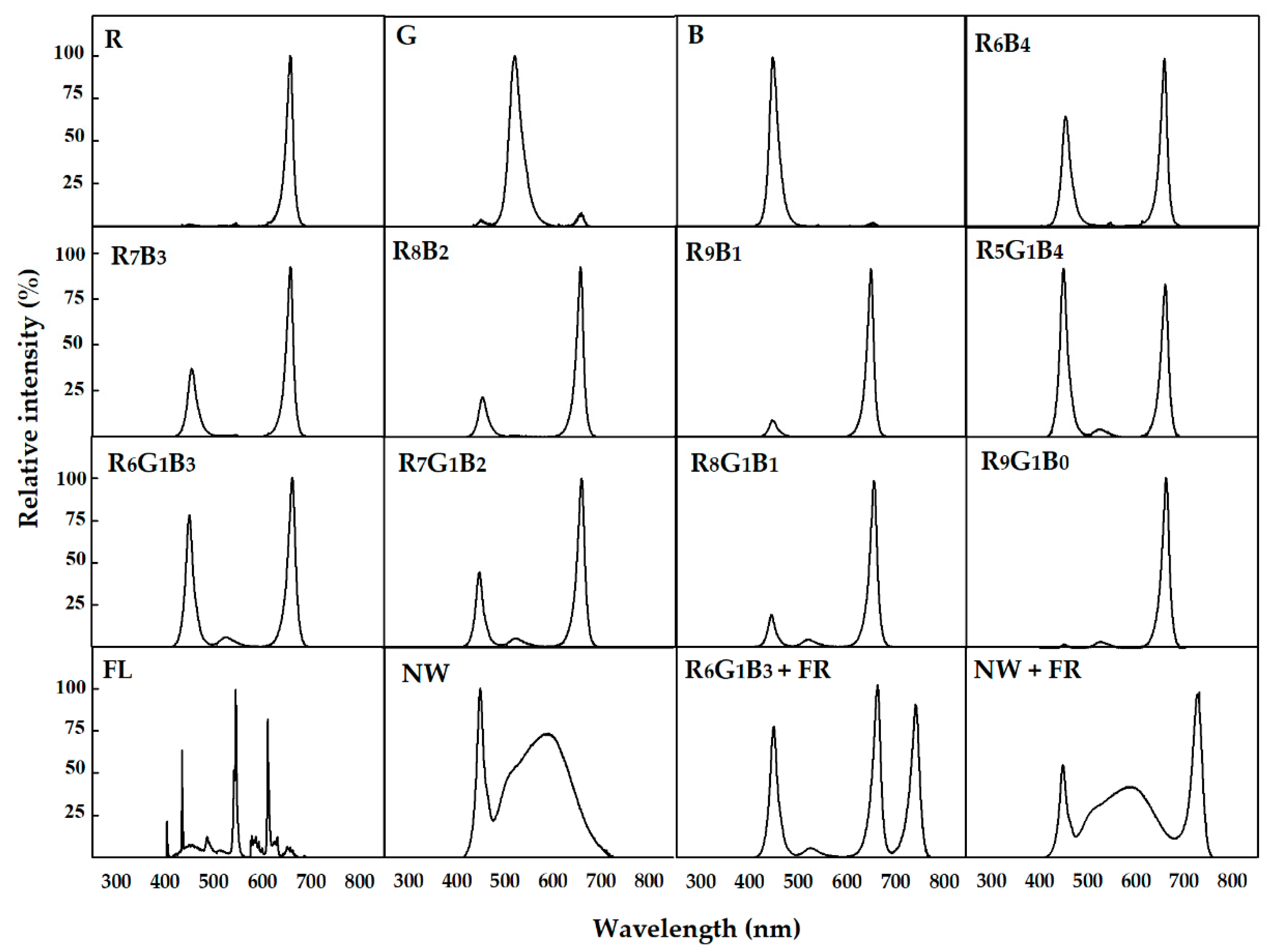
2.2. Light Treatments

Monochromatic group (mono group) with red (R, 656 nm), green (G, 520 nm), and blue (B, 451 nm) LEDs (L-PEC, Jeonju, Korea), a RB combination group (R6B4, R7B3, R8B2, and R9B1; base on chip number), and a RGB combination group (R5G1B4, R6G1B3, R7G1B2, R8G1B1, and R9G1B0) were used as light spectral treatments in this study (Figure S1). The white group (W group) was treated with fluorescent lamps (FL; HYG-FPL 36 W-R; Nam Yung, Seoul, Korea) and white LEDs (natural white, NW, Bissol LED, Seoul, Korea). The supplemental far-red group (FR group) was further investigated in R6G1B3 and NW using far-red LEDs (Itswell, Incheon, Korea; peak 740 nm), and the R/FR ratio of R6G1B3 + FR and NW + FR was adjusted to 1.2. In total, sixteen light treatments were created and tested simultaneously in a plant factory. The light period and photosynthetic photon flux density (PPFD) were set at 16 h and 80 µmol m−2 s−1, respectively. Light spectral distributions were measured using a spectroradiometer (JAZ-EL 200, Ocean optics, Dunedin, FL, USA) by dividing the cultivation area [54 × 58 cm (L × W)] into 6 areas for each light treatment based on the height of the canopy (Figure 1). As the canopy height of the crops for each treatment was differentiated, the distance between the crop and the light source was kept constant to remove the factor of change in light intensity according to the irradiation distance from the light source. To minimize experimental error due to the non-uniform distribution of the irradiated light source, the plants were rotated every 1–2 days.

2.3. Growth Characteristics

To compare the growth of ginseng sprouts under various light quality treatments, growth characteristics were measured at 5 weeks after planting. The shoot and root were separated by cutting the area around the rhizome with a knife, and the fresh weights of the shoot and root were measured using an electronic scale (Si-234, Denver Instrument, Bohemia, NY, USA). The length from the shoot of the rhizome to the part where the stem was split was measured by a ruler, and the leaf area was measured using a leaf area meter (LI-2050A, Li-Cor, Lincoln, NE, USA). T/R ratio was defined as the fresh weight ratio of top (shoot) to root. The shoot and root were freeze-dried for more than 96 h in a freeze dryer (Alpha 2–4 LSC plus, CHRIST, Osterode am Harz, Germany) at −75 °C, and then the dry weights of shoot and root were measured with an electronic scale.

2.4. Photosynthetic Parameters

After 4 weeks of planting, the photosynthetic rate, transpiration rate, and conductance of H2O of the fully extended leave were measured using a photosynthetic machine (LI-6400, LI-COR, Lincoln, NE, USA) equipped with a clear bottom chamber (2 × 3 cm) (LI-6400-08, Li-Cor, Lincoln, NE, USA). The conditions of the measurement chamber were set to 400 μmol mol−1 CO2, 23 °C air temperature, and 300 μmol s−1 flow rate. The photosynthetic parameters were measured at a point that light intensity was 80 µmol m−2 s−1 under each light source, and the measurements were performed for 3–6 h after the light was turned on. SPAD value reflecting relative chlorophyll content was measured by a portable chlorophyll meter (SPAD-502, Konica Minolta, Tokyo, Japan).

2.5. Total Saponin Content

To analyze the saponin content, the shoot and root were separately harvested at 5 weeks after planting and freeze-dried for 96 h in a freeze dryer. All samples were ground to powder using a mill (Tube mill control, IKA, Wilmington, NC, USA). For saponin extraction, approximately 100 mg of dry powder of each sample and 5 mL 1-BuOH (99.8%, v/v) were added to a 15 mL conical tube and then vortexed for 10 s. Thereafter, the extracted samples were incubated at 4 °C for more than 24 h and then sonicated in a sonication bath at 50 °C for 60 min (SK5210HP, KUDOS, Shanghai, China). After centrifuging (2195× g) at 4 °C for 15 min, the supernatant was evaporated at 40–50 °C for approximately 2 h using a nitrogen evaporator (N-EVAP 111, Organomation, Berlin, MA, USA), and then 5 mL of MeOH (99%, v/v) was added. After that, the total saponin content was analyzed according to a method described by [22]. The standard curve of total saponin content was obtained using the ginsenoside standard Re (ASB-00007210-005, ChromaDex, Los Angeles, CA, USA). Shoot and root saponin contents were expressed as milligrams of saponin per gram dry weight of shoot and root (mg g−1). Total saponin content was calculated by the sum of the saponin content per shoot dry weight and the saponin content per root dry weight, and this value was expressed as milligrams (mg).

2.6. Ginsenosides Content

Dry powder (45 mg) of each sample was mixed with 4.5 mL of ethanol and extracted in a sonication bath at 27–30 °C for 1 h. The extract was centrifuged at 2195× g at 20 °C for 10 min, and the supernatant was used for analysis. After evaporating the solvent of the supernatant by a nitrogen evaporator, 2.5 mL of distilled water and 2.5 mL of diethyl ether (99.5%, v/v) were added, mixed, and then incubated for 30 min. The supernatant diethyl ether was removed from the two layers, and the process was repeated. After that, 1 mL distilled water and 1 mL 1-butyl alcohol (99.8%, v/v) were added and mixed, followed by incubating for 30 min. The upper layer was transferred to a new tube, and the process was repeated. After evaporating the supernatant with a nitrogen evaporator, 600 µL of methanol was added and filtered using a syringe filter (BS20-PC13, BioFACT, Daejeon, Korea).
The ginsenosides content was measured using a HPLC system (YL9100, Youngin Chromass, Anyang, Korea) equipped with a UV/VIS detector (YL9120, Youngin Chromass, Anyang, Korea). A Fortis C18 column (F18-050905, 250 × 4.6 mm, 5 μM, Chromex Scientific, Dronfield, UK) was used, and samples were injected in 20 μL increments. Solvent A was 100% acetonitrile, and solvent B was deionized water. The flow rate of the mobile phases was maintained at 0.6 μL min−1, and the column temperature was 35 °C. Ginsenosides were detected at an absorbance of 203 nm. The ratio of solvent A was as follows; 30% (0–10 min), 30–40% (10–50 min), 40–80% (50–55 min), 80–20% (55–61 min) and 20–30% (61–70 min). The commercial grade ginsenosides consisting of protopanaxadiol (PD; Rb1, Rb2, Rc, and Rd) and protopanaxatriol (PT; Rg1, Rg2, Rg3, Re, and Rf) were purchased from ChromaDex (Los Angeles, CA, USA). Shoot and root ginsenosides contents were expressed as milligrams of ginsenosides per gram dry weight of shoot and root (mg g−1). Total ginsenosides content was calculated as above total saponin content, and this value was expressed as milligrams (mg).

2.7. Statistical Analysis

The growth characteristics and photosynthetic parameters were measured in 10 and 4 replicates, respectively. Total saponin and ginsenosides contents were measured in 6 and 5 replicates, respectively, using freeze-dried powder by combining 10 samples per treatment. For statistical analysis, ANOVA was performed using the SAS statistical program (Statistical Analysis System, 9.4 Version, SAS Institute, Cary, NC, USA). Significant differences in all treatments were verified at p < 0.05, using Duncan’s multiple range test. Graphs were created using the SigmaPlot program (Exact Graphs and Data Analysis, 12 Version, Systat Software Inc., San Jose, CA, USA).

3. Results

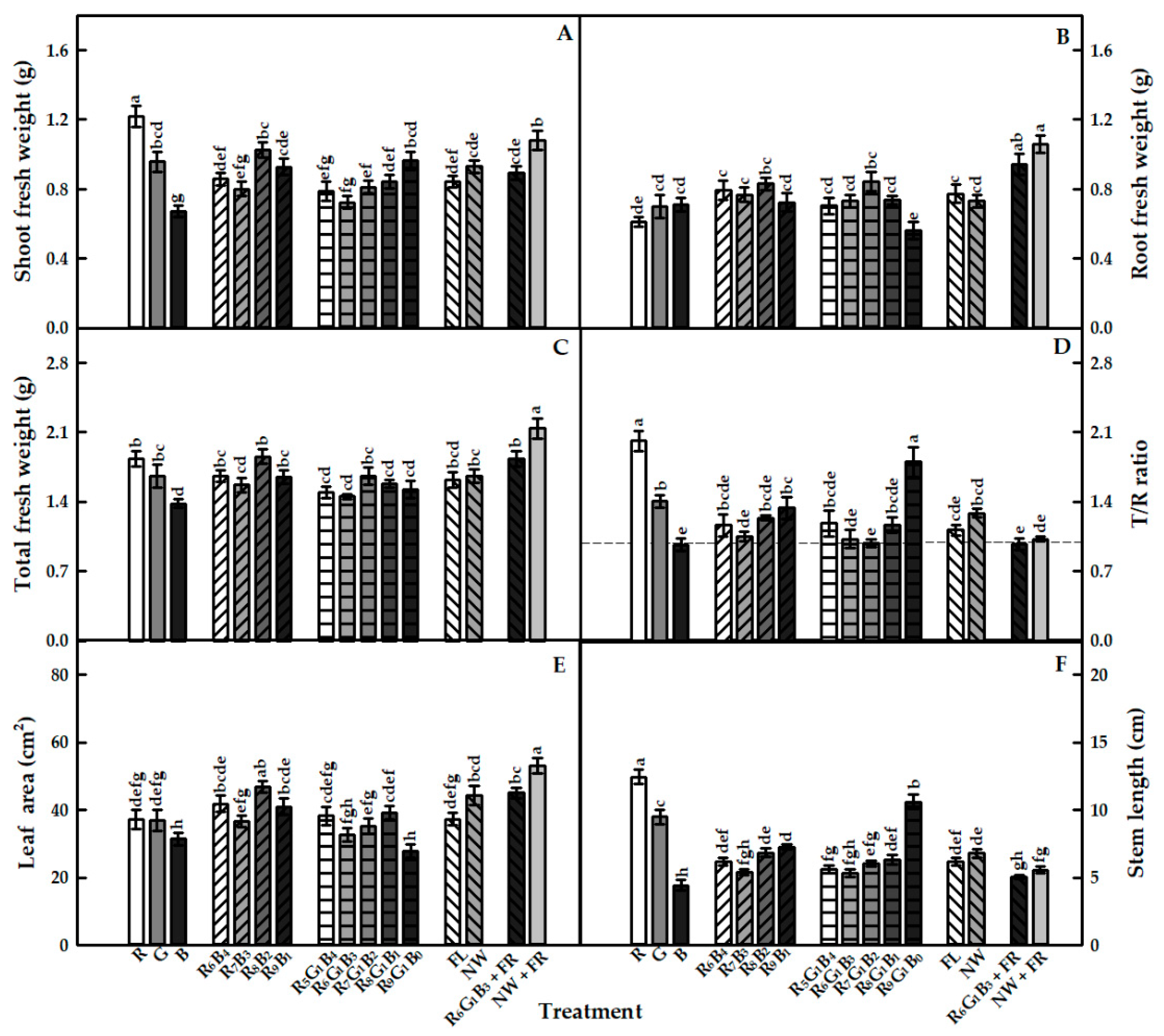
3.1. Growth Characteristics

In the monochromatic group, the growth characteristics of ginseng sprouts was highest under R and was lowest under B (excepted for root fresh weight); particularly, R exhibited 2.0 times higher shoot fresh weight and T/R ratio and 3.0 times higher stem length compared to B. Meanwhile, G showed lower shoot fresh weight, T/R ratio, stem length, and no significant difference in root fresh weight, total fresh weight, and leaf area comparing to R light (Figure 2). The combination of RB and RGB groups showed different effects on growth parameters of ginseng sprouts. In the RB group, R8B2 resulted in higher shoot fresh weight compared to R6B4 and R7B3, whereas in the RGB group, R9G1B0 exhibited the highest value (Figure 2A). There was no significant difference in root fresh weight among the RB group, while in the RGB group, R9G1B0 showed a lowest value (Figure 2B). The higher ratio of R resulted in a higher T/R ratio, and the highest value was obtained in R9G1B0 (Figure 2D). R8B2 had the highest leaf area in the RB group, and in the RGB group, R9G1B0 showed the lowest value (Figure 2E). Stem length under R9B1 was higher compared to R7B3 in the RB group, and R9G1B0 showed the highest value among treatments in the RGB group (Figure 2F). There were no significant differences in all of growth characteristics between FL and NW treatments. Both the supplement FR treatments (R6G1B3 + FR and NW + FR) had a significantly higher shoot and root fresh weights, total fresh weight, and leaf area compared to those under those in the respective treatments without FR (R6G1B3 and NW) (Figure 2A–C,E). Overall, R and the supplement of FR in NW showed the efficient improvement of ginseng sprout growth.

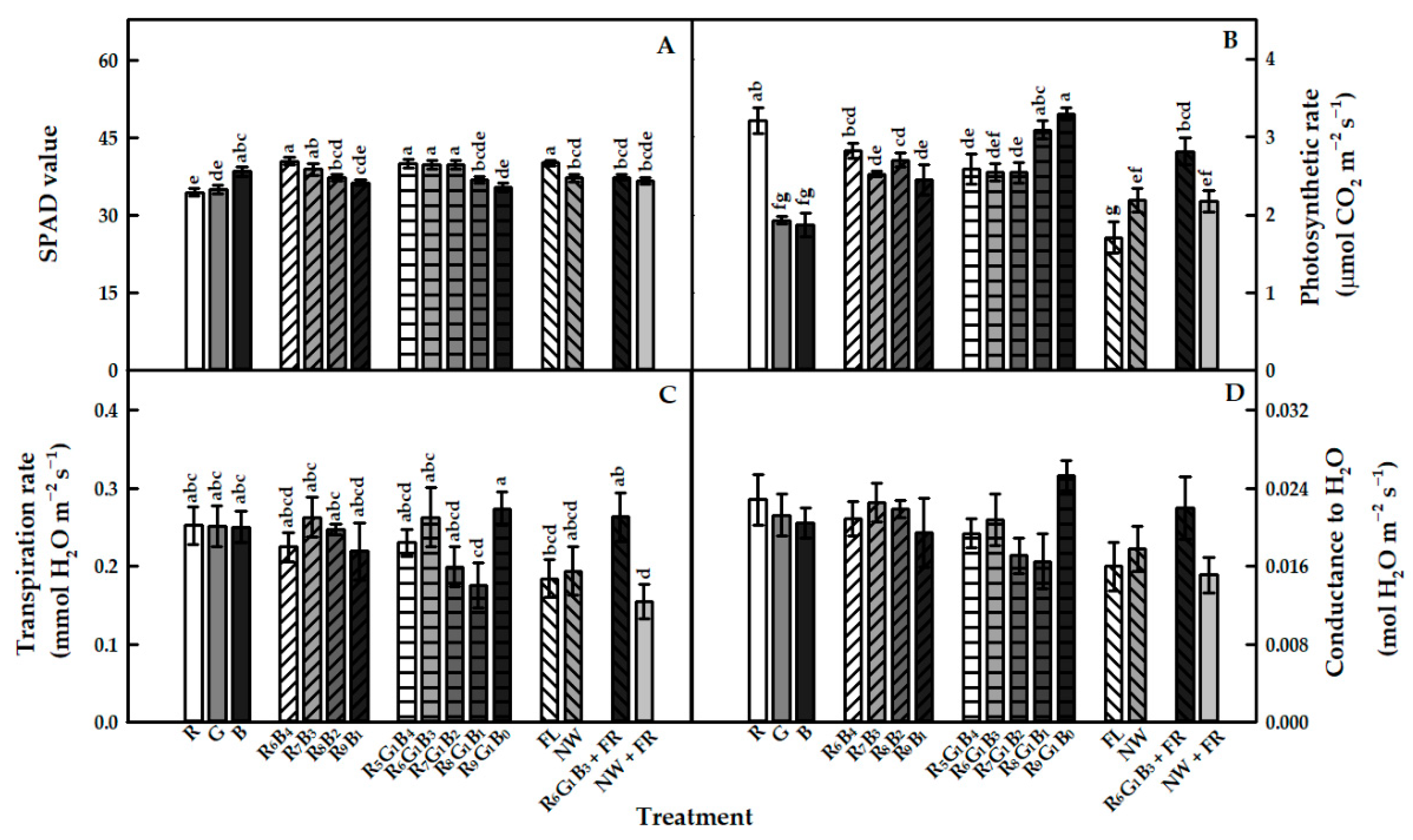
3.2. Photosynthetic Parameters

The result of SPAD value presented an opposite trend to that of growth characteristics. The SPAD value was slightly higher in B than in R and G, and in RB and RGB groups, the higher B ratio resulted in the higher value (Figure 3A). In the W group, this value in FL was higher than that in NW, while in the supplement FR, R6G1B3 + FR resulted in a lower value than R6G1B3, and no significant difference in SPAD was observed between NW + FR and NW. Photosynthetic rate of plants under R was approximately 2 times higher that under B and G (Figure 3B). There was no significant difference in the photosynthesis rate in the RB group, whereas this value under R8G1B1 and R9G1B0 was higher than that under other treatments in the RGB group. In the W group, FL had a lower value compared to NW, while the supplement of FR (R6G1B3 + FR and NW + FR) showed no difference from treatments without FR (R6G1B3 and NW). Regarding transpiration rate, no significant difference was observed among the mono, RB, and W groups (Figure 3C). In the RGB group, R9G1B0 had a higher transpiration rate than R8G1B1, whereas in the FR group, R6G1B3 + FR was 167% higher than NW + FR, and there was no significant difference in this value in R6G1B3 + FR and NW + FR compared to R6G1B3 and NW, respectively. The conductance of H2O showed a similar trend to the transpiration rate, but there was no difference among all treatments (Figure 3D).

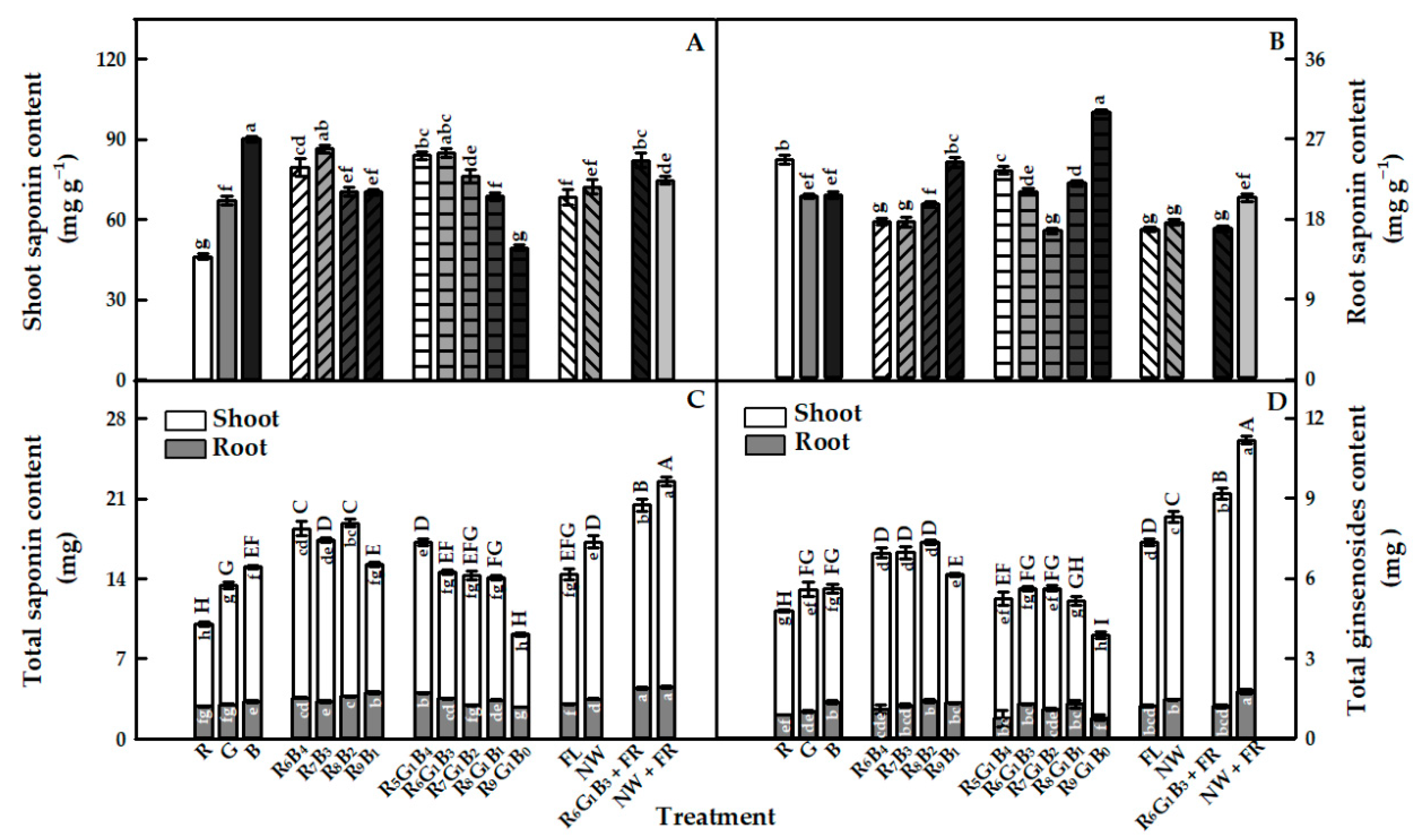
3.3. Total Saponin Content

The saponin content per gram of shoot dry weight (referred as shoot saponin content) under B was 1.95 and 1.34 times higher than that under R and G, respectively (Figure 4A). As the B ratio in RB and RGB groups increased, the shoot saponin content markedly enhanced. There was no significant difference in shoot saponin content in the W group. Supplemental FR was not effective in increasing shoot saponin content comparing to respective treatments without FR. The root saponin content per gram of root dry weight (referred as root saponin content) showed an opposite trend to the shoot saponin content (Figure 4B). Root saponin content under R was slightly higher than that under B and G, and this value markedly increased with the increase in the R proportion in the RB group. In the RGB group, R9G1B0 showed the highest value among treatments. There was no significant difference in root saponin content between the W treatments. This value in R6G1B3 + FR was lower than that in R6G1B3, while NW + FR resulted in a higher value compared to NW. Total saponin content per plant dry weight (referred as total saponin content) showed a similar trend to shoot saponin content (Figure 4C). B and a higher ratio of B induced higher total saponin content. Interestingly, supplementing with FR was effective in enhancing the total saponin content.

3.4. Ginsenosides Content

For the ginsenosides content per gram of shoot dry weight (referred as shoot ginsenosides content), in the mono group, the total value was highest in B, and this value was 1.55 times higher than that in R (Table 1). In particular, Rb1 and Re, the major ginsenosides, in B were 238% and 295%, respectively, higher than those in R. In the RB group, R7B3 showed higher Rb1 and Re compared to other treatments; the total value under R7B3 was 1.27 and 1.16 times higher than R8B2 and R9B1, respectively. In the RGB group, Rb1, Rg1, and Re in R6G1B3 were 1.74, 1.43, and 1.68 times, respectively, higher than those in R9G1B0. The W group showed no significant differences in Rb1, Rg1, and total value between treatments. In the supplement FR, R6G1B3 + FR exhibited a higher total value than R6G1B3 and was the highest value among all treatments; while NW + FR had no significant difference from NW, but this treatment also showed a very high value.
Regarding the ginsenosides content per gram of root dry weight (referred as root ginsenosides content), the total value in B was highest in mono group although there was no statistically significant difference compared to R (Table 2). The Re content under B was 4.21 and 5.15 times higher than that under R and G, respectively. In the RB group, R9B1 resulted in higher contents of major ginsenosides Rb1 and Rg1 compared to those under R6B4, which led to induce an increase in root ginsenosides content. In the RGB group, R9G1B0 and R8G1B1 showed higher values than R7G1B2. There were no significant differences in Rb1, Rg1, and total value in W group, whereas in the supplement FR, R6G1B3 + FR showed lower values compared to R6G1B3, and no significant difference between NW + FR and NW was obtained. The total ginsenosides content per plant dry weight (referred as total ginsenosides content) showed a similar trend to the total saponin content (Figure 4D). R and a high ratio of R reduced the total ginsenosides content. The total ginsenosides contents in R6G1B3 + FR and NW + FR were highest among all treatments with 9.18 and 11.17 mg, respectively; therefore, the supplement of FR was considerably effective in enhancing ginsenosides content.

4. Discussion

4.1. Growth Characteristics

In this study, R and the higher ratio of R in LEDs treatments had a positive effect on shoot growth of ginseng sprouts, which was considered due to the high light utilization efficiency of red light. Many studies proved that red light promotes plant biomass such as fresh and dry weights, plant height, and leaf area [23]. In the present study, the leaf area and stem length showed a similar pattern to the shoot fresh weight; therefore, it can be considered that they were the main factors in determining the total fresh weight. Meanwhile, monochromatic B suppresses hypocotyl elongation compared to monochromatic R, and the high proportion of B decreases the stem length [24]. In this study, R and B lights also showed a contrasting effect on stem growth. The higher ratio of R light resulted in a higher stem length. This was consistent with the results of previous studies on the growth of various crops under the combination of red and blue [20,25].
In the W group, although FL and NW had different light spectra, the ratio of R, G, and B light were almost the same at 28:44:27 and 29:48:23 (data not shown), respectively; therefore, it seemed not to be significantly different. Likewise, G light also did not show the positive effect on ginseng growth. Green light has a lower absorbance and higher transmittance and reflectance than other wavelengths, showing inefficiently impacts on the growth and development as well as photosynthesis of plants [26], which was observed in the present study with the lower photosynthesis rate under green light. In contrast, the supplement of FR LEDs increased both shoot and root of the ginseng sprouts, especially in leaf area, which corresponded to previous studies that showed a growth-promoting effect in lettuce and Crepidiastrum denticulatum [25,27]. The ratio of R/FR used in this experiment (1.2) was similar to the value during solar noon, resulting in greater plant height, internode length, and node number, which contributed to higher plant fresh weight in many crops [25,27]. However, although FR LEDs presented a higher ginseng sprouts growth, it was not effective on the stem length. Noticeably, in dicotyledonous plants, the low R/FR (in a shaded environment) results in the rapid elongation of the stem [28,29,30]. Since ginseng is a shade plant, the stem growth caused by the shade avoidance response did not occur in the FR group. Park and Runkle reported that shade-avoidant plants such as geranium, petunia, and snapdragon showing sensitive changes in stem length according to phytochrome photoequilibrium (PPE) directly affected by the R/FR ratio, but impatiens, a shade-tolerant plant, was not responsive [31]. In this study, both supplemental FR treatments showed significantly higher leaf area compared to the respective treatments without FR; therefore, FR light was effective in enhancing the leaf area of ginseng sprouts.
Regarding root growth of ginseng sprouts, since the cultivation period (5 weeks) of this experiment was not enough for the transportation of the nutrients produced from leave to the root, the difference in root weight according to visible light treatments seemed to be insignificant. Supplemental FR treatment increased the root fresh weight. Gelderen et al. stated that the auxin and gibberellic acid produced under FR in shoots may move toward the roots, which led to a positive effect on root development [32]. In general, the total fresh weight was similar to the shoot fresh weight because the change in shoot fresh weight was much greater than that of the root fresh weight according to light treatments. The T/R ratio was also comparable to the pattern of the shoot fresh weight. Both treatments with supplemented FR showed a T/R ratio of approximately 1 by significantly improving root growth.

4.2. Photosynthetic Parameters

The results of photosynthetic parameters supported the growth of ginseng sprouts. In the monochromatic and RGB groups, the positive effect of R light on enhancing the photosynthetic rate was confirmed. McCree also reported that red light resulted in higher photosynthetic efficiency compared to blue light [33]. Therefore, the treatments with a high ratio of red light increased the photosynthesis rate, which was thought to have a major effect on the increase in total fresh weight. Meanwhile, FR light had little impact on the improvement of the photosynthesis rate per unit leaf area, but it resulted in the expansion of the leaf area, which led to an increase in the photosynthetic rate and fresh weight of the whole plant. The transpiration rate and conductance of H2O increased as the B ratio increased in the combination groups. Although there was a difference in the chlorophyll content (SPAD value) according to the light treatments, the difference did not contribute to the photosynthetic rate. The SPAD value increased as the ratio of B increased. In general, blue light induces an increase in tetrapyrrole precursor 5-aminolevulinic acid, which improves chlorophyll biosynthesis, whereas red light downregulates protein and gene expression of enzymes involved in chlorophyll biosynthesis [34,35]. Supplemental FR light did not affect the SPAD value. Shade-tolerant plant species, such as impatiens, adapted even in FR light environments and showed normal photosynthesis without reducing chlorophyll content [36].

4.3. Total Saponin and Ginsenosides Contents

It is well known that the type and efficient production of bioactive compounds in ginseng are mostly dependent on the tissues and the plant’s age [37]. The recent studies prove that the accumulation of saponin and ginsenosides produced in ginseng is considerably affected by environmental conditions [38]. B light was reported to enhance the content of secondary metabolites than R light in several crops [12,39], which was consistent with results of the present study with the increase in shoot saponin content, total saponin content, and shoot ginsenosides content under B. A high proportion of B particularly enhanced the major ginsenosides Rb1, Rg1, and Re, which led to an increase in the total ginsenosides content. In contrast, Fournier et al. reported that R contributed to the increase of Rd and Rg1 in 2-year-old American ginseng, but B did not affect ginsenosides accumulation [40]. The root saponin content was higher as the ratio of R increased, which might be explained by the differentiated distribution of assimilates to shoot and root according to light quality. In general, it is easy to have a dilution effect, such as a decrease in the concentration of secondary metabolites per unit dry weight due to growth promotion [27].
The supplemental FR light increased the growth of ginseng sprouts as well as saponin and ginsenosides content in ginseng sprouts. These results corresponded to previous study reporting that FR light resulted in the variation in ginsenosides contents and the higher levels of FR light increased ginsenosides production [40]. The root ginsenosides content also showed the same tendency as the root saponin content, which was different from that in the shoot. The ratio of PD/PT showed a difference between shoot and root in the RGB and W groups that contained green light. The effect of light quality on ginsenosides synthesis is unclear, but it shows the possibility that PD/PT may vary depending on the light quality. The total ginsenosides content was similar to the pattern of the shoot ginsenosides content because the shoot was about 4.4 times higher than that of the root.

5. Conclusions

According to the results of this study, R light was effective on enhancing growth characteristics as well as photosynthesis parameters, while B light had a positive effect on improving the total saponin and ginsenosides content. The supplemental far-red light did not affect the stem length in ginseng sprouts, but it improved the leaf area and induced balanced growth between the shoot and root. This growth improvement by supplemental FR light led to an increase in total saponin and ginsenosides content. In particular, NW + FR was the most effective treatment. These results suggest that the growth and ginsenosides content of ginseng sprouts can be enhanced according to the composition of light quality. These findings can be applied to ensure the economic efficiency of ginseng sprout production.

Supplementary Materials

The following are available online at https://www.mdpi.com/2073-4395/10/12/1979/s1, Figure S1: Ginseng seedlings were cultivated under various light treatments for 5 weeks.

Author Contributions

Conceptualization, methodology; software, formal analysis, investigation, resources, data curation, writing—original draft preparation, Y.-J.K. and T.K.L.N.; supervision, validation, funding acquisition, writing—review and editing, M.-M.O. All authors have read and agreed to the published version of the manuscript.

Funding

This work was carried out with the support of Cooperative Research Program for Agriculture Science and Technology Development (project no. PJ01385201) Rural Development Administration, Korea.

Acknowledgments

In this section you can acknowledge any support given which is not covered by the author contribution or funding sections. This may include administrative and technical support, or donations in kind (e.g., materials used for experiments).

Conflicts of Interest

Authors declare no conflict of interest.

References

  1. Kim, D.H.; Moon, Y.S.; Lee, T.H.; Jung, J.S.; Suh, H.W.; Song, D.K. The inhibitory effect of ginseng saponins on the stress-induced plasma interleukin-6 level in mice. J. Neurosci. Lett. 2003, 353, 13–16. [Google Scholar] [CrossRef] [PubMed]
  2. López, M.V.N.; Cuadrado, M.P.G.-S.; Ruiz-Poveda, O.M.P.; Del Fresno, A.M.V.; Accame, M.E.C. Neuroprotective effect of individual ginsenosides on astrocytes primary culture. Biochim. Biophys. Acta Gen. Subj. 2007, 1770, 1308–1316. [Google Scholar] [CrossRef] [PubMed]
  3. Vuksan, V.; Sung, M.-K.; Sievenpiper, J.L.; Stavro, P.M.; Jenkins, A.L.; Di Buono, M.; Lee, K.-S.; Leiter, L.A.; Nam, K.Y.; Arnason, J.T.; et al. Korean red ginseng (Panax ginseng) improves glucose and insulin regulation in well-controlled, type 2 diabetes: Results of a randomized, double-blind, placebo-controlled study of efficacy and safety. Nutr. Metab. Cardiovasc. Dis. 2008, 18, 46–56. [Google Scholar] [CrossRef] [PubMed]
  4. Chen, F.; Luo, J.-G.; Kong, L. Determination of 10 ginsenosides in Panax ginseng of different harvest times based on HPLC fingerprints and principal component analysis. Nat. Prod. Res. 2013, 27, 851–854. [Google Scholar] [CrossRef] [PubMed]
  5. Li, X.G.; Yan, Y.Z.; Jin, X.; Kim, Y.K.; Uddin, M.R.; Kim, Y.B.; Bae, H.; Kim, Y.C.; Lee, S.W.; Park, S.U. Ginsenoside content in the leaves and roots of Panax ginseng at different ages. Life Sci. 2012, 9, 670–683. [Google Scholar]
  6. Kim, G.S.; Hyun, D.Y.; Kim, Y.O.; Lee, S.E.; Kwon, H.; Cha, S.W.; Park, C.B.; Kim, Y.B. Investigation of ginsenosides in different parts of Panax ginseng cultured by hydroponics. Korean J. Hortic. Sci. 2010, 28, 216–226. [Google Scholar]
  7. Choi, S.Y.; Cho, C.-W.; Lee, Y.-M.; Kim, S.-S.; Lee, S.-H.; Kim, K.-T. Comparison of Ginsenoside and Phenolic Ingredient Contents in Hydroponically-cultivated Ginseng Leaves, Fruits, and Roots. J. Ginseng. Res. 2012, 36, 425–429. [Google Scholar] [CrossRef] [Green Version]
  8. Kozai, T. Resource use efficiency of closed plant production system with artificial light: Concept, estimation and application to plant factory. Proc. Jpn. Acad. Ser. B 2013, 89, 447–461. [Google Scholar] [CrossRef] [Green Version]
  9. Bian, Z.H.; Yang, Q.C.; Liu, W.K. Effects of light quality on the accumulation of phytochemicals in vegetables produced in controlled environments: A review. J. Sci. Food Agric. 2015, 95, 869–877. [Google Scholar] [CrossRef]
  10. Schmitt, J.; Wulff, R.D. Light spectral quality, phytochrome and plant competition. Trends Ecol. Evol. 1993, 8, 47–51. [Google Scholar] [CrossRef]
  11. Smith, H. Light Quality, Photoperception, and Plant Strategy. Annu. Rev. Plant Physiol. 1982, 33, 481–518. [Google Scholar] [CrossRef]
  12. Son, K.-H.; Oh, M.M. Leaf Shape, Growth, and Antioxidant Phenolic Compounds of Two Lettuce Cultivars Grown under Various Combinations of Blue and Red Light-emitting Diodes. HortScience 2013, 48, 988–995. [Google Scholar] [CrossRef]
  13. Kozai, T. Smart Plant Factory: The Next Generation Indoor Vertical Farms, 1st ed.; Springer: Berlin/Heidelberg, Germany, 2018; pp. 3–14. [Google Scholar]
  14. Jeon, Y.-M.; Son, K.-H.; Kim, S.-M.; Oh, M.M. Growth and bioactive compounds as affected by irradiation with various spectrum of light-emitting diode lights in dropwort. Hortic. Environ. Biotechnol. 2017, 58, 467–478. [Google Scholar] [CrossRef]
  15. Kim, E.-Y.; Park, S.-A.; Park, B.-J.; Lee, Y.; Oh, M.M. Growth and antioxidant phenolic compounds in cherry tomato seedlings grown under monochromatic light-emitting diodes. Hortic. Environ. Biotechnol. 2014, 55, 506–513. [Google Scholar] [CrossRef]
  16. Lee, J.W.; Son, K.H.; Lee, J.H.; Kim, Y.J.; Oh, M.M. Growth and Biochemical Responses of Ice Plant Irradiated by Various Visible Light Spectra in Plant Factories. Hortic. Sci. Technol. 2019, 37, 598–608. [Google Scholar]
  17. Carvalho, R.F.; Takaki, M.; Azevedo, R.A. Plant pigments: The many faces of light perception. Acta Physiol. Plant. 2010, 33, 241–248. [Google Scholar] [CrossRef]
  18. Sullivan, J.A.; Deng, X.-W. From seed to seed: The role of photoreceptors in Arabidopsis development. Dev. Biol. 2003, 260, 289–297. [Google Scholar] [CrossRef] [Green Version]
  19. Li, J.; Hikosaka, S.; Goto, E. Effects of light quality and photosynthetic photon flux on growth and carotenoid pigments in spinach (Spinacia oleracea L.). Acta Hortic. 2011, 105–110. [Google Scholar] [CrossRef]
  20. Pennisi, G.; Blasioli, S.; Cellini, A.; Maia, L.; Crepaldi, A.; Braschi, I.; Spinelli, F.; Nicola, S.; Fernandez, J.A.; Stanghellini, C.; et al. Unraveling the Role of Red:Blue LED Lights on Resource Use Efficiency and Nutritional Properties of Indoor Grown Sweet Basil. Front. Plant Sci. 2019, 10, 305. [Google Scholar] [CrossRef] [Green Version]
  21. Zhang, X.; He, D.; Niu, G.; Yan, Z.; Song, J. Effects of environment lighting on the growth, photosynthesis, and quality of hydroponic lettuce in a plant factory. Int. J. Agric. Biol. Eng. 2018, 11, 33–40. [Google Scholar] [CrossRef]
  22. Hiai, S.; Oura, H.; Nakajima, T. Color reaction of some sapogenins and saponins with vanillin and sulfur1c acid. Planta Med. 1976, 29, 116–122. [Google Scholar] [CrossRef]
  23. Wang, J.; Lu, W.; Tong, Y.; Yang, Q. Leaf Morphology, Photosynthetic Performance, Chlorophyll Fluorescence, Stomatal Development of Lettuce (Lactuca sativa L.) Exposed to Different Ratios of Red Light to Blue Light. Front. Plant Sci. 2016, 7, 250. [Google Scholar] [CrossRef] [PubMed] [Green Version]
  24. Nanya, K.; Ishigami, Y.; Hikosaka, S.; Goto, E. Effects of blue and red light on stem elongation and flowering of tomato seedlings. Acta Hortic. 2012, 956, 261–266. [Google Scholar] [CrossRef]
  25. Bae, J.-H.; Park, S.-Y.; Oh, M.M. Supplemental irradiation with far-red light-emitting diodes improves growth and phenolic contents in Crepidiastrum denticulatum in a plant factory with artificial lighting. Hortic. Environ. Biotechnol. 2017, 58, 357–366. [Google Scholar] [CrossRef]
  26. Folta, K.M.; Maruhnich, S.A. Green light: A signal to slow down or stop. J. Exp. Bot. 2007, 58, 3099–3111. [Google Scholar] [CrossRef] [PubMed]
  27. Lee, M.-J.; Son, K.-H.; Oh, M.M. Increase in biomass and bioactive compounds in lettuce under various ratios of red to far-red LED light supplemented with blue LED light. Hortic. Environ. Biotechnol. 2016, 57, 139–147. [Google Scholar] [CrossRef]
  28. Morgan, D.C.; Smith, H. Linear relationship between phytochrome photoequilibrium and growth in plants under simulated natural radiation. Nat. Cell Biol. 1976, 262, 210–212. [Google Scholar] [CrossRef]
  29. Morgan, D.C.; Smith, H. The relationship between phytochrome-photoequilibrium and Development in light grown Chenopodium album L. Planta 1978, 142, 187–193. [Google Scholar] [CrossRef]
  30. Child, R.; Morgan, D.C.; Smith, H. Control of development in Chenopodium album l. by shadelight: The effect of light quality (red:far-red ratio) on morphogenesis. New Phytol. 1981, 89, 545–555. [Google Scholar] [CrossRef]
  31. Park, Y.; Runkle, E.S. Far-red radiation promotes growth of seedlings by increasing leaf expansion and whole-plant net assimilation. Environ. Exp. Bot. 2017, 136, 41–49. [Google Scholar] [CrossRef] [Green Version]
  32. Van Gelderen, K.; Kang, C.; Pierik, R. Light Signaling, Root Development, and Plasticity. Plant Physiol. 2018, 176, 1049–1060. [Google Scholar] [CrossRef] [PubMed] [Green Version]
  33. McCree, K. Test of current definitions of photosynthetically active radiation against leaf photosynthesis data. Agric. Meteorol. 1972, 10, 443–453. [Google Scholar] [CrossRef]
  34. Sood, S.; Gupta, V.; Tripathy, B.C. Photoregulation of the Greening Process of Wheat Seedlings Grown in Red Light. Plant Mol. Biol. 2005, 59, 269–287. [Google Scholar] [CrossRef] [PubMed]
  35. Tanaka, A.; Ito, H.; Tanaka, R.; Tanaka, N.K.; Yoshida, K.; Okada, K. Chlorophyll a oxygenase (CAO) is involved in chlorophyll b formation from chlorophyll a. Proc. Natl. Acad. Sci. USA 1998, 95, 12719–12723. [Google Scholar] [CrossRef] [Green Version]
  36. Park, Y.; Runkle, E.S. Far-red radiation and photosynthetic photon flux density independently regulate seedling growth but interactively regulate flowering. Environ. Exp. Bot. 2018, 155, 206–216. [Google Scholar] [CrossRef]
  37. Shi, W.; Wang, Y.; Li, J.; Zhang, H.; Ding, L. Investigation of ginsenosides in different parts and ages of Panax ginseng. Food Chem. 2007, 102, 664–668. [Google Scholar] [CrossRef]
  38. Lim, W.; Mudge, K.W.; Vermeylen, F. Effects of Population, Age, and Cultivation Methods on Ginsenoside Content of Wild American Ginseng (Panax quinquefolium). J. Agric. Food Chem. 2005, 53, 8498–8505. [Google Scholar] [CrossRef]
  39. Lee, J.G.; Oh, S.S.; Cha, S.H.; Jang, Y.A.; Kim, S.Y.; Um, Y.C.; Cheong, S.R. Effects of red/blue light ratio and short-term light quality conversion on growth and anthocyanin contents of baby leaf lettuce. Prot. Hortic. Plant Fact. 2010, 19, 351–359. [Google Scholar]
  40. Fournier, A.R.; Proctor, J.T.; Gauthier, L.; Khanizadeh, S.; Bélanger, A.; Gosselin, A.; Dorais, M. Understory light and root ginsenosides in forest-grown Panax quinquefolius. Phytochemistry 2003, 63, 777–782. [Google Scholar] [CrossRef]
Figure 1. Relative spectral distribution of sixteen light treatments with red LEDs (R), green LEDs (G), blue LEDs (B), fluorescent lamps (FL), natural white LEDs (NW), and far-red LEDs (FR) used in this study.
Agronomy 10 01979 g001
Figure 2. Shoot fresh weight (A), root fresh weight (B), total fresh weight (C), T/R ratio (D), leaf area (E) and stem length (F) of ginseng sprouts grown under various light treatments for 5 weeks. Different letters indicate significant differences at p < 0.05 (n = 10). The dashed line in figure (D) indicates a T/R ratio of 1.
Agronomy 10 01979 g002
Figure 3. SPAD value (A) of ginseng sprouts at 5 weeks after planting and photosynthetic rate (B), transpiration rate (C), and conductance to H2O (D) of ginseng sprouts grown under various light treatments at 4 weeks after planting. Different letters indicate significant differences at p < 0.05 (n = 4).
Agronomy 10 01979 g003
Figure 4. Shoot saponin content (A), root saponin content (B), total saponin content (C) and total ginsenosides content (D) of ginseng sprouts grown under various light treatments for 5 weeks. Different letters indicate a significant difference at p < 0.05, and capital letters in figure (C,D) indicate a significant difference in total contents of shoot and root (n = 6).
Agronomy 10 01979 g004
Table 1. Ginsenosides content of shoots in ginseng sprouts grown under various light treatments for 5 weeks.
Shoot Ginsenosides Content (mg g−1)
Group 1TreatmentPanaxadiol (PD) Panaxatriol (PT)Total ValuePD/PT
Rb1Rb2RcRd Rg1Rg2Rg3ReRf
MonoR1.36 j 20.68 fgh0.25 gh3.34 fg 10.40 f1.02 h0.10 h3.65 l0.14 f20.93 j0.37
G1.98 hi1.18 d0.28 bcde5.81 bc 13.93 cd1.67 def0.12 g4.35 k0.11 g29.44 fgh0.46
B3.25 bc1.11 de0.15 i4.86 d 10.43 f1.58 ef0.15 ef10.78 a0.24 a32.55 defg0.40
RBR6B42.59 fg1.49 c0.27 cdefg5.77 bc 13.71 cde2.15 c0.17 de5.08 ij0.11 g31.33 efgh0.48
R7B33.11 cd1.20 d0.24 gh4.84 d 14.24 bcd1.88 cde0.14 fg9.23 b0.21 bcd35.10 bcde0.37
R8B22.39 g0.97 de0.29 bc3.79 ef 12.98 de1.57 ef0.15 ef5.40 i0.15 f27.69 hi0.37
R9B12.52 g0.89 ef0.29 bc4.02 e 14.10 bcd1.45 fg0.16 de6.49 fg0.21 bcd30.13 fgh0.34
RGBR5G1B42.87 de0.44 hi0.25 gh2.40 h 14.89 bc1.00 h0.18 cd6.24 g0.16 ef28.42 gh0.26
R6G1B33.05 cd0.54 ghi0.30 b2.83 gh 17.17 a1.06 h0.17 cd7.77 d0.19 cde33.08 cdef0.26
R7G1B22.78 ef0.50 ghi0.25 fgh2.59 h 14.30 bcd1.10 h0.21 ab8.30 c0.21 abc30.24 fgh0.25
R8G1B12.10 h0.31 i0.26 defgh1.81 i 13.24 cde0.64 i0.18 cd6.01 gh0.21 abc24.77 j0.22
R9G1B01.75 i0.71 fg0.26 efgh3.49 ef 12.02 ef1.15 gh0.14 fg4.63 jk0.15 f24.30 ij0.34
WFL3.12 de1.14 c0.24 a5.34 b 15.90 a1.69 cd0.17 a9.07 hi0.23 ef36.89 bcd0.36
NW2.93 cd1.44 d0.33 h6.07 cd 17.32 ab1.94 def0.21 cde5.61 b0.16 ab36.02 bc0.43
FRR6G1B3 + FR3.50 a2.52 a0.27 cdef7.88 a 15.90 ab3.50 a0.19 bc7.08 e0.19 cde41.03 a0.53
NW + FR3.40 ab1.77 b0.29 bcd6.17 b 17.44 a2.54 b0.18 cd6.90 ef0.18 de38.87 ab0.43
1 Light treatments with red LEDs (R), green LEDs (G), blue LEDs (B), fluorescent lamps (FL), natural white LEDs (NW), and far-red LEDs (FR), 2 Mean separation within columns according to Duncan’s multiple range test at p < 0.05 (n = 5).
Table 2. Ginsenosides content of roots in ginseng sprouts grown under various light treatments for 5 weeks.
Root Ginsenosides Content (mg g−1)
Group 1TreatmentPanaxadiol (PD) Panaxatriol (PT)Total ValuePD/PT
Rb1Rb2RcRd Rg1Rg2ReRf
MonoR1.15 abc 20.60 cdef0.12 bcdef0.33 cdefg 3.86 abc0.72 a0.33 c0.34 bc7.45 abc0.42
G0.98 bcd0.45 ghi0.10 cdef0.28 fg 3.59 abcd0.53 c0.27 c0.34 bc6.54 bcd0.39
B1.14 abc0.83 a0.10 def0.31 defg 3.00 def0.77 a1.39 a0.52 a8.06 a0.42
RB R6B40.65 f0.39 i0.13 bcdef0.29 efg 2.26 fg0.36 d0.27 c0.34 bc4.69 e0.45
R7B30.91 de0.50 fgh0.07 ef0.32 cdefg 2.79 efg0.53 e0.98 b0.42 ab6.52 bcd0.38
R8B20.91 de0.54 efg0.13 bcde0.36 bcde 3.21 bcde0.57 bc0.28 c0.29 cd6.29 cd0.45
R9B11.29 a0.72 b0.17 abc0.42 a 3.44 abcde0.78 a0.35 c0.34 bc7.51 abc0.53
RGB R5G1B41.11 abcd0.71 bc0.21 a0.40 ab 3.31 bcde0.74 a0.27 c0.27 cd7.02 abcd0.53
R6G1B31.16 ab0.67 bcd0.17 abcd0.33 cdefg 3.43 abcde0.68 ab0.34 c0.30 bcd7.06 abcd0.49
R7G1B20.91 de0.54 ef0.12 bcdef0.27 g 2.84 defg0.56 bc0.47 c0.25 cd5.96 d0.45
R8G1B11.24 a0.72 b0.17 ab0.36 abcd 4.14 a0.76 a0.32 c0.33 bc8.04 a0.45
R9G1B01.25 a0.73 b0.12 bcdef0.34 bcdef 3.92 ab0.75 a0.33 c0.31 bcd7.77 ab0.46
W FL0.99 bcd0.67 bcde0.10 bcd0.31 abc 2.85 cde0.74 ab0.42 c0.28 cd6.37 bcd0.48
NW1.00 bcd0.64 bcd0.14 cdef0.38 defg 3.11 defg0.69 a0.25 c0.23 cd6.44 bcd0.50
FR R6G1B3 + FR0.72 ef0.40 hi0.06 f0.27 g 2.17 g0.40 d0.43 c0.19 d4.65 e0.45
NW + FR0.93 cd0.57 def0.15 abcd0.33 cdefg 2.89 defg0.57 bc0.28 c0.28 cd6.01 d0.49
1 Light treatments with red LEDs (R), green LEDs (G), blue LEDs (B), fluorescent lamps (FL), natural white LEDs (NW), and far-red LEDs (FR); 2 Mean separation within columns according to Duncan’s multiple range test at p < 0.05 (n = 5).
Publisher’s Note: MDPI stays neutral with regard to jurisdictional claims in published maps and institutional affiliations.

