
Milk Protein Polymer and Its Application in Environmentally Safe Adhesives


Abstract
:
1. Introduction
2. Physicochemical Properties of Milk Proteins
2.1. Caseins
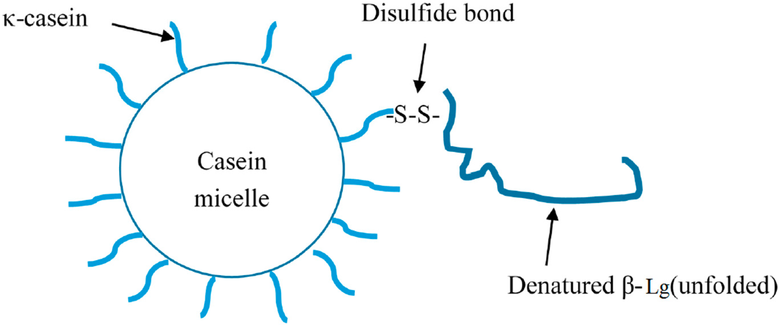
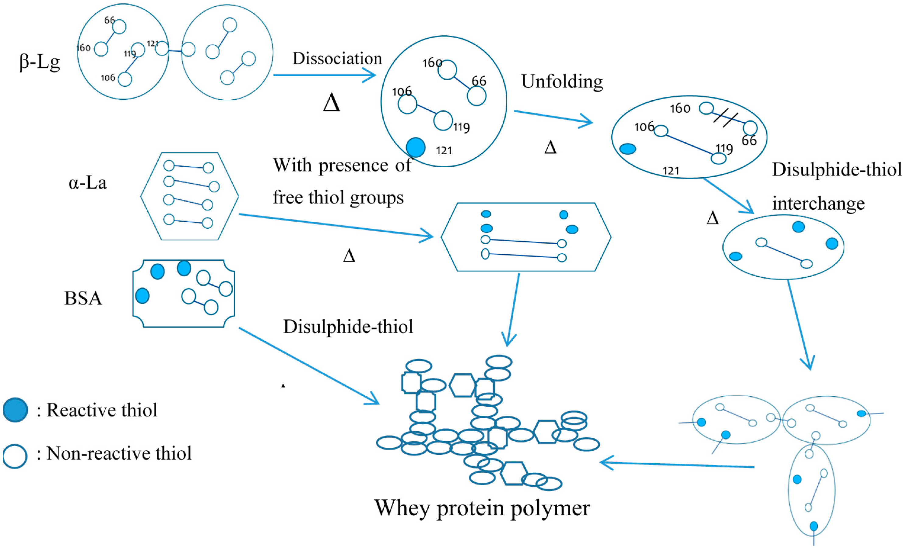
2.2. Whey Proteins
3. Milk Protein Products
3.1. Casein Products
3.2. Whey Protein Products
3.3. Membrane Filtered Milk Protein
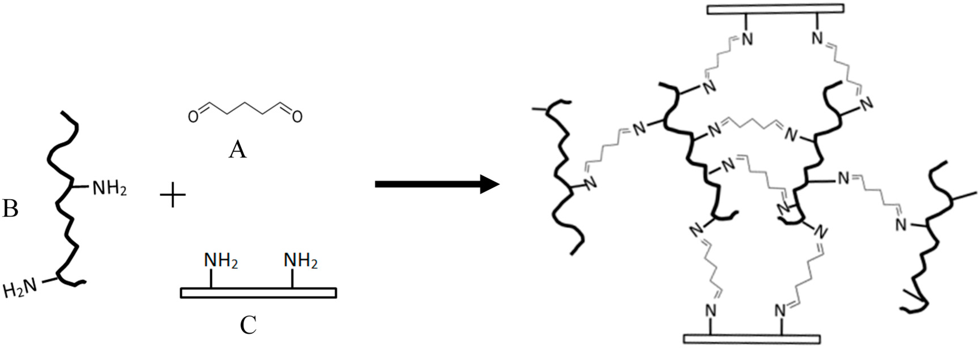
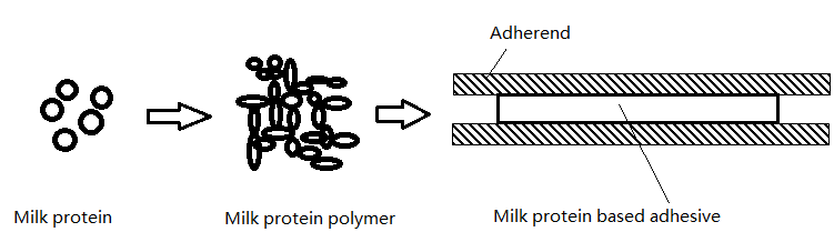
4. Milk Protein Polymerization
5. Milk-Protein-Based Adhesives Applications
5.1. Casein-Based Adhesives
5.2. Whey-Protein-Based Adhesives
6. Summary
Conflicts of Interest
References
- Alexander, J. Casein, its preparation, chemistry and technical utilization (Tague, E.L.). J. Chem. Educ. 1927, 4, 132. [Google Scholar] [CrossRef]
- Audic, J.; Chaufer, B.; Daufin, G. Non-food applications of milk components and dairy co-products: A review. Le Lait 2003, 83, 417–438. [Google Scholar] [CrossRef]
- Strube, O.I.; Rüdiger, A.A.; Bremser, W. Buildup of biobased adhesive layers by enzymatically controlled deposition on the example of casein. Int. J. Adhes. Adhes. 2015, 63, 9–13. [Google Scholar] [CrossRef]
- Yang, M.; Jin, Q.; Zhang, G. Natural Environment-Friendly Modified Casein Glue and Preparation Process Thereof. CN104531059 A, 22 April 2015. [Google Scholar]
- De Kruif, C.G.; Holt, C. Casein Micelle Structure, Functions and Interactions; Kluwer Academic/Plenum Publishers: New York, NY, USA, 2003. [Google Scholar]
- Horne, D.S. Casein micelle structure: Models and muddles. Curr. Opin. Colloid Interface Sci. 2006, 11, 148–153. [Google Scholar] [CrossRef]
- Wu, L.C.; Peng, Z.-Y.; Kim, P.S. Bipartite structure of the α-lactalbumin molten globule. Nat. Struct. Mol. Biol. 1995, 2, 281–286. [Google Scholar] [CrossRef]
- Ewbank, J.J.; Creighton, T.E. Structural characterization of the disulfide folding intermediates of bovine α-lactalbumin. Biochemistry 1993, 32, 3694–3707. [Google Scholar] [CrossRef] [PubMed]
- Qi, P.X. Studies of casein micelle structure: The past and the present. Le Lait 2007, 87, 363–383. [Google Scholar] [CrossRef]
- Eckelman, C.A. Brief Survey of Wood Adhesives; Purdue University Cooperative Extension Service: West Lafayette, IN, USA, 1999. [Google Scholar]
- Fox, P.F.; Kelly, A.L. The Caseins; Woodhead Publishing: Cambridge, UK, 2004. [Google Scholar]
- Walstra, P. On the stability of casein micelles1. J. Dairy Sci. 1990, 73, 1965–1979. [Google Scholar] [CrossRef]
- Semo, E.; Kesselman, E.; Danino, D.; Livney, Y.D. Casein micelle as a natural nano-capsular vehicle for nutraceuticals. Food Hydrocolloids 2007, 21, 936–942. [Google Scholar] [CrossRef]
- Horne, D.S. Casein structure, self-assembly and gelation. Curr. Opin. Colloid Interface Sci. 2002, 7, 456–461. [Google Scholar] [CrossRef]
- Anema, S.G.; Li, Y. Association of denatured whey proteins with casein micelles in heated reconstituted skim milk and its effect on casein micelle size. J. Dairy Res. 2003, 70, 73–83. [Google Scholar] [CrossRef] [PubMed]
- Walstra, P. Casein sub-micelles: Do they exist? Int. Dairy J. 1999, 9, 189–192. [Google Scholar] [CrossRef]
- Dumas, B.R.; Garnier, J. Structure of the casein micelle. The accessibility of the subunits to various reagents. J. Dairy Res. 1970, 37, 269–278. [Google Scholar] [CrossRef]
- Horne, D.S. Casein interactions: Casting light on the black boxes, the structure in dairy products. Int. Dairy J. 1998, 8, 171–177. [Google Scholar] [CrossRef]
- Holt, C.; de Kruif, C.G.; Tuinier, R.; Timmins, P.A. Substructure of bovine casein micelles by small-angle X-ray and neutron scattering. Colloids Surf. A 2003, 213, 275–284. [Google Scholar] [CrossRef]
- McMahon, D.J.; Oommen, B.S. Supramolecular structure of the casein micelle. J. Dairy Sci. 2008, 91, 1709–1721. [Google Scholar] [CrossRef] [PubMed]
- Holt, C.; Dalgleish, D.G. Electrophoretic and hydrodynamic properties of bovine casein micelles interpreted in terms of particles with an outer hairy layer. J. Colloid Interface Sci. 1986, 114, 513–524. [Google Scholar] [CrossRef]
- De Kruif, C.G. Casein micelle interactions. Int. Dairy J. 1999, 9, 183–188. [Google Scholar] [CrossRef]
- Dalgleish, D.G. Casein micelles as colloids: Surface structures and stabilities. J. Dairy Sci. 1998, 81, 3013–3018. [Google Scholar] [CrossRef]
- Dalgleish, D.G.; Spagnuolo, P.A.; Goff, H.D. A possible structure of the casein micelle based on high-resolution field-emission scanning electron microscopy. Int. Dairy J. 2004, 14, 1025–1031. [Google Scholar] [CrossRef]
- Bonnaillie, L.M.; Tomasula, P.M. Whey protein fractionation. In Whey Processing, Functionality and Healthy Benefits; Onwulata, C., Huth, P.J., Eds.; Wiley-Blackwell: Oxford, UK, 2008. [Google Scholar]
- Holt, C.; Davies, D.T.; Law, A.J.R. Effects of colloidal calcium phosphate content and free calcium ion concentration in the milk serum on the dissociation of bovine casein micelles. J. Dairy Res. 1986, 53, 557–572. [Google Scholar] [CrossRef]
- Aoki, T.; Yamada, N.; Tomita, I.; Kako, Y.; Imamura, T. Caseins are cross-linked through their ester phosphate groups by colloidal calcium phosphate. Biochim. Biophys. Acta (BBA) 1987, 911, 238–243. [Google Scholar] [CrossRef]
- Aoki, T.; Kako, Y.; Imamura, T. Separation of casein aggregates cross-linked by colloidal calcium phosphate from bovine casein micelles by high performance gel chromatography in the presence of urea. J. Dairy Res. 1986, 53, 53–59. [Google Scholar] [CrossRef]
- Aoki, T.; Sakamoto, H.; Kako, Y. Cross-linking of caseins by colloidal calcium phosphate in the presence of urea. Int. Dairy J. 1991, 1, 67–75. [Google Scholar] [CrossRef]
- Ozcan, T.; Horne, D.; Lucey, J.A. Effect of increasing the colloidal calcium phosphate of milk on the texture and microstructure of yogurt. J. Dairy Sci. 2011, 94, 5278–5288. [Google Scholar] [CrossRef] [PubMed]
- Augustin, M.A.; Oliver, C.M.; Hemar, Y. Casein, Caseinates and Milk Protein Concentrates; Wiley-Blackwell: Oxford, UK, 2011. [Google Scholar]
- Noelken, M.; Reibstein, M. Conformation of β-casein B. Arch. Biochem. Biophys. 1968, 123, 397–402. [Google Scholar] [CrossRef]
- Andrews, A.L.; Atkinson, D.; Evans, M.T.A.; Finer, E.G.; Green, J.P.; Phillips, M.C.; Robertson, R.N. The conformation and aggregation of bovine β-casein a. I. Molecular aspects of thermal aggregation. Biopolymers 1979, 18, 1105–1121. [Google Scholar] [CrossRef]
- Thompson, M.P.; Boswell, R.T.; Martin, V.; Jenness, R.; Kiddy, C.A. Casein-pellet-solvation and heat stability of individual cow’s milk. J. Dairy Sci. 1969, 52, 796–798. [Google Scholar] [CrossRef]
- Holt, C.; Sawyer, L. Caseins as rheomorphic proteins: Interpretation of primary and secondary structures of the αs1-, β- and κ-caseins. J. Chem. Soc. 1993, 89, 2683–2692. [Google Scholar] [CrossRef]
- Holt, C.; Wahlgren, M.N.; Drakenberg, T. Ability of a β-casein phosphopeptide to modulate the precipitation of calcium phosphate by forming amorphous dicalcium phosphate nanoclusters. Biochem. J. 1996, 314, 1035–1039. [Google Scholar] [CrossRef] [PubMed]
- Farrell, H.M., Jr.; Malin, E.L.; Brown, E.M.; Qi, P.X. Casein micelle structure: What can be learned from milk synthesis and structural biology? Curr. Opin. Colloid Interface Sci. 2006, 11, 135–147. [Google Scholar] [CrossRef]
- Livney, Y.D.; Schwan, A.L.; Dalgleish, D.G. A study of β-casein tertiary structure by intramolecular crosslinking and mass spectrometry. J. Dairy Sci. 2004, 87, 3638–3647. [Google Scholar] [CrossRef]
- Creamer, L.K.; Parry, D.A.D.; Malcolm, G.N. Secondary structure of bovine β-lactoglobulin B. Arch. Biochem Biophys. 1983, 227, 98–105. [Google Scholar] [CrossRef]
- Qin, B.Y.; Bewley, M.C.; Creamer, L.K.; Baker, H.M.; Baker, E.N.; Jameson, G.B. Structural basis of the tanford transition of bovine β-lactoglobulin. Biochemistry 1998, 37, 14014–14023. [Google Scholar] [CrossRef] [PubMed]
- Kuwata, K.; Era, S.; Hoshino, M.; Forge, V.; Goto, Y.; Batt, C.A. Solution structure and dynamics of bovine β-lactoglobulin A. Protein Sci. 1999, 8, 2541–2545. [Google Scholar] [CrossRef] [PubMed]
- Hoffmann, M.A.M.; van Mil, P.J.J.M. Heat-induced aggregation of β-lactoglobulin: Role of the free thiol group and disulfide bonds. J. Agric. Food Chem. 1997, 45, 2942–2948. [Google Scholar] [CrossRef]
- Qi, X.L.; Holt, C.; Mcnulty, D.; Clarke, D.T.; Brownlow, S.; Jones, G.R. Effect of temperature on the secondary structure of β-lactoglobulin at pH 6.7, as determined by CD and IR spectroscopy: A test of the molten globule hypothesis. Biochem. J. 1997, 324, 341–346. [Google Scholar] [CrossRef] [PubMed]
- Galani, D.; Apenten, R.K.O. Heat-induced denaturation and aggregation of β-lactoglobulin: Kinetics of formation of hydrophobic and disulphide-linked aggregates. Int. J. Food Sci. Technol. 1999, 34, 467–476. [Google Scholar] [CrossRef]
- Elofsson, C.; Dejmek, P.; Paulsson, M.; Burling, H. Characterization of a cold-gelling whey protein concentrate. Int. Dairy J. 1997, 7, 601–608. [Google Scholar] [CrossRef]
- Kumosinski, T.F.; Timasheff, S.N. Molecular interactions in β-lactoglobulin. X. The stoichiometry of the β-lactoglobulin mixed tetramerization1. J. Am. Chem. Soc. 1966, 88, 5635–5642. [Google Scholar] [CrossRef]
- Gottschalk, M.; Nilsson, H.; Roos, H.; Halle, B. Protein self-association in solution: The bovine β-lactoglobulin dimer and octamer. Protein Sci. 2003, 12, 2404–2411. [Google Scholar] [CrossRef] [PubMed]
- Greene, L.H.; Grobler, J.A.; Malinovskii, V.A.; Tian, J.; Acharya, K.R.; Brew, K. Stability, activity and flexibility in α-lactalbumin. Protein Eng. 1999, 12, 581–587. [Google Scholar] [CrossRef] [PubMed]
- Ewbank, J.J.; Creighton, T.E. Pathway of disulfide-coupled unfolding and refolding of bovine α-lactalbumin. Biochemistry 1993, 32, 3677–3693. [Google Scholar] [CrossRef] [PubMed]
- Kuwajima, K. The molten globule state of α-lactalbumin. FASEB J. 1996, 10, 102–109. [Google Scholar] [PubMed]
- Hirayama, K.; Akashi, S.; Furuya, M.; Fukuhara, K.-I. Rapid confirmation and revision of the primary structure of bovine serum albumin by esims and frit-FAB LC/MS. Biochem. Biophys. Res. Commun. 1990, 173, 639–646. [Google Scholar] [CrossRef]
- Bachmann, H.-P. Cheese analogues: A review. Int. Dairy J. 2001, 11, 505–515. [Google Scholar] [CrossRef]
- Desobry-Banon, S.; Richard, F.; Hardy, J. Study of acid and rennet coagulation of high pressurized milk. J. Dairy Sci. 1994, 77, 3267–3274. [Google Scholar] [CrossRef]
- Punidadas, P.; Rizvi, S. Separation of milk proteins into fractions rich in casein or whey proteins by cross flow filtration. Food Res. Int. 1998, 31, 265–272. [Google Scholar] [CrossRef]
- Zhou, N.; Mulvaney, S.J. The effect of milk fat, the ratio of casein to water, and temperature on the viscoelastic properties of rennet casein gels. J. Dairy Sci. 1998, 81, 2561–2571. [Google Scholar] [CrossRef]
- Panesar, P.S.; Kennedy, J.F.; Gandhi, D.N.; Bunko, K. Bioutilisation of whey for lactic acid production. Food Chem. 2007, 105, 1–14. [Google Scholar] [CrossRef]
- Marwaha, S.S.; Kennedy, J.F. Whey-pollution problem and potential utilization. Int. J. Food Sci. Technol. 1988, 23, 323–336. [Google Scholar] [CrossRef]
- Mavropoulou, I.P.; Kosikowski, F.V. Composition, solubility, and stability of whey powders. J. Dairy Sci. 1973, 56, 1128–1134. [Google Scholar] [CrossRef]
- Pearce, R.J. Whey processing. In Whey and Lactose Processing; Springer: Berlin, Germany, 1992. [Google Scholar]
- Wronkowska, M.; Jadacka, M.; Soral-Śmietana, M.; Zander, L.; Dajnowiec, F.; Banaszczyk, P.; Jeliński, T.; Szmatowicz, B. Acid whey concentrated by ultrafiltration a tool for modeling bread properties. LWT-Food Sci. Technol. 2015, 61, 172–176. [Google Scholar] [CrossRef]
- Chen, G.Q.; Eschbach, F.I.I.; Weeks, M.; Gras, S.L.; Kentish, S.E. Removal of lactic acid from acid whey using electrodialysis. Sep. Purif. Technol. 2016, 158, 230–237. [Google Scholar] [CrossRef]
- Chandrapala, J.; Duke, M.C.; Gray, S.R.; Weeks, M.; Palmer, M.; Vasiljevic, T. Nanofiltration and nanodiafiltration of acid whey as a function of pH and temperature. Sep. Purif. Technol. 2016, 160, 18–27. [Google Scholar] [CrossRef]
- Smithers, G.W. Whey-ing up the options—Yesterday, today and tomorrow. Int. Dairy J. 2015, 48, 2–14. [Google Scholar] [CrossRef]
- Vasiljevic, T.; Duke, M. Whey protein concentrate: Overview and membrane operations. In Encyclopedia of Membranes; Drioli, E., Giorno, L., Eds.; Springer: Berlin, Germany, 2015. [Google Scholar]
- Lagrange, V.; Whitsett, D.; Burris, C. Global market for dairy proteins. J. Food Sci. 2015, 80, A16–A22. [Google Scholar] [CrossRef] [PubMed]
- Okawa, T.; Shimada, M.; Ushida, Y.; Seki, N.; Watai, N.; Ohnishi, M.; Tamura, Y.; Ito, A. Demineralisation of whey by a combination of nanofiltration and anion-exchange treatment: A preliminary study. Int. J. Dairy Technol. 2015, 68, 478–485. [Google Scholar] [CrossRef]
- Scott, S.N.; Krishnapillai, A. Isolation and Purification of Components of Whey. US8932659 B2, 13 January 2015. [Google Scholar]
- Bhaskar, G.V.; Singh, H.; Blazey, N.D. Milk Protein Products and Processes. US7157108 B2, 2 January 2007. [Google Scholar]
- Saboyainsta, L.V.; Maubois, J.-L. Current developments of microfiltration technology in the dairy industry. Le Lait 2000, 80, 541–553. [Google Scholar] [CrossRef]
- Evans, J.; Zulewska, J.; Newbold, M.; Drake, M.A.; Barbano, D. Comparison of composition and sensory properties of 80% whey protein and milk serum protein concentrates. J. Dairy Sci. 2010, 93, 1824–1843. [Google Scholar] [CrossRef] [PubMed]
- Lu, C.; Wang, G.; Li, Y.; Zhang, L. Effects of homogenisation pressures on physicochemical changes in different layers of ultra-high temperature whole milk during storage. Int. J. Dairy Technol. 2013, 66, 325–332. [Google Scholar] [CrossRef]
- Anema, S.G. On heating milk, the dissociation of κ-casein from the casein micelles can precede interactions with the denatured whey proteins. J. Dairy Res. 2008, 75, 415–421. [Google Scholar] [CrossRef] [PubMed]
- Anema, S.G.; Li, Y. Effect of pH on the association of denatured whey proteins with casein micelles in heated reconstituted skim milk. J. Agric. Food Chem. 2003, 51, 1640–1646. [Google Scholar] [CrossRef] [PubMed]
- Gezimati, J.; Creamer, L.K.; Singh, H. Heat-induced interactions and gelation of mixtures of β-lactoglobulin and α-lactalbumin. J. Agric. Food Chem. 1997, 45, 1130–1136. [Google Scholar] [CrossRef]
- Hines, M.E.; Foegeding, E.A. Interactions of α-lactalbumin and bovine serum albumin with β-lactoglobulin in thermally induced gelation. J. Agric. Food Chem. 1993, 41, 341–346. [Google Scholar] [CrossRef]
- Schorsch, C.; Wilkins, D.K.; Jones, M.G.; Norton, I.T. Gelation of casein-whey mixtures: Effects of heating whey proteins alone or in the presence of casein micelles. J. Dairy Res. 2001, 68, 471–481. [Google Scholar] [CrossRef] [PubMed]
- Vachon, C.; Yu, H.-L.; Yefsah, R.; Alain, R.; St-Gelais, D.; Lacroix, M. Mechanical and structural properties of milk protein edible films cross-linked by heating and γ-irradiation. J. Agric. Food Chem. 2000, 48, 3202–3209. [Google Scholar] [CrossRef] [PubMed]
- Matheis, G.; Whitaker, J.R. A review: Enzymatic cross-linking of proteins applicable to foods. J. Food Biochem. 1987, 11, 309–327. [Google Scholar] [CrossRef]
- Strube, O.I.; Bremser, W.; Rüdiger, A.A. Method for Coating Surfaces by Enzymatic Reaction. WO2015150368 A1, 31 March 2015. [Google Scholar]
- Gerrard, J.A. Protein-protein crosslinking in food: Methods, consequences, applications. Trends Food Sci. Technol. 2002, 13, 391–399. [Google Scholar] [CrossRef]
- Huang, J.; Li, K. A new soy flour-based adhesive for making interior type II plywood. J. Am. Oil Chem. Soc. 2008, 85, 63–70. [Google Scholar] [CrossRef]
- Guo, M.; Wang, G. Whey protein polymerisation and its applications in environmentall safe adhesives. Int. J. Dairy Technol. 2016. [Google Scholar] [CrossRef]
- Gruendler, G.J. Adhesive. US633834 A, 26 September 1899. [Google Scholar]
- Ross, J.H.; Ross, C.D. Improvement in Processes of Preparing Glue. US183024 A, 10 October 1876. [Google Scholar]
- Shisler, G.M. Adhesive Material Containing Casein. US1886750 A, 8 November 1932. [Google Scholar]
- Corwin, J.F.; White, R.C. Bottle Labeling Adhesive. US2351109 A, 13 June 1944. [Google Scholar]
- Dalgleish, D.G.; Law, A.J.R. PH-induced dissociation of bovine casein micelles. II. Mineral solubilization and its relation to casein release. J. Dairy Res. 1989, 56, 727–735. [Google Scholar] [CrossRef]
- Vaia, B.; Smiddy, M.A.; Kelly, A.L.; Huppertz, T. Solvent-mediated disruption of bovine casein micelles at alkaline pH. J. Agric. Food Chem. 2006, 54, 8288–8293. [Google Scholar] [CrossRef] [PubMed]
- Bye, C.N. Casein and mixed protein adhesives. In Handbook of Adhesives; Skeist, I., Ed.; Springer US: Boston, MA, USA, 1990. [Google Scholar]
- Umemura, K.; Inoue, A.; Kawai, S. Development of new natural polymer-based wood adhesives I: Dry bond strength and water resistance of konjac glucomannan, chitosan, and their composites. J. Wood Sci. 2003, 49, 221–226. [Google Scholar] [CrossRef]
- Girdhar, B.K.; Hansen, P.M.T. Soluble casein by adsorption of ammonia. J. Food Sci. 1974, 39, 1237–1243. [Google Scholar] [CrossRef]
- McGann, T.C.A.; Fox, P.F. Physico-chemical properties of casein micelles reformed from urea-treated milk. J. Dairy Res. 1974, 41, 45–53. [Google Scholar] [CrossRef]
- Dheret, C.; Hayes, P.J.; Smith, S.J.; Bladon, S.L.; Harrington, J.; Sardina, M. Aqueous Adhesive Compositions Useful as Bottle Labeling Adhesives. US6590019 B2, 8 July 2003. [Google Scholar]
- Broich, L.; Herlfterkamp, B.; Onusseit, H. Water-Containing Adhesive Based on Casein. US5455066 A, 3 October 1995. [Google Scholar]
- Van der Leeden, M.C.; Rutten, A.A.C.M.; Frens, G. How to develop globular proteins into adhesives. J. Biotechnol. 2000, 79, 211–221. [Google Scholar] [CrossRef]
- Tschabold, G.L.; Mueller, D.L. Adhesive from Whey and a Method of Making It. US2624679 A, 6 January 1953. [Google Scholar]
- Zhao, Z.; Gao, Z.; Wang, W.; Guo, M. Formulation designs and characterisations of whey-protein based API adhesives. Pigment Resin Technol. 2011, 40, 410–417. [Google Scholar] [CrossRef]
- Gao, Z.; Wang, W.; Zhao, Z.; Guo, M. Novel whey protein-based aqueous polymer-isocyanate adhesive for glulam. J. Appl. Polym. Sci. 2011, 120, 220–225. [Google Scholar] [CrossRef]
- Gao, Z.; Yu, G.; Bao, Y.; Guo, M. Whey-protein based environmentally friendly wood adhesives. Pigment Resin Technol. 2011, 40, 42–48. [Google Scholar] [CrossRef]
- Wang, W.; Zhao, Z.; Gao, Z.; Guo, M. Whey protein-based water-resistant and environmentally safe adhesives for plywood. BioResources 2011, 6, 3339–3351. [Google Scholar]
- Wang, W.; Zhao, Z.; Gao, Z.; Guo, M. Water-resistant whey protein based wood adhesive modified by post-treated phenol-formaldehyde oligomers (PFO). BioResource 2012, 7, 1972–1983. [Google Scholar] [CrossRef]
- Wang, G.; Cheng, J.; Zhang, L.; Guo, M. Physicochemical and functional properties, microstructure, and storage stability of whey protein/polyvinylpyrrolidone based glue sticks. BioResources 2012, 7, 5422–5434. [Google Scholar] [CrossRef]
- Wang, G.; Zhang, T.; Ahmad, S.; Cheng, J.; Guo, M. Physicochemical and adhesive properties, microstructure and storage stability of whey protein-based paper glue. Int. J. Adhes. Adhes. 2013, 41, 198–205. [Google Scholar] [CrossRef]
- Wang, G.; Guo, M. Property and storage stability of whey protein-sucrose based safe paper glue. J. Appl. Polym. Sci. 2014. [Google Scholar] [CrossRef]
- Brew, K.; Vanaman, T.C.; Hill, R.L. Comparison of the amino acid sequence of bovine α-lactalbumin and hens egg white lysozyme. J. Biol. Chem. 1967, 242, 3747–3748. [Google Scholar] [PubMed]
- Fox, P.F.; McSweeney, P.L.H. Advanced Dairy Chemistry; Kluwer Academic/Plenum Publishers: New York, NY, USA, 2003. [Google Scholar]
- Quinn, J.; Wells, G.; Sutcliffe, T.; Jarmuske, M.; Maw, J.; Stiell, I.; Johns, P. Tissue adhesive versus suture wound repair at 1 year: Randomized clinical trial correlating early, 3-month, and 1-year cosmetic outcome. Ann. Emerg. Med. 1998, 32, 645–649. [Google Scholar] [CrossRef]
- Quinn, J.V. Tissue Adhesives in Clinical Medicine; BC Decker Inc.: Hamilton, ON, Canada, 2005. [Google Scholar]
- LeMaire, S.A.; Schmittling, Z.C.; Coselli, J.S.; Undar, A.; Deady, B.A.; Clubb, F.J.; Fraser, C.D. Bioglue surgical adhesive impairs aortic growth and causes anastomotic strictures. Ann. Thorac. Surg. 2002, 73, 1500–1505. [Google Scholar] [CrossRef]
- Chao, H.-H.; Torchiana, D.F. Bioglue: Albumin/Glutaraldehyde Sealant in Cardiac Surgery. J. Card. Surg. 2003, 18, 500–503. [Google Scholar] [CrossRef] [PubMed]
- Nadler, R.B.; Loeb, S.; Rubenstein, R.A.; Vardi, I.Y. Use of bioglue in laparoscopic partial nephrectomy. Urology 2006, 68, 416–418. [Google Scholar] [CrossRef] [PubMed]
- Hidas, G.; Kastin, A.; Mullerad, M.; Shental, J.; Moskovitz, B.; Nativ, O. Sutureless nephron-sparing surgery: Use of albumin glutaraldehyde tissue adhesive (BioGlue). Urology 2006, 67, 697–700. [Google Scholar] [CrossRef] [PubMed]
- Gaberel, T.; Borgey, F.; Thibon, P.; Lesteven, C.; Lecoutour, X.; Emery, E. Surgical site infection associated with the use of bovine serum albumine-glutaraldehyde surgical adhesive (BioGlue®) in cranial surgery: A case-control study. Acta Neurochir. 2011, 153, 156–163. [Google Scholar] [CrossRef] [PubMed]
- Ranu, H.; Gatheral, T.; Sheth, A.; Smith, E.E.J.; Madden, B.P. Successful endobronchial seal of surgical bronchopleural fistulas using BioGlue. Ann. Thorac. Surg. 2009, 88, 1691–1692. [Google Scholar] [CrossRef] [PubMed]
- Kumar, R.; Choudhary, V.; Mishra, S.; Varma, I.; Mattiason, B. Adhesives and plastics based on soy protein products. Ind. Crops Prod. 2002, 16, 155–172. [Google Scholar] [CrossRef]


| Proteins | Content, g/L |
|---|---|
| Casein | 25 |
| α-Casein | 12 |
| β-Casein | 9 |
| κ-Casein | 3.25 |
| Minor constituents | 0.75 |
| Whey Protein | 5.4 |
| β-lactoglobulin | 2.70 |
| α-lactoalbumin | 1.20 |
| Serum albumin | 0.65 |
| Minor constituents | 0.85 |
© 2016 by the authors. Licensee MDPI, Basel, Switzerland. This article is an open access article distributed under the terms and conditions of the Creative Commons Attribution (CC-BY) license ( http://creativecommons.org/licenses/by/4.0/).
Share and Cite
Guo, M.; Wang, G. Milk Protein Polymer and Its Application in Environmentally Safe Adhesives. Polymers 2016, 8, 324. https://doi.org/10.3390/polym8090324
Guo M, Wang G. Milk Protein Polymer and Its Application in Environmentally Safe Adhesives. Polymers. 2016; 8(9):324. https://doi.org/10.3390/polym8090324
Chicago/Turabian StyleGuo, Mingruo, and Guorong Wang. 2016. "Milk Protein Polymer and Its Application in Environmentally Safe Adhesives" Polymers 8, no. 9: 324. https://doi.org/10.3390/polym8090324
APA StyleGuo, M., & Wang, G. (2016). Milk Protein Polymer and Its Application in Environmentally Safe Adhesives. Polymers, 8(9), 324. https://doi.org/10.3390/polym8090324