Article

Repair Effect of Seaweed Polysaccharides with Different Contents of Sulfate Group and Molecular Weights on Damaged HK-2 Cells

1
Department of Chemistry, Jinan University, Guangzhou 510632, China
2
Institute of Biomineralization and Lithiasis Research, Jinan University, Guangzhou 510632, China
*
Author to whom correspondence should be addressed.
These authors contributed equally to this work.
Polymers 2016, 8(5), 188; https://doi.org/10.3390/polym8050188
Submission received: 2 April 2016 / Revised: 28 April 2016 / Accepted: 9 May 2016 / Published: 19 May 2016
(This article belongs to the Collection Polysaccharides)

Abstract

:
The structure–activity relationships and repair mechanism of six low-molecular-weight seaweed polysaccharides (SPSs) on oxalate-induced damaged human kidney proximal tubular epithelial cells (HK-2) were investigated. These SPSs included Laminaria japonica polysaccharide, degraded Porphyra yezoensis polysaccharide, degraded Gracilaria lemaneiformis polysaccharide, degraded Sargassum fusiforme polysaccharide, Eucheuma gelatinae polysaccharide, and degraded Undaria pinnatifida polysaccharide. These SPSs have a narrow difference of molecular weight (from 1968 to 4020 Da) after degradation by controlling H2O2 concentration. The sulfate group (–SO3H) content of the six SPSs was 21.7%, 17.9%, 13.3%, 8.2%, 7.0%, and 5.5%, respectively, and the –COOH contents varied between 1.0% to 1.7%. After degradation, no significant difference was observed in the contents of characteristic –SO3H and –COOH groups of polysaccharides. The repair effect of polysaccharides was determined using cell-viability test by CCK-8 assay and cell-morphology test by hematoxylin-eosin staining. The results revealed that these SPSs within 0.1–100 μg/mL did not express cytotoxicity in HK-2 cells, and each polysaccharide had a repair effect on oxalate-induced damaged HK-2 cells. Simultaneously, the content of polysaccharide –SO3H was positively correlated with repair ability. Furthermore, the low-molecular-weight degraded polysaccharides showed better repair activity on damaged HK-2 cells than their undegraded counterpart. Our results can provide reference for inhibiting the formation of kidney stones and for developing original anti-stone polysaccharide drugs.

Graphical Abstract

1. Introduction

Kidney stone is a common disease which occurrence is closely related to the damage of renal tubular epithelial cells. A high concentration of oxalate in the urine can damage renal tubular epithelial cells to trigger a series of oxidative stress reactions and eventually lead to the formation of kidney stones [1,2]. Urinary glycosaminoglycans (GAGs) can protect renal tubular epithelial cells against oxidative damage [3,4] and thus inhibit the formation of calcium oxalate stones.
Seaweed polysaccharides (SPSs) comprise repeating disaccharide sugar chains, similar to GAGs. Therefore, SPSs may be used to repair damaged renal tubular epithelial cells and inhibit the formation of stones. Polysaccharides from the seaweeds Fucus [5] and Sargassum [6] reportedly play significant protective roles in renal-tissue injury.
Faggio et al. [7] evaluated the anticoagulant activity of two algal sulfate polysaccharides extracted from Ulva fasciata (Chlorophyta) and Agardhiella subulata (Rhodophyta). No cytotoxicity was observed for the two SPS on human red blood cells, and both of them had significant anticoagulant activity. Polysaccharide extracted from brown alga Undaria pinnatifida also has an anticoagulant effect on human blood in vitro and are not cytotoxic [8].
The biochemical activity of SPS is determined by its chemical structure, especially molecular weight, as well as the content of sulfate groups and uronic acid [9,10,11,12]. In general, low-molecular-weight SPSs show effective biological activities. Zhao et al. [10] confirmed that low-molecular-weight degraded Porphyran polysaccharide has stronger scavenging ability of hydroxyl radical and DPPH radical.
Additionally, the higher content of anionic groups of polysaccharide such as the sulfate and uronic acid group is related to the beneficial biological activity. The Laminaria polysaccharide with higher uronic acid displayed stronger antioxidant activity [10]. Zhang et al. [13] and Wang et al. [14] also found that the high sulfate content of polysaccharide significantly increased its antioxidant activity. The –SO3H content of polysaccharides extracted from Ascophyllum nodosum at 150, 120, and 90 °C for 30 min were 7.82%, 16.87%, and 28.60%, respectively [15], and their scavenging capacity to DPPH radical is proportional to their –SO3H content, i.e., the component with 28.60% –SO3H content has the highest scavenging capacity of about 15%, whereas the component with 7.82% –SO3H content only has scavenging capacity of about 2%. Wang et al. [12] reported that sulfated polysaccharides from Cyclocarya paliurus present significantly higher repair capacity on H2O2-induced damaged RAW264.7 cells than non-sulfate polysaccharide samples. To date, few reports exist about the repair effect of SPSs on oxalate-induced cytotoxicity, especially the structure–activity relationships remain unknown.
To comparatively investigate the structure–activity relationships of SPSs and their repair ability on injured renal epithelial cells, in the present work, we selected six low-molecular-weight SPSs with narrow differences but various acidic sulfate group (−OSO3) content (21.7%, 17.9%, 13.3%, 8.2%, 7.0%, and 5.5%, respectively). We focused on studying the effect of –SO3H content of SPSs on their ability to repair injured human kidney proximal tubular epithelial cells (HK-2). Our results can provide reference for inhibiting the formation of kidney stones and for developing original anti-stone polysaccharide drugs.

2. Experimental Methods

2.1. Reagents and Apparatus

Laminaria japonica polysaccharide (Laminaria-1), Porphyra yezoensis polysaccharide (Porphyra-2), Gracilaria lemaneiformis polysaccharide (Gracilaria-3), Sargassum fusiforme polysaccharide (Sargassum-4), Eucheuma polysaccharide (Eucheuma-5), and Undaria pinnatifida polysaccharide (Undaria-6) were produced by Beijing newprobe Bioscience & Technology Co., Ltd (Beijing, China).
The cell proliferation assay kit (Cell Couting Kit 8, CCK-8) was purchased from Dojindo Laboratory (Kumamoto, Japan). Other chemical reagents were analytical grade and purchased from Guangzhou chemical reagent company (Guangzhou, China).
The apparatus included a Ubbelohde capillary viscometer (0.4–0.5, Qihang Glass Instrument Factory, Shanghai, China), an Ultraviolet-Visible spectrophotometer (Cary 500, Varian, Palo Alto, CA, USA), a Fourier Transform Infrared Spectrometer (EQUINOX 55, Bruker, Karlsruhe, Germany), a conductivity meter (DDS-11A, LEICI, Shanghai, China), Enzyme Mark Instrument (Safire2, Tecan, Männedorf, Switzerland), and an upright electric fluorescence microscope (22DI-E-D282, Leica, Solms, Germany).

2.2. Degradation of Polysaccharides

According to the molecular weight of natural polysaccharides, H2O2 was used to degrade high-molecular-weight SPSs. Taking Porphyra-2 for example, the degradation process was as follows. About 1.2 g of Porphyra-2 was weighed accurately and dissolved in 40 mL of distilled water at 70 °C. After heating to 90 °C, 27 mL of 30% (vol) H2O2 solution was quickly added to the reaction system. The degradation reaction was allowed to proceed for 2 h, at which point the solution pH was adjusted to 7.0 by adding 2 mol/L NaOH solution. The degraded SPS solution was then concentrated to one-third of its original volume at 60 °C. The product was precipitated by three times addition of anhydrous ethanol to the actual volume. The solution was stored at 4 °C in a refrigerator overnight and then filtered. The filtrate was washed with anhydrous ethanol twice and dried in a vacuum.
The degradation conditions of all the six SPSs are listed in Table 1.

2.3. Average Molecular Weight Determination

According to reference [16], the Ubbelohde viscosity method was used to measure the viscosity of samples in aqueous solutions at 30 ± 0.2 °C. After measuring the falling time in the viscometer before and after polysaccharide degradation, relative (ηr) and specific (ηsp) viscosity could be calculated from ηr = Ti/T0 and ηsp = ηr − 1, where Ti and T0 are the falling time of SPS solutions and deionized water, respectively. Intrinsic viscosity (η) could be calculated according to the one-point method, i.e., η = (2(ηsp − lnηr))1/2/c, in which c is the concentration of samples to be tested. The molecular weight of SPS was calculated through its value η. The relationship between η and the molecular weight M of the polymer solutions could be described by the Mark–Houwink empirical equation η = κMα, where κ and α are constants related to polymer nature, solvent, and temperature.

2.4. Measurement of Protein Content

2.4.1. Ultraviolet-Absorption Method

Polysaccharide solutions were scanned at a UV–VIS spectrophotometer to observe absorption situation between 260 and 280 nm. If the polysaccharide did not show a characteristic absorption peak, then it did not contain protein ingredients.

2.4.2. Ninhydrin Method

A drop of 1.0 mg/mL polysaccharide solution was dropped onto a filter paper and dried. Then, 2.5% ninhydrin was sprayed on the paper. If the paper maintained its original pale-yellow color, then the sample did not contain proteins or amino acids; if the paper became purple, the sample contained proteins or amino acids [17].

2.5. Analysis of Sulfate-Group Content

The sulfate-group (–SO3H) content of polysaccharides was determined by the BaCl2-gelatin turbidity method [18]. In a typical procedure, 0.3% gelatin solution was prepared in hot water (60–70 °C) and stored at 4 °C overnight. Two grams of BaCl2 was dissolved in gelatin solution and allowed to stand for 2–3 h at 25 °C. About 0.20 mL of polysaccharide solution (1.4 mg/mL) was added to 3.8 mL of 0.5 M HCl and 1 mL of BaCl2-gelatin reagent, and the mixture was allowed to stand for 10–20 min. A blank was prepared with 0.2 mL of water instead of SPS solution. The released barium sulfate suspension was measured at λ = 360 nm by UV–VIS spectrophotometry using potassium sulfate as standard, and the standard curve was shown in Figure S1. This experiment was performed in triplicate.

2.6. Analysis of Carboxylic Group Content

The carboxylic group (–COOH) content of SPS was measured by conductometric titration [19]. A conductivity titration curve was drawn using the value of conductivity as the Y-axis and the corresponding volume of the used NaOH as the X-axis. The conductivity titration curve could be divided into three sections (Figure 1): the conductivity decreasing stage (A), the equilibrium stage (B), and the conductivity increasing stage (C). Three tangent lines were constructed from the curve of three stages, and the cross-points were the stoichiometric points. The cross-point of lines A and B gave the volume of NaOH (V1) that excess hydrochloric acid and –SO3H consumed; and the cross-point of lines B and C gave the volume of NaOH (V2) that excess hydrochloric acid and the –COOH of the polysaccharides consumed together, so V2V1 (platform portion) was the volume of NaOH that the –COOH of SPSs consumed alone. The –COOH content could be obtained according to the following formula:
COOH   ( % ) = C N a O H × ( V 2 V 1 ) × 45 / 1000 C sample × 40 / 1000 × 100
where CNaOH (mol/L) represents the molar concentration of NaOH, CSample (g/L) represents the mass concentration of polysaccharide, 45 is the molar mass of –COOH and 40 (mL) is the volume of polysaccharide solution. The final value was the average of three parallel experiments.

2.7. Fourier Transform Infrared Spectrometry

A dried polysaccharide sample (2.0 mg) was mixed with KBr (200 mg). After grinding and pressing into a KBr pellet, scanning was performed between the ranges of 4000–400 cm−1 wave number.

2.8. Cytotoxicity Measurement of Polysaccharides

HK-2 cells were cultured in a DMEM culture medium containing 10% fetal bovine serum, 100 U/mL penicillin-100 μg/mL streptomycin antibiotics with pH 7.4 at 37 °C in a 5% CO2 humidified environment. Upon reaching 80%–90% confluent, cells were blown gently after trypsin digestion to form cell suspension for the following cell experiment.
To assess the cytotoxicity of polysaccharides as other authors have already performed [20,21,22], the cytotoxicities of six samples were determined as follows: Cell suspension with a cell concentration of 1 × 10⁵ cells/mL was inoculated per well in 96-well plates for 24 h. After the cells were confluent, the culture medium was removed by suction and the cells were washed twice with PBS. After that, 0.1 mL of 0, 0.1, 1, 10, and 100 μg/mL of different seaweed polysaccharides solutions were added respectively. Each experiment was repeated in three parallel wells. After incubation for 24 h, 10 μL CCK-8 was added to each well and incubated for 1.5 h. The OD values were measured using the enzyme mark instrument at 450 nm. Cell viability was determined using the following equation: Cell viability (%) = ODtreatment group/ODcontrol group × 100.

2.9. Repair Effect of Polysaccharide on Damaged HK-2 Cells

Cell suspension with a cell concentration of 1 × 10⁵ cells/mL was inoculated per well in 96-well plates, 0.1 mL of DMEM containing 10% fetal bovine serum was added per well and the cells were incubated for 24 h. The experimental model was divided into three groups [23]: (1) Control group: in which only serum-free culture medium was added; (2) Damaged group: in which serum-free culture medium with 2.8 mmol/L oxalate was added and incubated for 3 h; (3) Repair group: in which 60 μg/mL of different SPS solutions were added into the cells of damaged control group and repaired for 8 h. After completing the repairing, 10 μL of CCK-8 was added to each well and incubated for 1.5 h. The OD values were measured using the enzyme mark instrument at 450 nm to detect the repair capacity of polysaccharide.

2.10. Hematoxyline-Eosin Staining

HK-2 cells were grouped as in section 2.9. After the repair was completed, the supernatant was removed by suction and washed 2–3 times with PBS. Afterwards, the cells were fixed with 3.8% polyoxymethylene for 15 min at room temperature. Cells were washed three times with PBS. After fixation the cells were stained with hematoxylin stain and incubated for 15 min. Then cells were washed with distilled water (DW) for 2 min to remove excess stain. After that, the cells were stained with eosin staining solution for 5 min. The cells were washed with DW for 2 min to remove excess eosin. After treatment, the cells were observed under the microscope, the nucleus of the cells was stained purple or blue, whereas the cytoplasm was stained pink or red.

2.11. Statistical Analysis

Results were expressed as Mean ± SD from three independent experiments. The statistically significant differences were analyzed using Turkey’s test. p values < 0.05 were considered statistically significant, and p < 0.01 considered extremely significant.

3. Results and Discussion

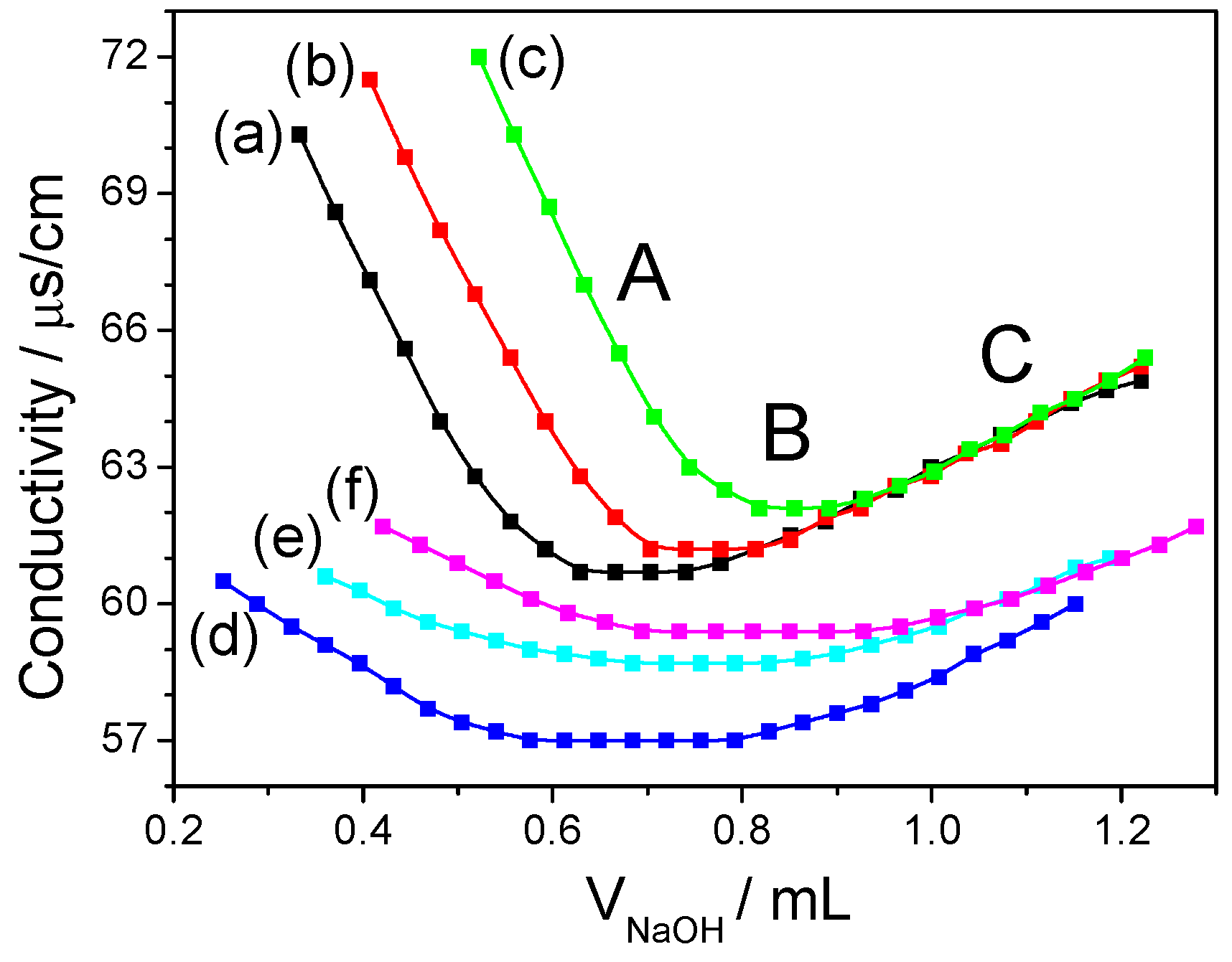
3.1. Degradation of Polysaccharides

Sulfated polysaccharides from different seaweeds were selected for this study and further degraded using H2O2. The natural polysaccharides were degraded for two reasons. First, the high molecular mass of natural polysaccharides hindered the biological activity exerted by polysaccharides. This is supported by a number of previous studies where activities of low-molecular-weight polysaccharides after degradation were often better than before [24,25]. Second, in the present work, we aimed to compare the ability of polysaccharides with different –SO3H contents to repair damaged renal epithelial cells, and the molecular weight of polysaccharides must be controlled to within a narrow range, thereby ensuring the accuracy of experimental research. This is confirmed by Zhao et al. [10], who suggested that when the molecular weight and molecular structure of polysaccharides were similar, the sulfate content of polysaccharides was the dominant factor affecting their biochemical activities. Therefore, polysaccharide degradation was necessary.
Accordingly, the initial molecular weight of some polysaccharide molecules was very high, the natural polysaccharides were degraded under different conditions (Table 1) to give the molecular weight of all polysaccharides with a narrow differences (from 1968 to 4020 Da). The molecular weight of polysaccharides was influenced by various degradation factors, such as H2O2 concentration, reaction temperature, and time. By changing H2O2 concentration in the degradation step, a suitable molecular weight of the polysaccharide can be obtained.

3.2. Chemical Characteristics

The molecular weights of six natural polysaccharides and four degraded polysaccharides are shown in Table 2. Although the molecular weights of all polysaccharides were significantly different before degradation, they had narrow differences (from 1968 to 4020 Da) after degradation under different conditions. The viscosity of all degraded polysaccharides decreased significantly (Table 1) and solubility increased compared with undegraded polysaccharides.
The –SO3H content of six kinds of natural SPSs ranged between 6.1%–21.7% (Table 2). Laminarin-1 contained the highest (21.7%) –SO3H content, whereas Undaria-6 (6.1%) contained the lowest. The –SO3H contents of polysaccharides were not significantly changed after degradation, which indicated that degradation did not exert a considerable impact on the –SO3H contents of polysaccharides.
The –SO3H contents of degraded Porphyra-2 and Gracilaria-3 (17.9% and 13.3%, respectively) were slightly higher than that of undegraded samples (17.3% and 12.7%, respectively). The hydroxyl radicals generated by the H2O2 system broke the sugar chains to expose the –SO3H inside the polysaccharide. This phenomenon led to higher levels of –SO3H content detected in the low-molecular-weight fragments after degradation [26]. Conversely, the –SO3H content (5.5%) of degraded Undaria-6 was found to be slightly lower than that of undegraded fragment (6.1%) because desulfonation frequently occurs in acidic medium or at elevated temperature [4]. In the present work, degradation was carried out in a weak acid medium of H2O2 at 90 °C, which may have led to the slight decrease in –SO3H content of SPSs.
The –COOH contents of all natural and degraded polysaccharides were measured by conductometric titration. The conductivity curves are illustrated in Figure 1, and the obtained value of uronic acid and conversion of uronic acid content are shown in Table 2. Akahane et al. [27] measured sulfate- and carboxyl-group content of a mixture alginic acid (–COOH only) and dextran sulfate (–SO3H only) with different proportions using the conductometric titration method. They found that both groups do not interfere with each other. Luo et al. [19] calculated the sulfate- and carboxyl- group content of sea-cucumber polysaccharide by conductometric titration. They showed that conductometric titration can simultaneously determine –SO3H and –COOH contents, but the curves of both groups differ in inflexion point. However, in the current study, we determined only –COOH content through the conductometric titration method. The –COOH content of six natural SPSs varied between 0.9% and 1.4%, in which Sargassum-4 (1.4%) had the highest –COOH content, and Porphyra-2 (0.9%) had the lowest. The –COOH content of SPSs only slightly changed after degradation.
All natural and degraded polysaccharides did not show any characteristic absorption peak at 260–280 nm and did not demonstrate a purple color with ninhydrin. Thus, all of them were devoid of proteins.

3.3. FT-IR Spectroscopy

Figure 2 shows the FT-IR spectrum of one representative algal polysaccharide. The FT-IR spectra of other polysaccharides are showed in Figure S2. The FT-IR spectra of all other SPSs were similar, and the main absorption peaks of the degraded and undegraded SPSs are shown in Table 3. A wide peak at about 3399.9–3429.2 cm−1 was due to the hydroxyl stretching vibration, and the peak at 2912.2–2939.6 cm−1 was due to C–H stretching vibration. The peak of 1380.2–1384.9 cm−1 could be assigned to deforming vibrations of the C–H bond [28]. The peak at about 1614.7–1637.3 cm−1 was attributed to the asymmetric and symmetric stretching vibrations of COOH. The peak at 1459.1 cm−1 was due to C–OH deformation vibration with contribution of O–C–O symmetric stretching vibration of carboxylate group. The peak at 1249.2–1262.8 cm−1 was due to the stretching vibration of S=O [29]. The peaks at about 1080.9–1086.9 and 1019.4–1025.6 cm−1 could be assigned to the stretching vibration of C–O. The peak at 931.8–935.3 cm−1 was attributed to the stretching vibration of C=O in uronic acid [30]. The peak at 881.2 cm−1 could be assigned to the C–H scissor vibration of β-type heterogeneous glycosidic bond [31].
The FT-IR spectrum of the degraded polysaccharide was very close to the undegraded one, which indicated that the overall structure of polysaccharide was not significantly affected by H2O2 degradation. However, the oxidative degradation of polysaccharides by H2O2 may result in the scission of carbohydrate chains and depolymerized polysaccharides by breaking glycosidic linkages [32]. Moreover, the –SO3H peak intensities of the four kinds of degradation products did not change after degradation, indicating that the –SO3H contents of all degraded polysaccharides had no significant variation. The relationship between the content of –SO3H in seaweed polysaccharides and the intensity of –SO3H absorption peak in FT-IR is shown in Figure S3 in the Supplementary Materials.

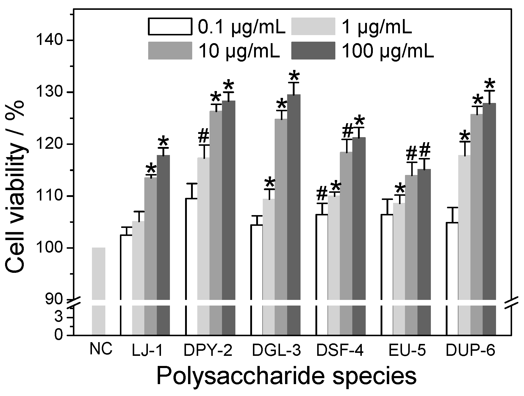
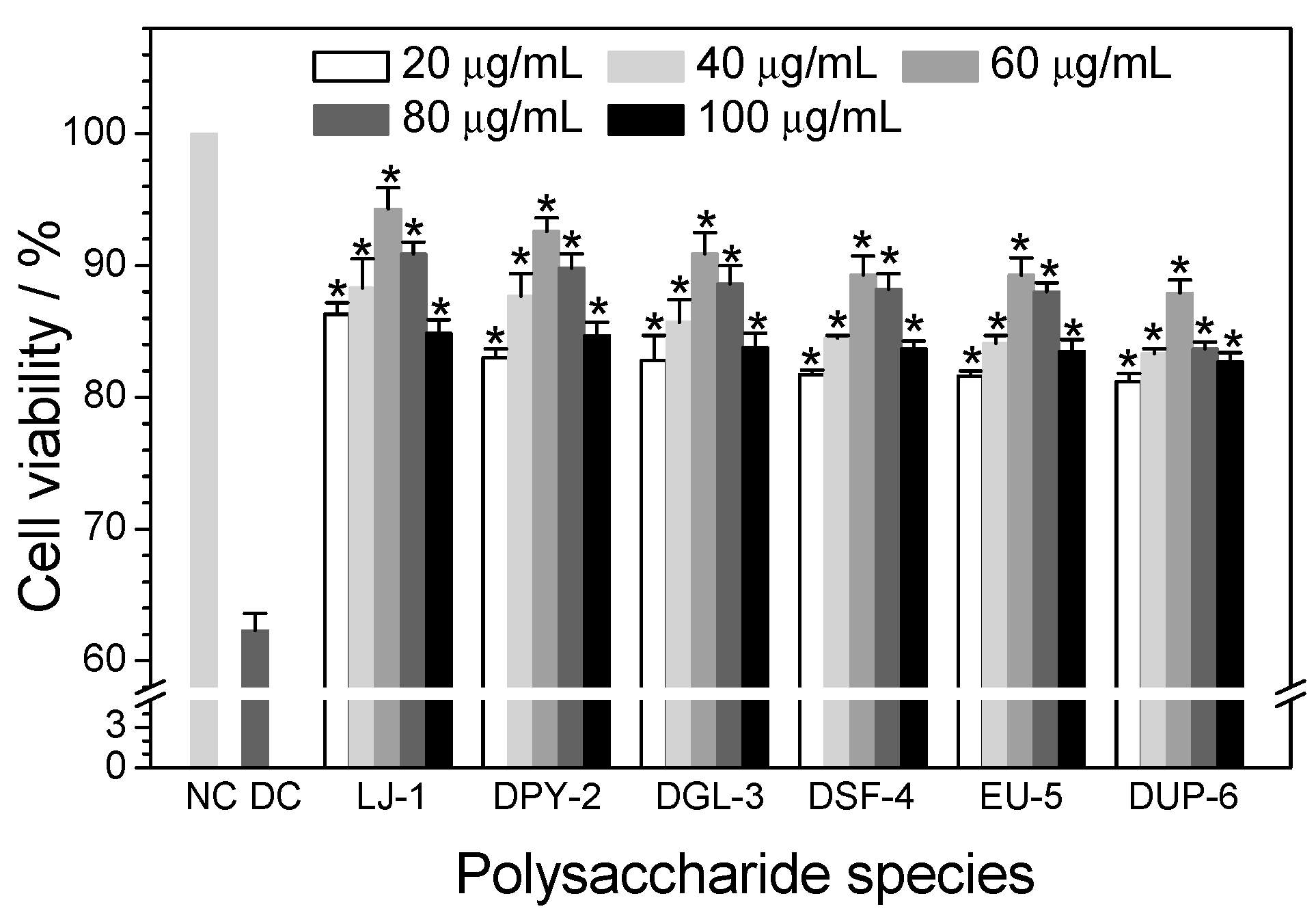
3.4. Cytotoxicity of Different SPSs on HK-2 Cells

The cytotoxicities of six kinds of SPSs with different contents of –SO3H group on normal HK-2 cells were detected by the CCK-8 method (Figure 3). When HK-2 cells were exposed to different concentrations of polysaccharides (0.1, 1, 10, and 100 μg/mL) for 24 h, the cell viability of all samples was above 100% and increased with increased polysaccharide concentration, indicating that these polysaccharides did not express any cytotoxicity within the range 0.1–100 μg/mL and also promoted cell growth. Possibly, the SPSs provided nutrition to cells, as cells divided rapidly due to sufficient nutrition and space. Therefore, cell viability was increased at the end of the assay. Similarly, Liang et al. [33] revealed that red SPSs (ι-carrageenan) exhibited no cytotoxicity and promoted the proliferation of human umbilical vein endothelial cells (HUVEC) within the range 5–1000 μg/mL, and the cell viability was more than 140% at the concentration of 800 μg/mL. Dore et al. [34] also found that the Sargassum vulgare polysaccharide increased the viability of normal rabbit aortic endothelial cells to above 100% in the concentration range of 25–100 μg/mL, and also promoted cell growth. Yao et al. [35] reported that Aloe polysaccharide speeded up the proliferation of fibroblasts significantly within the concentration range of 25–400 μg/mL.

3.5. Repair Ability of Different SPSs on Damaged HK-2 Cells

3.5.1. Effect of Polysaccharide Concentration

To optimize the repair effect of six SPSs on damaged HK-2 cells induced by 2.8 mmol/L oxalate, we performed a concentration-depended experiment in the concentration range from 20 to 100 μg/mL for 8 h (Figure 4). Oxalate is toxic to renal tubular cells by inducing inflammation, membrane damage, oxidative cell stress, DNA synthesis, and necrosis cell death at high concentrations (≥5 mmol/L) [36]. Figure 4 shows that all polysaccharides considerably repaired damaged cells compared with the damaged control group. Simultaneously, when damaged cells were repaired by different concentrations (20, 40, 60, 80, and 100 μg/mL) of polysaccharides, the cell viability of damaged cells was initially increased, reaching the maximum at 60 μg/mL, and then decreasing at higher concentrations (100 μg/mL), indicating that 60 μg/mL was adequate for the polysaccharides to play a role. Moreover, it was a remarkable fact that, although results of cell-viability between 60 and 80 μg/mL were similar for both degraded Sargassum-4 and Eucheuma-5, greater differences were found between results of 60 and 80 μg/mL for Laminaria-1 and degraded Undaria-6. Thus, in subsequent experiments, 60 μg/mL polysaccharides was chosen to repair cells and establish the effect of six SPSs to repair oxalate-induced damaged cells.

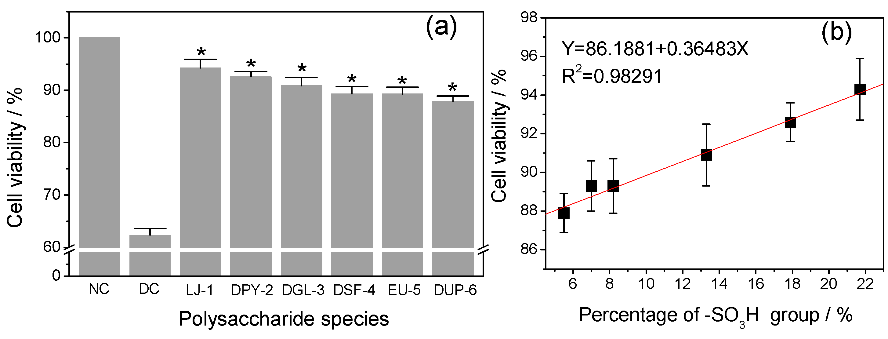
3.5.2. Effect of –SO3H Content

Six low-molecular-weight polysaccharides with narrow differences but different –SO3H contents were used to repair oxalate-induced damaged HK-2 cells. Results are shown in Figure 5a. All six polysaccharides at 60 μg/mL exerted a repair effect on injured cells to various degrees because the polysaccharides contained the negatively charged –SO3 and –COO groups, which could reduce the loss of the negative charge on the cell surface and repair the charge barrier [37]. Sun et al. [26] confirmed that the inhibitory effect of polysaccharides from two marine Chrysophyta on the hemolysis of RBCs may be due to the reducibility groups such as hydroxyl, sulfate, and uronic acid groups of the polysaccharides. These negatively-charged groups combine with the free radicals and restore the activity of cells. SPSs are exogenous antioxidants enhancing the antioxidant activity of cells by reducing free-radical generation and oxidative stress in renal epithelia cells. Hence, SPSs can protect and repair renal cells by increasing antioxidant capacity and blocking the injury pathway, which prevents the binding and retention of CaOx crystals to renal tissue, eventually reducing the occurrence of kidney-stone diseases. In the present work, when injured cells were repaired by the polysaccharides Laminaria-1, degraded Porphyra-2, degraded Gracilaria-3, degraded Sargassum-4, Eucheuma-5, and degraded Undaria-6 with different –SO3H contents (21.7%, 17.9%, 13.3%, 8.2%, 7.0%, and 5.5%, respectively), the cell viabilities of injured cells increased from 62.3% (damaged control) to 94.3%, 92.6%, 90.9%, 89.3%, 89.3%, and 87.9%, respectively. Meanwhile, results suggested that samples with higher –SO3H content had the maximum capacity to repair damaged cells, whereas samples with lower –SO3H content had modest activity. However, when compared the repair ability of all polysaccharides among each other, we did not find significant difference at p < 0.05. Furthermore, as aforementioned, the activity of polysaccharides also depended on –COOH content. Given that our samples had similar –COOH contents (between 1.0% and 1.7%), the effect of –COOH content could be avoided. Therefore, the repair abilities of polysaccharides were confirmed to be closely related to –SO3H content (Figure 5b). This finding was supported by the findings of Song et al. [38] on three polysaccharide fractions (B1, B2, and B3) from Bryopsis plumose with different –SO3H contents (7.56%, 6.71%, and 11.4%, respectively) that B3 with the highest –SO3H group content has the highest IC50 values 9.2 and 1.7 mg/mL in the scavenging activity of superoxide anion and DPPH free radicals, respectively. Therefore, the antioxidant ability of the three components was proportional to –SO3H content.

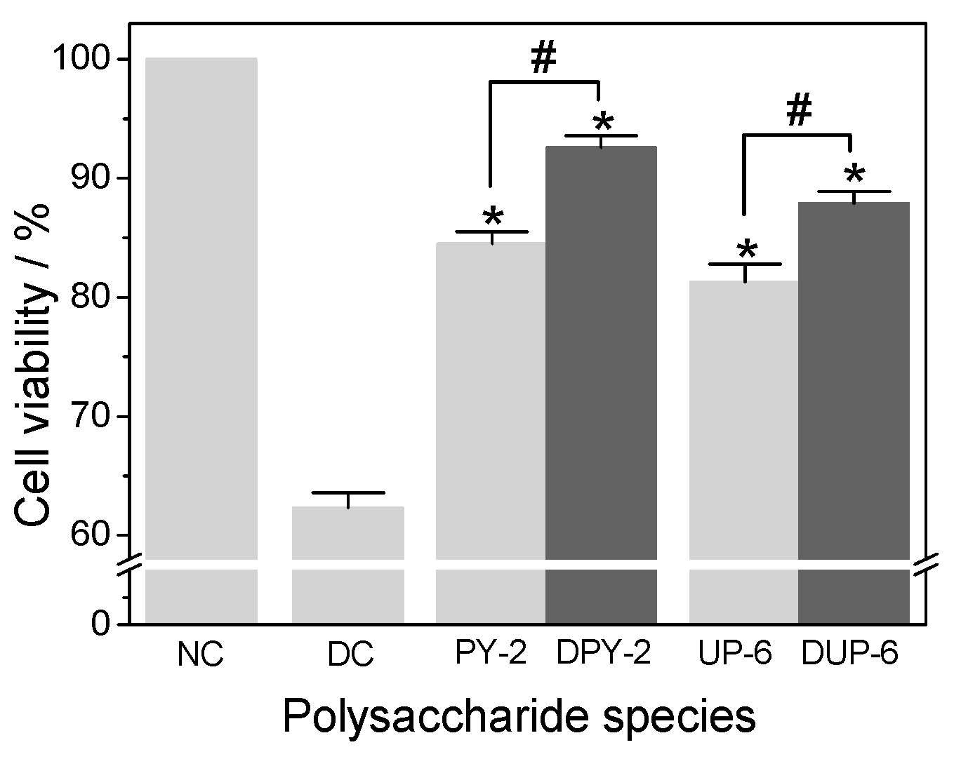
3.5.3. Effect of Molecular Weight

The influence of molecular weight of polysaccharides on the repair of oxalate-induced damaged cells is shown in Figure 6. Based on the –SO3H content, Porphyra-2 and Undaria-6 were selected to test the effect of molecular weight, because Porphyra-2 contained the highest –SO3H content, whereas Undaria-6 contained the lowest among the degraded samples. After depolymerization, the molecular weight of the polysaccharides Porphyra-2 and Undaria-6 decreased from 3.1 × 10⁴ to 4020 Da and 5.2 × 10⁶ to 3635 Da, respectively. However, both samples had similar –SO3H and –COOH contents before and after degradation (Table 2). Figure 6 suggests that the lower-molecular-weight fragments of Porphyra-2 and Undaria-6 exhibited stronger damage repair activity (92.6% and 87.9%, respectively) in HK-2 cells than their native polysaccharides (84.5% and 81.3%, respectively). There was significant difference occurred between Porphyra-2 before and after degradation (p < 0.05), and also for Undaria-6 before and after degradation (p < 0.05). In other words, degraded SPSs with low molecular weights showed better repair abilities. This finding corresponded with that of a previous study where low-molecular-weight fragments of Porphyridium cruentum polysaccharides after degradation showed stronger antioxidant activity than high-molecular-weight samples [39]. Clearly, the repair activity of polysaccharides was inversely related to their molecular weight. It was supposed that low molecular weight polysaccharides have more reductive hydroxyl group terminals to accept and remove the free radicals generated by oxalate [40]. However, it was not conducive for high molecular weight polysaccharides to cross cell-membranes, so it was difficult to exert their biological activities.

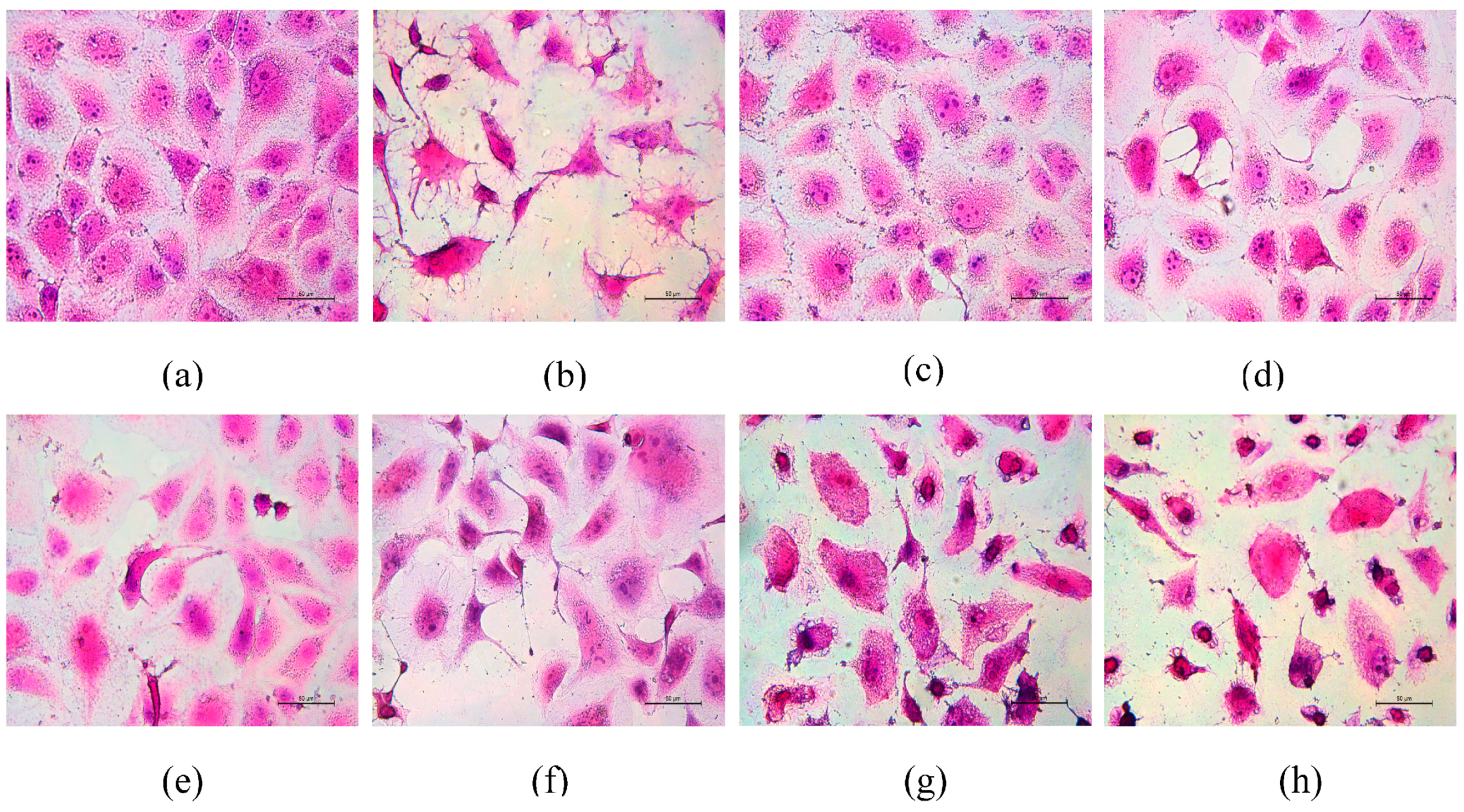
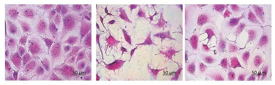
3.6. Morphological Changes of HK-2 Cells after Repair by Different Polysaccharides

The repair effect of the six SPSs on the morphology of damaged HK-2 cells was evaluated by hematoxylin and eosin staining. As shown in Figure 7a, HK-2 cells of the normal control group exhibited normal morphologies with an oval shape and round nucleus. Moreover, the integrity of tight junctions between cells and microvilli were clearly visible. When HK-2 cells were exposed to oxalate (2.8 mmol/L) for 3 h, the cells lost their natural shape and were shrunken into fibroblast like a spindle, cell connections were broken, and nuclear condensation and apoptotic bodies were clearly observed with a dense stain (Figure 7b). These abnormal morphological findings suggested that oxalate damaged the renal membrane, which may contribute to the development of stone disease by altering the properties of the renal epithelial cell membrane. After treating the damaged cells with various algal polysaccharides, cell morphology was gradually restored, and cellular connections tightened (Figure 7c–f), whereas Eucheuma-5 and degraded Undaria-6 showed less effect (Figure 7g,h) compare to other SPSs because of their low sulfate contents. All evaluated polysaccharide samples were able to restore the structure of damaged cells to varying degrees compared with the damaged control group. Laminaria-1 polysaccharide showed increased repair effect on cell morphology compared with all other polysaccharides. The cell-morphology-restoration activity of the other five components decreased in the order degraded Porphyra-2 > degraded Gracilaria-3 > degraded Sargassum-4 > Eucheuma-5 > degraded Undaria-6, the same as the order of sulfate contents of the samples. This finding indicated that –SO3H content affected the repair ability of polysaccharides, in accordance with the result of cell-viability assay.

4. Conclusions

Six types of low-molecular-weight seaweed polysaccharides with narrow differences (from 1968 to 4020 Da) but different contents of the characteristic –SO3H group (21.7%, 17.9%, 13.3%, 8.2%, 7%, and 5.5%, respectively) were selected to examine their impact on oxalate-induced damaged HK-2 cells. These polysaccharides did not express cytotoxicity within the 0.1–100 μg/mL dose range. When they were used to repair the damaged HK-2 cells induced by oxalate, the repair abilities were positively associated with the –SO3H content of polysaccharide and negatively associated with molecular weight. The polysaccharides had the best repair effect at 60 μg/mL. Thus, seaweed sulfated polysaccharides can be used to prevent or treat hyperoxaluria and renal-stone formation.

Supplementary Materials

Supplementary Materials can be found at www.mdpi.com/2073-4360/8/5/188/s1.

Acknowledgments

This work was supported by National Natural Science Foundation of China.

Author Contributions

Poonam Bhadja, Cai-Yan Tan and Jian-Ming Ouyang conceived and designed the experiments; Cai-Yan Tan performed the experiments; Poonam Bhadja and Cai-Yan Tan analyzed the data; Poonam Bhadja and Cai-Yan Tan contributed reagents/materials/analysis tools; Jian-Ming Ouyang and Kai Yu wrote the paper.

Conflicts of Interest

The authors declare no conflict of interest.

References

  1. Chen, S.; Gao, X.; Sun, Y.; Xu, C.; Wang, L.; Zhou, T. Analysis of HK-2 cells exposed to oxalate and calcium oxalate crystals: Proteomic insights into the molecular mechanisms of renal injury and stone formation. Urol. Res. 2010, 38, 7–15. [Google Scholar] [CrossRef] [PubMed]
  2. Farooq, S.M.; Boppana, N.B.; Asokan, D.; Sekaran, S.D.; Shankar, E.M.; Li, C.; Gopal, K.; Bakar, S.A.; Karthik, H.S.; Ebrahim, A.S. C-phycocyanin confers protection against oxalate-mediated oxidative stress and mitochondrial dysfunctions in MDCK cells. PLoS ONE 2014, 9, 93056–93064. [Google Scholar] [CrossRef] [PubMed]
  3. Boeve, E.R.; Cao, L.C.; Verkoelen, C.F.; Romijn, J.C.; de Bruijin, W.C.; Schroder, F.H. Glycosaminoglycans and other sulphated polysaccharides in calculogenesis of urinary stones. World J. Urol. 1994, 12, 43–48. [Google Scholar] [CrossRef] [PubMed] [Green Version]
  4. Ouyang, J.-M.; Wang, M.; Lu, P.; Tan, J. Degradation of sulfated polysaccharide extracted from algal Laminaria japonica and its modulation on calcium oxalate crystallization. Mater. Sci. Eng. C 2010, 30, 1022–1029. [Google Scholar] [CrossRef]
  5. Veena, C.K.; Josephine, A.; Preetha, S.P.; Varalakshmi, P.; Sundarapandiyan, R. Renal peroxidative changes mediated by oxalate: The protective role of fucoidan. Life Sci. 2006, 79, 1789–1795. [Google Scholar] [CrossRef] [PubMed]
  6. Zhang, C.-Y.; Kong, T.; Wu, W.-H.; Lan, M.-B. The protection of polysaccharide from the brown seaweed Sargassum graminifolium against ethylene glycol-induced mitochondrial damage. Mar. Drugs 2013, 11, 870–880. [Google Scholar] [CrossRef] [PubMed]
  7. Faggio, C.; Morabito, M.; Armeli, M.S.; Lo, P.G.; Pagano, M.; Genovese, G. Potential use of polysaccharides from the brown alga Undaria pinnatifida as anticoagulants. Braz. Arch. Biol. Technol. 2015, 58, 798–804. [Google Scholar] [CrossRef]
  8. Faggio, C.; Pagano, M.; Dottore, A.; Genovese, G.; Morabito, M. Evaluation of anticoagulant activity of two algal polysaccharides. Nat. Prod. Res. 2016, in press. [Google Scholar] [CrossRef] [PubMed]
  9. Zhao, T.-T.; Zhang, Q.-B.; Qi, H.-M.; Zhang, H.; Niu, X.-Z.; Xu, Z.-H.; Li, Z.-E. Degradation of porphyran from Porphyra haitanensis and the antioxidant activities of the degraded porphyrans with different molecular weight. Int. J. Biol. Macromol. 2006, 38, 45–50. [Google Scholar] [CrossRef] [PubMed]
  10. Zhao, X.; Xue, C.-H.; Li, B.-F. Study of antioxidant activities of sulfated polysaccharides from Laminaria japonica. J. Appl. Phycol. 2008, 20, 431–436. [Google Scholar] [CrossRef]
  11. Zhang, Z.S.; Zhang, Q.B.; Wang, J.; Song, H.F.; Zhang, H.; Niu, X.Z. Chemical modification and influence of function groups on the in vitro-antioxidant activities of porphyran from Porphyra haitanensis. Carbohydr. Polym. 2010, 79, 290–295. [Google Scholar] [CrossRef]
  12. Wang, Z.-J.; Xie, J.-H.; Kan, L.-J.; Wang, J.-Q.; Shen, M.-Y.; Li, W.-J.; Nie, S.-P.; Xie, M.-Y. Sulfated polysaccharides from Cyclocary apaliurus reduce H2O2-induced oxidative stress in RAW264.7 cells. Int. J. Biol. Macromol. 2015, 80, 410–417. [Google Scholar] [CrossRef] [PubMed]
  13. Zhang, H.; Wang, J.-Q.; Nie, S.-P.; Wang, Y.-X.; Cui, S.W.; Xie, M.-Y. Sulfated modification, characterization and property of a water-insoluble polysaccharide from Ganoderma atrum. Int. J. Biol. Macromol. 2015, 79, 248–255. [Google Scholar] [CrossRef] [PubMed]
  14. Wang, J.; Guo, H.; Zhang, J.; Wang, X.; Zhao, B.; Yao, J.; Wang, Y. Sulfated modification, characterization and structure–antioxidant relationships of Artemisia sphaerocephala polysaccharides. Carbohydr. Polym. 2010, 8, 897–905. [Google Scholar] [CrossRef]
  15. Yuan, Y.; Macquarrie, D. Microwave assisted extraction of sulfated polysaccharides (fucoidan) from Ascophyllum nodosum and its antioxidant activity. Carbohydr. Polym. 2015, 129, 101–107. [Google Scholar] [CrossRef] [PubMed]
  16. Spatareanu, A.; Bercea, M.; Budtova, T.; Harabagiu, V.; Sacarescu, L.; Coseri, S. Synthesis, characterization and solution behaviour of oxidized pullulan. Carbohydr. Polym. 2014, 111, 63–71. [Google Scholar] [CrossRef] [PubMed]
  17. Chutipongtanate, S.; Watcharatanyatip, K.; Homvises, T.; Jaturongkakul, K.; Thongboonkerd, V. Systematic comparisons of various spectrophotometric and colorimetric methods to measure concentrations of protein, peptide and amino acid: Detectable limits, linear dynamic ranges, interferences, practicality and unit costs. Talanta 2012, 98, 123–129. [Google Scholar] [CrossRef] [PubMed]
  18. Sakthivel, R.; Pandima Devi, K. Evaluation of physicochemical properties, proximate and nutritional composition of Gracilaria edulis collected from palk bay. Food Chem. 2015, 174, 68–74. [Google Scholar] [CrossRef] [PubMed]
  19. Luo, L.; Wu, M.-Y.; Xu, L.; Lian, W.; Xiang, J.-Y.; Lu, F.; Gao, N.; Xiao, C.; Wang, S.-M.; Zhao, J.-H. Comparison of physicochemical characteristics and anticoagulant activities of polysaccharides from three sea cucumbers. Mar. Drugs 2013, 11, 399–417. [Google Scholar] [CrossRef] [PubMed]
  20. Genovese, G.; Faggio, C.; Gugliandolo, C.; Torre, A.; Spanò, A.; Morabito, M.; Maugeri, T.L. In vitro evaluation of antibacterial activity of Asparagopsis taxiformis from the Straits of Messina against pathogens relevant in aquacultureq. Mar. Environ. Res. 2012, 73, 1–6. [Google Scholar] [CrossRef] [PubMed]
  21. Pagano, M.; Faggio, C. The use of erythrocyte fragility to assess xenobiotic cytotoxicity. Cell Biochem. Funct. 2015, 33, 351–355. [Google Scholar] [CrossRef] [PubMed]
  22. Teng, L.; Fu, H.; Wang, M.; Deng, C.; Song, Z.; Chen, J. Immunomodulatory activity of heparan sulfate mimetics from Escherichia coli K5 capsular polysaccharide in vitro. Carbohydr. Polym. 2015, 115, 643–650. [Google Scholar] [CrossRef] [PubMed]
  23. Sun, X.-Y.; Ouyang, J.-M.; Zhu, W.-Y.; Li, Y.-B.; Gan, Q.-Z. Size-Dependent toxicity and interactions of calcium oxalate dihydrate crystal on Vero renal epithelial cells. J. Mater. Chem. B 2015, 3, 1864–1878. [Google Scholar] [CrossRef]
  24. Zhang, Z.; Wang, X.; Mo, X.; Qi, H. Degradation and the antioxidant activity of polysaccharide from Enteromorpha linza. Carbohydr. Polym. 2013, 92, 2084–2087. [Google Scholar] [CrossRef] [PubMed]
  25. Sheng, J.; Sun, Y. Antioxidant properties of different molecular weight polysaccharides from Athyrium multidentatum(Doll.) Ching. Carbohydr. Polym. 2014, 108, 41–45. [Google Scholar] [CrossRef] [PubMed]
  26. Sun, L.; Wang, L.; Li, J.; Liu, H. Characterization and antioxidant activities of degraded polysaccharides from two marine Chrysophyta. Food Chem. 2014, 160, 1–7. [Google Scholar] [CrossRef] [PubMed]
  27. Akahane, T.; Takeuchi, S.; Minakata, A. Conductimetric titration of polyelectrolytes having sulfate and carboxyl groups. Polym. Bull. 1990, 24, 437–444. [Google Scholar] [CrossRef]
  28. Ge, Q.; Mao, J.-W.; Zhang, A.-Q.; Wang, Y.-J.; Sun, P.-L. Purification, chemical characterization, and antioxidant activity of a polysaccharide from the fruiting bodies of sanghuang mushroom (Phellinus baumii Pilát). Food Sci. Biotechnol. 2013, 22, 301–307. [Google Scholar] [CrossRef]
  29. Immanuel, G.; Sivagnanavelmurugan, M.; Marudhupandi, T.; Radhakrishnan, S.; Palavesam, A. The effect of fucoidan from brown seaweed Sargassum wightiion WSSV resistance and immune activity in shrimp Penaeus monodon (Fab). Fish Shellfish Immunol. 2012, 32, 551–564. [Google Scholar] [CrossRef] [PubMed]
  30. Wang, B. Chemical characterization and ameliorating effect of polysaccharide from Chinese jujube on intestine oxidative injury by ischemia and reperfusion. Int. J. Biol. Macromol. 2011, 48, 386–391. [Google Scholar] [CrossRef] [PubMed]
  31. Ji, C.-F.; Ji, Y.-B.; Meng, D.-Y. Sulfated modifiation and anti‑tumor activity of laminarin. Experimen. Therapeut. Med. 2013, 6, 1259–1264. [Google Scholar]
  32. Schuchmann, M.N.; von Sonntag, C. Effect of oxygen on OH-radical-induced scission of glycosidic linkage of cellobiose. Int. J. Radiat. Biol. Relat. Stud. Phys. Chem. Med. 1978, 34, 397–400. [Google Scholar] [CrossRef]
  33. Liang, W.; Mao, X.; Peng, X.; Tang, S. Effects of sulfate group in red seaweed polysaccharides on anticoagulant activity and cytotoxicity. Carbohydr. Polym. 2014, 101, 776–785. [Google Scholar] [CrossRef] [PubMed]
  34. Dore, C.M.P.G.; Alves, M.G.C.F.; Santos, N.D.; Cruz, A.K.M.; Câmara, R.B.G.; Castro, A.J.G.; Alves, L.G.; Nader, H.B.; Leite, E.L. Antiangiogenic activity and direct antitumor effect from a sulfated polysaccharide isolated from seaweed. Microvasc. Res. 2013, 88, 12–18. [Google Scholar] [CrossRef] [PubMed]
  35. Yao, H.; Chen, Y.; Li, S.; Huang, L.; Chen, W.; Lin, X. Promotion proliferation effect of a polysaccharide from Aloe barbadensis Miller on human fibroblasts in vitro. Int. J. Biol. Macromol. 2009, 45, 152–156. [Google Scholar] [CrossRef] [PubMed]
  36. Schepers, M.S.J.; Van Ballegooijen, E.S.; Bangma, C.H.; Verkoelen, C.F. Oxalate is toxic to renal tubular cells only at supraphysiologic concentrations. Kidney Int. 2005, 68, 1660–1669. [Google Scholar] [CrossRef] [PubMed]
  37. Yao, X.-Q.; Ouyang, J.-M.; Peng, H.; Zhu, W.-Y.; Chen, H.-Q. Inhibition on calcium oxalate crystallization and repair on injured renal epithelial cells of degraded soybean polysaccharide. Carbohydr. Polym. 2012, 90, 392–398. [Google Scholar] [CrossRef] [PubMed]
  38. Song, H.; Zhang, Q.; Zhang, Z.; Wang, J. In vitro antioxidant activity of polysaccharides extracted from Bryopsis plumosa. Carbohydr. Polym. 2010, 80, 1057–1061. [Google Scholar] [CrossRef]
  39. Sun, L.; Wang, C.; Shi, Q.; Ma, C. Preparation of different molecular weight polysaccharides from Porphyridium cruentum and their antioxidant activities. Int. J. Biol. Macromol. 2009, 45, 42–47. [Google Scholar] [CrossRef] [PubMed]
  40. Wang, J.; Hu, S.; Nie, S.; Yu, Q.; Xie, M. Reviews on mechanisms of in vitro antioxidant activity of polysaccharides. Oxid. Med. Cell. Longev. 2016. [Google Scholar] [CrossRef] [PubMed]
Figure 1. Conductivity curve of –COOH content of Porphyra-2 polysaccharide before and after degradation. (A) electrical conductivity decreasing stage; (B) equilibrium stage; (C) electrical conductivity increasing stage. (ac) are the triplicate experiments before degradation; (df) are three parallel experiments after degradation.
Polymers 08 00188 g001
Figure 2. FT-IR spectra of representative seaweed polysaccharide degraded Sargassum-4.
Polymers 08 00188 g002
Figure 3. Cytotoxic effect of six low-molecular-weight polysaccharides with different contents of –SO3H group on HK-2 cells. Cells were incubated with different concentration of (0.1–100 μg/mL) polysaccharides for 24 h and then cell viability was determined by CCK-8 assay. The control represents 100% cell viability. Data are reported as Mean ± SD (n = 3), derived from three independent experiments. Compared with normal control group, # indicates p < 0.05, * indicates p < 0.01. NC: Normal control; LJ-1: Laminaria-1; DPY-2: degraded Porphyra-2; DGL-3: degraded Gracilaria-3; DSF-4: degraded Sargassum-4; EU-5: Eucheuma-5; DUP-6: degraded Undaria-6.
Polymers 08 00188 g003
Figure 4. Effect of different polysaccharide concentrations (20, 40, 60, 80, and 100 μg/mL) on the viability of oxalate-induced damaged HK-2 cells. The cells were incubated with 2.8 mmol/L oxalate for 3 h, and then treated with different concentrations of SPSs for 8 h. Afterwards, cell-viability was determined by CCK-8 assay. Each bar was derived from three independent experiments and data are reported as Mean ± SD. Compared with damaged control group, * indicates p < 0.01. NC: Normal control; DC: Damaged control; LJ-1: Laminaria-1; DPY-2: degraded Porphyra-2; DGL-3: degraded Gracilaria-3; DSF-4: degraded Sargassum-4; EU-5: Eucheuma-5; DUP-6: degraded Undaria-6.
Polymers 08 00188 g004
Figure 5. Repair capacity of six low-molecular-weight seaweed polysaccharides (SPSs) on oxalate-induced damaged HK-2 cells (a), and the relationship between –SO3H content and repair capacity of SPSs (b). Each bar is derived from three independent experiments and data are reported as Mean ± SD. Compared with damaged control group, * indicates p < 0.01. NC: Normal control; DC: Damaged control; LJ-1: Laminaria-1; DPY-2: degraded Porphyra-2; DGL-3: degraded Gracilaria-3; DSF-4: degraded Sargassum-4; EU-5: Eucheuma-5; DUP-6: degraded Undaria-6.
Polymers 08 00188 g005
Figure 6. Cell repair activity of selected polysaccharides of different molecular weight on damaged HK-2 cells. Data were expressed as Mean ± SD from three independent experiments. Compared with damaged control group, * indicates p < 0.01. # indicates p < 0.05. NC: Normal control; DC: Damaged control; LJ-1: Laminaria-1; DPY-2: degraded Porphyra-2; DGL-3: degraded Gracilaria-3; DSF-4: degraded Sargassum-4; EU-5: Eucheuma-5; DUP-6: degraded Undaria-6.
Polymers 08 00188 g006
Figure 7. Repair effect of six SPSs on the morphology of damaged HK-2 cells evaluated by hematoxylin and eosin (HE) staining: (a) normal control; (b) damaged HK-2 cells; (c) Laminaria-1; (d) degraded Porphyra-2; (e) degraded Gracilaria-3; (f) degraded Sargassum-4; (g) Eucheuma-5; and (h) degraded Undaria-6. Oxalate-induced damaged HK-2 cells were treated with 60 μg/mL polysaccharides for 8 h, and then cell morphology was determined by HE staining under a microscope. The nucleus was stained blue or purple, and the cytoplasm was pink or red. Magnification: 400×.
Polymers 08 00188 g007
Table 1. Degradation conditions and physico-chemical properties of different seaweed polysaccharides (SPSs).
Polysaccharide speciesConcentration of H2O2Constant of Mark-Houwink equationIntrinsic viscosity η (mL/g)
καBefore degradationAfter degradation
LJ-18.21 × 10−30.7823.1
PY-212%7.86 × 10−30.6265.11.4
GL-310%7.00 × 10−20.7201,03924.1
SF-412%1.92 × 10−41.2326.34.9
EU-51.92 × 10−41.233.4
UP-615%8.21 × 10−30.78214604.9
LJ-1: Laninaria-1, PY-2: Porphyra-2, GL-3: Gracilaria-3, SF-4: Sargassum-4, EU-5: Eucheuma-5, UP-6: Undaria-6.
Table 2. Molecular weight, –SO3H content, and –COOH content of native and degraded SPSs.
SPSsMean molecular weights M/Da–SO3H contents/%–COOH contents/%Uronic acid contents/%
BD *AD *BDADBDADBDAD
Laminaria-11,96821.71.25.2
Porphyra-23.1 × 1044,02017.317.90.91.73.97.3
Gracilaria-36.2 × 1053,34312.713.31.01.04.34.3
Sargassum-41.6 × 1043,8288.48.21.41.36.05.4
Eucheuma-52,8507.01.25.0
Undaria-65.2 × 1063,6356.15.51.31.25.65.2
BD *: before degradation; AD *: after degradation.
Table 3. FT-IR characteristic absorption peaks of native and degraded seaweed polysaccharides.
SPSs–SO3H contents/%–COOH contents/%Characteristic absorption peaks of groups/cm−1
–OH–COOH–OSO3Sugar ring
LJ-121.71.23,400.91,632.11,262.32,912.2, 1,460, 1,380.3, 1,082.5, 1,019.4, 881.2
PY-217.30.93,436.31,632.91,249.22,922.0, 1,383.3, 1,080.9
DPY-217.91.73,429.21,618.41,250.02,925.5, 1,384.9, 1,080.8
GL-312.71.03,419.21,641.11,257.22,921.5, 1,378.4, 1,024.3
DGL-313.31.03,415.31,614.71,261.12,921.4, 1,383.8, 1,025.6
SF-48.41.43,415.11,630.91,260.22,931.8, 1,460.0, 1,380.2, 1,084.4, 1,021.6, 928.1
DSF-48.21.33,423.31,637.31,247.72,927.0, 1,421.2, 1,382.5, 1,081.2, 1,024.0, 935.3
EU-57.01.23,399.91,629.51,256.32,939.6, 1,459.1, 1,381.1, 1,086.9, 1,022.0, 931.8
UP-66.11.33,419.41,632.71,262.32,917.3, 1,022.5
DUP-65.51.23,413.21,615.81,262.82,921.4, 1,024.7
LJ-1: Laninaria-1; PY-2: Porphyra-2; DPY-2: Degraded Porphyra-2; GL-3: Gracilaria-3; DGL-3: Degraded Gracilaria-3; SF-4: Sargassum-4; DSF-4: Degraded Sargassum-4; EU-5: Eucheuma-5; UP-6: Undaria-6; DUP-6: Degraded Undaria-6.

