1. Introduction
Bacterial cellulose has emerged as an attractive biomaterial for applications including textiles, composite materials, wound dressings, personal care products, and food additives due to its high crystallinity, flexibility, porosity, purity, and ease of production compared with plant-derived cellulose [
1]. Considerable effort has focused on functionalizing bacterial cellulose to impart new properties, including antimicrobial activity [
2], magnetic properties [
3], bioelectric potential [
4], and bioethanol purification [
5]; however, these modifications typically require extensive downstream chemical processing. Genetic engineering of cellulose-producing microorganisms offers an alternative approach by enabling functionalization during cellulose biosynthesis. This one-step strategy could reduce the need for post-production chemical modification and simplify the production of functionalized bacterial cellulose.
Members of the genus
Komagataeibacter are among the highest-yielding bacterial cellulose producers and have become important hosts for engineering functionalized bacterial cellulose [
6]. Recent advances have produced strains with improved cellulose production [
7], altered fiber morphology [
8], and in vivo incorporation of chitin monomers [
9]. At the same time, extensive genetic toolkits have expanded the available plasmids, promoters, reporters, and secretion systems for this genus [
10,
11].
Despite these advances, significant challenges remain in developing engineered bacterial cellulose materials. Conventional functionalization strategies often require extensive post-production processing, while practical limitations continue to reduce the efficiency of genetic engineering in
Komagataeibacter and restrict implementation of desirable functional phenotypes. One of these is the difficulty of rapidly identifying correctly engineered transformants following genomic modification, possibly due to excessive cellulose, which inhibits DNA extraction [
10] (Supplementary Information). Previous studies have demonstrated that deletion of the pyrroloquinoline quinone (PQQ)-dependent glucose dehydrogenase (
gdh) reduces medium acidification and increases bacterial cellulose production in multiple
Komagataeibacter species when grown on glucose [
12,
13]. The acidic extracellular environment characteristic of acetic acid bacteria also limits functionalization strategies that depend on acid-sensitive enzymes. One example is the production of melanated bacterial cellulose through expression of the
Bacillus megaterium tyrosinase, encoded by
tyr1, which has previously required manual pH neutralization following pellicle formation [
14] because the enzyme is inhibited under acidic conditions [
15]. These observations suggest that the reduced-acidification phenotype resulting from
gdh deletion could provide a practical solution to both strain engineering and acid-sensitive functionalization.
In this study, we investigated whether the reduced-acidification phenotype resulting from gdh deletion could be exploited to address two practical challenges in engineering Komagataeibacter. We developed a rapid, inexpensive phenotype-based screening strategy for identifying engineered transformants and demonstrated that reduced acidification enabled one-step production of melanated bacterial cellulose without manual pH neutralization. Together, these findings establish reduced acidification as a practical engineering strategy that simplifies strain engineering while expanding opportunities for acid-sensitive bacterial cellulose functionalization.
2. Materials and Methods
2.1. Microbial Growth and Cellulose Production
Komagataeibacter sucrofermentans ATCC 700178 was obtained from the American Type Culture Collection (Manassas, VA, USA). Routine growth was performed in Hestrin-Schramm (HS) medium containing 5 g/L Phytone peptone (Becton Dickinson, Franklin Lakes, NJ, USA), 5 g/L yeast extract (Becton Dickinson, Franklin Lakes, NJ, USA), 2.7 g/L sodium phosphate dibasic heptahydrate (Thermo Fisher Scientific, Waltham, MA, USA), 1.5 g/L sodium citrate tribasic dihydrate (Sigma-Aldrich, Burlington, MA, USA), 0.6 g/L anhydrous magnesium sulfate (Thermo Fisher Scientific, Waltham, MA, USA), and either 20 g/L D-glucose (HSG; Sigma-Aldrich, Burlington, MA, USA) or 25 g/L D-mannitol (HSM; PhytoTech Labs, Lenexa, KS, USA). Where indicated, media were supplemented with 0.5 g/L L-tyrosine (HSGT or HSMT; Sunrise Science Products, Knoxville, TN, USA). Liquid media were supplemented with 5 U/mL cellulase from Trichoderma reesei (designated “-Cel”; Sigma-Aldrich, Burlington, MA, USA) and sterilized by filtration through a 0.22 μm syringe filter. Medium pH was adjusted to 5.7 with citric acid. Solid media contained 1.5% (w/v) agar, and all media were sterilized by autoclaving at 121 °C for 15 min. Routine starter cultures were grown overnight at 30 °C in 5 mL medium in 50 mL conical tubes.
2.2. Pellicle Production and Processing
Unless otherwise indicated, overnight starter cultures were washed twice with the appropriate cellulase-free medium, resuspended in the same medium, and inoculated into pellicle production media. Pellicles were incubated statically at 30 °C in a humidity-controlled chamber with 65–75% relative humidity. When required, pellicles were treated by incubation in two successive 1-h washes with 500 mL deionized water followed by a 2-h incubation in 0.1 M NaOH at 85 °C with shaking at 35 rpm. Following NaOH treatment, pellicles were washed twice with deionized water until the rinsate reached neutral pH, dried at 80 °C to constant weight, and weighed.
2.3. Genetic Engineering
DNA parts and primers used in this study are included in
Table S1, and gBlock sequences are included in
Table S2. pPETS_003 was constructed in pUC19, and pPETS_051 and pPETS_014 were constructed in pBBR1. NEB 5-alpha competent
E. coli cells (catalog no. C2987H; New England Biolabs, Ipswich, MA, USA) were used for plasmid construction. A gBlock containing the
BBa_J23104 promoter, the
BBa_B0035 ribosomal binding site (RBS), the
tyr1 coding sequence from
Bacillus megaterium, and the
rrnB1 terminator was ordered from Integrated DNA Technologies (Coralville, IA, USA). This sequence is the same as that used for
tyr1 expression in
K. rhaeticus by Walker et al. [
14]. All genetic components were amplified using the Platinum SuperFi II PCR Master Mix (catalog no. 12368050; Thermo Fisher Scientific; Waltham, MA, USA) and assembled using the NEBuilder HiFi DNA Assembly Master Mix (catalog no. E2621L; New England Biolabs; Ipswich, MA, USA). Plasmid sequences were verified by Oxford Nanopore Sequencing (Plasmidsaurus; South San Francisco, CA, USA).
K. sucrofermentans electrocompetent cells were prepared as previously described [
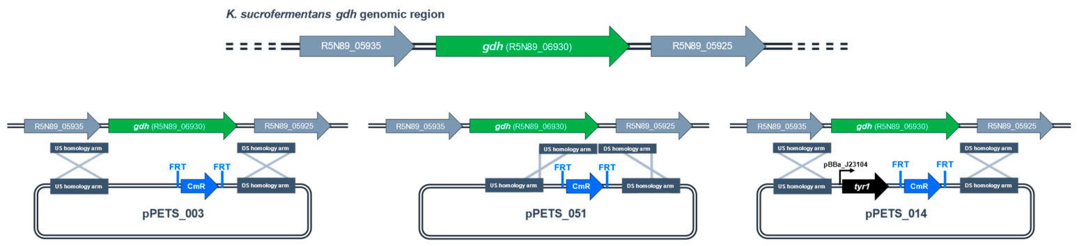
10] (Supplementary Information). Cells (100 µL) were transformed by electroporation (2485V, 50 µF, 100 Ω, 1 mm electrocuvette) with 1 μg PCR product composed of inserts of interest flanked by approximately 1 kb homology arms (
Figure 1;
Table S3). Transformants were recovered in 1 mL HSM-Cel overnight at 30 °C with 225 rpm shaking, and selected on HSG agar plates containing 350 μg mL
−1 chloramphenicol (HSG-Cm) unless otherwise noted. Strains were screened for integration into the
gdh locus as described and were verified by whole-genome sequencing (Plasmidsaurus; South San Francisco, CA, USA).
2.4. Colony PCR Evaluation
Five independent cultures each of wild-type (WT)
K. sucrofermentans were grown in HSM-Cel for two days, and
E. coli DH5α was grown in LB for one day. Cultures were adjusted to an OD
600 of 0.1 and serially diluted to OD
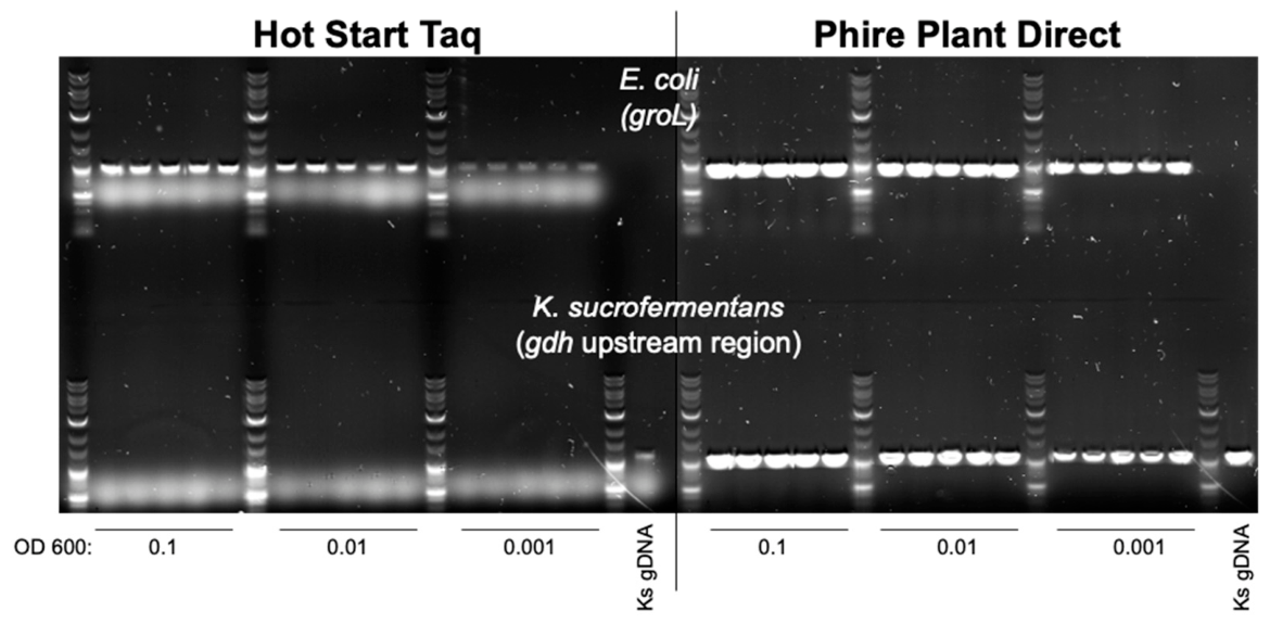
600 values of 0.01 and 0.001 in phosphate-buffered saline (PBS; catalog no. 10010023; Gibco, Waltham, MA, USA) or Phire Dilution Buffer (from the Thermo Scientific Phire Plant Direct PCR Master Mix, catalog no. F160L; Thermo Fisher Scientific, Waltham, MA, USA). One microliter of each suspension was used as a template for PCR using Hot Start Taq DNA Polymerase (catalog no. M0495L; New England Biolabs, Ipswich, MA, USA) or Phire Plant Direct PCR Master Mix (catalog no. F160L; Thermo Fisher Scientific, Waltham, MA, USA) according to the manufacturers’ instructions. Primers PETS_ETH_107/PETS_ETH_109 amplified a 940 bp region of the
E. coli groL gene, whereas primers O_PETS_11/O_PETS_17 amplified a 1080 bp region upstream of the
K. sucrofermentans gdh (
Table S3). PCR products were separated on 1% agarose gels containing ethidium bromide.
As a positive control, K. sucrofermentans colony biomass was dispersed in PBS containing 10 U/mL cellulase for 2 h at room temperature, pelleted, resuspended in Phire Dilution Buffer, and boiled for 20 min. Following centrifugation, 1 μL of the supernatant was used as a template for PCR.
2.5. Phenotype-Based Transformant Screening
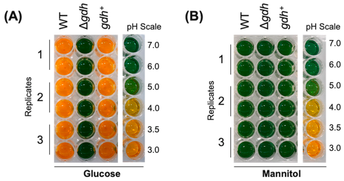
A pH-responsive indicator medium was prepared by supplementing HSG or HSM with 40 mg/L bromocresol green sodium salt (catalog no. A17503.06; Thermo Fisher Scientific, Waltham, MA, USA) and 80 mg/L thymol blue sodium salt (catalog no. 861367-5G; Sigma-Aldrich, Burlington, MA, USA) before autoclaving, yielding HSG-pH and HSM-pH, respectively. WT, Δ
gdh, and
gdh+ starter cultures were prepared as described in
Section 2.1, adjusted to an OD
600 of 1, and inoculated into duplicate 0.5 mL cultures of HSG-Cel, HSG-pH-Cel, HSM-Cel, or HSM-pH-Cel at an initial OD
600 of 0.01 in 48-well plates. Plates were sealed with Breathe-Easier membrane (catalog no. 9126-2000; USA Scientific, Ocala, FL, USA) and incubated at 30 °C with shaking at 250 rpm for 7 days. Colorimetric pH standards were prepared by adjusting sterile HSG-pH and HSM-pH to pH values of 7.0, 6.0, 5.0, 4.0, 3.5, and 3.0 using hydrochloric acid or sodium hydroxide.
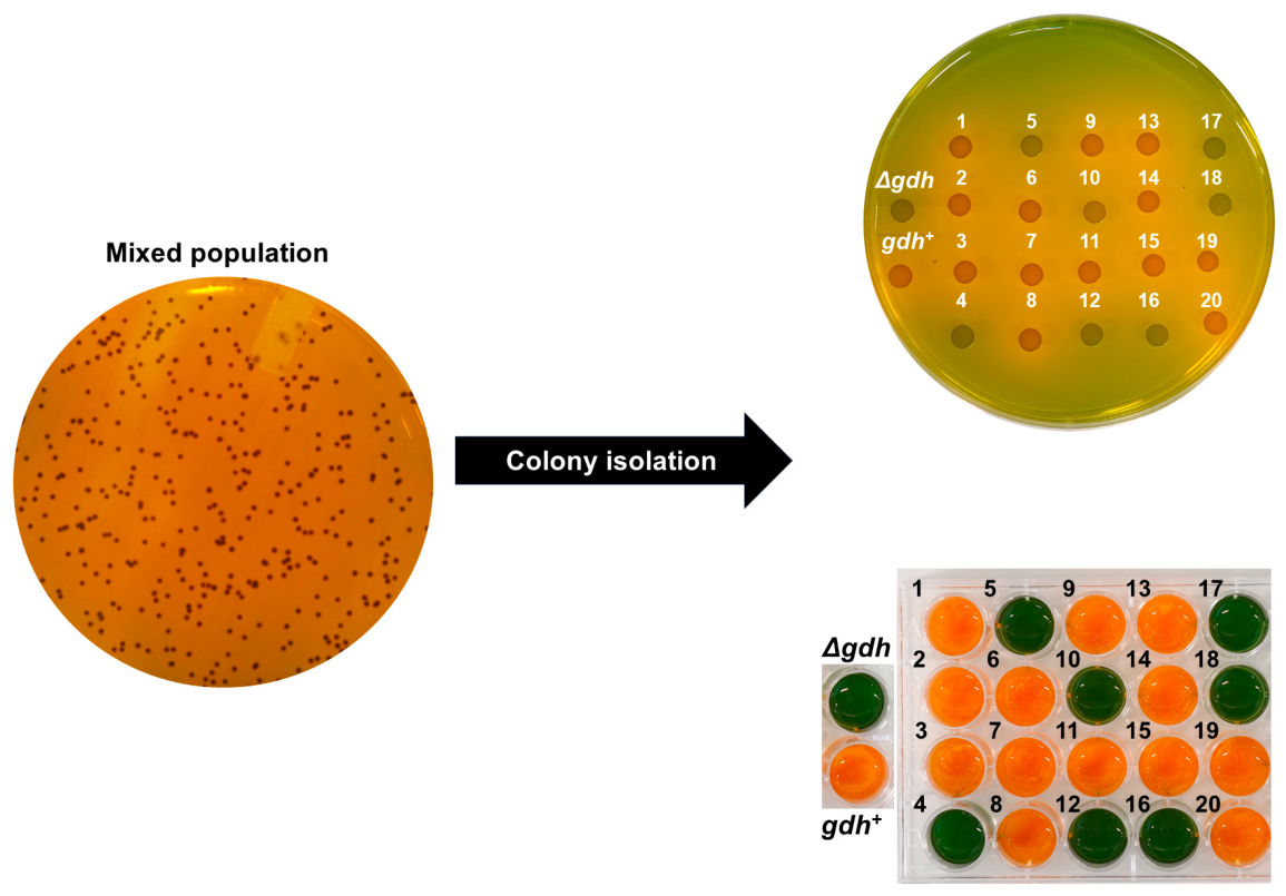
To validate phenotype-based transformant screening, equal optical densities of Δ
gdh and
gdh+ cultures were combined to generate a 1:1 mock community. Serial dilutions were plated on HSG-Cm-pH agar and incubated for 7 days. Twenty randomly selected colonies, together with known Δ
gdh and
gdh+ controls, were transferred to fresh HSG-Cm-pH agar and HSG-Cm-pH-Cel liquid medium, and colony identities were assigned based on medium color. PCR products were generated using primers O_PETS_11 and PETS_ETH_4 (
Table S3), and selected amplicons were sequenced by Plasmidsaurus (South San Francisco, CA, USA). Sequence alignments were performed using SnapGene software Version 8.1 (GSL Biotech, San Diego, CA, USA) [
16]. The screening workflow was subsequently applied during construction of the Δ
gdh::tyr1 strain by screening transformants on HSG-Cm-pH medium prior to whole-genome sequencing.
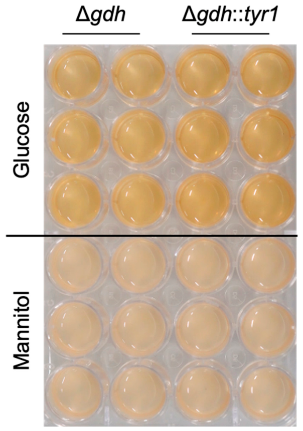
2.6. Establishing Conditions for One-Step Production of Melanated Bacterial Cellulose
Initial one-step melanation conditions were evaluated using previously reported culture conditions [
14]. Δ
gdh and Δ
gdh::tyr1 starter cultures were prepared as described above, adjusted to an OD
600 of 1, and inoculated at an initial OD
600 of 0.05 into duplicate 1 mL cultures of HSGT or HSMT supplemented with 10 μM CuSO
4·5H
2O (Thermo Fisher Scientific; Waltham, MA, USA) in 24-well plates. Cultures were incubated for 7 days as described in
Section 2.2.
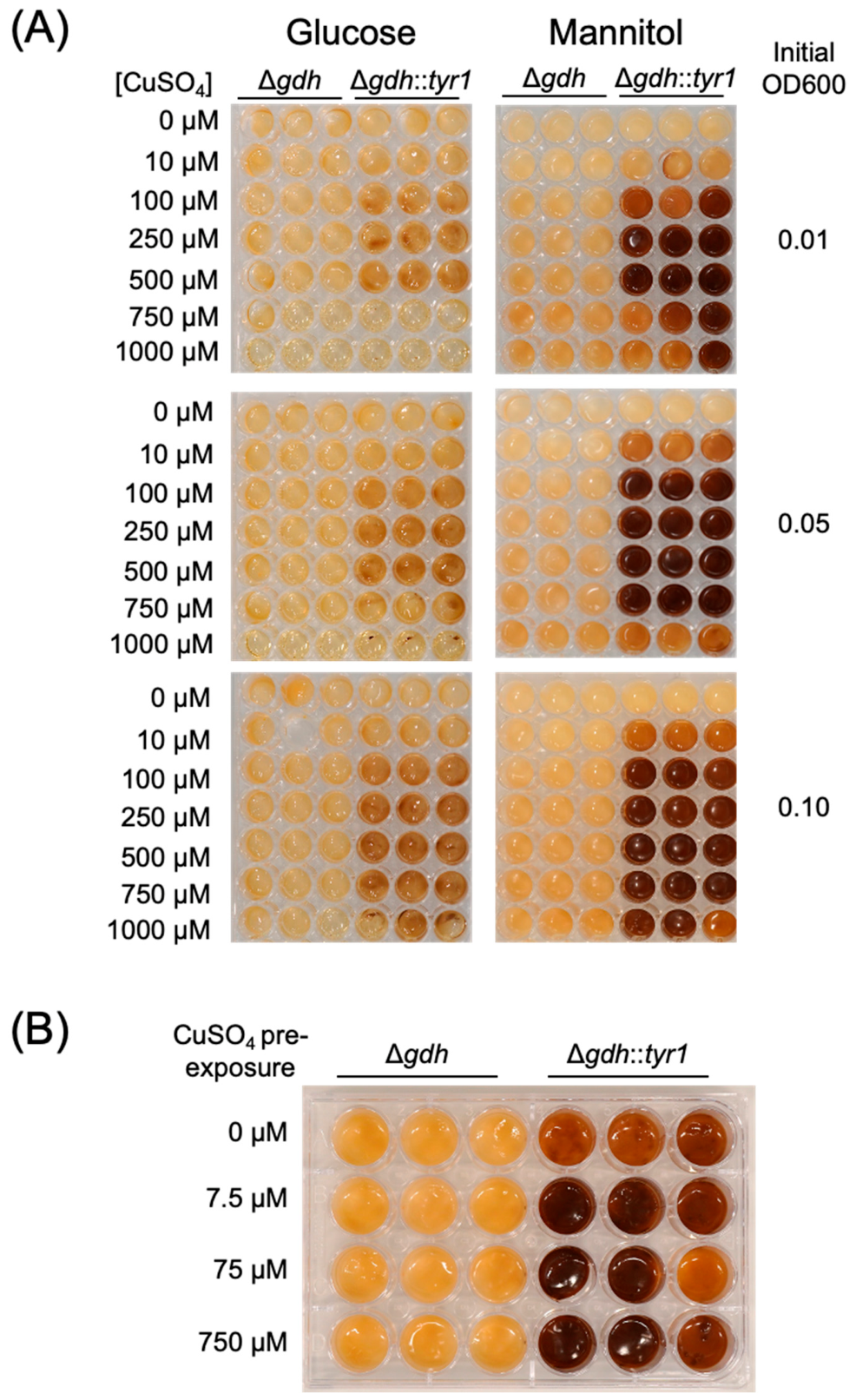
Melanation conditions were subsequently optimized by varying carbon source, CuSO
4 concentration, and inoculation density. Δ
gdh and Δ
gdh::
tyr1 starter cultures were inoculated at initial OD
600 values of 0.01, 0.05, or 0.1 into 0.5 mL HSGT or HSMT supplemented with 0, 10, 100, 250, 500, 750, or 1000 μM CuSO
4·5H
2O in 48-well plates. Cultures were incubated for 7 days as described in
Section 2.2.
To evaluate the effect of copper pre-exposure, starter cultures were adjusted to an OD600 of 0.05, inoculated into 1 mL of HSMT-Cel containing 0, 7.5, 75, or 750 μM CuSO4·5H2O, and incubated for 2 days. Cells equivalent to 0.1 OD units were then washed twice with HSMT and inoculated into 1 mL of HSMT supplemented with 750 μM CuSO4·5H2O for pellicle production.
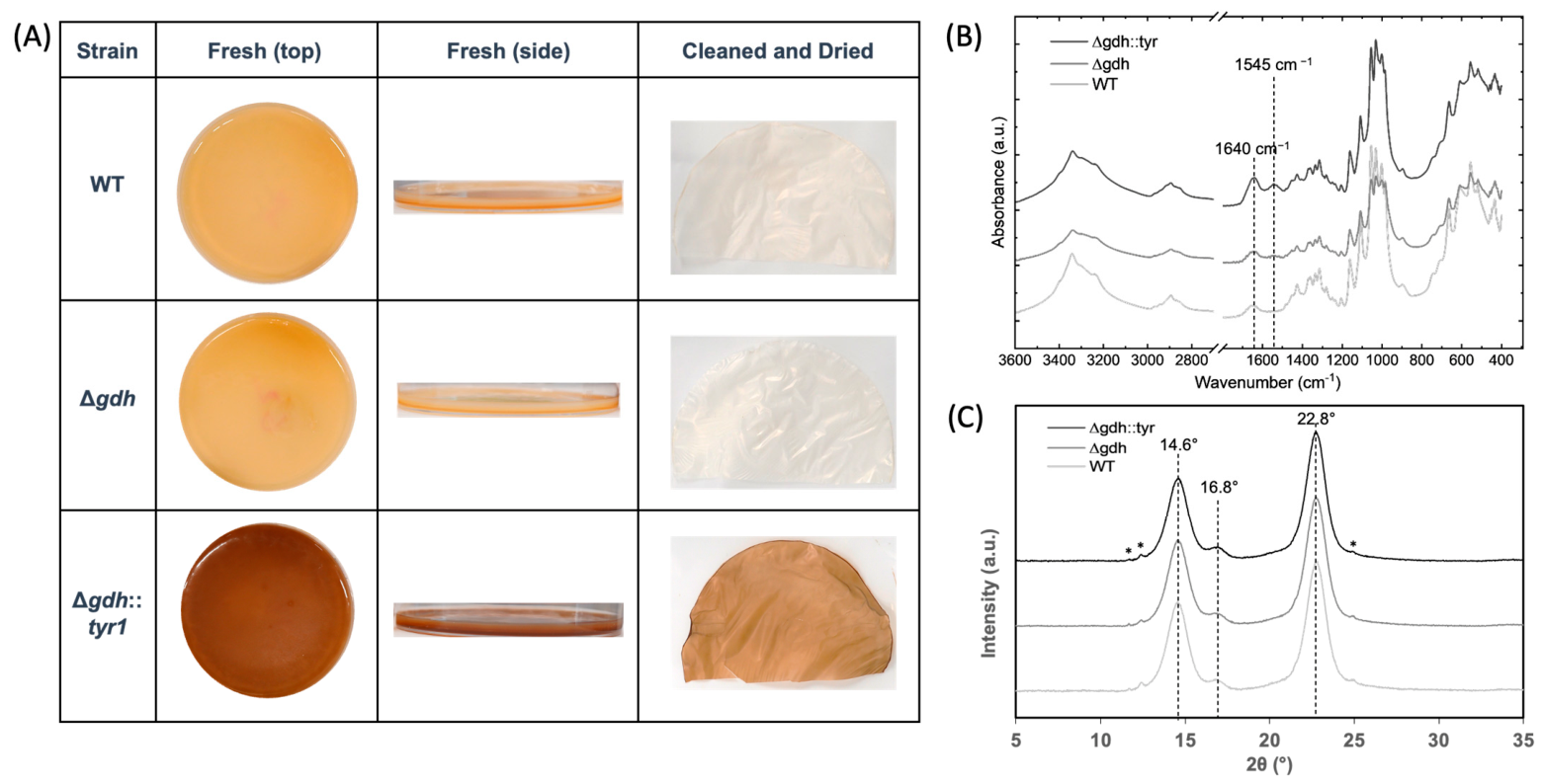
2.7. Characterization of Bacterial Cellulose
Pellicles produced by the WT, Δ
gdh, and Δ
gdh::
tyr1 strains under the optimized one-step melanation conditions described in
Section 2.6 were processed as described in
Section 2.2 and characterized by dry weight determination, Fourier transform infrared (FTIR) spectroscopy, and X-ray diffraction (XRD). FTIR was conducted using a Nicolet Summit FTIR (Thermo Fisher Scientific, Waltham, MA, USA) with a diamond attenuated total reflectance accessory. Each sample was measured in three places using 32 scans at 4 cm
−1 resolution in the range of 4000 to 400 cm
−1.
XRD patterns were measured with an Empyrean X-ray diffractometer (Malvern Panalytical, Malvern, United Kingdom) using Copper Kα
1 radiation (1.540598 Å; 45 kV and 40 mA). XRD patterns were measured from 2θ = 5–50°, with scan steps of 0.017° at 0.5°/min. The Divergence Slit was 0.435°, the anti-scatter slit was 5.5 mm, and the Soller slits were set to 0.04 rad. The monochromator and collimator were not used. Dried cellulose films were mounted onto glass cover slides lightly coated in Vaseline to anchor the films, and the slides were mounted onto standard XRD sample holders. The X-ray background was subtracted from the measured XRD patterns before cellulose crystallinity analysis. Cellulose crystallinity was calculated using the Segal peak height method [
17]:
where I
200 and I
am are the background-subtracted peak intensities at 2θ = 22.2 and 18.6°, corresponding to the crystal plane (200) and amorphous region, respectively.
2.8. Statistical Analysis
All quantitative experiments were performed using three biological replicates unless otherwise noted. Values are reported as mean ± standard deviation (SD). Statistical significance was determined using two-way ANOVA followed by Tukey’s multiple-comparison test, with p < 0.05 considered statistically significant.
4. Discussion
Genetic engineering of Komagataeibacter has enabled improved bacterial cellulose production and the development of novel functionalized materials; however, efficient strain engineering remains limited by practical challenges in screening candidate engineered transformants and in implementing acid-sensitive modifications. In this study, we demonstrate that the reduced-acidification phenotype resulting from gdh deletion provides a practical solution to both challenges. Specifically, reduced acidification enabled the development of a rapid, inexpensive, phenotype-based screening strategy for identifying engineered transformants and eliminated the need for manual pH neutralization, allowing one-step production of melanated bacterial cellulose. Together, these findings establish reduced acidification as a useful engineering phenotype that simplifies strain engineering and enables acid-sensitive functionalization of bacterial cellulose.
One practical challenge in engineering Komagataeibacter is efficiently identifying correctly engineered transformants. Although molecular confirmation remains essential for verifying genomic modifications, screening large numbers of transformants by colony PCR can become labor-intensive and costly, particularly when amplification is inconsistent. By exploiting the reduced-acidification phenotype associated with gdh deletion, we developed a simple phenotypic screening strategy that identifies transformants carrying the desired genomic modification. This approach enables direct whole-genome sequencing of candidate transformants, bypassing the need for intermediate PCR-based screening. This workflow is particularly useful for cellulose-producing strains because extracellular cellulose can interfere with rapid DNA-based screening methods and make colony PCR unreliable.
The effect of
gdh deletion on bacterial cellulose production has been investigated previously, primarily in the context of improving cellulose yield from glucose. Prior studies have shown that disruption of membrane-bound glucose dehydrogenase reduces oxidation of glucose to gluconic acid, thereby improving conversion of glucose for bacterial cellulose production [
12]. More broadly, bacterial cellulose yield and carbon conversion are strongly influenced by pH, buffer conditions, and carbon metabolism [
23]. In the present study, deletion of
gdh did not compromise cellulose production on mannitol, supporting its use as an engineering chassis under the conditions used for melanated bacterial cellulose production. This distinction is important because the objective of the present work was not solely to increase cellulose yield, but to use reduced acidification as an enabling phenotype for strain engineering and acid sensitive functionalization.
In addition to simplifying strain engineering, the reduced-acidification phenotype of Δ
gdh expands the range of acid-sensitive modifications that can be implemented in
Komagataeibacter. Previous studies demonstrated that production of melanated bacterial cellulose requires manual pH neutralization following pellicle formation because tyrosinase activity is inhibited under acidic conditions [
14]. By coupling
gdh deletion with the expression of
tyr1, we eliminated the need for this post-production neutralization step, thereby enabling one-step production of melanated bacterial cellulose. Although optimization of culture conditions was required to achieve consistent melanation, manual pH neutralization was no longer necessary. This distinction is important because it shifts melanation from a post-growth processing step to a genetically enabled production phenotype. More broadly, this strategy could facilitate implementation of other acid-sensitive enzymes for bacterial cellulose modification, expanding the range of biologically mediated bacterial cellulose functionalization strategies.
Structural characterization demonstrated that melanin incorporation did not measurably alter bacterial cellulose yield or crystallinity, indicating that the one-step melanation strategy is compatible with native cellulose production. Under the optimized conditions used in this study, melanated bacterial cellulose production was achieved without detectable reduction in cellulose yield (dry weight) relative to the corresponding non-melanated control. Furthermore, retention of pigmentation following NaOH treatment is consistent with previous reports demonstrating stable incorporation of melanin within the cellulose matrix. Together, these findings suggest that reduced-acidification-enabled melanation can produce functionalized bacterial cellulose without substantially altering its underlying structural properties.
This work should be interpreted as a proof-of-concept strain engineering study rather than a fully optimized bioprocess. Accordingly, dry cellulose yield was measured as the primary production metric, but substrate conversion yield and volumetric productivity were not determined. Future process-focused studies should evaluate carbon conversion, production rate, media cost, reactor configuration, and scale-up performance under optimized cultivation conditions. Such studies will be necessary to determine the economic feasibility of reduced-acidification-enabled production of functionalized bacterial cellulose at the industrial scale.