Article

Preparation and Paclitaxel-Loading of Regenerated Chitin Nanoparticles

1
School of Food and Pharmacy, Zhejiang Ocean University, Zhoushan 316022, China
2
School of Biological and Chemical Engineering, NingboTech University, Ningbo 315199, China
3
School of Materials and Chemical Engineering, Ningbo University of Technology, Ningbo 315211, China
*
Authors to whom correspondence should be addressed.
Polymers 2025, 17(22), 3036; https://doi.org/10.3390/polym17223036
Submission received: 24 October 2025 / Revised: 13 November 2025 / Accepted: 14 November 2025 / Published: 16 November 2025

Abstract

This study aims to investigate biodegradable and biocompatible chitin as a natural carrier for loading the hydrophobic anti-cancer drug paclitaxel (PTX) for the first time, resulting in a new loading system of chitin nanoparticles–paclitaxel (ChNps–PTX). The well-dispersed chitin nanoparticles (ChNps) and ChNps–PTX were prepared via a water-dripping regeneration method. The functional groups, crystal form, and the high degree of acetylation (DA = 96.03%) of ChNps did not change during regeneration, suggesting that ChNps retained the complete molecular structure of chitin. The average particle size of ChNps–PTX was approximately 93.11 nm, which was larger than that of ChNps (84.06 nm) because of loading PTX (the drug loading was approximately 8.01%). TEM and CLSM were employed to confirm PTX existence in ChNps–PTX, and the nano-strip PTX (30.52 ± 6.78 nm in length and 16.02 ± 2.77 nm in width) was found for the first time. Loading of PTX resulted in ChNps–PTX presenting a new characteristic peak at 1734 cm−1 in FT-IR spectra, a new peak at 2θ = 5.3° in XRD pattern, and a new exothermic peak (252 °C) in DSC curve. While ChNps–PTX showed a lower crystallinity (19.86%) compared with that of ChNps (24.11%) and chitin (36.77%), utilizing the chitin carrier and mild regeneration method to load PTX were highly beneficial for pharmaceutical fields.

Graphical Abstract

1. Introduction

Chitin is widely found in the exoskeletons of crustaceans and mollusks [1], and shrimp and crab shells are the primary commercially available source. It is a biocompatible and biodegradable linear polysaccharide composed of β- (1,4) -2-acetamido-2-deoxy-β-D-glucose monomer units [2]. Chitin is generally insoluble in common solvents, resulting in a much lower usable frequency compared to other natural polysaccharides. Nevertheless, the advantages such as stability, dense structure, excellent mechanical strength, good compatibility with hydrophobic drugs, and resistance to acids and alkali, all of which enable it to protect and load hydrophobic drugs or active substances, originate from its insolubility. Chitin nanoparticles (ChNps) are widely used in fields such as agriculture, functional food, medicine, and composite materials [3]. Additionally, ChNps display small particle size and superior surface properties, which may be conducive to their application in the field of drug loading.
The preparation methods of ChNps play a decisive role in their key properties such as particle size, morphology, and structural characteristics, which in turn affects the practical application of chitin. Ordinary preparation methods include mechanical treatment, acid hydrolysis, emulsification, and regeneration. Mechanical treatment consumes large amounts of physical energy [4]. Acid hydrolysis is prone to reduce the degree of acetylation (DA) of chitin and further to enhance the hydrophilicity of chitin [5]. The emulsifying process can accompany high energy consumption [6]. Regeneration is one of the most convenient methods for preparing ChNps. Its preparation principle lies in disrupting the dissolved state of chitin solution, thereby inducing the desolvation, aggregation, and phase separation of the chitin molecular units to spontaneously arrange into various forms such as nanoparticles [7]. Previous papers have reported that regenerating ChNps can be prepared by adjusting pH, or by adding anti-solvent to its original solvent like water and methanol [8,9,10]. The regeneration method can also facilitate the direct preparation of drug-loaded nanoparticles. For example, curcumin-loaded nanoparticles were obtained using methanol as the anti-solvent [11]. However, the regeneration method of adjusting pH often requires the use of high concentration acids and alkalis, which may cause a slight decrease in the DA of chitin [12]. On the other hand, nanoparticles prepared by conventional methods exhibited poor aggregative stability, which may be associated with factors such as pH, entropy, and microgravity [13,14,15]. The agglomeration could exert adverse impacts on practical applications [16]. The occurrence of aggregation in ChNps has been verified by the SEM results reported in previous literature [17]. The regeneration method of adding water drop-by-drop might be an ideal method for preparing ChNps, because it is more likely to retain the original hydrophobic structure of chitin and improve the aggregative stability.
Paclitaxel (PTX) is a hydrophobic drug with a high melting point and one of the most effective natural anti-cancer drugs as is known to all [18]. However, the direct use of PTX can lead to toxicity and low bioavailability [19]; thus, an appropriate drug loading is needed. The particle size and surface properties of carriers are crucial factors affecting drug bioavailability [20]. Drug-loaded nanoparticles can achieve transmembrane transport to enter the humoral circulation and reach target cells, thereby improving the bioavailability of the drugs, which was widely applied in small intestinal drug delivery [21,22,23]. Chitin with acetyl hydrophobic groups is suitable for loading PTX. Previous papers have reported PTX loaded into low DA chitin that is under 58.7% (chitosan), and the carriers were modified or cross-linked [24,25,26]. Not only does this cause chitin to lose its inherent excellent properties, but also there are potential health risks associated with the metabolism of the loaded drug. There has been no report related to the direct loading of PTX using chitin with a high DA so far. Therefore, it is urgently needed to develop a simple method for preparing small and uniform high DA ChNps loaded with PTX.
In this work, a new water-dripping regeneration method was established for preparing ChNps and ChNps–PTX. The mild preparation method can completely retain the molecular structure of chitin and ensure well-dispersed nanoparticles. Moreover, it achieved the effective loading of paclitaxel nanoparticles into chitin with a high DA for the first time. The microstructure of ChNps and ChNps–PTX was systematically characterized using Scanning Electron Microscopy (SEM), Transmission Electron Microscopy (TEM), Fourier Transform Infrared (FT-IR), Differential Scanning Calorimetry (DSC), X-ray Diffraction (XRD), and Confocal Laser Scanning Microscopy (CLSM). The research is expected to expand the application of chitin in the functional food and pharmaceutical fields and promote the development of PTX in drug delivery.

2. Materials and Methods

2.1. Materials

Chitin (with a DA of 96%) was purchased from Zhejiang Jinke Biochemical Co. (Yuhuan, China). Paclitaxel (PTX) was purchased from Xi’an Jiahe Biotechnology Co. (Xi’an, China). Methanol and Calcium chloride dihydrate were obtained from Sinopharm Chemical Reagent Co. (Shanghai, China). Deionized water (with a conductivity of approximately 0.32 µS/cm) was used throughout the experiment.

2.2. Preparation of Chitin Solution

Following the method described by Hiroshi Tamura et al. [27] with slight modifications, 82 g calcium chloride was mixed with 100 mL methanol. The mixture was stirred at 30 °C for several hours until an insoluble precipitate remained. After centrifugation, 0.4 g chitin was added to the supernatant (the initial chitin solution concentration was around 4 mg/mL). The mixture was stirred at 30 °C for 24 h, after which the precipitate was removed by centrifugation to obtain the chitin solution.

2.3. Preparation of ChNps

ChNps were prepared from the chitin solution using the regeneration method. The chitin was dissolved in a saturated methanol-calcium chloride solution with water used as the anti-solvent and added in a dropwise manner to induce the precipitation of chitin. The method was carried out based on the work of Zhang et al. and Sabitha M et al. [9,11] with improvements. In total, 10 mL deionized water was added dropwise into 10 mL chitin solution at 4500 rpm. The mass ratio of water and chitin solution was 1:1. The mixture was stirred at 1000 rpm for 30 min to make the solution turbid. The precipitate was collected by centrifugation and cleansed with water to remove methanol and calcium chloride until the conductivity approached that of the deionized water tested. The precipitate was then freeze-dried to obtain ChNps.

2.4. Preparation of ChNps–PTX

In total, 0.01 g PTX was dissolved in 10 mL chitin solution to form a Chitin–PTX co-solution. Then, 10 mL deionized water was added dropwise into the co-solution at 4500 rpm. The mass ratio of water and Chitin–PTX co-solution was 1:1. Then the mixture was stirred at 1000 rpm for 30 min, during which the solution gradually changed from clear to turbid. The precipitate was collected by centrifugation and rinsed repeatedly with deionized water to remove residual methanol and calcium chloride. Subsequently, ethanol was added to wash off unbounded PTX remaining on the surface of the sample. Finally, deionized water was used again to rinse away ethanol, and the conductivity of the rinse solution was measured to confirm thorough cleaning. The cleaned precipitate was freeze-dried to obtain ChNps–PTX.

2.5. Determination of DA for Chitin and ChNps

The DA of chitin was determined via Proton Nuclear Magnetic Resonance Spectroscopy (1H-NMR spectra). The sample was freeze-dried repeatedly using 99% D2O (pH = 4) which maximized the reduction in water interference through deuterium atoms [28]. The sample was analyzed using a Bruker ARX 300 NMR spectrometer. The DA of chitin was calculated according to Equation (1) [29].
DA = (1/3 × ICH3)/(1/6 × I(H2–H6))
where ICH3 is the integral area ratio of -CH3 signal (located at 1.94–2.46 ppm), and I(H2H6) refer to the total integral area of H2–H6 protons (H2, H3, H4, H5, H6) (3.17–4.34 ppm).

2.6. Determination of Drug Loading (DL)

The content of PTX was determined by high-performance liquid chromatography (HPLC) utilizing a C18 reversed-phase chromatographic column (Kromasil 100-5-C18, 4.6 mm × 250 mm, Nouryon Specialty Chemicals). (Amsterdam, The Netherlands). The column temperature was maintained at approximately 25 °C, and the ultraviolet detection wavelength for PTX was 227 nm. In total, 10 μL solution was injected into the HPLC instrument. The mobile phase consisted of methanol, deionized water, and acetonitrile with a volume ratio of 52.5:25:22.5, and the flow rate was set at 0.5 mL/min [30].
In total, 0.005 g PTX (10 μg/mL) was dissolved in 50 mL methanol to obtain the PTX solution. Subsequently, 5 mL of the PTX solution was taken and made up to 10 mL with methanol. Using this method, the solution was gradually diluted until standard solutions with PTX concentrations ranging from 10 to 0.625 μg/mL were obtained. The peak areas of PTX were determined by HPLC, which were used to construct the standard curve with peak area versus concentration.
The DL of ChNps–PTX could be calculated using the following method. After regeneration of ChNps–PTX, 1 mL of the mixed solution was taken and diluted with methanol to a constant volume of 100 mL. The content of free paclitaxel (PTX) was determined by high-performance liquid chromatography (HPLC), so as to calculate the mass of paclitaxel loaded in ChNps–PTX. The equation for DL referred to Equation (2).
DL = (m1m2)/m3
where m1 is the addition weight of PTX in the ChNps–PTX preparation process, m2 refers to the remaining weight of PTX after preparation of ChNps–PTX, and m3 is the weight of ChNps–PTX.

2.7. Characterization

SEM images were obtained using a Phenom Pro SEM system (Phenom Scientific Instrument Co.) (Eindhoven, The Netherlands). The sample surface was sputter-coated with Nano gold, and observations were performed at an accelerating voltage of 10 kV to determine the apparent morphology and particle size. A Tecnai JEM-F200 microscope (JEOL Ltd., Tokyo, Japan) was used for TEM analysis. The particle size was measured using the particle size analysis tool (Nano Measure) (Beihang University, V1.2), and 20 sets of data were selected for the calculation of the average value and standard deviation. ChNps–PTX was ultrasonically dispersed in ethanol, and the suspension was sampled onto a hole-free carbon film, followed by drying prior to detection. CLSM (Model TCS SP8 X, Leica Microsystems) (Wetzlar, Germany) was used to obtain fluorescence images of sample component distribution. ChNps–PTX was pre-stained with Nile Blue and Nile Red, and excess dye on the sample surface was rinsed off with deionized water. After uniform ultrasonic dispersion, the samples were observed under the excitation wavelengths of the two dyes (488 and 633 nm), respectively. Fourier FT-IR spectroscopy was conducted using a Thermo Scientific NICOLET iS10 spectrometer (Thermo Fisher Scientific, Massachusetts, USA) in the range of 400–4000 cm−1 with 36 scans. Samples were prepared at a ratio of sample to potassium bromide (KBr) = 1:100, ground, pressed into pellets, and then analyzed. XRD measurements were carried out using a Bruker D8 Advance diffractometer (Bruker Corporation, Massachusetts, USA) with a scanning range of 0° to 60° (2θ), operating at a tube voltage of 40 kV and a tube current of 30 mA. DSC was performed using a 214 Polyma instrument (NETZSCH-Gerätebau GmbH, Selb, Germany) under a nitrogen atmosphere (50 mL/min). The heating rate was 10 °C/min, with a temperature range of 30–280 °C.

3. Results and Discussion

For the anti-solvent regeneration method, the solute molecules were induced to precipitation when a kind of anti-solvent was added to its original solution to disrupt the dissolve state, and the nucleation and nucleus aggregation occurred simultaneously [31]. Adding a large amount of anti-solvent directly tends to cause agglomeration of the product [32], whereas adding the anti-solvent via a dripping method was conducive to promoting the slow occurrence of the nucleation process and was beneficial for the formation of small-sized nanoparticles. Moreover, a shear force generated by vigorous stirring was beneficial to the dispersion of nanoparticles [8]. Thus, a water-dripping regeneration method was innovatively used to prepare the small and uniform ChNps and ChNps–PTX and avoid the destruction of the original hydrophobic structure of chitin. ChNps–PTX was prepared by the regeneration of PTX and chitin after they formed a co-solution, where the preparation of nanoparticles and the drug-loading process occur simultaneously. The DL was approximately 8.01%.

3.1. DA Analysis of Chitin and ChNps

Losing the hydrophobic groups of chitin has an adverse effect on the loading of hydrophobic drugs, so the acetyl groups of ChNps should be retained during the production process. 1H-NMR was a highly accurate method for determining the DA of chitin. The CF3COOD solvent enabled the detection of chitin over a wide DA range, as it can dissolve chitin with high DA [33]. Figure 1 presented the 1H-NMR spectra of raw chitin and ChNps. The repeat unit of chitin contained key proton environments: H1 (the anomeric proton on the glucose ring) exhibited a chemical shift in the range of 4.5–5.5 ppm. The methyl protons of the acetyl group (CH3CO-) (linked to the carbonyl and nitrogen atoms) presented a chemical shift between 1.94 and 2.46 ppm. H2–H6 (other protons on the glucose ring) showed chemical shifts in the range of 3.17–4.34 ppm. The spectra of the two samples were basically consistent, indicating that the molecular backbone structure of chitin was unchanged after regeneration. The slight differences in peak shape and intensity between the two spectra might be attributed to factors such as the particle size and surface properties of ChNps. The integral areas and the calculated DA values of raw chitin and ChNps were summarized in Table 1. The results displayed that the DA values of raw chitin and ChNps were both 96.03%, confirming that the dissolution and subsequent regeneration process did not reduce the DA of chitin. It has been reported in a previous paper that chitin dissolved in phosphoric acid led to a decrease in DA from 91% to 77%~87% [34]. ChNps loaded with curcumin showed a DA of 72.4% [11]. And the DA of two kinds of chitosan determined by 1H-NMR spectrum was 28% and 3%, respectively [29]. The dissolution of chitin in saturated calcium chloride-methanol solution and the preparation of ChNps via water-dripping regeneration method were both physical processes, thus remaining at a high DA equal to the raw chitin. The ChNps still retained a high content of hydrophobic groups, preserving the structural advantages of chitin and facilitating its combination with the hydrophobic drug PTX.

3.2. Morphological Analysis

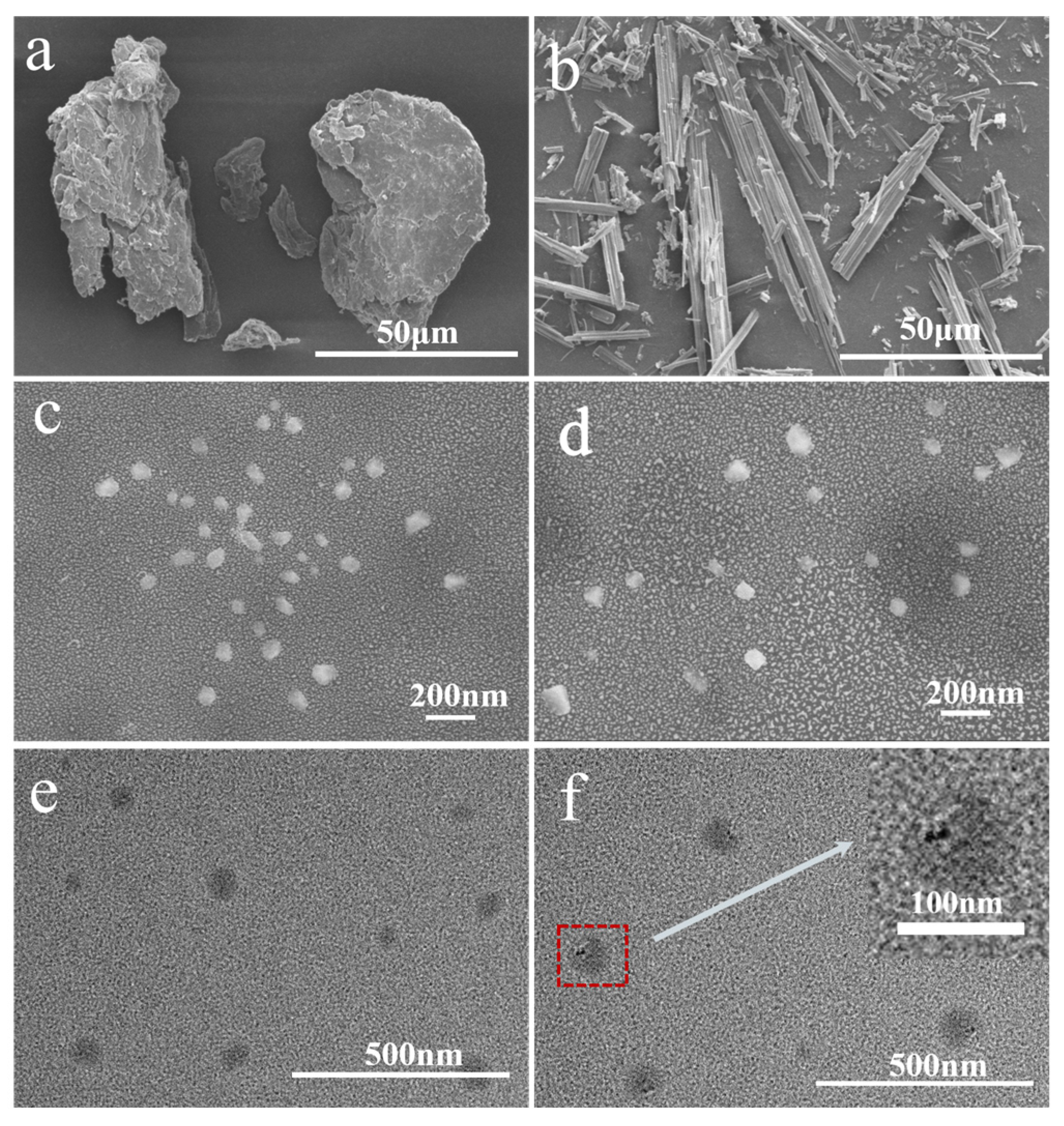
The SEM was used to observe the apparent morphology of the samples, while the TEM was applied to directly observe the PTX loading status. Figure 2 presented the SEM and TEM results of chitin, PTX, ChNps, and ChNps–PTX. SEM images showed that chitin (Figure 2a) exhibited an irregular blocky structure, and PTX (Figure 2b) presented a long crystalline strip morphology. The ChNps (Figure 2c) and ChNps–PTX (Figure 2d) prepared by the water-dripping regeneration method showed relatively uniform nano-quasi-spherical particles and excellent dispersibility. The morphology of ChNps (Figure 2e) and ChNps–PTX (Figure 2f) observed by TEM was consistent with that of SEM. The particle sizes of ChNps, ChNps–PTX, and the loaded PTX were obtained using the particle size measurement tool (Nano Measure), which has been previously adopted in existing studies [35]. The average particle size of ChNps–PTX (approximately 93.11 nm) was larger than that of ChNps (approximately 84.06 nm) due to the loading of PTX. In comparison, the ChNps prepared in this work exhibited a smaller particle size and better dispersibility than the ChNps with a particle size of approximately 150 nm prepared by the ionic gelation method reported in previous studies [17,36]. It has been reported that well-dispersed ChNps with a particle size of 70~80 nm were regenerated via the dropwise addition of methanol [11]. These results indicated that water-dripping and vigorous stirring were efficient approaches to promote the reduction in particle size and achieve uniform dispersion. The loading of PTX into ChNps was directly observed for the first time, and the PTX exhibited nano-strips 30.52 ± 6.78 nm in length and 16.02 ± 2.77 nm in width, as seen in Figure 2f. The loading schematic diagram of ChNps–PTX was shown in Figure 3. During the precipitation process, chitin chains and PTX may bind to each other due to the presence of interactions such as hydrogen bonds and van der Waals forces.

3.3. CLSM Analysis

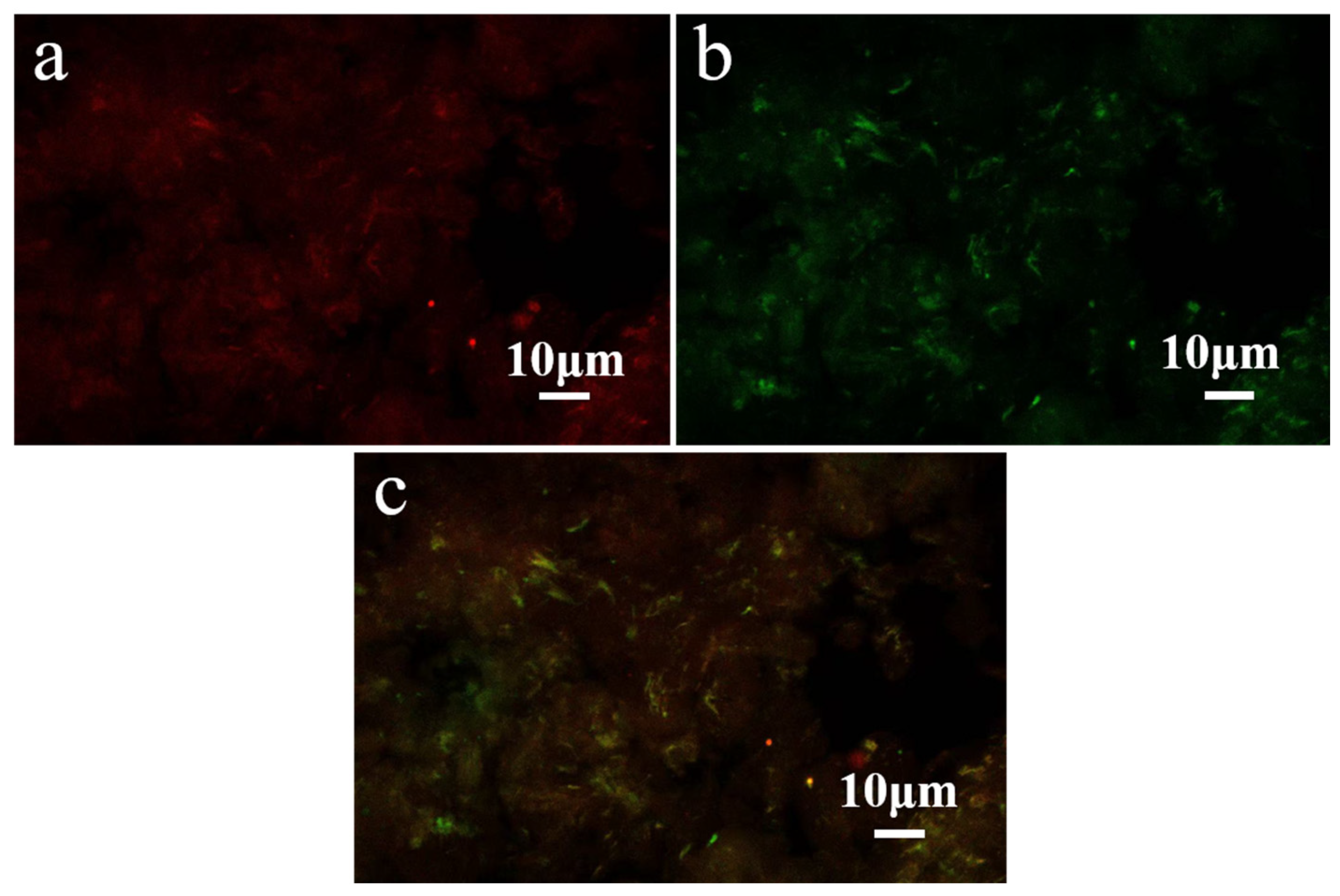
CLSM was used to characterize the loading system of ChNps–PTX, and the results are presented in Figure 4. ChNps–PTX stained with Nile Red exhibited red dots or filaments under excitation at 633 nm, while ChNps–PTX stained with Nile Blue showed green dots or filaments under excitation at 488 nm. As can be seen in Figure 4c, the merged laser channel image of the two signals displayed an orange-yellow color, which directly confirmed the successful loading of PTX in ChNps. This result was consistent with the findings of Xuechen Zhuang et al. [30], who successfully achieved loading PTX into debranched corn starch.

3.4. Structural Characterization

3.4.1. FT-IR Analysis

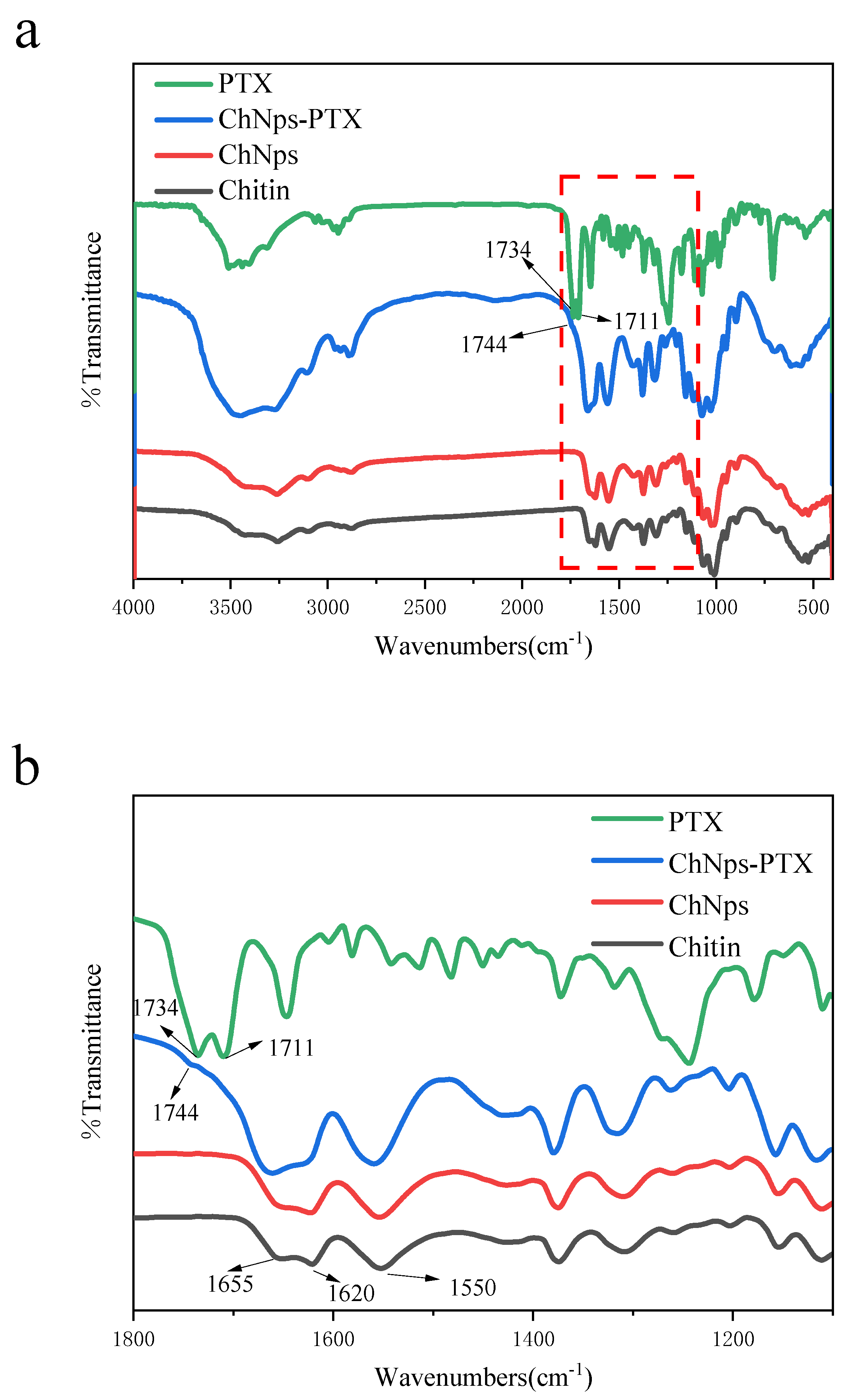
Figure 5 displays the FT-IR spectra of chitin, ChNps, ChNps–PTX, and PTX. The peak bands in the 3430–3100 cm−1 region represented the stretching of -OH and -NH groups, which were characteristic of intermolecular hydrogen bond networks [37]. The vibration regions of the chitin and ChNps curves were basically consistent, indicating that the chemical structure of ChNps prepared by the water-dripping regeneration method was unchanged. Moreover, the amide I band (C=O) observed around 1655 and 1620 cm−1 and the amide II band (N-H) observed at 1550 cm−1 of the ChNps spectrum were similar to that of the raw chitin, proving that the crystal form of α-chitin has not changed after regeneration [38]. This result was consistent with the previous report [39], attributed to the unchanged functional groups of chitin during the regeneration process. For PTX, the peak at 3456 cm−1 was the specific peak of PTX’s hydroxyl groups (O-H), and the peaks at 2850 cm−1 and 2917 cm−1 correspond to the C-H stretching vibration [8]. The peak shown at 1646 cm−1 was N-H stretching vibration, and 1243 cm−1 was C-N stretching vibration. The peak at 709 cm−1 was C-C stretching vibration. The peaks at 1734 and 1711 cm−1 of PTX represented the carbonyl (C=O) stretching vibration [18]. In Figure 5b, ChNps–PTX exhibited a new peak at 1744 cm−1, which indicated the presence of C=O signal of PTX. This result confirmed the successful loading of PTX into ChNps.

3.4.2. XRD Analysis

Figure 6 presented the XRD patterns of chitin, ChNPs, ChNps–PTX, and PTX. For the chitin and ChNps curves, distinct diffraction peaks appeared at 2θ = 9.3°, 12.6°, 19.5°, 22.8°, 23.3°, and 26.5°, representing that their crystalline forms were both α-chitin [40]. This was consistent with the FT-IR results (Section 3.4.1). Compared with chitin, the diffraction peaks of ChNPs and ChNps–PTX located at 2θ = 20.9° and 23.4° disappeared, and the degree of crystallinity of chitin, ChNps, and ChNps–PTX was 36.77%, 24.11%, and 19.86%, respectively. These results indicated a reduction in crystallinity of ChNPs and ChNps–PTX, which was consistent with previous papers related to the regenerated chitin [38,41]. The decreased crystallinity of ChNps was attributed to the rapid arrangement of chitin during the regeneration process, which led to the inadequate development of crystallization and chitin chains [42]. Furthermore, ChNps–PTX exhibited even lower crystallinity, which may be related to the disruption of the original crystalline structure by the interaction between chitin and PTX. PTX exhibited obvious characteristic diffraction peaks at multiple 2θ values, including 5.56°, 8.8°, and 12.4°. Compared with chitin and ChNps, ChNps–PTX showed a new diffraction peak at 2θ = 5.3°, further proving the successful loading of PTX into ChNps.

3.4.3. DSC Analysis

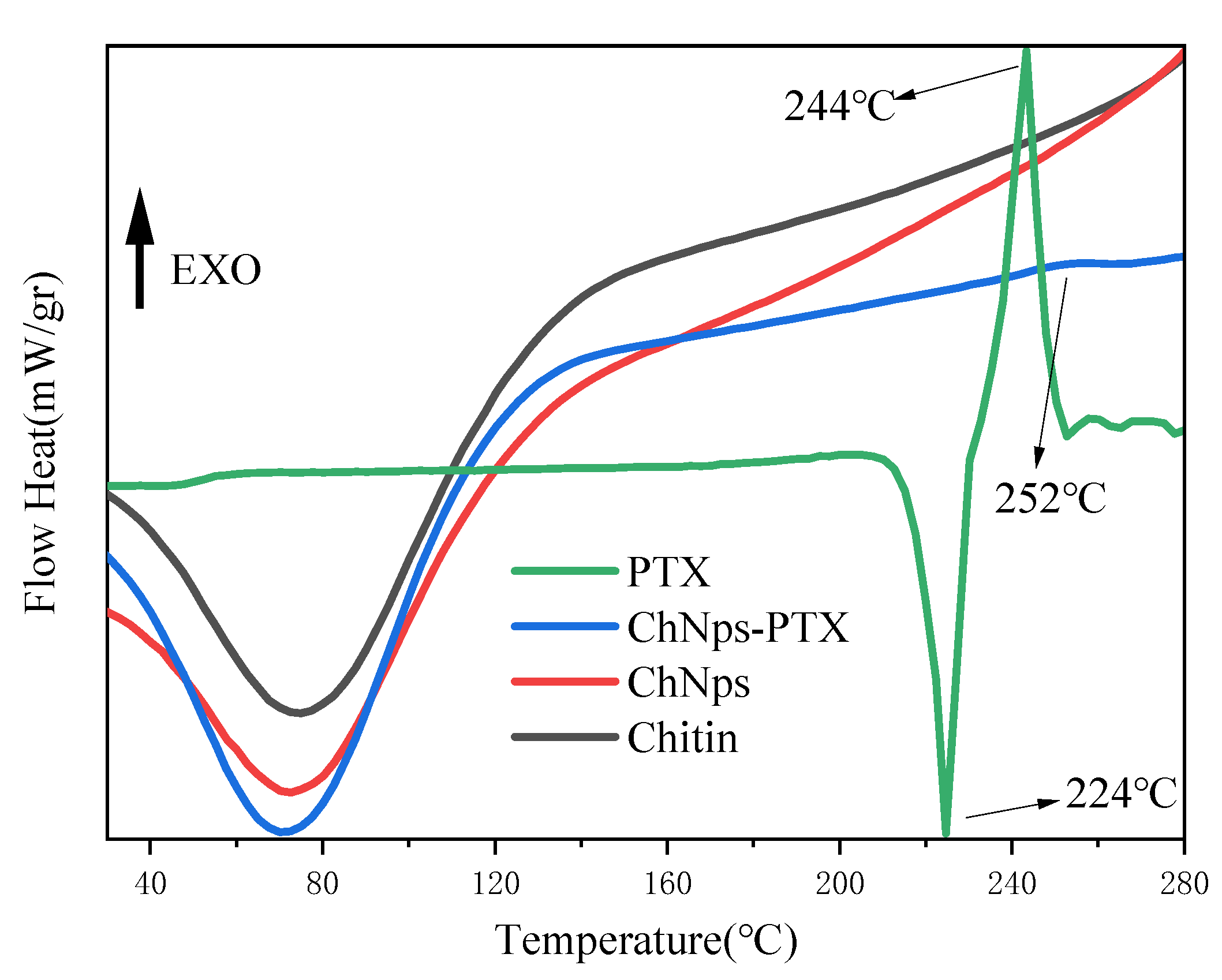
Chitin possessed good thermal stability due to its rigid hydrogen bond structure and acetyl groups. Figure 7 displayed the DSC thermograms of chitin, ChNps, ChNps–PTX, and PTX. Since polysaccharides tend to bind with water [43], the first endothermic peaks of chitin, ChNps, and ChNps–PTX appearing below 100 °C were attributed to the evaporation of adsorbed water [44], which was in agreement with the conclusions reported in a previous paper [45]. As the temperature continued to rise, thermal decomposition and release of volatile products occurred. For PTX, the endothermic peak at 224 °C and exothermic peak at 244 °C corresponded to its melting and decomposition processes, respectively [46]. A new exothermic peak of ChNps–PTX emerging at 252 °C compared with chitin and ChNps may be caused by the interaction between chitin and PTX, which also confirmed the successful loading of PTX into ChNps.

4. Conclusions

In this research, PTX and chitin were dissolved in the same solvent to form a co-solution, and the water-dripping regeneration method was used to prepare ChNps and ChNps–PTX with a relatively uniform particle size of approximately 84.06 and 93.11 nm, respectively. This study realized the loading of PTX into ChNps in the form of nano-strips (30.52 ± 6.78 nm in length and 16.02 ± 2.77 nm in width) for the first time, and the DL was approximately 8.01%. A series of characterizations demonstrated that the functional groups, DA, and crystal form of chitin were unchanged after regeneration, which was different from previous papers. 1H NMR spectra demonstrated that chitin and ChNps both showed high DA of 96.03%, indicating that the ChNps retained the acetyl groups. TEM and CLSM directly confirmed that PTX was loaded into ChNps rather than existing in a free state. The results of FT-IR and XRD displayed that the water-dripping regeneration method did not damage the molecular structure of chitin, and ChNps still had α-chitin structure. For ChNps–PTX, the new peak of 1734 cm−1 in the FT-IR spectrum, 2θ = 5.3° in the XRD pattern, and a new exothermic peak of 252 °C in the DSC curve confirmed the successful loading of PTX into ChNps. Thus, ChNps ensure stable PTX loading through simple methods, which will provide a feasible solution for the improvement of bioavailability of PTX. It is also beneficial for the clinical application of PTX. We believe that the ChNps and ChNps–PTX prepared in this work will provide a reference for the research of chitin carriers and broaden the applications of chitin in the food and pharmaceutical fields. The water-dripping regeneration method can further expand the research and development of composite carriers for loading fat-soluble drugs and construct a multifunctional drug delivery system.

Author Contributions

Y.L.: investigation (Lead); methodology (Equal); writing—original draft (Lead). D.Q. and X.Z.: writing—review and editing (Lead). Z.P. and T.L.: validation (Lead). S.D.: supervision (Lead). All authors have read and agreed to the published version of the manuscript.

Funding

This research was supported by Zhejiang Province’s Special Support Program (2023R5232), Zhejiang Provincial Natural Science Foundation of China under Grant No. LMS25B060006, Key R & D Program Project of Ningbo City (2025Z112), Specialty Fund of Zhejiang Institute Tianjin University (grant number ZITJU2024-ZYHT004), and Chinese College Students’ Innovation and Entrepreneurship Training Program (202513022052).

Institutional Review Board Statement

Not applicable.

Data Availability Statement

The original contributions presented in this study are included in the article. Further inquiries can be directed to the corresponding authors.

Acknowledgments

The FTIR and 1H-NMR were supported by the Analysis and Testing Center of Sci-Compass. DSC and TEM were supported by the Analysis and Testing Center of Yong Jiang Laboratory. XRD, SEM, and HPLC were supported by the Analysis and Testing Center of Ningbo University of Technology.

Conflicts of Interest

The authors declare no conflicts of interest.

Abbreviations

The following abbreviations are used in this manuscript:
ChNpsChitin nanoparticles
PTXPaclitaxel
ChNps–PTXChitin nanoparticles–paclitaxel
DAdegree of acetylation
DLdrug loading
FT-IRFourier Transform Infrared
DSCDifferential Scanning Calorimetry
XRDX-ray Diffraction
CLSMConfocal Laser Scanning Microscopy
TEMTransmission Electron Microscopy
SEMScanning Electron Microscopy
1H-NMRProton Nuclear Magnetic Resonance Spectroscopy
HPLChigh-performance liquid chromatography

References

  1. Olugbojo, J.A.; Akinyemi, A.A.; Obasa, S.O.; Dare, E.O. Extraction and Utilization of Chitin and Chitosan from Waste Yields of Economically Important Crustaceans and Molluscs for Improved Fish Production. Asian J. Fish. Aquat. Res. 2024, 26, 95–114. [Google Scholar] [CrossRef]
  2. Ngasotter, S.; Xavier, K.M.; Meitei, M.M.; Waikhom, D.; Pathak, J.; Singh, S.K. Crustacean shell waste derived chitin and chitin nanomaterials for application in agriculture, food, and health–A review. Carbohydr. Polym. Technol. Appl. 2023, 6, 100349. [Google Scholar] [CrossRef]
  3. Joseph, B.; Sam, R.M.; Balakrishnan, P.; Maria, H.J.; Gopi, S.; Volova, T.; Fernandes, S.C.M.; Thomas, S. Extraction of Nanochitin from Marine Resources and Fabrication of Polymer Nanocomposites: Recent Advances. Polymers 2020, 12, 1664. [Google Scholar] [CrossRef]
  4. Dutta, A.K.; Yamada, K.; Izawa, H.; Morimoto, M.; Saimoto, H.; Ifuku, S. Preparation of Chitin Nanofibers from Dry Chitin Powder by Star Burst System: Dependence on Number of Passes. J. Chitin Chitosan Sci. 2013, 1, 59–64. [Google Scholar] [CrossRef]
  5. Ben Cheikh, F.; Ben Mabrouk, A.; Magnin, A.; Putaux, J.L.; Boufi, S. Chitin nanocrystals as Pickering stabilizer for O/W emulsions: Effect of the oil chemical structure on the emulsion properties. Colloids Surf. B Biointerfaces 2021, 200, 111604. [Google Scholar] [CrossRef]
  6. Zhang, J.; Zhu, W.; Liang, J.; Li, L.; Zheng, L.; Shi, X.; Wang, C.; Dong, Y.; Li, C.; Zhu, X. In Situ Synthesis of Gold Nanoparticles from Chitin Nanogels and Their Drug Release Response to Stimulation. Polymers 2024, 16, 390. [Google Scholar] [CrossRef]
  7. Rigual, V.; Ovejero-Perez, A.; Martinez-Mangas, A.; Garcia-Sánchez, B.; Dominguez, J.; Alonso, M.V.; Oliet, M.; Rodriguez, F. Comparison of bottom-up and top-down precipitation strategies for lignin nanoparticle obtention from organosolv and ionosolv Eucalyptus globulus liquors. Faraday Discuss. 2025. [Google Scholar] [CrossRef]
  8. Zewude, D.A.; Noguchi, T.; Sato, K.; Izawa, H.; Ifuku, S. Production of chitin nanoparticles by bottom-up approach from alkaline chitin solution. Int. J. Biol. Macromol. 2022, 210, 123–127. [Google Scholar] [CrossRef]
  9. Zhang, Y.; Chen, Z.; Bian, W.; Feng, L.; Wu, Z.; Wang, P.; Zeng, X.; Wu, T. Stabilizing oil-in-water emulsions with regenerated chitin nanofibers. Food Chem. 2015, 183, 115–121. [Google Scholar] [CrossRef]
  10. Kadokawa, J.I. A Mini-Review: Fabrication of Polysaccharide Composite Materials Based on Self-Assembled Chitin Nanofibers. Materials 2024, 17, 1898. [Google Scholar] [CrossRef]
  11. Mangalathillam, S.; Rejinold, N.S.; Nair, A.; Lakshmanan, V.K.; Nair, S.V.; Jayakumar, R. Curcumin loaded chitin nanogels for skin cancer treatment via the transdermal route. Nanoscale 2012, 4, 239–250. [Google Scholar] [CrossRef] [PubMed]
  12. Zhong, Y.; Cai, J.; Zhang, L. A Review of Chitin Solvents and Their Dissolution Mechanisms. Chin. J. Polym. Sci. 2020, 38, 1047–1060. [Google Scholar] [CrossRef]
  13. Kätelhön, E.; Sokolov, S.V.; Bartlett, T.R.; Compton, R.G. The Role of Entropy in Nanoparticle Agglomeration. ChemPhysChem 2017, 18, 51–54. [Google Scholar] [CrossRef]
  14. Pyttlik, A.; Kuttich, B.; Kraus, T. Microgravity Removes Reaction Limits from Nonpolar Nanoparticle Agglomeration. Small 2022, 18, 2204621. [Google Scholar] [CrossRef] [PubMed]
  15. Al-Gebory, L.; Mengüç, M.P. The effect of pH on particle agglomeration and optical properties of nanoparticle suspensions. J. Quant. Spectrosc. Radiat. Transf. 2018, 219, 46–60. [Google Scholar] [CrossRef]
  16. Diephuis, W.R.; Molloy, A.L.; Boltz, L.L.; Porter, T.B.; Orozco, A.A.; Duron, R.; Crespo, D.; George, L.J.; Reiffer, A.D.; Escalera, G.; et al. The Effect of Agglomeration on Arsenic Adsorption Using Iron Oxide Nanoparticles. Nanomaterials 2022, 12, 1598. [Google Scholar] [CrossRef]
  17. Dhanasekaran, S.; Rameshthangam, P.; Venkatesan, S.; Singh, S.K.; Vijayan, S.R. In Vitro and In Silico Studies of Chitin and Chitosan Based Nanocarriers for Curcumin and Insulin Delivery. J. Polym. Environ. 2018, 26, 4095–4113. [Google Scholar] [CrossRef]
  18. Kumar, M.; Goswami, P.; Jha, A.; Manjit, M.; Satpute, A.P.; Koch, B.; Mishra, B. Formulation and evaluation of cetuximab functionalized phospholipid modified nanocrystals of paclitaxel for non-small cell lung cancer therapy. Sci. Rep. 2024, 14, 29114. [Google Scholar] [CrossRef]
  19. Yang, F.; Zhang, Q.; Liang, Q.; Wang, S.; Zhao, B.; Wang, Y.; Cai, Y.; Li, G. Bioavailability Enhancement of Paclitaxel via a Novel Oral Drug Delivery System: Paclitaxel-Loaded Glycyrrhizic Acid Micelles. Molecules 2015, 20, 4337–4356. [Google Scholar] [CrossRef]
  20. Wang, Y.; Zhao, Y.; Cui, Y.; Zhao, Q.; Musetti, S.; Kinghorn, K.; Wang, S. Overcoming multiple gastrointestinal barriers by bilayer modified hollow mesoporous silica nanocarriers. Acta Biomater. 2018, 65, 405–416. [Google Scholar] [CrossRef] [PubMed]
  21. Liu, G.; Zhou, Y.; Chen, L. Intestinal Uptake of Barley Protein Based Nanoparticles for β-Carotene Delivery. Acta Pharm. Sin. B 2019, 9, 87–96. [Google Scholar] [CrossRef]
  22. Ravindran, S.; Suthar, J.K.; Rokade, R.; Deshpande, P.; Singh, P.; Pratinidhi, A.; Khambadkhar, R.; Utekar, S. Pharmacokinetics, Metabolism, Distribution and Permeability of Nanomedicine. Curr. Drug Metab. 2018, 19, 327–334. [Google Scholar] [CrossRef]
  23. Zhang, D.; Jiang, F.; Ling, J.; Ouyang, X.; Wang, G. Delivery of curcumin using a zein-xanthan gum nanocomplex: Fabrication, characterization, and in vitro release properties. Colloids Surf. B Biointerfaces 2021, 204, 111827. [Google Scholar] [CrossRef]
  24. Nsereko, S.; Amiji, M. Localized delivery of paclitaxel in solid tumors from biodegradable chitin microparticle formulations. Biomaterials 2002, 23, 2723–2731. [Google Scholar] [CrossRef]
  25. Smitha, K.T.; Anitha, A.; Furuike, T.; Tamura, H.; Nair, S.V.; Jayakumar, R. In vitro evaluation of paclitaxel loaded amorphous chitin nanoparticles for colon cancer drug delivery. Colloids Surf. B Biointerfaces 2013, 104, 245–253. [Google Scholar] [CrossRef]
  26. Lin, H.; Chen, Y.; Wu, Y.; Lin, Y. Glycol chitin/PAA hydrogel composite incorporated bio-functionalized PLGA microspheres intended for sustained release of anticancer drug through intratumoral injection. Biotech Week 2018, 29, 1839–1858. [Google Scholar] [CrossRef]
  27. Tamura, H.; Nagahama, H.; Tokura, S. Preparation of chitin hydrogel under mild conditions. Cellulose 2006, 13, 357–364. [Google Scholar] [CrossRef]
  28. Heux, L.; Brugnerotto, J.; Desbrières, J.; Versal, M.F.; Rinaudo, M. Solid state NMR for determination of degree of acetylation of chitin and chitosan. Biomacromolecules 2000, 1, 746–751. [Google Scholar] [CrossRef]
  29. Hirai, A.; Odani, H.; Nakajima, A. Determination of degree of deacetylation of chitosan by 1H NMR spectroscopy. Polym. Bull. 1991, 26, 87–94. [Google Scholar] [CrossRef]
  30. Zhuang, X.; Pang, M.; Qiu, D.; Chen, Y.; Mao, L.; Zhang, Z.; Xian, S.; Tang, X. Preparation, characterization and evaluation of loading paclitaxel into debranched corn starch. Ind. Crops Prod. 2025, 229, 120969. [Google Scholar] [CrossRef]
  31. Chu, L.; Niu, W.; Wu, S.; Ma, W.; Tang, B.; Zhang, S.; Li, H.; Liu, Z. A two-step approach for size controlled preparation of monodisperse polysaccharide-based nanospheres. Mater. Res. Express 2019, 6, 055013. [Google Scholar] [CrossRef]
  32. Zhuang, X.; Zhang, S.; Li, H. Multiple Ethanolic-Precipitation: An Approach to the Separation of Dextrin Fractions with Narrow Molecular Weight Distributions from Acid-Hydrolyzed Waxy Corn Starch. Starch-Starke 2020, 72, 11–12. [Google Scholar] [CrossRef]
  33. Kasaai, M.R. Determination of the degree of N -acetylation for chitin and chitosan by various NMR spectroscopy techniques: A review. Carbohydr. Polym. 2009, 79, 801–810. [Google Scholar] [CrossRef]
  34. Wu, T.; Wang, G.; Gao, C.; Chen, Z.; Feng, L.; Wang, P.; Zeng, X.; Wu, Z. Phosphoric acid-based preparing of chitin nanofibers and nanospheres. Cellulose 2016, 23, 477–491. [Google Scholar] [CrossRef]
  35. Zuo, R.; Kong, X.; Wang, Y.; Deng, S.G.; Zhuang, X.; Qiu, D. Isolation and characterization of natural nano starch from amaranth starch. Int. J. Biol. Macromol. 2024, 260 Pt 1, 129525. [Google Scholar] [CrossRef]
  36. Gayathri, N.K.; Aparna, V.; Maya, S.; Biswas, R.; Jayakumar, R.; Mohan, C.G. Preparation, characterization, drug release and computational modelling studies of antibiotics loaded amorphous chitin nanoparticles. Carbohydr. Polym. 2017, 177, 67–76. [Google Scholar] [CrossRef] [PubMed]
  37. Falgayrac, A.; Pellerin, V.; Terrol, C.; Fernandes, S.C.M. Turning black soldier fly rearing by-products into valuable materials: Valorisation through chitin and chitin nanocrystals production. Carbohydr. Polym. 2024, 344, 122545. [Google Scholar] [CrossRef]
  38. Li, J.; Hao, Z.; Wang, B.; Feng, X.; Mao, Z.; Sui, X. High-tensile chitin films regenerated from cryogenic aqueous phosphoric acid. Carbohydr. Polym. 2023, 312, 120826. [Google Scholar] [CrossRef]
  39. Tyshkunova, I.V.; Chukhchin, D.G.; Gofman, I.V.; Pavlova, E.N.; Ushakov, V.A.; Vlasova, E.N.; Poshina, D.N.; Skorik, Y.A. Chitin Cryogels Prepared by Regeneration from Phosphoric Acid Solutions. Materials 2021, 14, 5191. [Google Scholar] [CrossRef]
  40. Ding, B.; Zhao, D.; Song, J.; Gao, H.; Xu, D.; Xu, M.; Cao, X.; Zhang, L.; Cai, J. Light weight, mechanically strong and biocompatible α-chitin aerogels from different aqueous alkali hydroxide/urea solutions. Sci. China Chem. 2016, 59, 1405–1414. [Google Scholar] [CrossRef]
  41. Kato, Y.; Kaminaga, J.; Matsuo, R.; Isogai, A. TEMPO-mediated oxidation of chitin, regenerated chitin and N -acetylated chitosan. Carbohydr. Polym. 2004, 58, 421–426. [Google Scholar] [CrossRef]
  42. Yu, P.; He, H.; Luo, Y.; Jia, D.; Dufresne, A. Elastomer Reinforced with Regenerated Chitin from Alkaline/Urea Aqueous System. ACS Appl. Mater. Interfaces 2017, 9, 26460–26467. [Google Scholar] [CrossRef] [PubMed]
  43. Ngasotter, S.; Xavier, K.A.M.; Porayil, L.; Balange, A.; Nayak, B.B.; Eapen, S.; Adarsh, K.J.; Sreekala, M.S.; Sharma, R.; Ninan, G. Optimized high-yield synthesis of chitin nanocrystals from shrimp shell chitin by steam explosion. Carbohydr. Polym. 2023, 16, 121040. [Google Scholar] [CrossRef] [PubMed]
  44. Ilyas, H.N.; Zia, K.M.; Rehman, S.; Ilyas, R.; Sultana, S. Utilization of Shellfish Industrial Waste for Isolation, Purification, and Characterizations of Chitin from Crustacean’s Sources in Pakistan. J. Polym. Environ. 2021, 29, 2337–2348. [Google Scholar] [CrossRef]
  45. Farokhi, N.M.; Milani, J.M.; Amiri, Z.R. Production and comparison of structural, thermal and physical characteristics of chitin nanoparticles obtained by different methods. Sci. Rep. 2024, 14, 14594. [Google Scholar] [CrossRef]
  46. Zhang, C.; Ping, Q.; Zhang, H. Self-assembly and characterization of paclitaxel-loaded N-octyl-O-sulfate chitosan micellar system. Colloids Surf. B Biointerfaces 2004, 39, 69–75. [Google Scholar] [CrossRef]
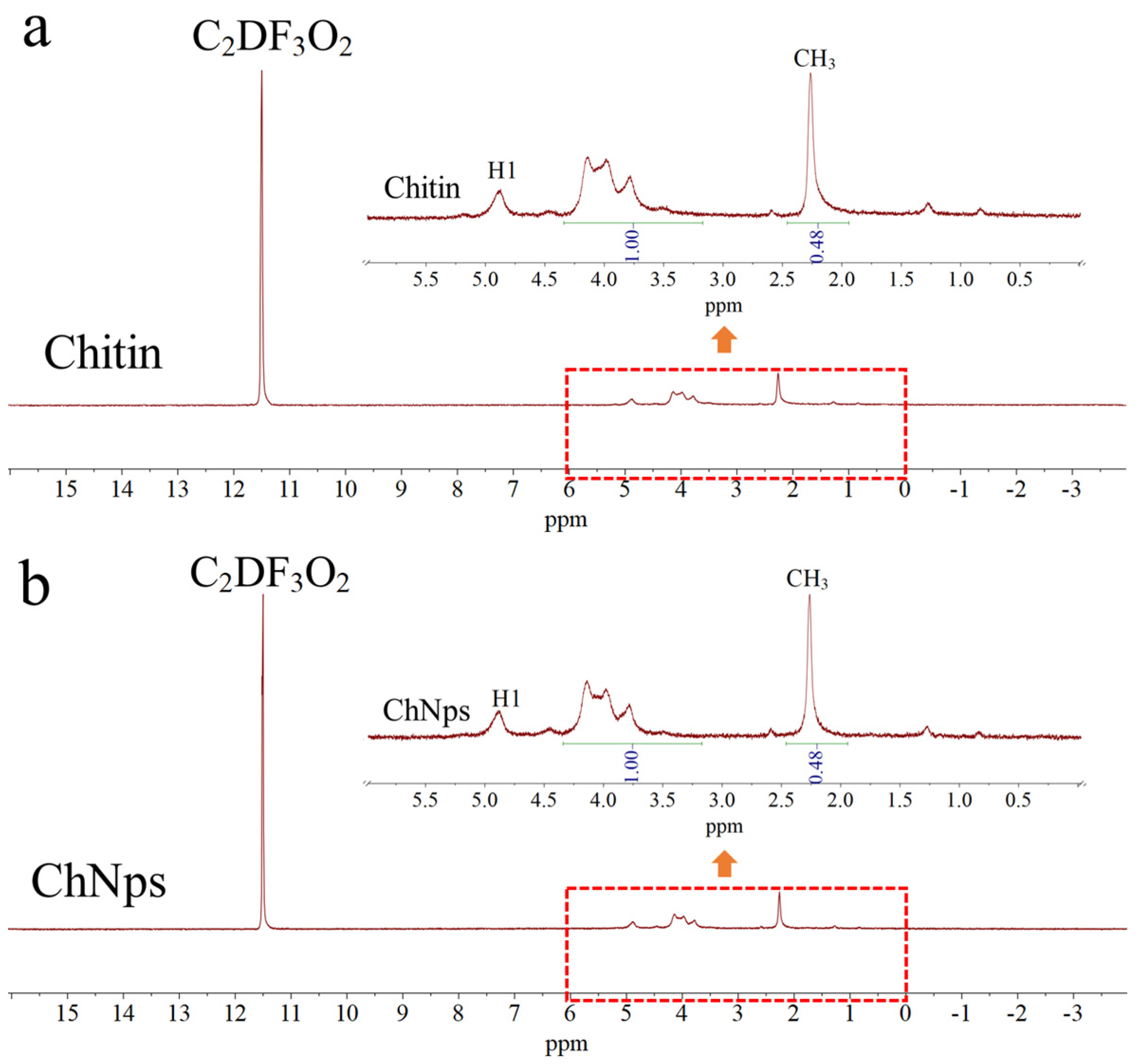
Figure 1. 1H-NMR spectra and of chitin (a) and ChNps (b).
Polymers 17 03036 g001
Figure 2. SEM images of chitin (a), PTX (b), ChNps (c), and ChNps–PTX (d); TEM images of ChNps (e) and ChNps–PTX (f).
Polymers 17 03036 g002
Figure 3. The loading schematic diagram of ChNps–PTX.
Polymers 17 03036 g003
Figure 4. CLSM images of ChNps–PTX under irradiation with 633 nm (a) and 488 nm (b) wavelengths, and superimposed laser channels (c).
Polymers 17 03036 g004
Figure 5. FT-IR spectra (a) and partially enlarged FT-IR spectra from 1800 to 1100 cm−1 (b) of chitn, ChNps, ChNps–PTX, and PTX.
Polymers 17 03036 g005
Figure 6. XRD patterns of chitin, ChNps, ChNps–PTX, and PTX.
Polymers 17 03036 g006
Figure 7. DSC thermograms of chitin, ChNps, ChNps–PTX, and PTX.
Polymers 17 03036 g007
Table 1. Integral areas and DA of chitin and ChNps.
SampleIntegral Area of -CH3 (1.94–2.64 ppm)Integral Area of H2–H6 (3.17–4.34 ppm)DA, %
Chitin0.481.0096.03
ChNps0.481.0096.03
Disclaimer/Publisher’s Note: The statements, opinions and data contained in all publications are solely those of the individual author(s) and contributor(s) and not of MDPI and/or the editor(s). MDPI and/or the editor(s) disclaim responsibility for any injury to people or property resulting from any ideas, methods, instructions or products referred to in the content.