
Effect of Silver/Reduced Graphene Oxide@Titanium Dioxide (Ag/rGO@TiO2) Nanocomposites on the Mechanical Characteristics and Biocompatibility of Poly(Styrene-co-Methyl Methacrylate)-Based Bone Cement

Abstract
1. Introduction
2. Experimental
2.1. Chemicals
2.2. Preparation of Ag/rGO@TiO2
2.3. Preparation of (Ag/rGO@TiO2)/(PS-PMMA) Nanocomposite
2.4. Ball-Milling of (Ag/rGO@TiO2)/(PS-PMMA) Nanocomposite
2.5. Preparation of the (Ag/rGO@TiO2)/(PS-PMMA)/PMMA Bone Cement
2.6. Characterizations
2.6.1. Fourier Transform-Infrared Spectroscopy (FT-IR)
2.6.2. X-Ray Diffraction (XRD)
2.6.3. Nanoindentation Tests
2.6.4. High-Resolution Transmission Electron Microscopy (HR-TEM)
2.7. In Vitro Studies
2.7.1. Cell-Culturing
2.7.2. Passaging
2.7.3. Alamar Blue Assay
3. Results and Discussion
3.1. Silver/Reduced Graphene Oxide@Titanium Dioxide/(Poly(Styrene-co-Methyl Methacrylate)/Polymethyl Methacrylate) ((Ag/rGO@TiO2)/(PS-PMMA)/PMMA) Nanocomposite Bone Cement
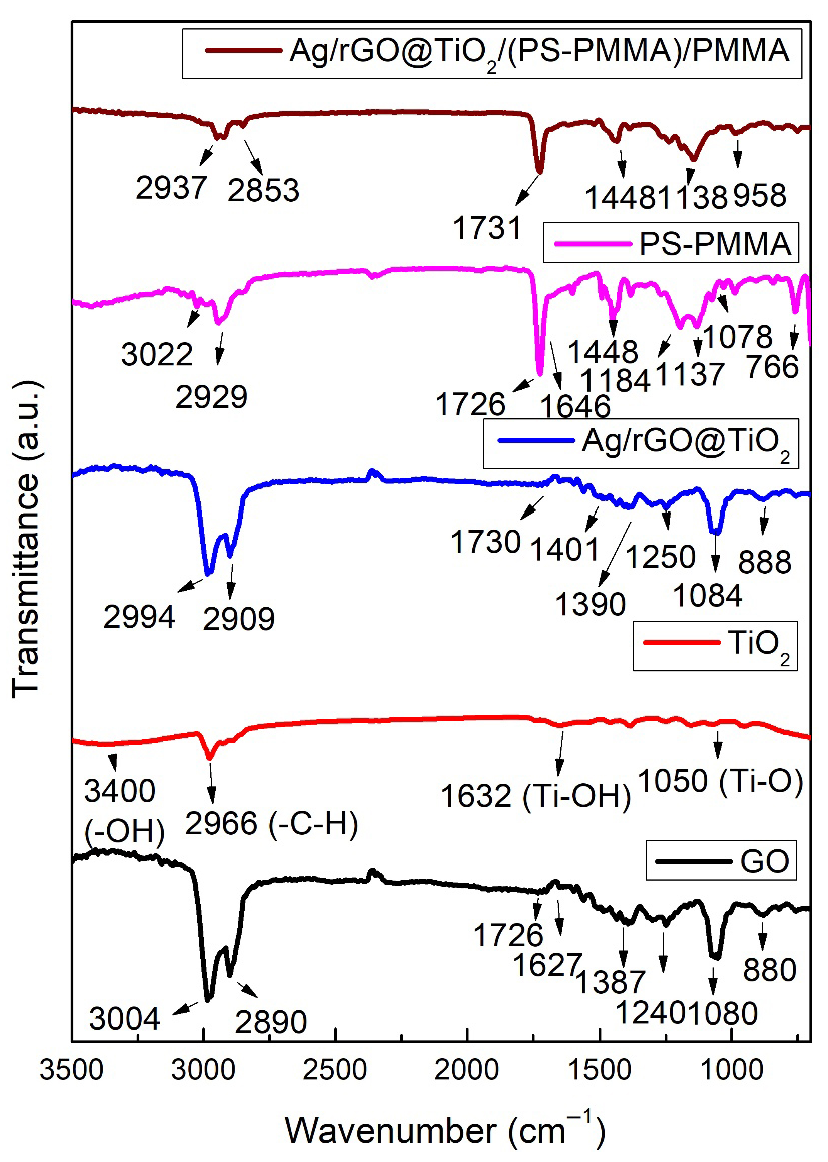
3.2. FT-IR
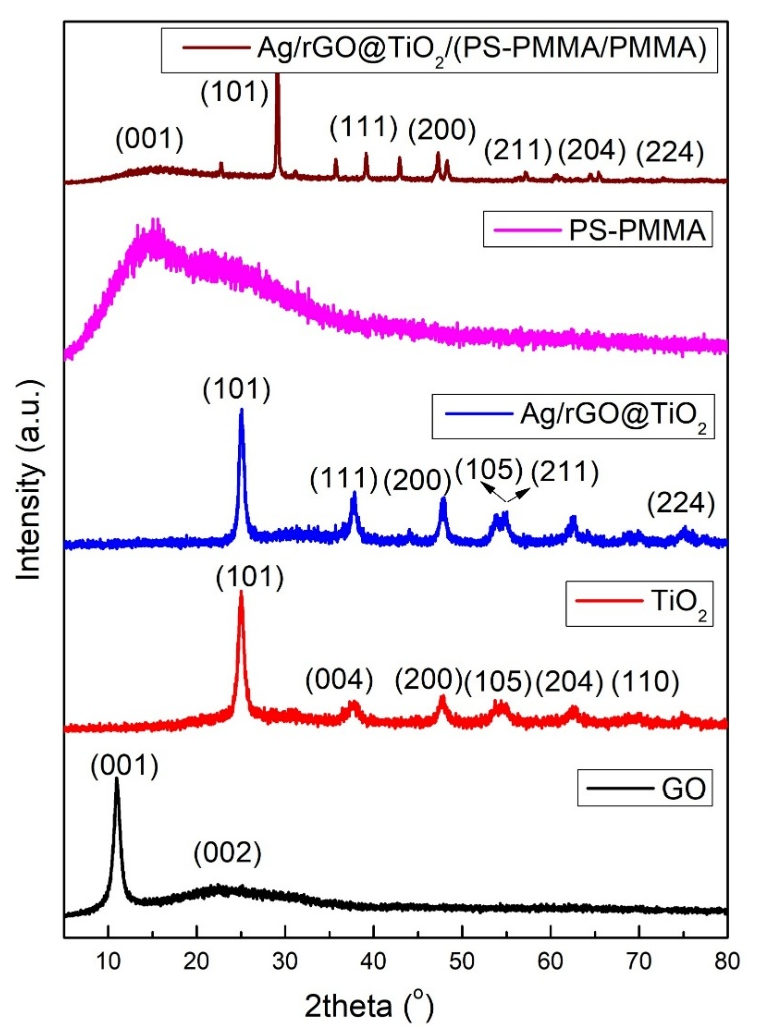
3.3. XRD
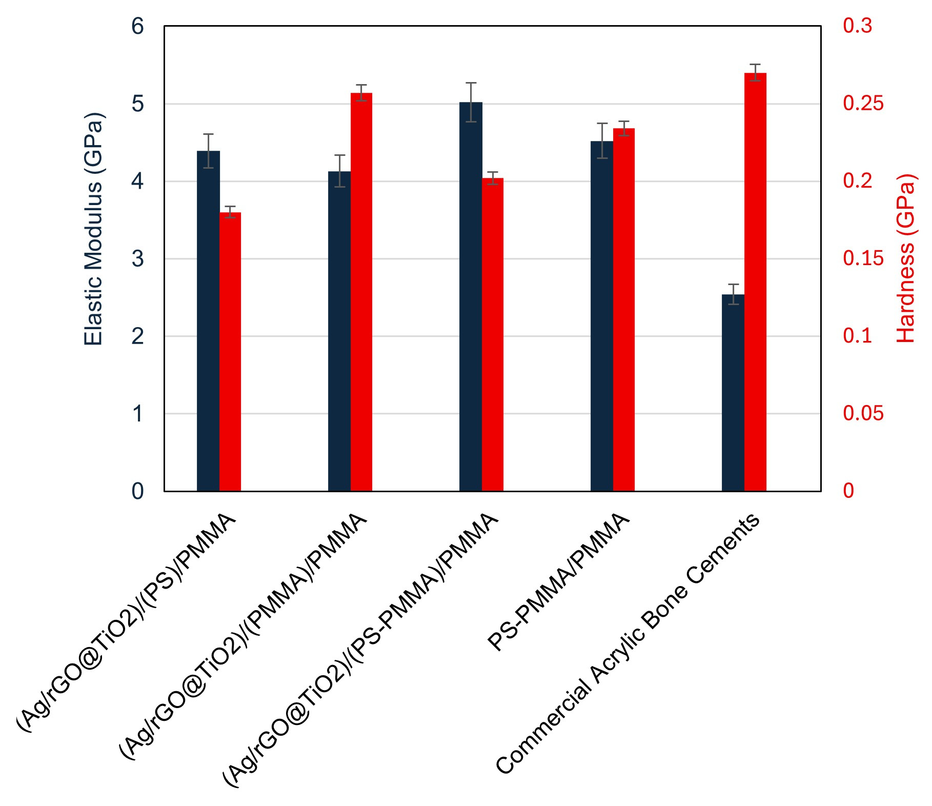
3.4. Nanomechanical Properties
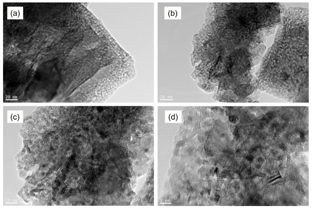
3.5. HR-TEM
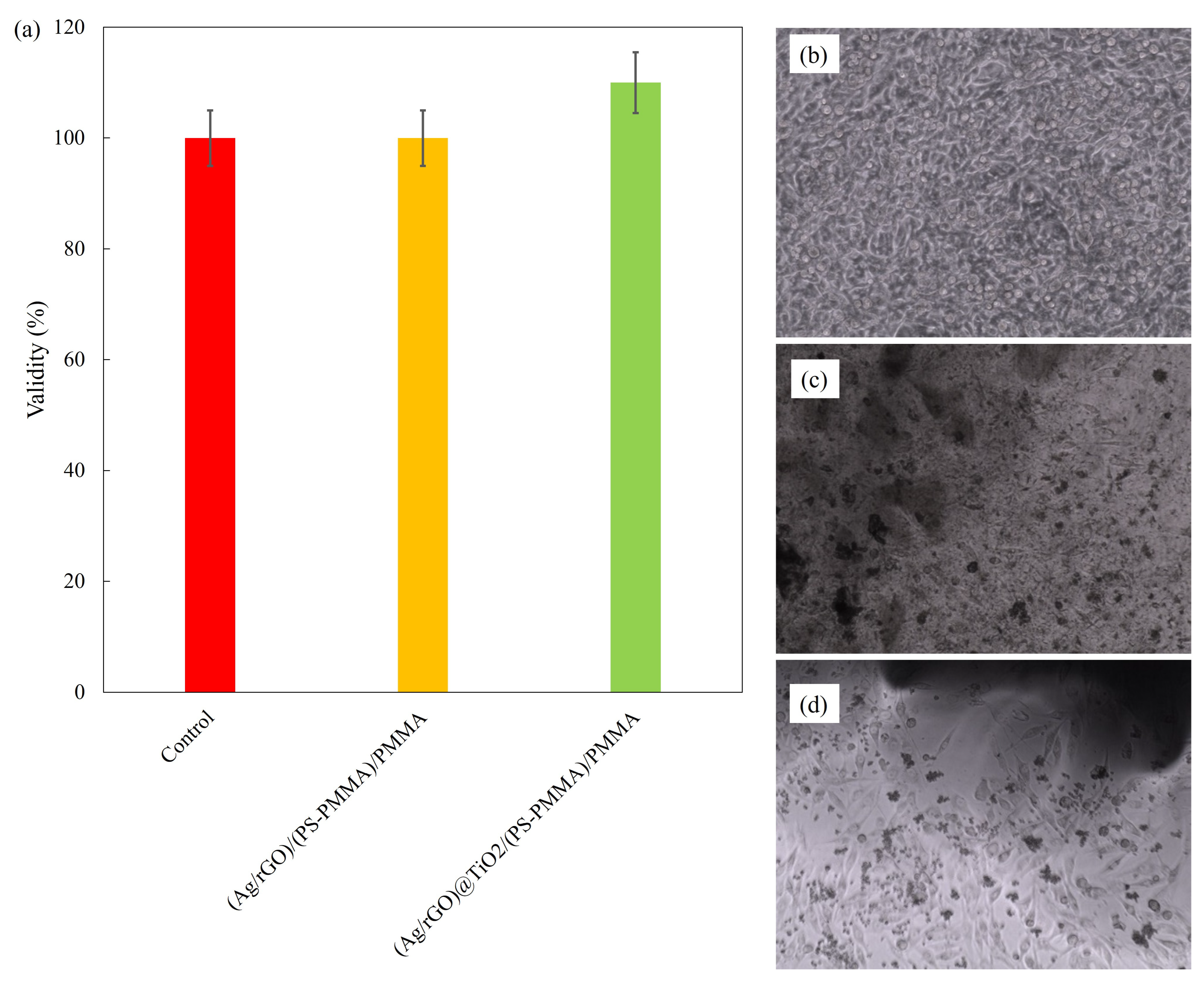
3.6. In Vitro Studies
4. Conclusions
Author Contributions
Funding
Institutional Review Board Statement
Data Availability Statement
Acknowledgments
Conflicts of Interest
References
- Currey, J. 1—The Structure and Mechanical Properties of Bone. In Bioceramics and Their Clinical Applications; Kokubo, T., Ed.; Woodhead Publishing: Sawston, UK, 2008; pp. 3–27. ISBN 978-1-84569-204-9. [Google Scholar]
- Rho, J.-Y.; Kuhn-Spearing, L.; Zioupos, P. Mechanical Properties and the Hierarchical Structure of Bone. Med. Eng. Phys. 1998, 20, 92–102. [Google Scholar] [CrossRef] [PubMed]
- Fyhrie, D.P. The Mechanical Properties of Bone. In Osteoporosis in Men; Elsevier: Amsterdam, The Netherlands, 2010; pp. 51–67. [Google Scholar]
- Currey, J.D. The Structure and Mechanics of Bone. J. Mater. Sci. 2012, 47, 41–54. [Google Scholar] [CrossRef]
- Currey, J. Measurement of the Mechanical Properties of Bone: A Recent History. Clin. Orthop. Relat. Res. 2009, 467, 1948–1954. [Google Scholar] [CrossRef] [PubMed]
- Cowin, S.C. The Mechanical and Stress Adaptive Properties of Bone. Ann. Biomed. Eng. 1983, 11, 263–295. [Google Scholar] [CrossRef] [PubMed]
- Robey, P.G.; Boskey, A.L. The Composition of Bone. Primer Metab. Bone Dis. Disord. Miner. Metab. 2008, 7, 32–38. [Google Scholar]
- Huang, J.-F.; Wu, Q.-N.; Zheng, X.-Q.; Sun, X.-L.; Wu, C.-Y.; Wang, X.-B.; Wu, C.-W.; Wang, B.; Wang, X.-Y.; Bergman, M. The Characteristics and Mortality of Osteoporosis, Osteomyelitis, or Rheumatoid Arthritis in the Diabetes Population: A Retrospective Study. Int. J. Endocrinol. 2020, 2020, 8821978. [Google Scholar] [CrossRef] [PubMed]
- Lewis, G. Properties of Acrylic Bone Cement: State of the Art Review. J. Biomed. Mater. Res. 1997, 38, 155–182. [Google Scholar] [CrossRef]
- Dunne, N.; Orr, J. Curing Characteristics of Acrylic Bone Cement. J. Mater. Sci. Mater. Med. 2002, 13, 17–22. [Google Scholar] [CrossRef] [PubMed]
- Saleh, K.J.; El Othmani, M.M.; Tzeng, T.H.; Mihalko, W.M.; Chambers, M.C.; Grupp, T.M. Acrylic Bone Cement in Total Joint Arthroplasty: A Review. J. Orthop. Res. 2016, 34, 737–744. [Google Scholar] [CrossRef] [PubMed]
- Dunne, N.; Orr, J. Influence of Mixing Techniques on the Physical Properties of Acrylic Bone Cement. Biomaterials 2001, 22, 1819–1826. [Google Scholar] [CrossRef] [PubMed]
- Lewis, G. Alternative Acrylic Bone Cement Formulations for Cemented Arthroplasties: Present Status, Key Issues, and Future Prospects. J. Biomed. Mater. Res. Part B Appl. Biomater. 2008, 84, 301–319. [Google Scholar] [CrossRef] [PubMed]
- Anagnostakos, K.; Becker, S.L.; Sahan, I. Antifungal-Loaded Acrylic Bone Cement in the Treatment of Periprosthetic Hip and Knee Joint Infections: A Review. Antibiotics 2022, 11, 879. [Google Scholar] [CrossRef] [PubMed]
- Williams, T.D.; Adler, T.; Smokoff, L.; Kaur, A.; Rodriguez, B.; Prakash, K.J.; Redzematovic, E.; Baker, T.S.; Rapoport, B.I.; Yoon, E.S. Bone Cements Used in Vertebral Augmentation: A State-of-the-Art Narrative Review. J. Pain Res. 2024, 1029–1040. [Google Scholar] [CrossRef] [PubMed]
- Taghizadeh, E.; Navaei-Nigjeh, M.; Mirkazemi, M.; Rad-Malekshahi, M. Comparison of Physico-Chemical Properties of Different Types of Orthopedic Acrylic Cement. J. Biomater. Sci. Polym. Ed. 2025, 36, 1343–1363. [Google Scholar] [CrossRef] [PubMed]
- Lin, H.; Gao, Z.; Shan, T.; Asilebieke, A.; Guo, R.; Kan, Y.; Li, C.; Xu, Y.; Chu, J. A Review on the Promising Antibacterial Agents in Bone Cement–From Past to Current Insights. J. Orthop. Surg. Res. 2024, 19, 673. [Google Scholar] [CrossRef] [PubMed]
- Venkatesan, K.; Karthik, K.S.; Mathew, A.M.; Sreya, P.; Mallick, S.P.; Pattanayak, D.K. Facile Synthesis of Silver Loaded Bioactive Glass Ceramic and Reinforced Composite Scaffold Using Acrylic Polymer for Bone Tissue Engineering Applications. Adv. Powder Technol. 2025, 36, 104892. [Google Scholar] [CrossRef]
- Mackert, J.; El-Shewy, M.; Pannu, D.; Schoenbaum, T. Prosthetic Complications and Survival Rates of Metal-Acrylic Implant Fixed Complete Dental Prostheses: A Retrospective Study up to 10 Years. J. Prosthet. Dent. 2024, 132, 766–771. [Google Scholar] [CrossRef] [PubMed]
- Quezada, M.M.; Fernandes, C.; Montero, J.; Correia, A.; Salgado, H.; Fonseca, P. A Different Approach to Analyzing the Surface Roughness of Prosthetic Dental Acrylic Resins. Appl. Sci. 2024, 14, 619. [Google Scholar] [CrossRef]
- Raszewski, Z.; Nowakowska-Toporowska, A.; Nowakowska, D.; Więckiewicz, W. Update on Acrylic Resins Used in Dentistry. Mini Rev. Med. Chem. 2021, 21, 2130–2137. [Google Scholar] [CrossRef] [PubMed]
- Angelara, K.; Bratos, M.; Sorensen, J.A. Comparison of Strength of Milled and Conventionally Processed PMMA Complete-Arch Implant-Supported Immediate Interim Fixed Dental Prostheses. J. Prosthet. Dent. 2023, 129, 221–227. [Google Scholar] [CrossRef] [PubMed]
- Souza, L.F.B.; Pires, T.S.; Kist, P.P.; Valandro, L.F.; Moraes, R.R.; Özcan, M.; Pereira, G.K.R. 3D Printed, Subtractive, and Conventional Acrylic Resins: Evaluation of Monotonic versus Fatigue Behavior and Surface Characteristics. J. Mech. Behav. Biomed. Mater. 2024, 155, 106556. [Google Scholar] [CrossRef] [PubMed]
- Mounika, C.; Tadge, T.; Keerthana, M.; Velyutham, R.; Kapusetti, G. Advancements in Poly (Methyl Methacrylate) Bone Cement for Enhanced Osteoconductivity and Mechanical Properties in Vertebroplasty: A Comprehensive Review. Med. Eng. Phys. 2023, 120, 104049. [Google Scholar] [CrossRef] [PubMed]
- Edo, G.I.; Ndudi, W.; Ali, A.; Yousif, E.; Zainulabdeen, K.; Onyibe, P.N.; Akpoghelie, P.O.; Ekokotu, H.A.; Isoje, E.F.; Igbuku, U.A. An Updated Review on the Modifications, Recycling, Polymerization, and Applications of Polymethyl Methacrylate (PMMA). J. Mater. Sci. 2024, 59, 20496–20539. [Google Scholar] [CrossRef]
- Arora, M.; Chan, E.K.; Gupta, S.; Diwan, A.D. Polymethylmethacrylate Bone Cements and Additives: A Review of the Literature. World J. Orthop. 2013, 4, 67. [Google Scholar] [CrossRef] [PubMed]
- Awadhesh, A.P.; Lingala, H. Bone Cement. In Bioimplants Manufacturing: Fundamentals and Advances; CRC Press: Boca Raton, FL, USA, 2024; p. 48. [Google Scholar]
- Seesala, V.S.; Sheikh, L.; Basu, B.; Mukherjee, S. Mechanical and Bioactive Properties of PMMA Bone Cement: A Review. ACS Biomater. Sci. Eng. 2024, 10, 5939–5959. [Google Scholar] [CrossRef] [PubMed]
- Elbakyan, L.; Zaporotskova, I. Composite Nanomaterials Based on Polymethylmethacrylate Doped with Carbon Nanotubes and Nanoparticles: A Review. Polymers 2024, 16, 1242. [Google Scholar] [CrossRef] [PubMed]
- Ayora-Gutiérrez, G.; Canché-Escamilla, G.; Cervantes-Uc, J.M.; Uribe-Calderon, J.A. PMMA-Bead Decorated Graphene Oxide, a Novel Strategy to Improve GO Dispersion in Bone Cements. Int. J. Polym. Mater. Polym. Biomater. 2025, 1–14. [Google Scholar] [CrossRef]
- Xing, J.; Liu, S. Application of Loaded Graphene Oxide Biomaterials in the Repair and Treatment of Bone Defects: A Review. Bone Jt. Res. 2024, 13, 725. [Google Scholar] [CrossRef] [PubMed]
- Ayora-Gutiérrez, G.; Abreu-Rejón, A.D.; May-Pat, A.; Guerrero-Bermea, C.; Fernández-Escamilla, V.V.; Rodríguez-Fuentes, N.; Cervantes-Uc, J.M.; Uribe-Calderon, J.A. Effect of Surface Modification of Graphene Oxide with a Reactive Silane Coupling Agent on the Mechanical Properties and Biocompatibility of Acrylic Bone Cements. J. Biomater. Sci. Polym. Ed. 2024, 35, 345–363. [Google Scholar] [CrossRef] [PubMed]
- Krishnan, M.R.; Alsharaeh, E.H. Methodological Impact on Curing Kinetics of Bone Cement Based on Poly (Styrene-co-Methyl Methacrylate)–2D Nanofiller Nanocomposites. Polymers 2025, 17, 116. [Google Scholar] [CrossRef] [PubMed]
- Tavakoli, M.; Najafinezhad, A.; Mirhaj, M.; Karbasi, S.; Varshosaz, J.; Al-Musawi, M.H.; Madaninasab, P.; Sharifianjazi, F.; Mehrjoo, M.; Salehi, S. Graphene Oxide-Encapsulated Baghdadite Nanocomposite Improved Physical, Mechanical, and Biological Properties of a Vancomycin-Loaded PMMA Bone Cement. J. Biomater. Sci. Polym. Ed. 2024, 35, 823–850. [Google Scholar] [CrossRef] [PubMed]
- Krishnan, M.R.; Alsoughayer, S.; Michael, F.M.; Alsharaeh, E.H. Poly(Styrene-co-Methyl Methacrylate)-Silver/Reduced Graphene Oxide-Nano Hydroxyapatite Nanocomposites for Bone Cement Applications. Int. J. Polym. Mater. Polym. Biomater. 2025, 74, 657–668. [Google Scholar] [CrossRef]
- Gamal, S.; Mikhail, M.; Salem, N.; El-Wakad, M.T.; Abdelbaset, R. Effect of Using Nano-Particles of Magnesium Oxide and Titanium Dioxide to Enhance Physical and Mechanical Properties of Hip Joint Bone Cement. Sci. Rep. 2024, 14, 2838. [Google Scholar] [CrossRef] [PubMed]
- Gamal, S.; Mikhail, M.; Salem, N.; EL-Wakad, M.T.; Abdelbaset, R. Enhanced Bone Cement for Fixation of Prosthetic Joint Utilizing Nanoparticles. J. Mater. Sci. Mater. Med. 2025, 36, 10. [Google Scholar] [CrossRef] [PubMed]
- Serov, D.A.; Gritsaeva, A.V.; Yanbaev, F.M.; Simakin, A.V.; Gudkov, S.V. Review of Antimicrobial Properties of Titanium Dioxide Nanoparticles. Int. J. Mol. Sci. 2024, 25, 10519. [Google Scholar] [CrossRef] [PubMed]
- Younis, A.B.; Haddad, Y.; Kosaristanova, L.; Smerkova, K. Titanium Dioxide Nanoparticles: Recent Progress in Antimicrobial Applications. Wiley Interdiscip. Rev. Nanomed. Nanobiotechnol. 2023, 15, e1860. [Google Scholar] [CrossRef] [PubMed]
- Sharma, P.; Kumari, R.; Yadav, M.; Lal, R. Evaluation of TiO2 Nanoparticles Physicochemical Parameters Associated with Their Antimicrobial Applications. Indian J. Microbiol. 2022, 62, 338–350. [Google Scholar] [CrossRef] [PubMed]
- Ikram, M.; Javed, B.; Hassan, S.W.U.; Satti, S.H.; Sarwer, A.; Raja, N.I.; Mashwani, Z.U.R. Therapeutic Potential of Biogenic Titanium Dioxide Nanoparticles: A Review on Mechanistic Approaches. Nanomedicine 2021, 16, 1429–1446. [Google Scholar] [CrossRef] [PubMed]
- Kawassaki, R.K.; Romano, M.; Dietrich, N.; Araki, K. Titanium and Iron Oxide Nanoparticles for Cancer Therapy: Surface Chemistry and Biological Implications. Front. Nanotechnol. 2021, 3, 735434. [Google Scholar] [CrossRef]
- Kumaravel, V.; Nair, K.M.; Mathew, S.; Bartlett, J.; Kennedy, J.E.; Manning, H.G.; Whelan, B.J.; Leyland, N.S.; Pillai, S.C. Antimicrobial TiO2 Nanocomposite Coatings for Surfaces, Dental and Orthopaedic Implants. Chem. Eng. J. 2021, 416, 129071. [Google Scholar] [CrossRef] [PubMed]
- Aslam Khan, M.U.; Al-Arjan, W.S.; Binkadem, M.S.; Mehboob, H.; Haider, A.; Raza, M.A.; Abd Razak, S.I.; Hasan, A.; Amin, R. Development of Biopolymeric Hybrid Scaffold-Based on AAc/GO/nHAp/TiO2 Nanocomposite for Bone Tissue Engineering: In-Vitro Analysis. Nanomaterials 2021, 11, 1319. [Google Scholar] [CrossRef] [PubMed]
- Salarian, M.; Xu, W.Z.; Wang, Z.; Sham, T.-K.; Charpentier, P.A. Hydroxyapatite–TiO2-Based Nanocomposites Synthesized in Supercritical CO2 for Bone Tissue Engineering: Physical and Mechanical Properties. ACS Appl. Mater. Interfaces 2014, 6, 16918–16931. [Google Scholar] [CrossRef] [PubMed]
- Zhang, G.; Hou, X.; Geng, Z.; Yusoff, M.; Roslan, N.A.; Razali, M.H. Enhanced Bone Regeneration Using Sodium Alginate and Polyvinyl Alcohol Incorporating TiO2 Nanoparticles Composite Film for Orthopedic Application. Results Chem. 2025, 13, 102049. [Google Scholar] [CrossRef]
- Khan, S.; Garg, M.; Chockalingam, S.; Gopinath, P.; Kundu, P.P. TiO2 Doped Chitosan/Poly (Vinyl Alcohol) Nanocomposite Film with Enhanced Mechanical Properties for Application in Bone Tissue Regeneration. Int. J. Biol. Macromol. 2020, 143, 285–296. [Google Scholar] [CrossRef] [PubMed]
- Noreen, S.; Wang, E.; Feng, H.; Li, Z. Functionalization of TiO2 for Better Performance as Orthopedic Implants. Materials 2022, 15, 6868. [Google Scholar] [CrossRef] [PubMed]
- Qi, L.; Guo, B.; Lu, Q.; Gong, H.; Wang, M.; He, J.; Jia, B.; Ren, J.; Zheng, S.; Lu, Y. Preparation and Photocatalytic and Antibacterial Activities of Micro/Nanostructured TiO2-Based Photocatalysts for Application in Orthopedic Implants. Front. Mater. 2022, 9, 914905. [Google Scholar] [CrossRef]
- Zhang, L.; Jin, Z. Antibacterial Activities of Titanium Dioxide (TiO2) Nanotube with Planar Titanium Silver (TiAg) to Prevent Orthopedic Implant Infection. J. Orthop. Surg. Res. 2024, 19, 144. [Google Scholar] [CrossRef] [PubMed]
- Liu, T.; Yang, G.; Li, T.; Wang, Q.; Liu, H.; He, F. Preparation of Ag@3D-TiO2 Scaffolds and Determination of Its Antimicrobial Properties and Osteogenesis-Promoting Ability. Orthop. Surg. 2024, 16, 1445–1460. [Google Scholar] [CrossRef] [PubMed]
- Yao, Y.; Lin, P.; Ye, D.; Miao, H.; Cao, L.; Zhang, P.; Xu, J.; Dai, L. Enhanced Long-Term Antibacterial and Osteogenic Properties of Silver-Loaded Titanium Dioxide Nanotube Arrays for Implant Applications. Int. J. Nanomed. 2025, 20, 3749–3764. [Google Scholar] [CrossRef] [PubMed]
- Alkhayal, A.; Fathima, A.; Alhasan, A.H.; Alsharaeh, E.H. PEG Coated Fe3O4/RGO Nano-Cube-like Structures for Cancer Therapy via Magnetic Hyperthermia. Nanomaterials 2021, 11, 2398. [Google Scholar] [CrossRef] [PubMed]
- Alsharaeh, E.H.; Othman, A.A. Microwave Irradiation Synthesis and Characterization of RGO-AgNPs/Polystyrene Nanocomposites. Polym. Compos. 2014, 35, 2318–2323. [Google Scholar] [CrossRef]
- Mussa, Y.; Ahmed, F.; Arsalan, M.; Alsharaeh, E. Two Dimensional (2D) Reduced Graphene Oxide (RGO)/Hexagonal Boron Nitride (h-BN) Based Nanocomposites as Anodes for High Temperature Rechargeable Lithium-Ion Batteries. Sci. Rep. 2020, 10, 1882. [Google Scholar] [CrossRef] [PubMed]
- Krishnan, M.R.; Almohsin, A.; Alsharaeh, E.H. Syntheses and Fabrication of Mesoporous Styrene-co-Methyl Methacrylate-Graphene Composites for Oil Removal. Diam. Relat. Mater. 2022, 130, 109494. [Google Scholar] [CrossRef]
- Krishnan, M.R.; Aldawsari, Y.F.; Alsharaeh, E.H. Three-Dimensionally Cross-Linked Styrene-Methyl Methacrylate-Divinyl Benzene Terpolymer Networks for Organic Solvents and Crude Oil Absorption. J. Appl. Polym. Sci. 2021, 138, 49942. [Google Scholar] [CrossRef]
- Kurtz, S.; Villarraga, M.; Zhao, K.; Edidin, A. Static and Fatigue Mechanical Behavior of Bone Cement with Elevated Barium Sulfate Content for Treatment of Vertebral Compression Fractures. Biomaterials 2005, 26, 3699–3712. [Google Scholar] [CrossRef] [PubMed]
- Hernandez, L.; Muñoz, M.E.; Goñi, I.; Gurruchaga, M. New Injectable and Radiopaque Antibiotic Loaded Acrylic Bone Cements. J. Biomed. Mater. Res. Part B Appl. Biomater. 2008, 87, 312–320. [Google Scholar] [CrossRef] [PubMed]
- López, A.; Hoess, A.; Thersleff, T.; Ott, M.; Engqvist, H.; Persson, C. Low-Modulus PMMA Bone Cement Modified with Castor Oil. Bio-Med. Mater. Eng. 2011, 21, 323–332. [Google Scholar] [CrossRef] [PubMed]
Disclaimer/Publisher’s Note: The statements, opinions and data contained in all publications are solely those of the individual author(s) and contributor(s) and not of MDPI and/or the editor(s). MDPI and/or the editor(s) disclaim responsibility for any injury to people or property resulting from any ideas, methods, instructions or products referred to in the content. |
© 2025 by the authors. Licensee MDPI, Basel, Switzerland. This article is an open access article distributed under the terms and conditions of the Creative Commons Attribution (CC BY) license (https://creativecommons.org/licenses/by/4.0/).
Share and Cite
Krishnan, M.R.; Alshabib, R.M.; Alsharaeh, E.H. Effect of Silver/Reduced Graphene Oxide@Titanium Dioxide (Ag/rGO@TiO2) Nanocomposites on the Mechanical Characteristics and Biocompatibility of Poly(Styrene-co-Methyl Methacrylate)-Based Bone Cement. Polymers 2025, 17, 1970. https://doi.org/10.3390/polym17141970
Krishnan MR, Alshabib RM, Alsharaeh EH. Effect of Silver/Reduced Graphene Oxide@Titanium Dioxide (Ag/rGO@TiO2) Nanocomposites on the Mechanical Characteristics and Biocompatibility of Poly(Styrene-co-Methyl Methacrylate)-Based Bone Cement. Polymers. 2025; 17(14):1970. https://doi.org/10.3390/polym17141970
Chicago/Turabian StyleKrishnan, Mohan Raj, Reem M. Alshabib, and Edreese H. Alsharaeh. 2025. "Effect of Silver/Reduced Graphene Oxide@Titanium Dioxide (Ag/rGO@TiO2) Nanocomposites on the Mechanical Characteristics and Biocompatibility of Poly(Styrene-co-Methyl Methacrylate)-Based Bone Cement" Polymers 17, no. 14: 1970. https://doi.org/10.3390/polym17141970
APA StyleKrishnan, M. R., Alshabib, R. M., & Alsharaeh, E. H. (2025). Effect of Silver/Reduced Graphene Oxide@Titanium Dioxide (Ag/rGO@TiO2) Nanocomposites on the Mechanical Characteristics and Biocompatibility of Poly(Styrene-co-Methyl Methacrylate)-Based Bone Cement. Polymers, 17(14), 1970. https://doi.org/10.3390/polym17141970