Review

Recent Advances in PEEK for Biomedical Applications: A Comprehensive Review of Material Properties, Processing, and Additive Manufacturing

1
Biomedical Engineering Department, Widener University, One University Place, Chester, PA 19013, USA
2
Mechanical Engineering Department, Widener University, One University Place, Chester, PA 19013, USA
*
Author to whom correspondence should be addressed.
Polymers 2025, 17(14), 1968; https://doi.org/10.3390/polym17141968
Submission received: 29 May 2025 / Revised: 9 July 2025 / Accepted: 14 July 2025 / Published: 17 July 2025

Abstract

Polyetheretherketone (PEEK) is a high-performance thermoplastic polymer widely recognized for its distinct mechanical strength, chemical resistance, and biocompatibility. These characteristics make it suitable for a wide range of applications, particularly in medical, aerospace, chemical, and electronics fields. Conventional processing techniques, such as 3D printing, molding, and extrusion, are widely employed for PEEK fabrication. This review critically examines recent advancements in PEEK research, with an emphasis on additive manufacturing techniques that are expanding its applications in the medical field. We provide an in-depth analysis of PEEK’s intrinsic properties, diverse processing methods, and current challenges that hinder its wider adoption. In addition to evaluating PEEK’s performance, this review compares it with alternative biomaterials—such as titanium and ultra-high molecular weight polyethylene (UHMWPE)—to explore its advantages and limitations in biomedical applications. Furthermore, this review discusses cost considerations, regulatory constraints, long-term clinical performance challenges, and failure modes that are essential for validating and ensuring the reliability of PEEK in clinical use. By synthesizing the recent literature, particularly from the last decade, this review highlights the significant potential of PEEK and underscores ongoing research efforts aimed at overcoming its limitations, paving the way for its broader implementation in advanced technological applications.

1. Introduction

Polyetheretherketone (PEEK) is a semi-crystalline thermoplastic polymer belonging to the polyaryletherketone (PAEK) family, and is widely used in biomedical, aerospace, and industrial applications due to its exceptional mechanical, thermal, and chemical resistance properties [1]. PEEK is typically synthesized via step-growth polymerization through the nucleophilic aromatic substitution reaction between 4,4′-difluorobenzophenone and hydroquinone in the presence of a base such as potassium carbonate and high-boiling solvents like diphenyl sulfone under an inert atmosphere at temperatures ranging from 300 to 350 °C [2]. This reaction produces linear chains of PEEK with high molecular weight, resulting in its robust mechanical performance. At the molecular level, PEEK consists of repeating units of aromatic rings linked by ether and ketone groups (–C6H4–O–C6H4–O–C6H4–CO–). The rigid aromatic backbone provides high thermal stability, while ether linkages contribute to its processability [3]. PEEK exhibits a semi-crystalline structure with crystallinity typically ranging from 30% to 40%, depending on processing conditions. Its crystalline phase is orthorhombic with tightly packed chains, enhancing its chemical resistance and mechanical strength [2]. Additionally, PEEK has a glass transition temperature (Tg) of approximately 143 °C and a melting temperature (Tm) around 343 °C [2]. Its high thermal stability allows sterilization via autoclaving without degradation, making it suitable for reusable biomedical devices. The crystallization temperature ranges between 160 and 310 °C, depending on cooling rates during processing, which influences its degree of crystallinity and thus mechanical performance [4].
These properties make PEEK highly desirable in various industries, including aerospace, electronics, and particularly medicine [5]. In biomedical applications, PEEK’s elastic modulus is comparable to that of cortical bone, and resistance to physiological environments makes it a compelling alternative to traditional materials such as titanium and ultra-high molecular weight polyethylene (UHMWPE) [6]. While titanium is well-regarded for its strength and corrosion resistance, its high stiffness can lead to stress shielding and subsequent bone resorption. In contrast, PEEK offers mechanical compatibility with bone, enhancing implant integration and reducing complications [7]. These properties make PEEK comparable to bone in stiffness while providing sufficient toughness, thus making it attractive for orthopedic and spinal implants [2].
The material’s durability, thermal resistance, and lightweight nature further contribute to its suitability for orthopedic devices such as spinal cages, craniomaxillofacial prostheses, bone screws, dental implants, and joint replacements [5,8,9].
Overall, the combination of excellent thermal resistance, mechanical performance, chemical inertness, and biocompatibility makes PEEK an attractive material for biomedical applications, especially where metallic implants are limited by weight, imaging artifacts, or lack of tailored properties. Figure 1 illustrates examples of PEEK applications in orthopedic implants [10].
PEEK can be processed via several manufacturing techniques, each offering distinct advantages depending on the application. Injection molding is suitable for the mass production of components with complex geometries due to its high melt strength and thermal stability. Extrusion is commonly used to produce rods, sheets, and filaments for machining or additive manufacturing. Subtractive Machining, including the milling and turning of extruded PEEK rods, enables the fabrication of high-precision implant geometries. While these conventional methods remain prevalent in industry, they offer limited flexibly in controlling key structural features such as porosity and surface roughness [11,12,13].
In recent years, additive manufacturing (AM), particularly three-dimensional (3D) printing, has emerged as a transformative approach, enabling the creation of customized implants with tailored characteristics, including surface texture, porosity, and mechanical strength [9,14]. Among AM techniques, fused deposition modeling (FDM)—also known as fused filament fabrication (FFF)—is the most widely used for polymer fabrication, owing to its low maintenance costs, material versatility, and accessibility for consumers [15,16]. Leveraging computer-aided design (CAD) and computer-aided manufacturing (CAM) technologies, PEEK implants can be precisely tailored to meet individual clinical requirements [17].
Overall, the combination of excellent thermal resistance, mechanical performance, chemical inertness, and biocompatibility makes PEEK an attractive material for biomedical applications, especially where metallic implants are limited by weight, imaging artifacts, or a lack of tailored properties. However, while significant efforts have been made to enhance the mechanical properties of polymers commonly used in FDM—such as polylactic acid (PLA), acrylonitrile butadiene styrene (ABS), and polyethylene terephthalate glycol (PETG)—further research is still needed to fully optimize the mechanical performance of PEEK [18]. This paper aims to review recent advancements in PEEK research, with a particular focus on biomedical applications and the identification of current knowledge gaps.

2. Processing Techniques

2.1. Traditional Methods

As demand for PEEK continues to grow, additive manufacturing—particularly three-dimensional (3D) printing fabrication techniques—have been used to process this high-performance polymer. However, before the emergence of additive manufacturing, PEEK components for biomedical and industrial applications were primarily fabricated using conventional manufacturing techniques such as injection molding, compression molding, extrusion, and subtractive machining (milling and turning) [19,20,21]. Injection and compression molding are widely used for the mass production of PEEK components due to their high efficiency and capability to produce complex geometries with excellent dimensional accuracy and surface finish. Medical-grade PEEK can be injection molded into implants and device components; however, precise temperature control is essential due to its high melting temperature (~343 °C) and processing temperature range (350–400 °C) [2]. Special attention is also required for mold design and cooling to minimize residual stresses and warpage. Additionally, injection molding is commonly used to manufacture lightweight PEEK components for aerospace applications, offering energy efficiency and design flexibility [22,23]. Compression molding can also be combined with salt beads as porogens to fabricate porous PEEK structures, which demonstrates the feasibility of tailoring pore size for orthopedic applications [24].
Extrusion is another common method to produce PEEK in the form of rods, sheets, or filaments. Extruded PEEK rods are often used as stock materials for further machining into final implant geometries [25]. On the other hand, subtractive machining techniques such as milling, turning, and drilling are frequently employed to produce PEEK implants with high dimensional tolerances. PEEK’s machinability is considered favorable compared to metals due to its lower hardness and density, allowing for easier cutting with reduced tool wear. However, thermal management is critical during machining because excessive heat buildup can lead to local melting, surface damage, or dimensional inaccuracies [25].
Overall, conventional manufacturing techniques remain important for the production of standardized, mass-produced PEEK medical components. However, they are limited in producing patient-specific or highly complex porous structures without extensive tooling and secondary operations, a gap now addressed by additive manufacturing technologies. Additive manufacturing enables the fabrication of complex geometries, minimizes material waste, and supports the creation of customizable implants at lower production costs [26]. The following section explores the application of 3D printing in biomedical applications, with a focus on its impact on implant mechanical and biological performance of implants.

2.2. 3D Printing

Among the various fabrication techniques available for PEEK, this review focuses on 3D printing due to its growing relevance in biomedical applications. Additive manufacturing methods such as fused deposition modeling (FDM) and selective laser sintering (SLS) enable the layer-by-layer construction of customized implants. FDM, in particular, is more widely adopted because of its lower cost, simpler setup, and ease of operation [27]. Additionally, the compact size of most PEEK-compatible FDM printers makes them suitable for clinical and laboratory environments.
However, PEEK’s high melting point—requiring nozzle temperatures exceeding 340 °C—limits its compatibility with standard 3D printers. Unlike common polymers such as PLA, which print at 190–230 °C, PEEK demands specialized high-temperature printers [28]. These extreme conditions can lead to warping, shrinkage, delamination, and poor interlayer adhesion, all of which compromise mechanical performance [29,30]. To address these challenges, researchers have explored thermal process control, including precise adjustments of nozzle and chamber temperatures [31]. For example, Liu et al. (2024) printed PEEK specimens at 420 °C and applied post-print heat treatment at 300 °C for two hours, which improved both crystallinity and interlayer adhesion [32]. (Further discussion on crystallinity is provided in Section 4.3).
In another study, Xiaoyong et al. (2017) [28] printed 1BA-type tensile specimens with high precision using a 0.2 mm layer height, 0.8 mm shell thickness, and a 20 mm/s print speed. They varied bed and ambient temperatures to evaluate their effects on tensile strength. The highest mechanical performance was achieved with a 130 °C bed and 60 °C ambient temperature, highlighting the critical role of thermal conditions in producing high-strength PEEK components.
Beyond thermal parameters, mechanical properties are also influenced by print settings such as layer height and speed. Chithambaram and Senthilnathan (2024) demonstrated that a 0.15 mm layer height and a print speed of 20 mm/s optimized hardness and wear resistance in FFF-printed PEEK [33].
In contrast, non-thermal parameters such as build orientation and sample position appear to have a limited impact. Zarean et al. (2023) [14] evaluated mechanical performance across specimens printed in various orientations (XX, XZ, ZX) and positions. While overall strength remained consistent, the ZX orientation exhibited slightly lower tensile strength. Figure 2 illustrates the sample orientations evaluated.

3. Applications

PEEK finds application across multiple engineering domains, including biomedical, chemical, electrical, and mechanical fields, owing to its exceptional thermal stability, chemical inertness, radiation resistance, and semi-crystalline structure. In electrical engineering, it is used in components such as electrodes, printed circuit boards, fire shields, and dielectric films for wafer holders [19,34]. It is also used for plasma coatings on metal surfaces to enhance corrosion resistance, wear protection, and thermal insulation [35]. However, among all its applications, PEEK has seen particularly rapid growth in the biomedical field. The following subsections explore its evolving role in medical implants, followed by highlights of its continued use in other industries.

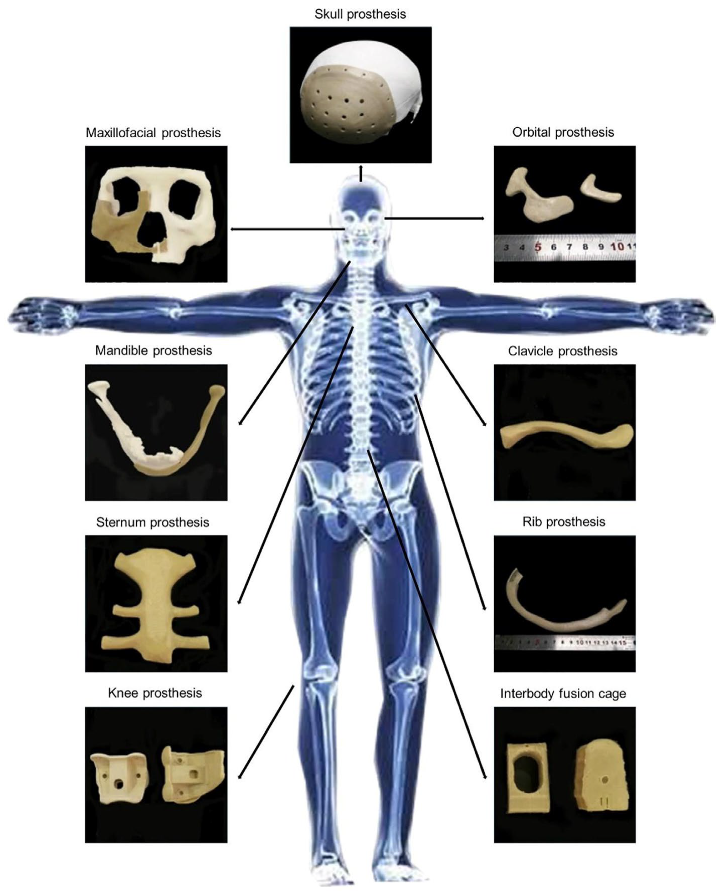
3.1. PEEK in Orthopedics Applications

PEEK’s excellent biocompatibility and mechanical properties have made it an increasingly successful option as an orthopedic implant biomaterial. Since receiving approval from the U.S. Food and Drug Administration (FDA) in 1998, PEEK has been recognized as a viable polymer for bone substitution [36]. It is now widely accepted as a radiolucent alternative to metallic biomaterials, and extensive research is being conducted to improve its physical and mechanical properties for various orthopedic applications [2]. Below are some key orthopedic areas where PEEK has been successfully utilized.

3.1.1. Craniomaxillofacial Reconstruction

Craniomaxillofacial (CMF) trauma resulting from sports injuries, accidents, disease, or tumor resections often requires complex reconstructive surgery [37,38]. A widely adopted approach involves the use of computer-aided design (CAD) and computer-aided manufacturing (CAM) technologies in conjunction with 3D printing to produce patient-specific implants. While metals, polymers, and ceramics have traditionally been utilized, PEEK has gained increasing attention due to its favorable mechanical properties, biocompatibility, and thermal stability—features that permit sterilization without material degradation [38]. These characteristics make PEEK particularly well-suited for craniofacial implants and cranioplasty procedures, which aim to reconstruct skull defects using synthetic or biological materials [39].
Compared to traditional autografts or allografts, synthetic materials such as PEEK offer reduced operative time and improved cosmetic outcomes [40]. PEEK plates are lightweight, flexible, cost-effective, and easily customized [41]. Moreover, its mechanical compatibility with human bone and energy-absorbing elasticity provide superior protection to brain tissue compared to titanium implants [42]. The accessibility of 3D printing has further accelerated the production of custom cranial implants, driving ongoing research into enhancing PEEK’s performance in CMF applications [43].
Despite these advantages, PEEK still faces notable challenges, including poor osseointegration and weak interfacial adhesion, which can contribute to implant loosening or long-term failure. Surface modification techniques—such as hydroxyapatite coatings—have shown promise in improving bone integration, although their long-term stability and effectiveness remain areas of active investigation [44]. Future studies should also explore the performance of PEEK implants under dynamic physiological stresses to better understand their behavior in post-operative conditions.

3.1.2. Spinal Implants

PEEK applications in spinal implants have also been extensively investigated. Basgul et al. (2018) demonstrated the use of 3D-printed PEEK cages in lumbar fusion, highlighting the material’s role in maintaining implant strength [45]. A subsequent study explored FFF-printed porous PEEK cages, focusing on the influence of printing parameters—nozzle size, print speed, layer thickness, and the number of cages per build—on thermal conditions during printing. The findings showed that optimizing these parameters reduced thermal gradient, improving interlayer bonding and mechanical performance [46].
Beyond optimizing printing parameters, PEEK can be reinforced with other materials such as carbon fiber to enhance implant properties. Lindtner et al. (2018) [47] investigated nonmetallic carbon fiber-reinforced PEEK (CF/PEEK) pedicle screws for spinal stabilization, comparing them to traditional titanium screws. The results showed that CF/PEEK screws performed comparably under cyclic loading until loosening, with screw stability further improved by the addition of PMMA. Additionally, the radiolucent nature of CF/PEEK minimizes imaging artifacts, offering a key advantage over metallic implants, as shown in Figure 3.

3.1.3. Joint Replacement

Polyethylene (PE) is widely used in knee replacement implants; however, wear particle generation remains a leading cause of revision surgeries [48]. Consequently, interest has shifted toward PEEK and its composites as alternatives materials [49]. Scholes & Unsworth (2009) evaluated the wear characteristics of PEEK and carbon fiber-reinforced PEEK (CFR-PEEK) using a multi-directional pin-on-plate machine with low carbon (LC) and high carbon (HC) CoCrMo alloy plates, finding that CFR-PEEK offers favorable wear performance and biocompatibility compared to UHMWPE [50].
A more recent controlled trial compared PEEK and traditional CoCrMo tibial prostheses, demonstrating ideal cement penetrations and no additional complications [51]. While PEEK shows significant promise, further research is needed to validate its long-term clinical performance in knee arthroplasty.
PEEK’s favorable mechanical properties, chemical resistance, and biocompatibility have also promoted its use in hip replacement applications. Early work showed that PEEK composites are viable bearing surfaces for acetabular cups, offering performance comparable to UHMWPE [52]. Recent advances incorporated reduced graphene oxide (rGO) and calcium hydroxyapatite (cHAp) into 3D-printed PEEK structures, with optimized lattice designs to enhance mechanical performance and biocompatibility. The results emphasized the critical role of porosity and lattice architecture in replicating the mechanical behavior of natural bone [53].
In another study, a comprehensive computational wear stimulation of cobalt chromium (CoCr) on CFR-PEEK bearing couples under gait cycle demonstrated superior wear performance of CoCr-on-CFR-PEEK compared to CoCr-on-cross-linked polyethylene (XLPE) in total hip replacement. However, CoCr-on-CFR-PEEK produced a higher release of metallic particles, raising greater biocompatibility concerns than polyethylene wear [54]. Further research is needed to minimize CoCr wear rate when paired with PEEK-based bearing surfaces.

3.1.4. Rib Prostheses

Custom-made PEEK rib prostheses have recently been utilized in chest wall reconstruction for conditions such as malignant tumors and congenital deformities [55]. In one study, a custom-designed rib prosthesis fabricated using FDM technology demonstrated mechanical behavior and stability comparable to natural human ribs [56]. Further research suggested that adjusting parameter combinations can enhance the mechanical properties of PEEK prostheses, providing flexibility to tailor design and strength based on specific prostheses requirements [57]. However, additional studies are needed to understand the correlation between prosthesis geometries, printing parameters, and mechanical properties.
In conclusion, PEEK and its composites demonstrate strong potential for orthopedic implant applications, though further research is needed to fully optimize their performance. Ongoing advancements continue to position PEEK as an emerging alternative to traditional biomaterials, with anticipated growth across various sectors.

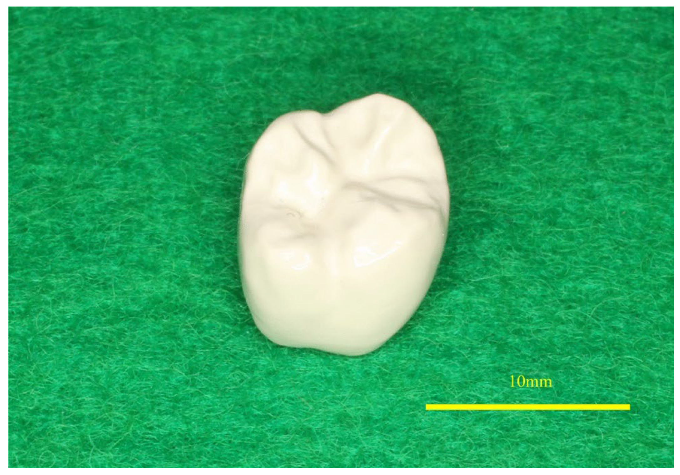
3.2. PEEK in Dental Implants

In addition to orthopedic applications, PEEK is gaining attention in dentistry due to its numerous advantages over traditional dental implant materials. It has been utilized in removable dentures, maxillofacial prostheses, and crowns owing to its low elastic modulus, high radiation resistance, excellent thermal stability, and insolubility in oral solvents [58]. PEEK’s elastic modulus (approximately 3–4 GPa) is closer to that of cortical bone (14 GPa) compared to metals like titanium (approximately 102–110 GPa), reducing stress shielding—a mechanical phenomenon in which extensive stiffness surrounding the implant leads to bone resorptions [59,60]. Furthermore, PEEK’s radiolucent nature results in fewer artifacts in magnetic resonance imaging (MRI) compared to metallic materials such as titanium [61].
Kimura et al. (2022) [62] conducted an in vivo study of PEEK crowns placed on the molars of nineteen subjects, assessing the condition of the crowns at cementation and after six months to evaluate patient satisfaction, masticatory ability, and occlusal force. Figure 4 illustrates the fabricated PEEK crown. The results showed no fractures, cracks, or detachment, and no additional interventions were required. Patient satisfaction was high, with complaints mainly related to mastication and esthetics. Occlusal pressure and force remained within the standard range between treated and untreated sides. In a more recent study, Yoshida et al. (2025) [63] hypothesized that PEEK implants would reduce stress shielding and promote bone modification compared to Ti implants. Mechanical testing on artificial bone surfaces proved that PEEK implants transmit more stress than Ti implants, reducing bone resorptions and stress shielding.
However, unmodified PEEK can be challenging in dental applications due to its bioinert nature and hydrophobicity. To address these limitations, research has shifted toward the development of modified PEEK. Najeeb et al. (2016) reviewed several nano-modification strategies, including the incorporation of bioactive apatite and the application of synthetic osteoconductive hydroxyapatite, to improve PEEK’s performance and bioactivity [64]. Additionally, surface modification techniques —such as plasma coating, chemical treatment, and physical treatment—have been shown to significantly enhance PEEK’s mechanical and biologic properties [65]. Further details on surface modification techniques are discussed in Section 5.1.

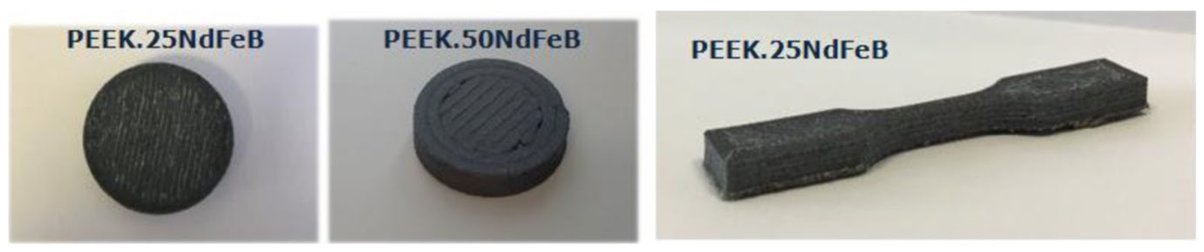
3.3. Other PEEK Applications

Beyond healthcare applications, PEEK’s unique properties have enabled its use across a wide range of industries. In aerospace, PEEK has gained attention for its chemical resistance and thermal stability [66]. Permanent rare earth magnets, such as neodymium–iron–boron (NdFeB), are currently used in space mechanisms and equipment—such as drive systems, motors, and electronic components—for their high magnetic performance and low volume and weight. However, their poor corrosion resistance makes them susceptible to degradation of magnetic properties. To address this issue, Pigliaru et al. (2020) [67] developed a PEEK-NdFeB composite filament to protect the magnetic alloy from corrosion. Figure 5 illustrates the printed samples of PEEK-NdFeB composites.
Carbon fiber-reinforced PEEK (CF/PEEK) filaments have shown promise in radar detection technologies and electromagnetic wave (EMW) absorption due to their outstanding mechanical and electrical properties. Zhang et al. (2025) reported that CF/PEEK-based pyramidal honeycomb metastructures exhibit superior mechanical performance alongside enhanced EMW absorption capabilities [68]. In the field of electronics, CF/PEEK composites have demonstrated improved electrical conductivity and thermal performance. For instance, Martin et al. (2024) [69] utilized CF/PEEK facesheets in the fabrication of sandwich panels via induction welding. This method enabled low-temperature joining, thereby reducing the risk of facesheet deconsolidation and core crushing—critical considerations for aerospace and automotive components where high strength, lightweight design, and weldability are paramount.
Beyond transportation and electronics, PEEK composites have also found applications in the nuclear industry. Wu et al. (2020) [70] explored 3D-printed boron carbide-PEEK composites for neutron radiation shielding and observed notable performance enhancements following appropriate heat treatment. These composites also exhibited increased tensile and flexural strength compared to conventional polyethylene-based shielding materials. In automotive engineering, PEEK is being positioned as a potential substitute for metals such as aluminum, brass, bronze, and titanium, primarily due to its superior strength-to-weight ratio and excellent resistance to grease, oil, acids, and other automotive fluids [71,72].
As research advances, PEEK continues to gain traction across a wide array of industries. Emerging applications are being explored in sectors such as textiles, oil and gas, and solar energy [73,74,75]. While both pure and composite forms of PEEK exhibit promising capabilities, many of these applications remain in the research and development phase. Continued studies are necessary to fully validate their performance and scalability in real-world environments.

4. Properties of PEEK

PEEK offers several advantageous attributes, positioning it as a promising alternative to traditional biomaterials [1]. Notably, its elastic modulus closely resembles that of the human bone, making it a suitable substitute for metallic biomaterials such as titanium. In addition, PEEK is radiolucent, resulting in fewer artifacts during magnetic resonance imaging [61]. However, despite these benefits, PEEK’s inherent bioinertness and low surface energy nature challenge its ability to fully replace traditional biomaterials in orthopedic and orthodontic applications [60]. To overcome these limitations, recent advancements in additive manufacturing have significantly improved the microstructure and tribological properties of PEEK, expanding its potential applications [76]. These improvements have enabled the creation of patient-specific implants, making PEEK an attractive option for load-bearing implants and orthopedic devices.
Nonetheless, the adoption of PEEK is influenced not only by its superior mechanical and biocompatible properties but also by its cost and scalability compared to traditional biomaterials such as titanium alloys or stainless steel. PEEK is a high-performance thermoplastic with a relatively higher raw material cost. For instance, medical-grade PEEK costs approximately USD 500–USD 800 per kilogram, which is substantially higher than the cost of titanium alloy powders (around USD 200–USD 400 per kilogram) or bulk stainless steel [2,77]. However, the processing and manufacturing costs must also be considered. While metals require subtractive machining, complex multi-step forming, or expensive powder bed fusion processes, PEEK can be additively manufactured via fused filament fabrication (FFF) or laser sintering with relatively simpler setups, lower energy consumption, and reduced post-processing needs [4]. Additionally, the additive manufacturing of PEEK allows for the production of patient-specific implants with minimal material wastage, which may offset its higher material cost in small-batch or customized implant production [4].
From a scalability perspective, the traditional injection molding of PEEK remains cost-effective for the mass production of standard devices, whereas additive manufacturing provides a scalable route for low-volume, customized, or complex porous structures. Recent studies suggest that as additive manufacturing technology for PEEK matures, the per-part cost is expected to decrease due to improvements in process efficiency and printer accessibility [4].
Overall, while PEEK remains a more expensive biomaterial in raw material cost compared to metals, its scalability through additive manufacturing for customized medical devices, along with reduced machining requirements and lighter weight, can make it a cost-effective option in specific biomedical applications where design complexity and biocompatibility outweigh raw material expenses. Table 1 illustrates the key properties of PEEK in comparison to titanium and UHMWPE.

4.1. Thermal and Radiative Properties

PEEK’s radiation resistance and thermal stability enhance its tolerance to high temperatures and gamma radiation, making it suitable for healthcare applications, particularly in the sterilization of medical devices [17,79]. In surgical settings, sterilization processes are routinely used to disinfect surgical devices and implants. These procedures expose materials to high temperatures, pressure, and humidity, which can compromise the dimensional stability and functional integrity of the implants. Therefore, considerable research has focused on evaluating the thermal resilience of medical-grade PEEK under sterilization conditions [79]. For example, Sharma et al. (2023) investigated the effects of steam sterilization on the dimensional characteristics of 3D-printed PEEK cranial implants and found that the material maintained high dimensional accuracy post-sterilization [43]. Similarly, Chai et al. (2022) [80] examined the effects of gamma irradiation on PEEK’s surface and internal structure, focusing on its tribological behavior. Their results showed that gamma-irradiated PEEK exhibited lower dynamic friction and increased surface oxidation, beneficial for lubrication applications. However, higher irradiation doses led to surface degradation, increased wear rate, and the formation of defects like micropores and microcracks, which negatively impact the surface hardness and wear resistance. These results underscore the need for further research to optimize irradiation doses and sterilization methods, ensuring that the mechanical and tribological properties or PEEK are preserved for critical biomedical applications [81].

4.2. Porosity

PEEK is widely recognized for its biocompatibility, which underpins its growing use in biomedical applications. However, a major limitation remains its relatively poor osseointegration, particularly in orthopedic and dental contexts. To address this, research has increasingly focused on strategies to enhance bone implant integration without compromising mechanical integrity.
A commonly investigated approach involves introducing controlled porosity into PEEK structures. By increasing surface area and promoting cellular activity, porous architectures can improve biological fixation. Evans et al. (2015) proposed that tailoring porosity could enhance bone ingrowth while preserving the material’s load-bearing capacity [82]. Building on this, Gummadi et al. (2022) [7] fabricated porous PEEK scaffolds using fused filament fabrication (FFF), evaluating pore sizes from 100 µm to 600 µm. Their findings identified 300 µm as the optimal pore size for orthopedic load-bearing applications, with larger pores resulting in diminished mechanical strength.
Similarly, Wong et al. (2021) [83] employed fused deposition modeling (FDM) to create cranium PEEK implants with varying porosities, shown in Figure 6. In vivo experiments indicated no adverse biological responses. Notably, implants with 40% porosity demonstrated optimal bone compatibility and osseointegration. These structures supported the adhesion of platelets, osteocytes, and monocytes, while the increased surface roughness further encouraged bone growth.
Beyond geometric design, printing parameters also significantly influence the porosity and mechanical performance of PEEK implants. Basgul et al. (2018) [45] examined lumbar fusion cages printed at varying speeds and assessed their compressive, shear, and torsional properties. Their results highlighted an optimal print speed between 1000 and 1500 mm/min, balancing reduced production time with mechanical integrity. Moreover, a direct correlation was observed between print speed and porosity, with faster speeds producing higher porosity.
In summary, modifying PEEK’s porosity—through both structural design and process parameters—can markedly enhance its osseointegration potential. Continued research is essential to refine printing techniques that yield porous structures without sacrificing mechanical performance. These advancements hold significant promise not only for orthopedic implants but also for applications in dental implants and tissue engineering.

4.3. Crystallinity

Crystallinity plays a critical role in determining the mechanical properties of PEEK. Achieving a well-ordered crystalline structure typically necessitates a controlled, gradual cooling process [84]. To enhance crystallinity in additively manufactured PEEK, numerous studies have explored post-processing treatments—particularly thermal annealing—as a means of improving both structural and mechanical performance [85].
Zhen et al. (2023) [86] examined the effects of post-processing heat treatment on 3D-printed PEEK components. Their approach involved preheating an oven to a target temperature, placing the PEEK parts inside for a designated period, and subsequently cooling them in ambient air. The results indicated that higher annealing temperatures and extended exposure times significantly improved both crystallinity and mechanical strength. In a related study, Adamson and Eslami (2025) [87] applied a similar annealing process to five 3D-printed PEEK specimens to assess changes in surface morphology and crystallization. Their results revealed that samples treated at 360 °C for six hours exhibited enhanced crystallinity and more uniform surface textures. Atomic force microscopy (AFM) imaging (Figure 7) confirmed the formation of larger and more distinct grain boundaries, indicating improved molecular ordering and surface stability.
While these studies highlight the value of post-processing heat treatments in promoting crystallinity and mechanical integrity, further investigations are warranted. Specifically, there is a growing need to evaluate how other processing parameters—such as raster angle, ambient temperature, nozzle temperature, and printing speed—influence the crystalline morphology and microstructural characteristics of PEEK components [88,89].
Shirani Bidabadi et al. (2024) [90] employed an alternative approach to enhance the crystallization of PEEK by using a hybrid nanomaterial system composed of cellulose nano-crystals (CNC) and graphene nanoplatelets (GNP). The CNC:GNP specimens were shown to be the optimal hybrid system in improving the tensile strength and modulus by altering the crystalline morphology. However, more work is required to fully understand the long-term mechanical performance and crystalline morphology of these hybrid systems.
Beyond heat treatment, other post-processing techniques such as surface polishing, metal coating, and UV curing have been explored to enhance the properties of polymer-based materials [91]. Overall, post-processing 3D-printed PEEK remains under investigation, and further research is essential to develop a more comprehensive understanding.

4.4. Fatigue and Fracture Behavior of PEEK

Another important aspect of processing PEEK is understanding its fatigue behavior, which is critical when designing implants subjected to cyclic loading [92]. Previous research has investigated the stress-life fatigue behavior and fracture characteristics of notched PEEK under cyclic loading, including the percentage of the lifetime spent in crack initiation versus propagation. The results showed that increasing the notch severity and cyclic stress level reduced the number of cycles to failure, with a significant portion of the lifetime spent in the crack initiation phase [93].
Another study focused on the impact of stress concentrations, such as notches and irregular shapes, on the fracture behavior of PEEK implants. The stress–strain curve of different notched radii, illustrated in Figure 8, revealed that increasing the radius from 0.5 mm to 4.0 mm reduced stress triaxiality. The study concluded that implant designs with reduced stress concentrations—such as smaller notches, stress-relieving grooves, and narrower notches—can enhance performance under cyclic loading [94].
Rendas et al. (2023) [95] studied the fatigue behavior of 3D-printed PEEK under tension–tension cyclic loading at varying stress levels ranging from 75% to 95% of its ultimate tensile strength. The material exhibited an above-average fatigue strength of about 65 MPa (~75% of ultimate tensile strength), highlighting PEEK’s ability to withstand high cyclic stress levels close to its ultimate tensile strength before failure. However, fracture surface analysis revealed crack formations during printing, suggesting that adjusting the printing parameters can significantly impact fatigue strength. Table 2 summarizes key advances in the fatigue and fracture behavior of PEEK in load-bearing applications.
Although significant research has investigated the fatigue behavior of PEEK, further studies are needed to fully understand its fatigue and fracture behaviors under cyclic loading conditions similar to those in the human body, including long-term tension, compression, and testing in environments that replicate physiological conditions. Similarly, it is crucial to identify the optimal printing parameters to enhance the performance of PEEK implants produced using additive manufacturing.

4.5. Biocompatibility and Physiological Toxicity

PEEK has been widely investigated and utilized in biomedical applications due to its excellent biocompatibility and negligible physiological toxicity. As a bioinert polymer, PEEK does not elicit significant adverse reactions when implanted in the human body. This is a critical advantage for orthopedic, spinal, and craniofacial implants where long-term stability and minimal immune response are required. Multiple in vitro and in vivo studies have demonstrated that PEEK exhibits minimal cytotoxicity, does not induce inflammatory responses, and maintains stable interactions with surrounding tissues [2]. For instance, Kurtz and Devine (2007) reported that PEEK implants showed similar or better cellular adhesion and proliferation compared to metallic implants when used in spinal cages and orthopedic devices [2]. Similarly, comparative studies have shown that PEEK exhibits cytotoxicity and pro-inflammatory responses similar to traditional dental implant materials such as titanium alloys and zirconia [78].
Additionally, PEEK is chemically stable within the physiological environment and does not release toxic degradation products under normal sterilization and implantation conditions [96]. Its radiolucency allows for clear post-operative imaging without artifacts, unlike metallic implants, providing additional clinical safety and diagnostic benefits [2]. Overall, the FDA approval of PEEK for various implantable medical devices underscores its demonstrated safety, biocompatibility, and favorable performance in long-term biomedical applications.
However, PEEK is inherently bioinert rather than bioactive, meaning that it does not naturally bond to bone. To enhance osseointegration, osteoconductivity, and cellular interactions, surface modifications such as plasma treatment, chemical etching, or hydroxyapatite coating are commonly employed [97]. Section 5 will explore these surface modification approaches in greater detail.

5. Limitations and Modifications of PEEK
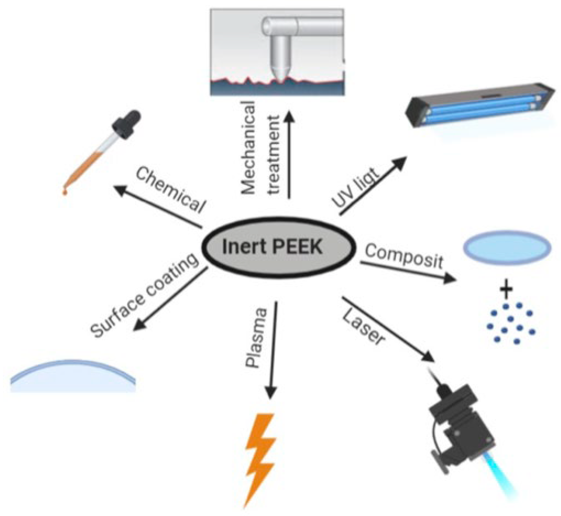
PEEK continues to gain attention in various fields due to its favorable attributes in different settings. However, ongoing research has identified several limitations that constrain its application in some areas. Researchers are actively exploring solutions, primarily through surface modification, to address these challenges. The following subsections review key limitations and the efforts made to overcome them.

5.1. Hydrophilicity

The inherent hydrophobicity of untreated PEEK surfaces significantly limits their use in medical implants by hindering cell and tissue adhesion, potentially leading to implant failure. Surface modification techniques—such as surface coating, surface roughening, plasma treatment, chemical treatment, and laser modification—have been employed to improve PEEK wettability and bioactivity [98,99,100]. Key modification techniques are illustrated in Figure 9.
One study reviewed various surface modification techniques, such as sandblasting, corona treatment, oxygen plasma treatment, argon plasma treatments, laser irradiation, and acid etching, to enhance the surface properties of copper-coated PEEK films. Although no single optimal technique was identified, each has its distinct advantages and drawbacks. Overall, these treatments enhanced the bonding strength, surface roughness, and oxygen-to-carbon (O/C) ratio and reduced the contact angle, all of which contribute to better cell integration [101].
Wang et al. (2022) also reviewed strategies to improve the adhesive properties of PEEK dental implants, concluding promising adhesion performance but emphasizing the need for further clinical trials to evaluate the long-term outcomes [102]. Despite extensive effort, there remains a gap in identifying the optimal approach to improve PEEK wettability [100]. In the following subsections are several promising surface modification techniques previously explored in the literature.

5.1.1. Physical Modifications

Physical surface modification has shown promising results in enhancing the surface wettability and tissue adhesion of PEEK. Several studies have focused on modifying PEEK’s surface characteristics by altering its architecture, surface morphology, and stiffness. Microscale and nanoscale surface modifications—such as sandblasting, plasma treatment, and accelerated neutral atom beam (ANAB) technology—have been shown to promote osteointegration by changing the structure and surface morphology of PEEK [103].
One study evaluated the influence of sandblasting on the wettability of PEEK disks, indicating that increased surface roughness led to a decrease in the contact angle, suggesting enhanced wettability [104]. In another in vitro study, the surface of PEEK copings on underlying dentin abutments was modified using sandblasting and UV radiation, revealing that the combination of sandblasting and UV radiation produced the highest retention strength, while the UV radiation-only specimens exhibited the lowest strength [105].
Similarly, Porrelli et al. (2021) demonstrated that combining sandblasting with air-plasma treatment significantly enhances the wettability and surface roughness of PEEK samples [106]. Other techniques, such as corona-discharge treatment and (ANAB) technology, have also been shown to reduce the contact angle and enhance the hydrophilicity of PEEK surfaces [107,108]. However, there is limited literature concerning these techniques, and further investigation is needed to fully understand their impact on long-term in vivo applications.
While individual approaches have shown promising results, combining multiple techniques appears more effective in enhancing bioactivity and promoting bone ingrowth on PEEK surfaces [109]. As a result, ongoing research continues to explore these combinations to optimize PEEK performance across various biomedical domains.

5.1.2. Chemical Modifications

Combining chemical and physical surface modification techniques has been shown to further enhance PEEK’s surface properties. Zhao et al. (2020) [110] proposed a two-step strategy that mimics the natural structure of joint cartilage by anchoring an acrylic acid-co-acryl amide hydrogel layer onto the surface of PEEK via UV-initiated polymerization. The hydrogel-coated PEEK substrate was then immersed in a ferric nitrate solution to promote cross-linkage between the iron ions and the carboxyl groups in the hydrogel, transforming the hydrophobic PEEK surface into a hydrophilic, low-friction surface with self-healing properties. Another promising technique explored by Luo et al. (2023) [111] involved femtosecond laser (FSL) treatment followed by hydroxylation, aimed at promoting osteogenic activity in PEEK implants in a fast, simple, and environmentally friendly approach. This approach increased surface roughness and significantly enhanced surface hydrophilicity, promoting cell proliferation and osteointegration.
Wang et al. (2019) [112] developed an optimal fast ambient temperature sulfonation treatment to PEEK surface hydrophilicity. Three treatment methods were tested: Treatment 1 involved sulfonation with sulfuric acid, Treatment 2 included post-sulfonation polishing with soft laboratory tissue, and Treatment 3 involved sulfonation followed by immersion in 6 wt% NaOH. The results showed that a 30 s sulfonation time with Treatment 3 was the most effective in achieving the highest hydrophilicity, as illustrated in Figure 10.
Similarly, dos Santos et al. (2021) demonstrated that sulfonation with sulfuric acid and piranha solution reduced PEEK’s water contact angle, promoted cell adhesion and proliferation, and maintained cytocompatibility, with no significant differences in cell viability compared to untreated PEEK [113].
In conclusion, the various surface modification techniques yielded different results, emphasizing the need to choose the right method—or combination of methods—based on the intended application. Table 3 summarizes these techniques and their respective performances.

5.2. Bioactivity

Despite its excellent mechanical strength and biocompatibility, polyetheretherketone (PEEK) is inherently bioinert, which limits its surface energy, protein adsorption, and cellular adhesion—ultimately resulting in poor osseointegration [103,114]. To overcome these limitations, researchers have investigated various surface modification techniques, including the application of bioactive coatings such as hydroxyapatite (HA), calcium silicate, and bioglass [115]. For instance, Johansson et al. (2018) [116] employed a spin coating method to apply HA onto PEEK implants, which were then implanted into the tibias and femurs of rabbits. Their findings demonstrated significantly improved bone formation and osseointegration, particularly during the early stages of healing (2–12 weeks). Similarly, Hong et al. (2018) used a dip coating technique to deposit a bioactive glass-chitosan (BG-CH) composite on PEEK surfaces, resulting in enhanced cell adhesion, proliferation, and differentiation—demonstrating its potential for orthopedic applications [117].
However, a major concern with coated implants is the potential for coating delamination over time. The detachment of surface layers may release particles into the surrounding tissue, potentially compromising implant stability and leading to adverse biological reactions [118]. To address this issue, alternative strategies such as scaffold fabrication using additive manufacturing have been explored. Roskies et al. (2016) [119] utilized selective laser sintering (SLS) to fabricate porous PEEK scaffolds and seeded them with bone marrow stromal cells (BMSCs) and adipose-derived mesenchymal stem cells (ADSCs) harvested from Sprague Dawley rats. The study revealed that these tissue-engineered PEEK scaffolds successfully supported mesenchymal cell growth and integration, thereby enhancing osteointegration—particularly for applications in craniofacial reconstruction. Given the broad array of surface modification approaches and their associated limitations, further research is essential to refine or eliminate suboptimal methods and to establish the most effective techniques for enhancing the bioactivity of PEEK implants without compromising their mechanical integrity.

6. Clinical Translation of PEEK Implants

6.1. Regulatory Considerations

The clinical translation of PEEK-based implants in the United States is governed primarily by the U.S. Food and Drug Administration (FDA) device classification and review processes. Since the first PEEK spinal and orthopedic implants received 510 (k) clearance in the late 1990s, most off-the-shelf PEEK devices leverage that pathway by demonstrating substantial equivalence to predicate devices [2,120]. Novel PEEK constructs lacking suitable predicates must instead undergo the more rigorous Premarket Approval (PMA) process, which requires comprehensive bench testing, animal studies, and clinical trials to establish safety and effectiveness. For patient-specific, 3D-printed implants manufactured in limited quantities, the Custom Device Exemption (CDE) may apply; devices under CDE must still comply with Good Manufacturing Practices (GMP) as defined in 21 CFR 820 and submit annual usage reports to the FDA [121]. Moreover, the FDA’s guidance on the additive manufacturing of medical devices emphasizes the need for validated process controls, full lot traceability, and rigorous post-build testing critical considerations given PEEK’s high processing temperatures and sensitivity to print parameters [122].
Despite these regulatory avenues, several practical challenges persist in bringing 3D-printed PEEK implants to the market. First, ensuring reproducibility and dimensional accuracy across multiple prints demands robust in-process monitoring and strict quality-control protocols. Second, although PEEK’s high melting point generally permits autoclave sterilization, each new geometry must undergo sterilization validation, comparing steam or gamma radiation, to confirm that no dimensional distortion or polymer embrittlement occurs. Finally, any novel surface treatments or bioactive coatings applied to PEEK implants necessitate biocompatibility testing in accordance with ISO 10993-1:2018 [123] to rule out cytotoxicity, sensitization, or adverse inflammatory responses, even when the base polymer has established safety profiles.

6.2. Long-Term Clinical Performance and Failure Modes

PEEK has demonstrated promising long-term outcomes in various biomedical applications, particularly in spinal, craniofacial, and joint implants. Clinical studies have reported high success rates for PEEK-based implants, such as a 93.7% success rate and a relatively low complication rate of 15.4% in cranioplasty procedures using 3D-printed PEEK implants [124]. Despite these encouraging results, several failure modes have been identified that warrant further investigation. One of the most significant challenges is PEEK’s inherent bioinertness, which can lead to poor osseointegration and implant loosening over time [125]. This limitation has prompted the development of surface modification techniques, including plasma treatment, sulfonation, and hydroxyapatite coatings, to enhance bone implant integration. Another concern is wear particle generation, particularly in joint replacements, where PEEK and its composites may release debris that can trigger inflammatory responses. Although carbon fiber-reinforced PEEK (CFR-PEEK) has shown improved wear resistance, the biological impact of long-term exposure to wear particles remains under scrutiny. Additionally, fatigue and fracture under cyclic loading are critical considerations for load-bearing implants. Studies have shown that optimizing implant geometry and additive manufacturing parameters can significantly improve fatigue resistance. Coating delamination is another potential failure mode, especially in implants with bioactive surface layers, as it may compromise implant stability and lead to adverse tissue reactions. Overall, while PEEK exhibits excellent mechanical and chemical stability, continued research is essential to address these long-term performance challenges and to validate its reliability across diverse clinical scenarios.

7. Conclusions

Polyetheretherketone (PEEK) is widely acknowledged as a high-performance polymer that combines exceptional mechanical, thermal, and chemical properties, positioning it as a highly versatile material across diverse industries. Over recent years, extensive research has sought to optimize PEEK’s structural, surface, and biological characteristics. This comprehensive review highlights recent advancements in enhancing PEEK and its composites, with a focus on biomedical applications, where precise control over material properties can profoundly influence patient outcomes.
PEEK’s favorable elastic modulus, radiolucency, chemical inertness, and fatigue resistance has led to its increasing adoption as an alternative to conventional biomaterials like titanium alloys and UHMWPE. It is widely used in clinical applications, including spinal cages, craniomaxillofacial reconstruction, joint replacements, dental prostheses, and rib implants, underscoring its versatility and clinical promise. Additionally, additive manufacturing techniques has further propelled the fabrication of patient-specific implants with controlled porosity and complex geometries, enabling tissue integration and load-bearing capabilities.
Despite these notable strides, PEEK’s bioinert nature and hydrophobic surface continue to pose significant challenges, limiting protein adsorption and cellular adhesion essential for osteointegration and long-term implant success. Surface modification strategies—including plasma treatment, sandblasting, acid etching, laser irradiation, and bioactive coatings such as hydroxyapatite and bioglass—have demonstrated promising results in enhancing PEEK’s bioactivity. However, these modifications must balance improved biological response with mechanical strength and stability, as issues like coating delamination and particle debris highlight the need for durable surface treatments that maintain performance under physiological conditions.
Additionally, the mechanical performance of PEEK in additive manufacturing remains highly sensitive to processing parameters such as printing speed, nozzle temperature, raster angle, and post-processing heat treatments. These factors significantly influence crystallinity, porosity, and microstructural morphology, which directly affect mechanical strength, fatigue resistance, and durability. While annealing and heat treatment protocols have improved structural properties, standardized processing guidelines are needed to optimize mechanical properties without compromising efficiency or introducing defects.
Another critical avenue lies in the development of PEEK composites, particularly carbon fiber-reinforced PEEK (CF/PEEK) and boron carbide reinforced PEEK, which enhances mechanical performance with functional properties such as electromagnetic wave absorption and radiation shielding. These composites expand PEEK’s application potential beyond biomedicine into aerospace, automotive, electronics, and nuclear industries. However, they also introduce manufacturing and performance challenges that require systematic investigations into interfacial bonding, thermal stability, and long-term behavior under operational stresses.
Looking ahead, additive manufacturing is poised to further revolutionize the fabrication of PEEK-based components. Its ability to produce highly customized, multi-material, and hierarchical structures opens exciting opportunities for personalized medicine, lightweight structural components, and multifunctional devices. Nevertheless, translating laboratory-scale innovations into clinical and industrial practice requires overcoming barriers related to reproducibility, scalability, regulatory approval, long-term clinical performance, and cost-effectiveness. Interdisciplinary collaboration among material scientists, biomedical engineers, clinicians, and manufacturers will be essential to address these challenges.
In conclusion, while PEEK demonstrates remarkable potential and versatility, further advancements are needed to understand the relationships between processing parameters, microstructure, mechanical behavior, and biological interactions. Expanding upon the promising findings related to increased porosity, future studies should prioritize the investigation of PEEK’s mechanical behavior at elevated porosity levels, specifically ranging from 50% to 80%. Additionally, significant research should prioritize the development of tailored manufacturing and modification techniques for specific applications, alongside comprehensive long-term in vivo evaluations to ensure safety and efficacy. By addressing current limitations, PEEK is well positioned to become a cornerstone material in the next generation of high-performance, biocompatible, and multifunctional devices.

Author Contributions

Conceptualization, B.E. and S.T.; methodology, S.T.; software, S.D.; validation, S.D.; formal analysis, B.E., S.D. and S.T.; investigation, S.D.; resources, B.E. and S.T.; data curation, S.D.; writing—original draft preparation, S.D.; writing—review and editing, B.E. and S.T.; visualization, S.D.; supervision, S.T.; project administration, S.T.; funding acquisition, B.E. and S.T. All authors have read and agreed to the published version of the manuscript.

Funding

This research received no external funding.

Institutional Review Board Statement

Not applicable.

Data Availability Statement

No new data were created or analyzed in this study. Data sharing is not applicable to this article.

Acknowledgments

The authors would like to thank Widener University for its support of this project.

Conflicts of Interest

The authors declare no conflicts of interest.

References

  1. Mbogori, M.; Vaish, A.; Vaishya, R.; Haleem, A.; Javaid, M. Poly-Ether-Ether-Ketone (PEEK) in Orthopaedic Practice- A Current Concept Review. J. Orthop. Rep. 2022, 1, 3–7. [Google Scholar] [CrossRef]
  2. Kurtz, S.M.; Devine, J.N. PEEK Biomaterials in Trauma, Orthopedic, and Spinal Implants. Biomaterials 2007, 28, 4845–4869. [Google Scholar] [CrossRef] [PubMed]
  3. Jin, L.; Ball, J.; Bremner, T.; Sue, H.-J. Crystallization Behavior and Morphological Characterization of Poly(Ether Ether Ketone). Polymer 2014, 55, 5255–5265. [Google Scholar] [CrossRef]
  4. Berretta, S.; Evans, K.; Ghita, O. Additive Manufacture of PEEK Cranial Implants: Manufacturing Considerations versus Accuracy and Mechanical Performance. Mater. Des. 2018, 139, 141–152. [Google Scholar] [CrossRef]
  5. Dua, R.; Rashad, Z.; Spears, J.; Dunn, G.; Maxwell, M. Applications of 3D-Printed PEEK via Fused Filament Fabrication: A Systematic Review. Polymers 2021, 13, 4046. [Google Scholar] [CrossRef]
  6. Verma, S.; Sharma, N.; Kango, S.; Sharma, S. Developments of PEEK (Polyetheretherketone) as a Biomedical Material: A Focused Review. Eur. Polym. J. 2021, 147, 110295. [Google Scholar] [CrossRef]
  7. Gummadi, S.K.; Saini, A.; Owusu-Danquah, J.S.; Sikder, P. Mechanical Properties of 3d-Printed Porous Poly-Ether-Ether-Ketone (Peek) Orthopedic Scaffolds. JOM 2022, 74, 3379–3391. [Google Scholar] [CrossRef]
  8. Lv, M.; Yang, X.; Gvetadze, S.R.; Gupta, A.; Li, J.; Sun, J. Accurate Reconstruction of Bone Defects in Orbital–Maxillary–Zygomatic (Omz) Complex with Polyetheretherketone (Peek). J. Plast. Reconstr. Aesthet. Surg. 2022, 75, 1750–1757. [Google Scholar] [CrossRef]
  9. Singh, S.; Prakash, C.; Ramakrishna, S. 3D Printing of Polyether-Ether-Ketone for Biomedical Applications. Eur. Polym. J. 2019, 114, 234–248. [Google Scholar] [CrossRef]
  10. Ma, H.; Suonan, A.; Zhou, J.; Yuan, Q.; Liu, L.; Zhao, X.; Lou, X.; Yang, C.; Li, D.; Zhang, Y. PEEK (Polyether-Ether-Ketone) and Its Composite Materials in Orthopedic Implantation. Arab. J. Chem. 2021, 14, 102977. [Google Scholar] [CrossRef]
  11. Jogi, B.F.; Tarekar, M.; Dhajekar, R.M.; Pawade, R. Multi Objective Optimization Using Taguchi Grey Relational Analysis (Gra) for Cnc Turning of Poly-Ether-Ether-Ketone (Peek) Polymer. Polym. Polym. Compos. 2016, 24, 523–528. [Google Scholar] [CrossRef]
  12. Moharil, S.; Reche, A.; Durge, K. Polyetheretherketone (PEEK) as a Biomaterial: An Overview. Cureus 2023, 15, e44307. [Google Scholar] [CrossRef] [PubMed]
  13. Pereira, T.; Kennedy, J.V.; Potgieter, J. A Comparison of Traditional Manufacturing vs Additive Manufacturing, the Best Method for the Job. Procedia Manuf. 2019, 30, 11–18. [Google Scholar] [CrossRef]
  14. Zarean, P.; Malgaroli, P.; Zarean, P.; Seiler, D.; de Wild, M.; Thieringer, F.M.; Sharma, N. Effect of Printing Parameters on Mechanical Performance of Material-Extrusion 3D-Printed PEEK Specimens at the Point-of-Care. Appl. Sci. 2023, 13, 1230. [Google Scholar] [CrossRef]
  15. Gonzalez Lugo, C.A.; Caputo, D.S.; Hutchinson, M.J.; Fouladi, K.; Eslami, B. Design and Development of an Environmentally Controlled Enclosure for a Commercial 3D Printer. Rapid Prototyp. J. 2022, 29, 780–791. [Google Scholar] [CrossRef]
  16. Mohammed Ali Al -Zaidi, A.; Al Gawhari, F. Types of Polymers Using in 3D Printing and Their Applications: A Brief Review. Eur. J. Theor. Appl. Sci. 2024, 1, 978–985. [Google Scholar] [CrossRef]
  17. Honigmann, P.; Sharma, N.; Okolo, B.; Popp, U.; Msallem, B.; Thieringer, F.M. Patient-Specific Surgical Implants Made of 3d Printed Peek: Material, Technology, and Scope of Surgical Application. BioMed Res. Int. 2018, 2018, 4520636. [Google Scholar] [CrossRef]
  18. Mendenhall, R.; Eslami, B. Experimental Investigation on Effect of Temperature on FDM 3D Printing Polymers: ABS, PETG, and PLA. Appl. Sci. 2023, 13, 11503. [Google Scholar] [CrossRef]
  19. Thiruchitrambalam, M.; Bubesh Kumar, D.; Shanmugam, D.; Jawaid, M. A Review on PEEK Composites–Manufacturing Methods, Properties and Applications. Mater. Today Proc. 2020, 33, 1085–1092. [Google Scholar] [CrossRef]
  20. Moby, V.; Dupagne, L.; Fouquet, V.; Attal, J.-P.; François, P.; Dursun, E. Mechanical Properties of Fused Deposition Modeling of Polyetheretherketone (PEEK) and Interest for Dental Restorations: A Systematic Review. Materials 2022, 15, 6801. [Google Scholar] [CrossRef]
  21. Li, T.; Song, Z.; Yang, X.; Du, J. Influence of Processing Parameters on the Mechanical Properties of Peek Plates by Hot Compression Molding. Materials 2023, 16, 36. [Google Scholar] [CrossRef]
  22. Czepiel, M.; Bańkosz, M.; Sobczak-Kupiec, A. Advanced Injection Molding Methods: Review. Materials 2023, 16, 5802. [Google Scholar] [CrossRef]
  23. Yang, J.; Xie, J.; Ji, K.; Wang, X.; Jiao, X.; Xu, Z.; Zhao, P. Microcellular Injection Molding of Polyether-Ether-Ketone. Polymer 2022, 251, 124866. [Google Scholar] [CrossRef]
  24. Siddiq, A.; Kennedy, A.R. Compression Moulding and Injection over Moulding of Porous PEEK Components. J. Mech. Behav. Biomed. Mater. 2020, 111, 103996. [Google Scholar] [CrossRef] [PubMed]
  25. Panayotov, I.V.; Orti, V.; Cuisinier, F.; Yachouh, J. Polyetheretherketone (PEEK) for Medical Applications. J. Mater. Sci. Mater. Med. 2016, 27, 118. [Google Scholar] [CrossRef] [PubMed]
  26. Khan, W. Comparison on Performance of Injection Moulding and 3D Printed Parts. Doctoral Dissertation, Mechanical Engineering, Edinburgh Napier University, Edinburgh, UK, 2020. [Google Scholar]
  27. Chen, M.; Luo, C.; Yuan, Y.; Zhou, H.; Li, Z.; Wang, Q.; Gong, B.; Li, Z.; Sun, H. Modification of PEEK for Implants: Strategies to Improve Mechanical, Antibacterial, and Osteogenic Properties. Rev. Adv. Mater. Sci. 2024, 63, 20240025. [Google Scholar] [CrossRef]
  28. Xiaoyong, S.; Liangcheng, C.; Honglin, M.; Peng, G.; Zhanwei, B.; Cheng, L. Experimental Analysis of High Temperature Peek Materials on 3d Printing Test. In Proceedings of the 2017 9th International Conference on Measuring Technology and Mechatronics Automation (ICMTMA), Changsha, China, 14–15 January 2017; IEEE: Piscataway, NJ, USA, 2017; pp. 13–16. [Google Scholar]
  29. Baek, I.; Kwon, O.; Lim, C.-M.; Park, K.Y.; Bae, C.-J. 3D PEEK Objects Fabricated by Fused Filament Fabrication (FFF). Materials 2022, 15, 898. [Google Scholar] [CrossRef]
  30. Hu, B.; Duan, X.; Xing, Z.; Xu, Z.; Du, C.; Zhou, H.; Chen, R.; Shan, B. Improved Design of Fused Deposition Modeling Equipment for 3D Printing of High-Performance PEEK Parts. Mech. Mater. 2019, 137, 103139. [Google Scholar] [CrossRef]
  31. Park, S.J.; Lee, J.E.; Park, J.; Lee, N.-K.; Son, Y.; Park, S.-H. High-Temperature 3D Printing of Polyetheretherketone Products: Perspective on Industrial Manufacturing Applications of Super Engineering Plastics. Mater. Des. 2021, 211, 110163. [Google Scholar] [CrossRef]
  32. Liu, G.; Hu, N.; Huang, J.; Tu, Q.; Xu, F. Experimental Investigation on the Mechanical and Dynamic Thermomechanical Properties of Polyether Ether Ketone Based on Fused Deposition Modeling. Polymers 2024, 16, 3007. [Google Scholar] [CrossRef]
  33. Chithambaram, K.; Senthilnathan, N. Effects of Printing Parameters on Hardness and Wear Characteristics of 3D Printed Polyetheretherketone (PEEK) Polymer. Mater. Lett. 2024, 356, 135588. [Google Scholar] [CrossRef]
  34. Ling, X.; Jing, X.; Zhang, C.; Chen, S. Polyether Ether Ketone (Peek) Properties and Its Application Status. IOP Conf. Ser. Earth Environ. Sci. 2020, 453, 012080. [Google Scholar] [CrossRef]
  35. Ahmad, K.; Batool, S.A.; Farooq, M.T.; Minhas, B.; Manzur, J.; Yasir, M.; Wadood, A.; Avcu, E.; Ur Rehman, M.A. Corrosion, Surface, and Tribological Behavior of Electrophoretically Deposited Polyether Ether Ketone Coatings on 316L Stainless Steel for Orthopedic Applications. J. Mech. Behav. Biomed. Mater. 2023, 148, 106188. [Google Scholar] [CrossRef]
  36. Chen, Z.; Chen, Y.; Wang, Y.; Deng, J.; Wang, X.; Wang, Q.; Liu, Y.; Ding, J.; Yu, L. Polyetheretherketone Implants with Hierarchical Porous Structure for Boosted Osseointegration. Biomater. Res. 2023, 27, 61. [Google Scholar] [CrossRef]
  37. Gassner, R.; Tuli, T.; Hächl, O.; Rudisch, A.; Ulmer, H. Cranio-Maxillofacial Trauma: A 10 Year Review of 9543 Cases with 21 067 Injuries. J. Cranio-Maxillo-Fac. Surg. Off. Publ. Eur. Assoc. Cranio-Maxillo-Fac. Surg. 2003, 31, 51–61. [Google Scholar] [CrossRef]
  38. Slavin, B.V.; Ehlen, Q.T.; Costello, J.P.I.; Nayak, V.V.; Bonfante, E.A.; Benalcázar Jalkh, E.B.; Runyan, C.M.; Witek, L.; Coelho, P.G. 3D Printing Applications for Craniomaxillofacial Reconstruction: A Sweeping Review. ACS Biomater. Sci. Eng. 2023, 9, 6586–6609. [Google Scholar] [CrossRef] [PubMed]
  39. Yao, S.; Zhang, Q.; Mai, Y.; Yang, H.; Li, Y.; Zhang, M.; Zhang, R. Outcome and Risk Factors of Complications after Cranioplasty with Polyetheretherketone and Titanium Mesh: A Single-Center Retrospective Study. Front. Neurol. 2022, 13, 926436. [Google Scholar] [CrossRef] [PubMed]
  40. Alkhaibary, A.; Alharbi, A.; Alnefaie, N.; Oqalaa Almubarak, A.; Aloraidi, A.; Khairy, S. Cranioplasty: A Comprehensive Review of the History, Materials, Surgical Aspects, and Complications. World Neurosurg. 2020, 139, 445–452. [Google Scholar] [CrossRef] [PubMed]
  41. Hughes, E.B.; Alfarone, J.; Chernov, E.S.; Debick, N.A.; Jalal, M.; Kim, Y.; Suryadevara, A.; Krishnamurthy, S. Polyetheretherketone (Peek) into the Future: Lowering Infection Rates in Cranioplasty. Cureus 2025, 17, c214. [Google Scholar] [CrossRef]
  42. Lethaus, B.; Safi, Y.; Ter Laak-Poort, M.; Kloss-Brandstätter, A.; Banki, F.; Robbenmenke, C.; Steinseifer, U.; Kessler, P. Cranioplasty with Customized Titanium and Peek Implants in a Mechanical Stress Model. J. Neurotrauma 2012, 29, 1077–1083. [Google Scholar] [CrossRef]
  43. Sharma, N.; Zubizarreta-Oteiza, J.; Tourbier, C.; Thieringer, F.M. Can Steam Sterilization Affect the Accuracy of Point-of-Care 3d Printed Polyetheretherketone (Peek) Customized Cranial Implants? An Investigative Analysis. J. Clin. Med. 2023, 12, 2495. [Google Scholar] [CrossRef]
  44. Kauke-Navarro, M.; Knoedler, L.; Knoedler, S.; Deniz, C.; Safi, A.-F. Surface Modification of PEEK Implants for Craniofacial Reconstruction and Aesthetic Augmentation—Fiction or Reality? Front. Surg. 2024, 11, 1351749. [Google Scholar] [CrossRef] [PubMed]
  45. Basgul, C.; Yu, T.; MacDonald, D.W.; Siskey, R.; Marcolongo, M.; Kurtz, S.M. Structure–Property Relationships for 3D-Printed PEEK Intervertebral Lumbar Cages Produced Using Fused Filament Fabrication. J. Mater. Res. 2018, 33, 2040–2051. [Google Scholar] [CrossRef] [PubMed]
  46. Basgul, C.; MacDonald, D.W.; Siskey, R.; Kurtz, S.M. Thermal Localization Improves the Interlayer Adhesion and Structural Integrity of 3D Printed PEEK Lumbar Spinal Cages. Materialia 2020, 10, 100650. [Google Scholar] [CrossRef] [PubMed]
  47. Lindtner, R.A.; Schmid, R.; Nydegger, T.; Konschake, M.; Schmoelz, W. Pedicle Screw Anchorage of Carbon Fiber-Reinforced PEEK Screws under Cyclic Loading. Eur. Spine J. 2018, 27, 1775–1784. [Google Scholar] [CrossRef]
  48. Cheppalli, N.; Metikala, S.; Albertson, B.S.; Yaw, K. Plastics in Total Knee Replacement: Processing to Performance. Cureus 2021, 13, e12969. [Google Scholar] [CrossRef]
  49. Koh, Y.-G.; Park, K.-M.; Lee, J.-A.; Nam, J.-H.; Lee, H.-Y.; Kang, K.-T. Total Knee Arthroplasty Application of Polyetheretherketone and Carbon-Fiber-Reinforced Polyetheretherketone: A Review. Mater. Sci. Eng. C 2019, 100, 70–81. [Google Scholar] [CrossRef]
  50. Scholes, S.C.; Unsworth, A. Wear Studies on the Likely Performance of CFR-PEEK/CoCrMo for Use as Artificial Joint Bearing Materials. J. Mater. Sci. Mater. Med. 2009, 20, 163–170. [Google Scholar] [CrossRef]
  51. Zhao, G.; Yao, S.; Sun, X.; Ma, J.; Wang, J. Consequences of Using Poly-Ether-Ether-Ketone versus Traditional Implant on Tibial Cement Penetration and Short-Term Clinical Outcomes during Total Knee Arthroplasty: A Randomized Controlled Trial. J. Orthop. Surg. 2023, 18, 589. [Google Scholar] [CrossRef]
  52. Wang, A.; Lin, R.; Polineni, V.K.; Essner, A.; Stark, C.; Dumbleton, J.H. Carbon Fiber Reinforced Polyether Ether Ketone Composite as a Bearing Surface for Total Hip Replacement. Tribol. Int. 1998, 31, 661–667. [Google Scholar] [CrossRef]
  53. Oladapo, B.I.; Zahedi, S.A.; Ismail, S.O. Mechanical Performances of Hip Implant Design and Fabrication with PEEK Composite. Polymer 2021, 227, 123865. [Google Scholar] [CrossRef]
  54. Alpkaya, A.; Mihcin, S. The Computational Approach to Predicting Wear: Comparison of Wear of Performance of CFR PEEK and XLPE Liners in Total Hip Replacement. Tribol. Trans. 2022, 66, 59–72. [Google Scholar] [CrossRef]
  55. Simal, I.; García-Casillas, M.; Cerdá, J.; Riquelme, Ó.; Lorca-García, C.; Pérez-Egido, L.; Fernández-Bautista, B.; Torre, M.; De Agustín, J. Three-Dimensional Custom-Made Titanium Ribs for Reconstruction of a Large Chest Wall Defect. Eur. J. Pediatr. Surg. Rep. 2016, 04, 026–030. [Google Scholar] [CrossRef] [PubMed]
  56. Kang, J.; Wang, L.; Yang, C.; Wang, L.; Yi, C.; He, J.; Li, D. Custom Design and Biomechanical Analysis of 3D-Printed PEEK Rib Prostheses. Biomech. Model. Mechanobiol. 2018, 17, 1083–1092. [Google Scholar] [CrossRef] [PubMed]
  57. Zhang, C.; Wang, L.; Kang, J.; Fuentes, O.M.; Li, D. Bionic Design and Verification of 3D Printed PEEK Costal Cartilage Prosthesis. J. Mech. Behav. Biomed. Mater. 2020, 103, 103561. [Google Scholar] [CrossRef]
  58. Akay, C.; Ersöz, M.B. PEEK in Dentistry, Properties and Application Areas. Int. Dent. Res. 2020, 10, 60–65. [Google Scholar] [CrossRef]
  59. Ouldyerou, A.; Merdji, A.; Aminallah, L.; Roy, S.; Mehboob, H.; Özcan, M. Biomechanical Performance of Ti-PEEK Dental Implants in Bone: An in-Silico Analysis. J. Mech. Behav. Biomed. Mater. 2022, 134, 105422. [Google Scholar] [CrossRef]
  60. Qin, L.; Yao, S.; Zhao, J.; Zhou, C.; Oates, T.W.; Weir, M.D.; Wu, J.; Xu, H.H.K. Review on Development and Dental Applications of Polyetheretherketone-Based Biomaterials and Restorations. Materials 2021, 14, 408. [Google Scholar] [CrossRef]
  61. Rahmitasari, F.; Ishida, Y.; Kurahashi, K.; Matsuda, T.; Watanabe, M.; Ichikawa, T. PEEK with Reinforced Materials and Modifications for Dental Implant Applications. Dent. J. 2017, 5, 35. [Google Scholar] [CrossRef]
  62. Kimura, H.; Morita, K.; Nishio, F.; Abekura, H.; Tsuga, K. Clinical Report of Six-Month Follow-up after Cementing PEEK Crown on Molars. Sci. Rep. 2022, 12, 19070. [Google Scholar] [CrossRef]
  63. Yoshida, E.; Nomoto, R.; Amitani, Y.; Hayakawa, T. PEEK Stress-Shielding with Artificial Bone for Dental Implants. Dent. Mater. J. 2025, 44, 121–127. [Google Scholar] [CrossRef]
  64. Najeeb, S.; Zafar, M.S.; Khurshid, Z.; Siddiqui, F. Applications of Polyetheretherketone (PEEK) in Oral Implantology and Prosthodontics. J. Prosthodont. Res. 2016, 60, 12–19. [Google Scholar] [CrossRef] [PubMed]
  65. Benakatti, V.B.; Sajjanar, J.A.; Acharya, A. Polyetheretherketone (PEEK) in Dentistry. J. Clin. Diagn. Res. 2019, 13, ZE10–ZE12. [Google Scholar] [CrossRef]
  66. Nobrega, G.; Cardoso, B.; Souza, R.; Pereira, J.; Pontes, P.; Catarino, S.O.; Pinho, D.; Lima, R.; Moita, A. A Review of Novel Heat Transfer Materials and Fluids for Aerospace Applications. Aerospace 2024, 11, 275. [Google Scholar] [CrossRef]
  67. Pigliaru, L.; Rinaldi, M.; Ciccacci, L.; Norman, A.; Rohr, T.; Ghidini, T.; Nanni, F. 3D Printing of High Performance Polymer-Bonded PEEK-NdFeB Magnetic Composite Materials. Funct. Compos. Mater. 2020, 1, 4. [Google Scholar] [CrossRef]
  68. Zhang, X.; Wei, G.; Huang, X.; Zhang, H.; Hao, X.; Tan, S.; Liu, K.; Ji, G. Optimized Pyramidal Honeycomb PEEK/CF Composites Metastructure through 3D Printing for Broadband Electromagnetic Wave Absorption. Mater. Today Phys. 2025, 50, 101620. [Google Scholar] [CrossRef]
  69. Martin, R.G.; Johansson, C.; Tavares, J.R.; Dubé, M. CF/PEEK Skins Assembly by Induction Welding for Thermoplastic Composite Sandwich Panels. Compos. Part B Eng. 2024, 284, 111676. [Google Scholar] [CrossRef]
  70. Wu, Y.; Cao, Y.; Wu, Y.; Li, D. Neutron Shielding Performance of 3d-Printed Boron Carbide Peek Composites. Materials 2020, 13, 2314. [Google Scholar] [CrossRef]
  71. Kumar, A.; Saxena, N.V. A Review on usage of peek material for wheels. Int. Res. J. Mod. Eng. Technol. Sci. 2021, 3, 844–846. [Google Scholar]
  72. Manivannan, R.; Rajasekar, R.; Vk, G. Modelling and Analysis of Four Wheel Rim by Using PEEK Composites. Mater. Sci. Eng. Int. J. 2018, 2, 177–183. [Google Scholar] [CrossRef]
  73. Badeghaish, W.; Wagih, A.; Rastogi, S.; Lubineau, G. Effect of High-Temperature Acid Aging on Microstructure and Mechanical Properties of PEEK. Polym. Test. 2024, 134, 108429. [Google Scholar] [CrossRef]
  74. Gupta, R.; Shinde, S.; Yella, A.; Subramaniam, C.; Saha, S.K. Thermomechanical Characterisations of PTFE, PEEK, PEKK as Encapsulation Materials for Medium Temperature Solar Applications. Energy 2020, 194, 116921. [Google Scholar] [CrossRef]
  75. Lu, C.; Xu, N.; Zheng, T.; Zhang, X.; Lv, H.; Lu, X.; Xiao, L.; Zhang, D. The Optimization of Process Parameters and Characterization of High-Performance CF/PEEK Composites Prepared by Flexible CF/PEEK Plain Weave Fabrics. Polymers 2019, 11, 53. [Google Scholar] [CrossRef] [PubMed]
  76. Zhang, H.; Duan, M.; Qin, S.; Zhang, Z. Preparation and Modification of Porous Polyetheretherketone (PEEK) Cage Material Based on Fused Deposition Modeling (FDM). Polymers 2022, 14, 5403. [Google Scholar] [CrossRef]
  77. Ramakrishna, S.; Mayer, J.; Wintermantel, E.; Leong, K.W. Biomedical Applications of Polymer-Composite Materials: A Review. Compos. Sci. Technol. 2001, 61, 1189–1224. [Google Scholar] [CrossRef]
  78. Peng, T.-Y.; Shih, Y.-H.; Hsia, S.-M.; Wang, T.-H.; Li, P.-J.; Lin, D.-J.; Sun, K.-T.; Chiu, K.-C.; Shieh, T.-M. In Vitro Assessment of the Cell Metabolic Activity, Cytotoxicity, Cell Attachment, and Inflammatory Reaction of Human Oral Fibroblasts on Polyetheretherketone (PEEK) Implant–Abutment. Polymers 2021, 13, 2995. [Google Scholar] [CrossRef]
  79. Kumar, A.; Yap, W.T.; Foo, S.L.; Lee, T.K. Effects of Sterilization Cycles on Peek for Medical Device Application. Bioengineering 2018, 5, 18. [Google Scholar] [CrossRef]
  80. Chai, L.; Zhang, B.; Qiao, L.; Wang, P.; Weng, L. Influence of Gamma Irradiation-Induced Surface Oxidation on Tribological Property of Polyetheretherketone (Peek). Polym. Bull. 2022, 79, 6513–6531. [Google Scholar] [CrossRef]
  81. Rakhadilov, B.; Ormanbekov, K.; Zhassulan, A.; Andybayeva, G.; Shynarbek, A.; Yeldos Mukhametov, Y.M. Influence of Irradiation Doses on the Mechanical and Tribological Properties of Polyetheretherketone Exposed to Electron Beam Treatment. Adv. Sci. Technol. Res. J. 2024, 19, 121–131. [Google Scholar] [CrossRef]
  82. Evans, N.T.; Torstrick, F.B.; Lee, C.S.D.; Dupont, K.M.; Safranski, D.L.; Chang, W.A.; Macedo, A.E.; Lin, A.; Boothby, J.M.; Whittingslow, D.C.; et al. High Strength, Surface Porous Polyether-Ether-Ketone for Load-Bearing Orthopaedic Implants. Acta Biomater. 2015, 13, 159–167. [Google Scholar] [CrossRef]
  83. Wong, K.I.; Zhong, Y.; Li, D.; Cheng, Z.; Yu, Z.; Wei, M. Modified Porous Microstructure for Improving Bone Compatibility of Poly-Ether-Ether-Ketone. J. Mech. Behav. Biomed. Mater. 2021, 120, 104541. [Google Scholar] [CrossRef]
  84. Vaezi, M.; Yang, S. Extrusion-Based Additive Manufacturing of PEEK for Biomedical Applications. Virtual Phys. Prototyp. 2015, 10, 123–135. [Google Scholar] [CrossRef]
  85. Gao, S.; Liu, R.; Xin, H.; Liang, H.; Wang, Y.; Jia, J. The Surface Characteristics, Microstructure and Mechanical Properties of PEEK Printed by Fused Deposition Modeling with Different Raster Angles. Polymers 2022, 14, 77. [Google Scholar] [CrossRef] [PubMed]
  86. Zhen, H.; Zhao, B.; Quan, L.; Fu, J. Effect of 3d Printing Process Parameters and Heat Treatment Conditions on the Mechanical Properties and Microstructure of Peek Parts. Polymers 2023, 15, 2209. [Google Scholar] [CrossRef] [PubMed]
  87. Adamson, M.; Eslami, B. Post-Processing PEEK 3D-Printed Parts: Experimental Investigation of Annealing on Microscale and Macroscale Properties. Polymers 2025, 17, 744. [Google Scholar] [CrossRef]
  88. Lee, A.; Wynn, M.; Quigley, L.; Salviato, M.; Zobeiry, N. Effect of Temperature History during Additive Manufacturing on Crystalline Morphology of PEEK. Adv. Ind. Manuf. Eng. 2022, 4, 100085. [Google Scholar] [CrossRef]
  89. Zanjanijam, A.R.; Major, I.; Lyons, J.G.; Lafont, U.; Devine, D.M. Fused Filament Fabrication of Peek: A Review of Process-Structure-Property Relationships. Polymers 2020, 12, 1665. [Google Scholar] [CrossRef]
  90. Shirani Bidabadi, B.; Motta De Castro, E.; Carrola, M.; Koirala, P.; Tehrani, M.; Asadi, A. Engineering the Crystalline Architecture for Enhanced Properties in Fast-Rate Processing of Poly(Ether Ether Ketone) (Peek) Nanocomposites. ACS Appl. Eng. Mater. 2024, 2, 2038–2054. [Google Scholar] [CrossRef]
  91. Dizon, J.R.C.; Gache, C.C.L.; Cascolan, H.M.S.; Cancino, L.T.; Advincula, R.C. Post-Processing of 3D-Printed Polymers. Technologies 2021, 9, 61. [Google Scholar] [CrossRef]
  92. Tang, S. Tension–Tension Fatigue Behavior of Hydroxyapatite Reinforced Polyetheretherketone Composites. Int. J. Fatigue 2004, 26, 49–57. [Google Scholar] [CrossRef]
  93. Sobieraj, M.C.; Murphy, J.E.; Brinkman, J.G.; Kurtz, S.M.; Rimnac, C.M. Notched Fatigue Behavior of PEEK. Biomaterials 2010, 31, 9156–9162. [Google Scholar] [CrossRef]
  94. Chen, F.; Gatea, S.; Ou, H.; Lu, B.; Long, H. Fracture Characteristics of PEEK at Various Stress Triaxialities. J. Mech. Behav. Biomed. Mater. 2016, 64, 173–186. [Google Scholar] [CrossRef] [PubMed]
  95. Rendas, P.; Imperadeiro, A.; Martins, R.F.; Soares, B.A.R. High-Cycle Fatigue Behaviour of Polyetheretherketone (Peek) Produced by Additive Manufacturing. Polymers 2023, 16, 18. [Google Scholar] [CrossRef] [PubMed]
  96. Toth, J.M.; Wang, M.; Estes, B.T.; Scifert, J.L.; Seim, H.B.; Turner, A.S. Polyetheretherketone as a Biomaterial for Spinal Applications. Biomaterials 2006, 27, 324–334. [Google Scholar] [CrossRef] [PubMed]
  97. Ma, R.; Tang, T. Current Strategies to Improve the Bioactivity of PEEK. Int. J. Mol. Sci. 2014, 15, 5426–5445. [Google Scholar] [CrossRef]
  98. Choudhury, S.S.; Pandey, M.; Bhattacharya, S. Recent Developments in Surface Modification of PEEK Polymer for Industrial Applications: A Critical Review. Rev. Adhes. Adhes. 2021, 9, 410–433. [Google Scholar]
  99. Li, W.; Sang, L.; Jian, X.; Wang, J. Influence of Sanding and Plasma Treatment on Shear Bond Strength of 3D-Printed PEI, PEEK and PEEK/CF. Int. J. Adhes. Adhes. 2020, 100, 102614. [Google Scholar] [CrossRef]
  100. Omrani, M.M.; Hadjizadeh, A.; Milani, A.; Kim, K. PEEK Surface Modification Methods and Effect of the Laser Method on Surface Properties. Biointerface Res. Appl. Chem. 2020, 10, 5132–5140. [Google Scholar] [CrossRef]
  101. Münstedt, H.; Grossmann, J. Surface Modifications of Polyetheretherketone (Peek): Results from the Literature and Special Studies of Copper-Coated Films. Polymers 2022, 14, 4797. [Google Scholar] [CrossRef]
  102. Wang, B.; Huang, M.; Dang, P.; Xie, J.; Zhang, X.; Yan, X. Peek in Fixed Dental Prostheses: Application and Adhesion Improvement. Polymers 2022, 14, 2323. [Google Scholar] [CrossRef]
  103. YU, D.; LEI, X.; ZHU, H. Modification of Polyetheretherketone (PEEK) Physical Features to Improve Osteointegration. J. Zhejiang Univ. Sci. B 2022, 23, 189–203. [Google Scholar] [CrossRef]
  104. Elawadly, T.; Radi, I.A.W.; El Khadem, A.; Osman, R.B. Can PEEK Be an Implant Material? Evaluation of Surface Topography and Wettability of Filled Versus Unfilled PEEK With Different Surface Roughness. J. Oral Implantol. 2017, 43, 456–461. [Google Scholar] [CrossRef] [PubMed]
  105. Mithran, A.; Upwanshi, V.R.; Alqahtani, I.M.; Alqahtani, S.J.M.; Nayak, P.P.; Alsharif, A.A.; Babu, J.S.; Swarnalatha, C.; Nayyar, A.S. Evaluation of the Impact of Surface Modification with Air Abrasion (Sandblasting) and Ultraviolet Radiation on the Retention Strength of Polyetherether Ketone (Peek) Coping in Relation to the Underlying Dentin Abutments: An in Vitro Study. J. Microsc. Ultrastruct. 2024. [Google Scholar] [CrossRef]
  106. Porrelli, D.; Mardirossian, M.; Crapisi, N.; Urban, M.; Ulian, N.A.; Bevilacqua, L.; Turco, G.; Maglione, M. Polyetheretherketone and Titanium Surface Treatments to Modify Roughness and Wettability–Improvement of Bioactivity and Antibacterial Properties. J. Mater. Sci. Technol. 2021, 95, 213–224. [Google Scholar] [CrossRef]
  107. Comyn, J.; Mascia, L.; Xiao, G.; Parker, B.M. Corona-Discharge Treatment of Polyetheretherketone (PEEK) for Adhesive Bonding. Int. J. Adhes. Adhes. 1996, 16, 301–304. [Google Scholar] [CrossRef]
  108. Khoury, J.; Maxwell, M.; Cherian, R.E.; Bachand, J.; Kurz, A.C.; Walsh, M.; Assad, M.; Svrluga, R.C. Enhanced Bioactivity and Osseointegration of PEEK with Accelerated Neutral Atom Beam Technology. J. Biomed. Mater. Res. B Appl. Biomater. 2017, 105, 531–543. [Google Scholar] [CrossRef]
  109. Chayanun, S.; Chanamuangkon, T.; Boonsuth, B.; Boccaccini, A.R.; Lohwongwatana, B. Enhancing PEEK Surface Bioactivity: Investigating the Effects of Combining Sulfonation with Sub-Millimeter Laser Machining. Mater. Today Bio 2023, 22, 100754. [Google Scholar] [CrossRef]
  110. Zhao, X.; Karthik, N.; Xiong, D.; Liu, Y. Bio-Inspired Surface Modification of PEEK through the Dual Cross-Linked Hydrogel Layers. J. Mech. Behav. Biomed. Mater. 2020, 112, 104032. [Google Scholar] [CrossRef]
  111. Luo, F.; Li, D.; Huang, Y.; Mao, R.; Wang, L.; Lu, J.; Ge, X.; Fan, Y.; Zhang, X.; Chen, Y.; et al. Efficient Osteogenic Activity of PEEK Surfaces Achieved by Femtosecond Laser–Hydroxylation. ACS Appl. Mater. Interfaces 2023, 15, 37232–37246. [Google Scholar] [CrossRef]
  112. Wang, W.; Luo, C.J.; Huang, J.; Edirisinghe, M. PEEK Surface Modification by Fast Ambient-Temperature Sulfonation for Bone Implant Applications. J. R. Soc. Interface 2019, 16, 20180955. [Google Scholar] [CrossRef]
  113. Dos Santos, F.S.F.; Vieira, M.; da Silva, H.N.; Tomás, H.; Fook, M.V.L. Surface Bioactivation of Polyether Ether Ketone (PEEK) by Sulfuric Acid and Piranha Solution: Influence of the Modification Route in Capacity for Inducing Cell Growth. Biomolecules 2021, 11, 1260. [Google Scholar] [CrossRef]
  114. Ma, T.; Zhang, J.; Sun, S.; Meng, W.; Zhang, Y.; Wu, J. Current Treatment Methods to Improve the Bioactivity and Bonding Strength of PEEK for Dental Application: A Systematic Review. Eur. Polym. J. 2023, 183, 111757. [Google Scholar] [CrossRef]
  115. Feng, X.; Ma, L.; Liang, H.; Liu, X.; Lei, J.; Li, W.; Wang, K.; Song, Y.; Wang, B.; Li, G.; et al. Osteointegration of 3d-Printed Fully Porous Polyetheretherketone Scaffolds with Different Pore Sizes. ACS Omega 2020, 5, 26655–26666. [Google Scholar] [CrossRef] [PubMed]
  116. Johansson, P.; Barkarmo, S.; Hawthan, M.; Peruzzi, N.; Kjellin, P.; Wennerberg, A. Biomechanical, Histological, and Computed X-ray Tomographic Analyses of Hydroxyapatite Coated PEEK Implants in an Extended Healing Model in Rabbit. J. Biomed. Mater. Res. A 2018, 106, 1440–1447. [Google Scholar] [CrossRef] [PubMed]
  117. Hong, W.; Guo, F.; Chen, J.; Wang, X.; Zhao, X.; Xiao, P. Bioactive Glass–Chitosan Composite Coatings on PEEK: Effects of Surface Wettability and Roughness on the Interfacial Fracture Resistance and in Vitro Cell Response. Appl. Surf. Sci. 2018, 440, 514–523. [Google Scholar] [CrossRef]
  118. Tsuchiya, A.; Toita, R.; Tsuru, K.; Ishikawa, K. Enhanced Osseointegration Capability of Poly(Ether Ether Ketone) via Combined Phosphate and Calcium Surface-Functionalization. Int. J. Mol. Sci. 2019, 21, 198. [Google Scholar] [CrossRef]
  119. Roskies, M.; Jordan, J.O.; Fang, D.; Abdallah, M.-N.; Hier, M.P.; Mlynarek, A.; Tamimi, F.; Tran, S.D. Improving PEEK Bioactivity for Craniofacial Reconstruction Using a 3D Printed Scaffold Embedded with Mesenchymal Stem Cells. J. Biomater. Appl. 2016, 31, 132–139. [Google Scholar] [CrossRef]
  120. U.S. Food and Drug Administration. 510(k) Premarket Notification; FDA: Silver Spring, MD, USA, 1998.
  121. Code of Federal Regulations. Title 21 CFR 820: Quality System Regulation; U.S. Government Publishing Office: Washington, DC, USA, 2016.
  122. U.S. Food and Drug Administration. Technical Considerations for Additive Manufactured Medical Devices: Guidance for Industry and FDA Staff; FDA: Silver Spring, MD, USA, 2017.
  123. ISO 10993-1:2018; Biological Evaluation of Medical Devices–Part 1: Evaluation and Testing Within a Risk Management Process. ISO: Geneva, Switzerland, 2018.
  124. MohanaSundaram, A.; Kamalakannan, Y.; Raja, V.; Mofatteh, M.; Haque, M.A. The World’s First 3D-Printed PEEK Cranial Implant: A New Horizon in Precision and Personalized Neurosurgery. Neurosurg. Rev. 2024, 47, 616. [Google Scholar] [CrossRef]
  125. Rendas, P.; Figueiredo, L.; Machado, C.; Mourão, A.; Vidal, C.; Soares, B. Mechanical Performance and Bioactivation of 3D-Printed PEEK for High-Performance Implant Manufacture: A Review. Prog. Biomater. 2023, 12, 89–111. [Google Scholar] [CrossRef]
Figure 1. Schematic illustrating the applications of PEEK implants in orthopedics. Reprinted from Ma et al. [10]. Licensed under CC BY-NC-ND 4.0 (https://www.sciencedirect.com/science/article/pii/S1878535220305384#f0005 (accessed on 13 July 2025)).
Polymers 17 01968 g001
Figure 2. Dog bone samples printed in different orientations. Adapted from “Effect of Printing Parameters on Mechanical Performance of Material-Extrusion 3D-Printed PEEK Specimens at the Point-of-Care,” by Zarean et al. [14]. Licensed under CC BY 4.0 (https://www.mdpi.com/2076-3417/13/3/1230 (accessed on 13 July 2025)).
Polymers 17 01968 g002
Figure 3. Axial fluoroscopic images of instrumented vertebrae. (a) CF/PEEK pedicle screws (right) and titanium control screw (left); (b) PMMA-augmented (right) and nonaugmented CF/PEEK (left) pedicle screws. Adapted from Lindtner et al. [47]. Licensed under CC BY 4.0 (https://link.springer.com/article/10.1007/s00586-018-5538-8 (accessed on 13 July 2025)).
Polymers 17 01968 g003
Figure 4. The occlusal surface of the PEEK crown. Adapted from Kimura et al. [62]. Licensed under CC BY 4.0 (https://www.nature.com/articles/s41598-022-23458-5 (accessed on 13 July 2025)).
Polymers 17 01968 g004
Figure 5. Three-dimensionally printed magnetic sample of PEEK NdFeB 25 (left), PEEK NdFeB 50 (center), and tensile test sample PEEK NdFeB25. Adapted from Pigliaru et al. [67]. Licensed under CC BY 4.0 (https://jmscomposites.springeropen.com/articles/10.1186/s42252-020-00006-w (accessed on 13 July 2025)).
Polymers 17 01968 g005
Figure 6. The modified porous PEEK implants. (A) The CAD of the modified implants; (B) the modified PEEK implants: from left to right: 40%, 50%, 60%, and solid; (C) side view of the implant; (D) front view of the implant. Reprinted from Wong et al. [83]. Licensed under CC BY-NC-ND 4.0 (https://www.sciencedirect.com/science/article/pii/S1751616121002241 (accessed on 13 July 2025)).
Polymers 17 01968 g006
Figure 7. AFM scans of PEEK annealed for 6 h at 360 °C: (a) height, (b) amplitude, and (c) phase scans. Adapted from Adamson et al. [87]. Licensed under CC BY 4.0 (https://www.mdpi.com/2073-4360/17/6/744 (accessed on 13 July 2025)).
Polymers 17 01968 g007
Figure 8. Equivalent plastic strain to fracture with the change in stress triaxiality. Adapted from Chen et al. [94]. Licensed under CC BY 4.0 (https://www.sciencedirect.com/science/article/pii/S1751616116302430 (accessed on 13 July 2025)).
Polymers 17 01968 g008
Figure 9. Various surface modification methods to improve the properties of PEEK. Adapted from Omrani et al. [100]. Licensed under CC BY 4.0 (https://biointerfaceresearch.com/wp-content/uploads/2020/01/20695837102132140.pdf (accessed on 13 July 2025)).
Polymers 17 01968 g009
Figure 10. Water contact angle on PEEK surfaces modified by different sulfonation procedures and reaction durations, (a,d,g,j,m) Treatment 1. (b,e,h,k,n) Treatment 2. (c,f,i,l,o) Treatment 3. (ac): 5 s, (df): 10 s, (gi): 30 s, (jl): 60 s, (mo): 90 s. Adapted from Wang et al. [112]. Licensed under CC BY 4.0 (https://royalsocietypublishing.org/doi/10.1098/rsif.2018.0955 (accessed on 13 July 2025)).
Polymers 17 01968 g010
Table 1. Comparison of key properties of PEEK, titanium, and UHMWPE.
ReferencesPropertyPEEKTitaniumUHMWPE
[2,20,47,60]Elasticity Modulus (GPa)3–4102–1130.8–1.6
[2,14,60]Tensile Strength (MPa)80–97954–97620–49
[2]Elongation at break (ductility)~30%--
[2]Flexural Strength (MPa)~140–160--
[50,54]Wear ResistanceGoodExcellentExcellent
[1,10,60,61]BiocompatibilityExcellentExcellentGood
[1,2,10,47,56]RadiolucencyRadiolucentRadiopaqueRadiolucent
[1,5,47]OsteointegrationPoorGoodPoor
[1,78]CytotoxicityLowLowModerate
[1,2]Raw Material CostHighModerateLow
[1,2]Manufacturing CostModerateHighLow
Table 2. Summary of key studies on the fatigue and fracture behavior of PEEK.
ReferenceFocusFindingsConclusion
[92]Understanding the fatigue behavior of PEEK and its composites in implant devices subjected to repeated loading. PEEK composites, such as HA/PEEK materials, exhibit promising fatigue resistance properties suitable for biomedical applications.Highlighted the promising future of PEEK composites in biomedical implants and stressed the need for further research into the development of fatigue damage in these materials.
[93]Investigating the stress-life fatigue behavior and fracture characteristics of notched PEEK specimens.More severe notch geometries and higher cyclic stress levels significantly reduced the number of cycles to failure.
Majority of the fatigue lifetime was spent in the crack initiation phase.
Emphasized the importance of considering design-related stress concentrations when developing PEEK-based implants undergoing cyclic loading.
[94]Examining the fracture behavior of PEEK specimens with varying notch radii under different stress triaxialities.Smaller notch radii resulted in higher stress triaxiality.
Designs with reduced stress concentrations demonstrated improved performance under cyclic loading conditions.
Highlighted stress triaxiality as a key design parameter for optimizing the performance of notched PEEK implants.
[95]Investigating the high-cycle fatigue behavior of 3D-printed PEEK specimens subjected to stress-controlled tension–tension cyclic loading, highlighting the significance of cyclic loading in load-bearing implant design.Three-dimensionally printed PEEK endured high cyclic stress levels up to 75% of its ultimate tensile strength before failure. Fracture surface analysis revealed that crack initiation occurred in voids formed during the printing process.Highlighted PEEK’s potential for load-bearing applications. Emphasized the need for further research to understand the correlation between printing parameters and fatigue behavior.
Table 3. Summary of various PEEK surface modification techniques.
Reference.Surface Modification TechniqueSummary
[101,104,105]Sandblasting/BlastingIncreased surface roughness without significantly altering the chemical composition of PEEK. Increased wettability and adhesion.
[101,107]Corona treatmentIncreased surface polarity and O/C ratio, improving adhesion and wettability. However, it decreased surface roughness.
[101]Oxygen plasma treatmentSignificantly increased the O/C ratio, enhancing adhesion and wettability. However, it decreased surface roughness.
[101]Argon plasma treatmentEnhanced surface adhesion without increasing oxygen content. Also reduced surface roughness.
[101,105,111]Laser treatmentReduced surface roughness and contact angle without oxygen content. Enhanced surface hydrophilicity. May introduce surface degradation
[101]UV radiationIncreased oxygen content on the surface PEEK, enhancing wettability and bond strength.
[101]Chemical treatmentStrongly increased the O/C ratio and surface roughness, significantly improving adhesion.
[110]Hydrogel-coated PEEKImproved surface wettability and lubrication. Demonstrated self-healing properties, and reduced wear in implants.
[112,113]SulfonationImproved hydrophilicity by significantly reducing the water contact angle. Created a nano-porous PEEK surface for enhanced adhesion and bioactivity.
Disclaimer/Publisher’s Note: The statements, opinions and data contained in all publications are solely those of the individual author(s) and contributor(s) and not of MDPI and/or the editor(s). MDPI and/or the editor(s) disclaim responsibility for any injury to people or property resulting from any ideas, methods, instructions or products referred to in the content.

