
Comparative Analysis of Reconstitution and Solubility of Two Poly-L-Lactic Acid Fillers for Medical Applications


Abstract
1. Introduction
2. Methods
- Capturing reconstitution processes on a shadowless table using a 4K camera (MX BrioTM, Logitech, 1015 Lausanne, Switzerland).
- Conducting analyses with a digital light microscope Levenhuk D95L LCD (Levenhuk, Inc., Tampa, FL, USA), which involved digital recording of both static and dynamic images using an 8 MP built-in camera,
- Utilizing certified micrometric sieves with hole diameters of 200 μm and 71 μm, coupled with digital image recording via a 12 MP camera.
- Following the validation process, the main phase of the study was carried out, culminating in the reporting of the findings.
3. Reconstitution Procedures
3.1. Reconstitution
- PLLA-LASYNPRO™
- PLLA-SCA
- For 8 mL reconstitution: 5 mL of water for injection was added to the vial, followed by vigorous shaking for 1 min. Then, an additional 3 mL of water was added, and the vial was shaken for a further 1 min.
- For 5 mL reconstitution: 5 mL of water for injection was added to the vial, and the vial was shaken vigorously for 1 min.
3.2. Observation Conditions
3.3. Sample Collection
- PLLA-LASYNPRO™: As the product is homogeneous, no separated layers were collected.
- PLLA-SCA: Due to its inhomogeneity and the clear instructions in the IFU, care was taken to avoid collecting any foam formed during reconstitution.
- 0.5 mL for microscopic examination.
- 1.5 mL for evaluation on a 200 μm micrometric sieve.
- 1.5 mL for evaluation on a 71 μm micrometric sieve.
3.4. Macroscopic Observations
3.4.1. Recording Setup
3.4.2. Microscopic Observations
3.4.3. Micrometric Sieve Observations
4. Results
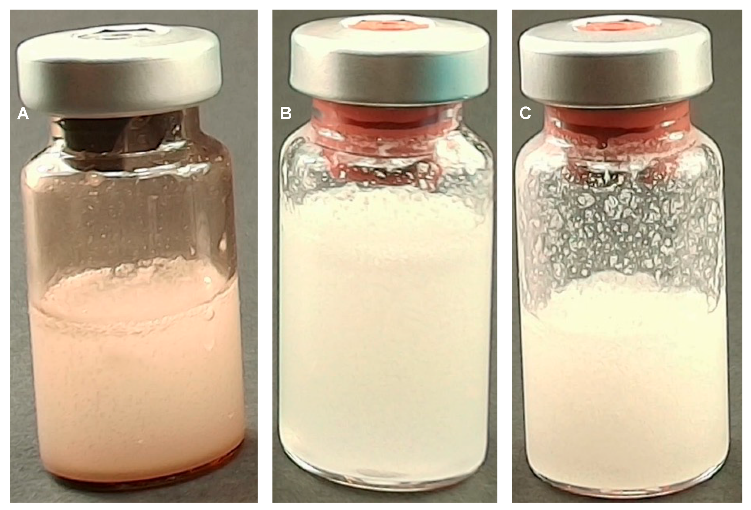
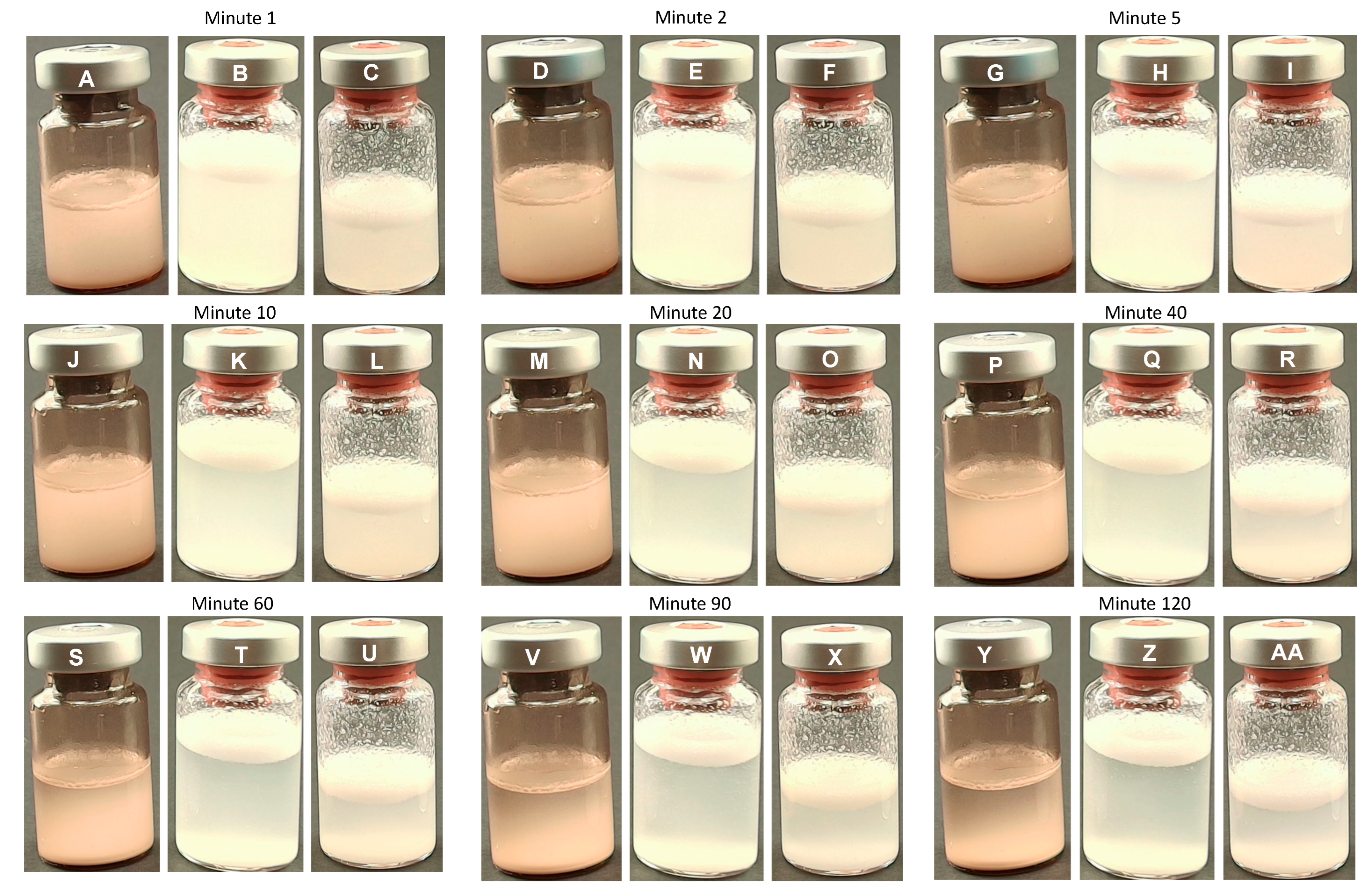
4.1. Macroscopic Observations
Immediately After Reconstitution
- Minutes 1–20
- Minutes 40–120
4.2. Microscopic Observations
4.2.1. PLLA-LASYNPRO™ Microscopic Characteristics
4.2.2. PLLA-SCA Microscopic Characteristics
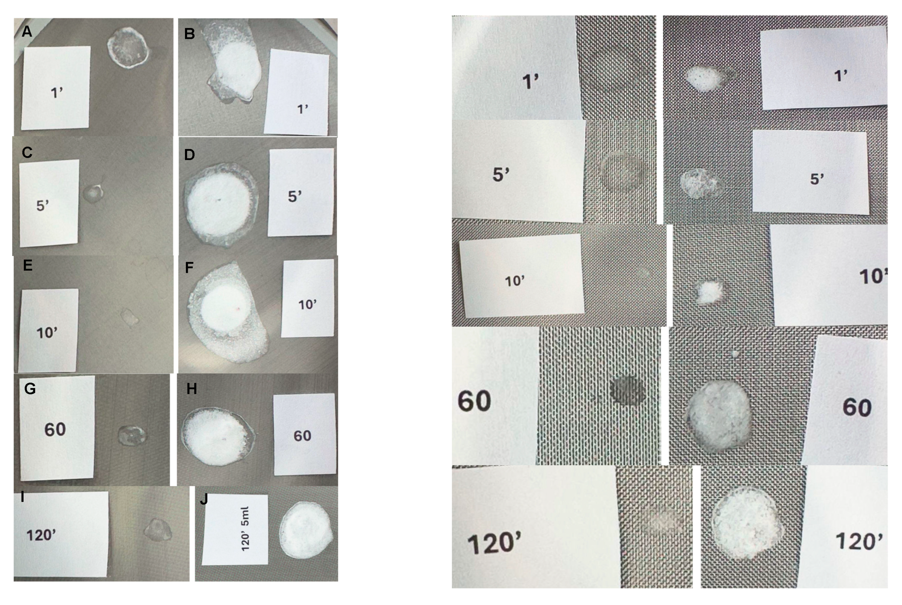
4.3. Micrometric Sieve Observations
- PLLA-LASYNPRO™
- PLLA-SCA
5. Discussion
- Macroscopic Observations
- Microscopic Observations
- Micrometric Sieve Observations
6. Conclusions
Supplementary Materials
Author Contributions
Funding
Institutional Review Board Statement
Data Availability Statement
Acknowledgments
Conflicts of Interest
References
- Li, K.; Meng, F.; Li, Y.R.; Tian, Y.; Chen, H.; Jia, Q.; Cai, H.; Jiang, H.B. Application of nonsurgical modalities in improving facial aging. Int. J. Dent. 2022, 2022, 8332631. [Google Scholar] [CrossRef] [PubMed] [PubMed Central]
- ISAPS International Survey on Aesthetic Cosmetic Procedures Performed in 2023. Available online: https://www.isaps.org/discover/about-isaps/global-statistics/global-survey-2023-full-report-and-press-releases/ (accessed on 1 March 2025).
- Fitzgerald, R.; Bass, L.M.; Goldberg, D.J.; Graivier, M.H.; Lorenc, Z.P. Physiochemical characteristics of poly-L-lactic acid (PLLA). Aesthet Surg. J. 2018, 38 (Suppl. S1), S13–S17. [Google Scholar] [CrossRef] [PubMed]
- Haddad, S.; Galadari, H.; Patil, A.; Goldust, M.; Al Salam, S.; Guida, S. Evaluation of the biostimulatory effects and the level of neocollagenesis of dermal fillers: A review. Int. J. Dermatol. 2022, 61, 1284–1288. [Google Scholar] [CrossRef] [PubMed]
- Xu, Q.; Sun, X.; Yang, W.; Bai, Y.; Tan, X. Comprehensive systematic review of poly-L-lactic Acid in facial clinical application. Aesthetic Plast. Surg. 2024, 48, 5115–5135. [Google Scholar] [CrossRef] [PubMed]
- Ao, Y.J.; Yi, Y.; Wu, G.H. Application of PLLA (poly-L-lactic acid) for rejuvenation and reproduction of facial cutaneous tissue in aesthetics: A review. Medicine 2024, 103, e37506. [Google Scholar] [CrossRef] [PubMed] [PubMed Central]
- Lorenc, Z.P. Techniques for the optimization of facial and nonfacial volumization with injectable poly-l-lactic acid. Aesthetic Plast. Surg. 2012, 36, 1222–1229. [Google Scholar] [CrossRef] [PubMed]
- Harper, J.; Avelar, L.; Haddad, A.; Novello, J.; Mest, D.; Guarnieri-Munia, C.; Rice, S.; Vega, J.; Andriopolous, B.; Nogueria, A.; et al. Expert recommendations on the use of injectable poly-L-lactic acid for contour deficiencies of the buttocks. J. Drugs Dermatol. 2022, 21, 21–26. [Google Scholar] [CrossRef] [PubMed]
- Avelar, L.E.; Nabhani, S.; Wüst, S. Unveiling the mechanism: Injectable poly-L-lactic acid’s evolving role—Insights from recent studies. J. Cosmet. Dermatol. 2025, 24, e16635. [Google Scholar] [CrossRef] [PubMed] [PubMed Central]
- Dunn, A.; Long, T.; Zarraga, M.; Aguilera, S.B. Nodules on the anterior neck following poly-L-lactic acid injection. Cutis 2022, 109, E15–E17. [Google Scholar] [CrossRef] [PubMed]
- Jeon, Y.J.; Koo, D.W.; Lee, J.S. Late onset foreign body reaction due to poly-L-lactic acid facial injections for cosmetic purpose. Ann. Dermatol. 2020, 32, 519–522. [Google Scholar] [CrossRef] [PubMed] [PubMed Central]
- Lee, T.Y. Lactate: A multifunctional signaling molecule. Yeungnam Univ. J. Med. 2021, 38, 183–193. [Google Scholar] [CrossRef] [PubMed] [PubMed Central]
- Sigrist, R.M.; de Noronha, M.G.O.; Borelli, S.S.; Teixeira, S.P.; Funes, H.L.X.; Lourenço, L.M. Dynamic ultrasound evaluation of body fillers and biostimulators in the buttocks of fresh-frozen specimen. J. Cosmet. Dermatol. 2022, 21, 5621–5627. [Google Scholar] [CrossRef] [PubMed]
- Sedush, N.G.; Kalinin, K.T.; Azarkevich, P.N.; Gorskaya, A.A. Physicochemical characteristics and hydrolytic degradation of polylactic acid dermal fillers: A comparative study. Cosmetics 2023, 10, 110. [Google Scholar] [CrossRef]
- Juläine. Instructions for Use. Available online: https://julaineofsweden.com/wp-content/uploads/2024/06/Instructions-for-Use-JULAINE-of-Sweden.pdf (accessed on 2 March 2025).
- Sculptra. Instructions for Use Version 4.7. Available online: Galderma.com/au/sites/default/files/SculptraIFUV4.7.pdf (accessed on 2 March 2025).
- Guo, J.; Fang, W.; Wang, F. Injectable fillers: Current status, physicochemical properties, function mechanism, and perspectives. RSC Adv. 2023, 13, 23841–23858. [Google Scholar] [CrossRef] [PubMed]
- Caicedo, M.S.; Samelko, L.; McAllister, K.; Jacobs, J.J.; Hallab, N.J. Increasing both CoCrMo-alloy particle size and surface irregularity induces increased macrophage inflammasome activation in vitro potentially through lysosomal destabilization mechanisms. J. Orthop. Res. 2013, 31, 1633–1642. [Google Scholar] [CrossRef]
- Baranov, M.V.; Kumar, M.; Sacanna, S.; Thutupalli, S.; van den Bogaart, G. Modulation of immune responses by particle size and shape. Front. Immunol. 2021, 11, 607945. [Google Scholar] [CrossRef] [PubMed] [PubMed Central]
- Wang, J.; Chen, H.J.; Hang, T.; Yu, Y.; Liu, G.; He, G.; Xiao, S.; Yang, B.R.; Yang, C.; Liu, F.; et al. Physical activation of innate immunity by spiky particles. Nat. Nanotechnol. 2018, 13, 1078–1086. [Google Scholar] [CrossRef] [PubMed] [PubMed Central]
- Goldberg, D.; Guana, A.; Volk, A.; Daro-Kaftan, E. Single-arm study for the characterization of human tissue response to injectable poly-L-lactic acid. Dermatol. Surg. 2013, 39, 915–922. [Google Scholar] [CrossRef] [PubMed]
- Ray, S.; Ta, H.T. Investigating the effect of biomaterials such as poly-(l-lactic acid) particles on collagen synthesis in vitro: Method is matter. J. Funct. Biomater. 2020, 11, 51. [Google Scholar] [CrossRef] [PubMed] [PubMed Central]
- Sk, M.S.; Mwangomo, R.; Daniel, L.; Gilmore, J. Solution blow spinning: An emerging nanomaterials-based wound-care technology. J. Biomed. Mater. Res. B Appl. Biomater. 2025, 113, e35513. [Google Scholar] [CrossRef] [PubMed]
- Middleton, J.C.; Tipton, A.J. Synthetic biodegradable polymers as orthopedic devices. Biomaterials 2000, 21, 2335–2346. [Google Scholar] [CrossRef] [PubMed]
- Narayanan, G.; Vernekar, V.N.; Kuyinu, E.L.; Laurencin, C.T. Poly (lactic acid)-based biomaterials for orthopaedic regenerative engineering. Adv. Drug Deliv. Rev. 2016, 107, 247–276. [Google Scholar] [CrossRef] [PubMed] [PubMed Central]
- Hu, Y.; Fu, X.; Chen, X.D.; Yang, J.; Yang, L.S.; Zhang, M.Q. Association behaviors between carboxymethyl cellulose and polylactic acid revealed by resonance light scattering spectra. Polym. Bull. 2009, 62, 549–559. [Google Scholar] [CrossRef]
- Nonhoff, M.; Puetzler, J.; Hasselmann, J.; Fobker, M.; Gosheger, G.; Schulze, M. The potential for foreign body reaction of implanted poly-L-lactic acid: A systematic review. Polymers 2024, 16, 817. [Google Scholar] [CrossRef] [PubMed] [PubMed Central]
- Yamoum, C.; Magaraphan, R. Effect of carboxymethylcellulose on plasticized polylactide. Adv. Mat. Res. 2013, 658, 19–24. [Google Scholar] [CrossRef]
- Sánchez-Bodón, J.; Ruiz-Rubio, L.; Hernáez-Laviña, E.; Vilas-Vilela, J.L.; Moreno-Benítez, M.I. Poly(l-lactide)-based anti-inflammatory responsive surfaces for surgical implants. Polymers 2020, 13, 34. [Google Scholar] [CrossRef] [PubMed] [PubMed Central]
- He, T.; Zhang, Z.; Zhang, X.; Niu, H.; Wang, S.; Wang, Q.; Lai, C. Effects of poly-L-lactic acid fillers on inflammatory response and collagen synthesis in different animal models. J. Cosmet. Dermatol. 2025, 24, e70000. [Google Scholar] [CrossRef] [PubMed] [PubMed Central]
- Oh, H.; Lee, S.; Na, J.; Kim, J.H. Comparative evaluation of safety and efficacy of a novel hyaluronic acid-polynucleotide/poly-L-lactic acid composite dermal filler. Aesthetic Plast. Surg. 2021, 45, 1792–1801. [Google Scholar] [CrossRef] [PubMed]
- Tabei, Y.; Sugino, S.; Eguchi, K.; Tajika, M.; Abe, H.; Nakajima, Y.; Horie, M. Effect of calcium carbonate particle shape on phagocytosis and pro-inflammatory response in differentiated THP-1 macrophages. Biochem. Biophys. Res. Commun. 2017, 490, 499–505. [Google Scholar] [CrossRef] [PubMed]
- Lebre, F.; Sridharan, R.; Sawkins, M.J.; Kelly, D.J.; O’Brien, F.J.; Lavelle, E.C. The shape and size of hydroxyapatite particles dictate inflammatory responses following implantation. Sci. Rep. 2017, 7, 2922. [Google Scholar] [CrossRef] [PubMed] [PubMed Central]
- Anderson, J.M.; Rodriguez, A.; Chang, D.T. Foreign body reaction to biomaterials. Semin. Immunol. 2008, 20, 86–100. [Google Scholar] [CrossRef] [PubMed] [PubMed Central]
- Magacho-Vieira, F.N.; Vieira, A.O.; Soares, A., Jr.; Alvarenga, H.C.L.; de Oliveira Junior, I.R.A.; Daher, J.A.C.; Napoli, J.V.M.P.; Serra, J.P.A.; Provázio, S.C. Consensus recommendations for the reconstitution and aesthetic use of poly-D,L-lactic acid microspheres. Clin. Cosmet. Investig. Dermatol. 2024, 17, 2755–2765. [Google Scholar] [CrossRef] [PubMed] [PubMed Central]
- Avelar, L.; Haddad, A.; Fabi, S.G.; Somenek, M.; Beleznay, K.; Aguilera, S.B.; Taylor-Barnes, K.; Morgan, P.; Mao, C.; Öhrlund, Å.; et al. Physicochemical Properties of Two Poly-L-Lactic Acid Injectable Implants: Potential Impact on Their Biological Properties. Preprints 2024, 2024121051. [Google Scholar] [CrossRef]

Disclaimer/Publisher’s Note: The statements, opinions and data contained in all publications are solely those of the individual author(s) and contributor(s) and not of MDPI and/or the editor(s). MDPI and/or the editor(s) disclaim responsibility for any injury to people or property resulting from any ideas, methods, instructions or products referred to in the content. |
© 2025 by the authors. Licensee MDPI, Basel, Switzerland. This article is an open access article distributed under the terms and conditions of the Creative Commons Attribution (CC BY) license (https://creativecommons.org/licenses/by/4.0/).
Share and Cite
Kubik, P.; Gruszczyński, W.; Filipowska, M. Comparative Analysis of Reconstitution and Solubility of Two Poly-L-Lactic Acid Fillers for Medical Applications. Polymers 2025, 17, 1778. https://doi.org/10.3390/polym17131778
Kubik P, Gruszczyński W, Filipowska M. Comparative Analysis of Reconstitution and Solubility of Two Poly-L-Lactic Acid Fillers for Medical Applications. Polymers. 2025; 17(13):1778. https://doi.org/10.3390/polym17131778
Chicago/Turabian StyleKubik, Pawel, Wojciech Gruszczyński, and Monika Filipowska. 2025. "Comparative Analysis of Reconstitution and Solubility of Two Poly-L-Lactic Acid Fillers for Medical Applications" Polymers 17, no. 13: 1778. https://doi.org/10.3390/polym17131778
APA StyleKubik, P., Gruszczyński, W., & Filipowska, M. (2025). Comparative Analysis of Reconstitution and Solubility of Two Poly-L-Lactic Acid Fillers for Medical Applications. Polymers, 17(13), 1778. https://doi.org/10.3390/polym17131778