Article

A Highly Selective and Sensitive Nano-Silver sol Sensor for Hg2+ and Fe3+: Green Preparation and Mechanism

1
College of Chemistry and Chemical Engineering, Shandong Collaborative Innovation Center of Marine Biobased Fibers and Ecological Textiles, Qingdao University, Qingdao 266071, China
2
Institute of Biomedical Engineering, College of Life Sciences, Qingdao University, Qingdao 266071, China
*
Author to whom correspondence should be addressed.
Polymers 2022, 14(18), 3745; https://doi.org/10.3390/polym14183745
Submission received: 28 July 2022 / Revised: 18 August 2022 / Accepted: 25 August 2022 / Published: 7 September 2022
(This article belongs to the Section Circular and Green Polymer Science)

Abstract

:
The development of highly selective and highly sensitive nanometer colorimetric chemical sensors is an urgent requirement in the immediate detection of heavy metal ions. In this work, silver-nanoparticle (Ag NPs)-based chemosensors were prepared by a simple and green method, in which the silver nitrate, carboxymethyl cellulose sodium (CMS) and Polyvinylpyrrolidone (PVP), and glucose are used as the silver source, double stabilizer and green reductant, respectively. The obtained colloidal CMS/PVP-Ag NPs showed a high dispersibility and stability, and creating a high selectivity and sensitivity to detect Hg2+ and Fe3+ with remarkable and rapid color variation. Low limits of detection (LOD) of 7.1 nM (0–20 μM) and 15.2 nM (20–100 μM) for Hg2+ and 3.6 nM for Fe3+ were achieved. More importantly, the CMS/PVP-Ag NPs has a high sensitivity even in a complex system with multiple heavy ions, the result of the practical ability to detect Hg2+ and Fe3+ in tap water and seawater reached a rational range of 98.33~104.2% (Hg2+) and 98.85~104.80% (Fe3+), indicating the great potential of the as-prepared nanocomposites colorimetric chemosensor for practical applications.

Graphical Abstract

1. Introduction

Facing the hazardous problem of heavy metal pollution in the air, water and even food, it is imperative to improve detection methods [1,2,3]. Among the various heavy metal ions, excess intake of foods containing divalent mercury (Hg2+) and ferric iron (Fe3+) in daily life can harm the human body, resulting in brain damage, Alzheimer’s disease, Parkinson’s disease and endocrine system disease [4,5,6,7,8,9]. Thus, developing analytical detection techniques for Hg2+ and Fe3+ has emerged as a significant research field [10,11,12,13,14]. Several conventional analytical technologies, such as atomic absorption spectrometry (AAS), inductively coupled plasma mass spectroscopy (ICP-MS) and high-phase liquid chromatography (HPLC), have been widely applied due to the excellent sensitivity and selectivity for the detection of heavy metal ions [15,16,17,18]. Unfortunately, the fact that those technologies involve high cost, are time consuming and have complicated operations has greatly affected their extensive application in the detection of metal ions [19,20,21].
Currently, colorimetric chemosensors have the prominent advantage of high sensitivity and being visible to the naked eye [22,23,24]. Intriguingly, benefiting from the optical, mechanical and electrochemical properties, the NPs manifest diverse applications in different fields (energy storage, catalysis, coating and absorption) [25,26]. To date, a series of metal NPs-based colorimetric sensors have been developed owing to the outstanding extinction coefficients in the visible area of colorimetric assays. Silver nanoparticles (Ag NPs) can provide higher extinction coefficients than gold nanoparticles (AuNPs) [27,28] and have a plasma resonance absorption (SPR) [29,30,31], and the sensitive central absorption peak of Ag NPs is located at 400 nm [32], suggesting the desirable applicability of colorimetric sensing of metal ions [33], which are attributed to the good interaction between Ag NPs and its analytes, thus changing the position of the absorption peak in the spectrum [34,35]. The construction of functional Ag NPs is essential in determining the properties of colorimetric sensing as a green-reducing agent [36], and the introduction of glucose offers the aldehyde groups to effectively reduce the silver nitrate to Ag NPs [37,38]. The above sensors have the defects of complex synthetic conditions, the release of harmful synthetic raw materials into the environment and a high LOD. Therefore, the development of a green, rapid, environmentally friendly and low-LOD detection method based on nano silver sol colorimetric chemical sensors has great potential application prospects.
Here, we fabricated an efficient colorimetric chemosensor (CMS/PVP-Ag NPs colloid) via a facile and green strategy. Using CMS and PVP as double green stabilizers, CMS/PVP-Ag NPs were prepared by a silver mirror reaction of glucose and AgNO3 and the Ag NPs were anchored on the stable CMS/PVP network via non-covalent bonds. As a result, the obtained CMS/PVP-Ag NPs colloid showed high dispersibility and stability and exhibited excellent sensitivity for Hg2+ and Fe3+ with a reasonable LOD value, selectivity and, in particular, the colorimetric chemosensors had superior selectivity in multiple ion systems. Of note, the colorimetric chemosensors could effectively probe the content of Hg2+ and Fe3+ in tap water and seawater, suggesting great potential applications of detection within complex water samples.

2. Experimental Procedures

2.1. Reagents and Apparatus

AgNO3, Glucose (Glu), Carboxymethyl cellulose sodium (CMS), Polyvinylpyrrolidone (K30, PVP), Al(NO3)3·9H2O, Cu(NO3)2·3H2O, Bi(NO3)3·5H2O, Ba(NO3)2, Ca(NO3)2·4H2O, Cd(NO3)2·4H2O, Co(NO3)2·6H2O, Fe(NO3)2·6H2O, Fe(NO3)3·9H2O, KNO3, Mg(NO3)2·6H2O, Ni(NO3)2·6H2O, Pb(NO3)2, Zn(NO3)2·6H2O, HgCl2 and NaNO3 reached analytical-reagent grade and were purchased from Sinopharm Chemical Reagent Co., LN (Shanghai, China).

2.2. Synthesis of CMS/PVP-Ag NPs Colloidal Solution

The detailed synthesized process of the CMS/PVP-Ag NPs colloid is illustrated in Scheme 1. At first, 50 μL of 0.1 M AgNO3 was added into CMS (0.1% [w/v], 50 mL) and PVP (1% [w/v], 5 mL) aqueous solution with vigorous magnetic stirring for 10 min. After that, glucose (0.1 M, 150 μL) and NaOH (1% [w/v], 800 μL) were transferred into the above mixture solution with continuous stirring for 1 h. The reaction was finished until no color changes and stored in dark conditions. All of the experimental processes were performed at a constant temperature of 70 °C.

2.3. Characterization of CMS/PVP-Ag NPs Colloidal Solution

The UV–VIS absorption spectra were recorded via UV–VIS spectrophotometer (UV-2450, Shimadzu, Kyoto, Japan) with a variable wavelength between 300 and 600 nm at room temperature. X-ray diffractometer (XRD) data were measured by a powder X-ray diffractometer (D/MAX-RB, Tokyo, Japan) using CuKα radiation (λ = 0.15418 nm) over the 2θ range of 30° to 80° with a step-size of 0.05°. Transmission electron microscopy (TEM) images were obtained using a JEOL JEM-2100 Plus microscope (JEOL, Tokyo, Japan) at an accelerating voltage of 200.0 kV.

2.4. Adsorption and Detection of Hg2+ and Fe3+

The CMS/PVP-Ag NPs colloidal solution was diluted twice with ultra-pure water, and then utilized for the adsorption and detection of Hg2+ and Fe3+. In order to investigate the adsorption and detection properties of the CMS/PVP-Ag NPs colloidal solution, various metal ions (60 µL, 0.001 M) including Mg2+, Co2+, Cu2+, Ni2+, Zn2+, Ca2+, Cd2+, Fe3+, Fe2+, K+, Ba2+, Na+, Hg2+, Al3+ and Pb2+ ions were added to the CMS/PVP-Ag NPs colloidal solution (3 mL), respectively. Then, the above solutions were shaken well to observe color changes with the naked eye and were measured by the UV-vis spectrophotometer. Compared with the blank solution, it was found that the Ag NPs solution with Hg2+ and Fe3+ had apparent changes in color and ultraviolet absorption. To further obtain the detection limit, different concentrations of Hg2+ ions (3–300 µL,1 mM) were added to the CMS/PVP-Ag NPs colloidal solution (3 mL) and the quantitative detection of Fe3+ (3–30 µL, 1 mM) was also completed according to the above procedure. Additionally, samples were collected from seawater and animal plasma to confirm the practical application of the CMS/PVP-Ag NPs colloidal solution.
The limit of detection (LOD) is the lowest concentration of the measured sample, and the calculation formula is:
LOD = 3 S / N
where S represents the standard deviation of the blank sample, and N is the slope of the standard linear.

3. Results and Discussion

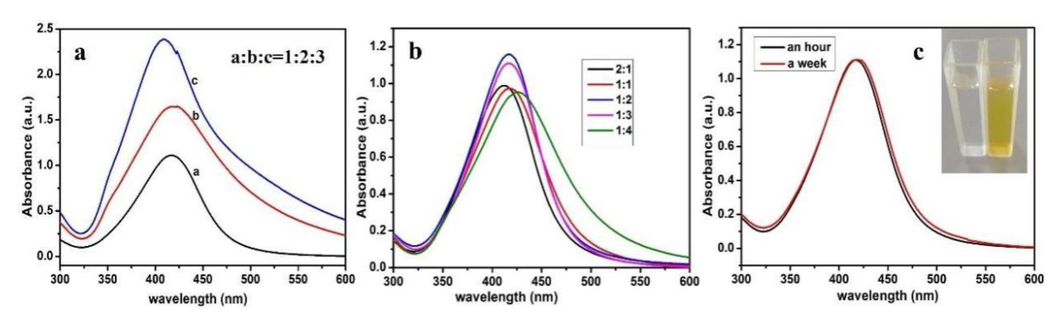
3.1. Characterization Studies of CMS/PVP-Ag NPs Colloidal Solution

The synthesis method of a silver-nanoparticle (Ag NPs)-based chemosensor used CMS as the stabilizing agent and glucose as the reducing agent [39]. The Ag NPs sol was synthesized by adding AgNO3 solutions of different concentrations and the obvious characterization adsorption was presented in the region of 410–430 nm. As shown in Figure 1a, the absorbance intensity of the Ag NPs sol prepared increased with the increase in AgNO3 concentration, and there was a small blue shift, indicating that sol aggregation occurred easily at high concentrations. Figure 1b shows the UV-vis spectra of silver sol prepared by AgNO3 and glucose with different molar ratios. From the absorption curve, there was no significant difference between the molar ratios 1:2 and 1:3. However, after a week, the absorption curve of AgNO3/glucose = 1/3 almost changed (Figure 1c), indicating that the CMS/PVP-Ag NPs colloid solution possessed the best stability.
Figure 2a shows the XRD pattern of Ag NPs to analyze the crystalline phase. It can be seen that there was an obvious crystal diffraction peak at 2θ = 38.5°, which can be assigned to the (111) of silver crystal according to the JCPDS card. In addition, there are three weak peaks at 2θ of 44.5°, 64.5° and 77.6°, corresponding to the (200), (220) and (311) crystal planes of the crystalline silver, respectively, indicating the formation of simple silver in the system [40,41]. The morphology of Ag NPs was characterized by the TEM image (Figure 2b). Clearly, Ag NPs exhibit well-defined spherical crystals, and the SAED pattern of CMS/Ag NPs shows bright circular rings, proving the polycrystalline characteristic of the synthesized Ag NPs [42]. These particle diameters are distributed in the range of 15 to 40 nm and the calculated average nanocrystal size of Ag NPs was approximately 25 nm (Figure 2c). Meanwhile, the EDS results suggest the distribution of the Ag element (Figure 2d) [43].

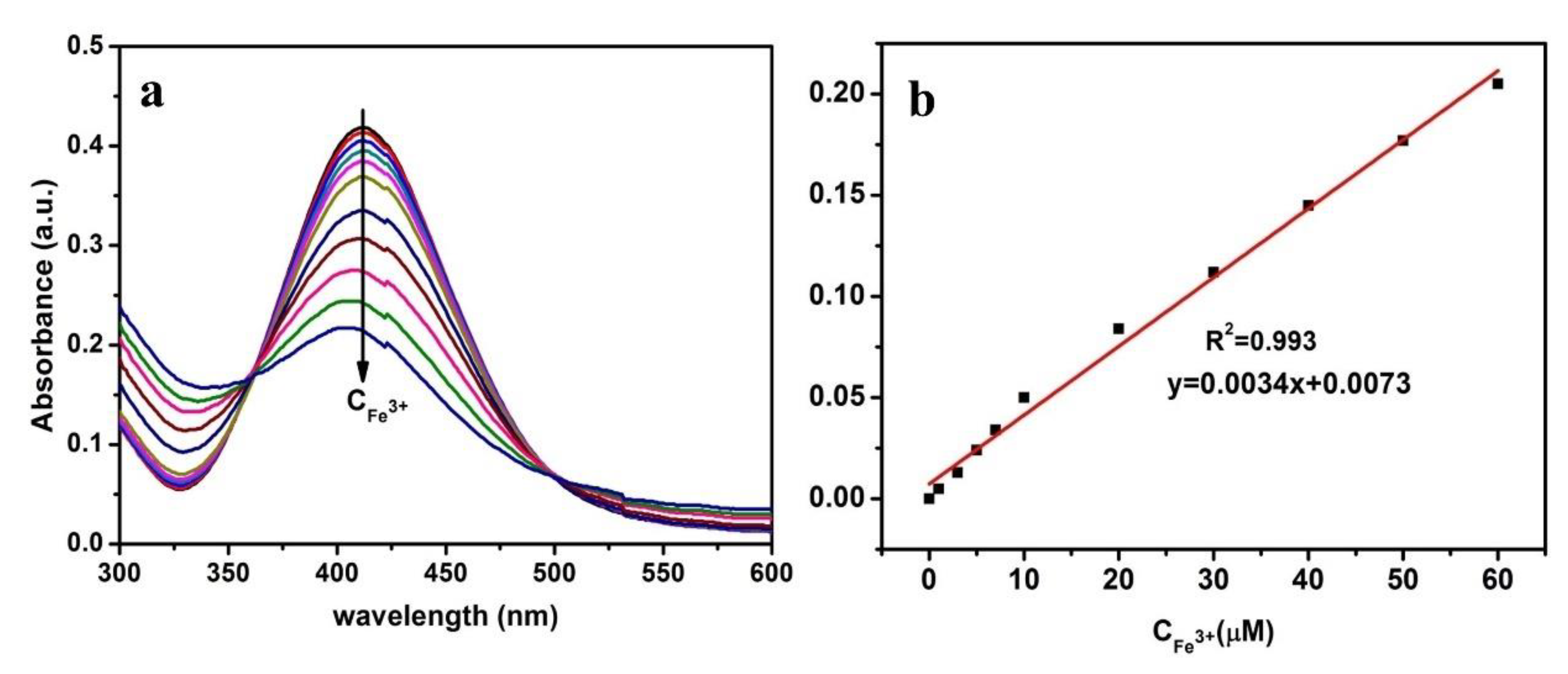
3.2. Detection Selectivity and Sensitivity of Hg2+ and Fe3+

The various metal ions were selected (such as Hg2+, Cd2+, Co2+, Cu2+, Fe3+, Fe2+, K+, Al3+, Ba2+, Ca2+, Mg2+, Na+, Ni2+, Pb2+ and Zn2+) to verify the selectivity of the CMS/PVP-Ag NPs colloidal solution. As shown in Figure 3a, all ions were treated as detected under the same conditions with a maximum concentration of 50 μM, and only Hg2+ and Fe3+ produced noticeable response curves and the absorption peak decreased with an obvious blue shift after the addition of Hg2+ and Fe3+. Moreover, the values of ΔA were found to be 0.62 and 0.3 in the presence of Hg2+ and Fe3+, respectively, and the obvious changes could not be found in other metal ions (Figure 3b). When Hg2+ and Fe3+ are added to 50 μM, the color of the CMS/PVP-Ag NPs colloidal solution tended to be colorless and lighter, respectively, further demonstrating that the CMS/PVP-Ag NPs colloidal solution has a unique selectivity for Hg2+ and Fe3+ rather than other metal ions [44].
Moreover, the sensitivity of the CMS/PVP-Ag NPs colloidal solution for Hg2+ and Fe3+ was further investigated to probe the optimum conditions. It was shown that the color of the sensing system gradually faded away with increasing concentrations of Hg2+, and when the concentration of Hg2+ increased to 50 μM, the color of the sensing system disappeared, providing an efficient platform for the colorimetric detection of Hg2+ (inset in Figure 4a). On the addition of Hg2+, the absorption peak of Ag NPs decreased and the maximum absorption wavelength of Ag NPs displayed a distinct blue shift (Figure 4a), which indicated the formation of a Ag–Hg amalgam on the surface of Ag NPs and the decrease in the concentration and size of the nano-silver particles [45]. The correlation of the ion concentration in the range of 0 to 100 μM and the absorption response were exhibited in Figure 4b; there were two good linear relationships (R1 = 0.998, R2 = 0.995) between ΔA and Hg2+ (0–100 μM), and the LOD for Hg2+ was estimated to be 7.1 nM (0–20 μM) and 15.2 nM (20–100 μM). Meanwhile, Figure 4c reveals two favorable linear relationships (R1 = 0.980, R2 = 0.991) between Δλ and the Hg2+ concentration in the range of 0 to 100 μM, and the LOD was approximately 0.2 nM (0–30 μM) and 0.7 nM (30–100 μM), respectively. The value of LOD, due to the addition of the small amount of Hg2+, could significantly decline when the absorbance was added, causing the higher sensitivity of detection, which is comparable with or superior to other detection methods in previous literature (Table S1) [46,47,48,49,50,51]. The excellent LOD value with small Hg2+ concentrations and the wide linear range implied a high sensitivity and potential application for Hg2+ detection.
As shown in Figure 5a, a slight decrease in the absorbance of Ag NPs could be observed with a gradual increase in the concentration of Fe3+. It is noted that the ΔA and the concentration of Fe3+ in the range of 0–60 μM maintained a good linear relationship (R = 0.993) and the value of LOD was approximately 3.6 nM, demonstrating that the detection ability of the system was relatively significant (Figure 5b). Table S2 shows a comparison of our sensors with those previously reported in literature [5,13,15,17,19,52,53,54,55]. The method is green, rapid, simple, low-cost and highly sensitive. More importantly, the as-prepared sensors showed good stability, and were highly sensitive and selective with a low LOD and the wide linear range for the detection of Fe3+.
The TEM image indicates the aggregation of Ag NPs after the addition of Hg2+, demonstrating that the addition of Hg2+ can induce the formation of a Ag–Hg amalgam (Figure 6b) [44]. Figure 6c reveals that the size of Ag NPs is estimated to be 50–150 nm, which corresponds with the TEM image. Additionally, the result of EDS shows three peaks of Hg (Figure 6d), which is consistent with the above results.
TEM was conducted to explore the morphology variation of the CMS/ PVP-Ag NPs sol when the Hg2+ was introduced. Figure 7a presents the image of CMS/PVP-Ag NPs with Hg2+, and Figure 7b and c reveals the existence of mercury and silver elements in the CMS/PVP-Ag NPs, respectively. A superimposed image of mercury and silver are shown in Figure 7d. It can be observed that the composites are hybridizing with each other, and adding Hg2+ into the CMS/PVP-Ag NPs can increase the size of Ag NPs to form a large cluster.

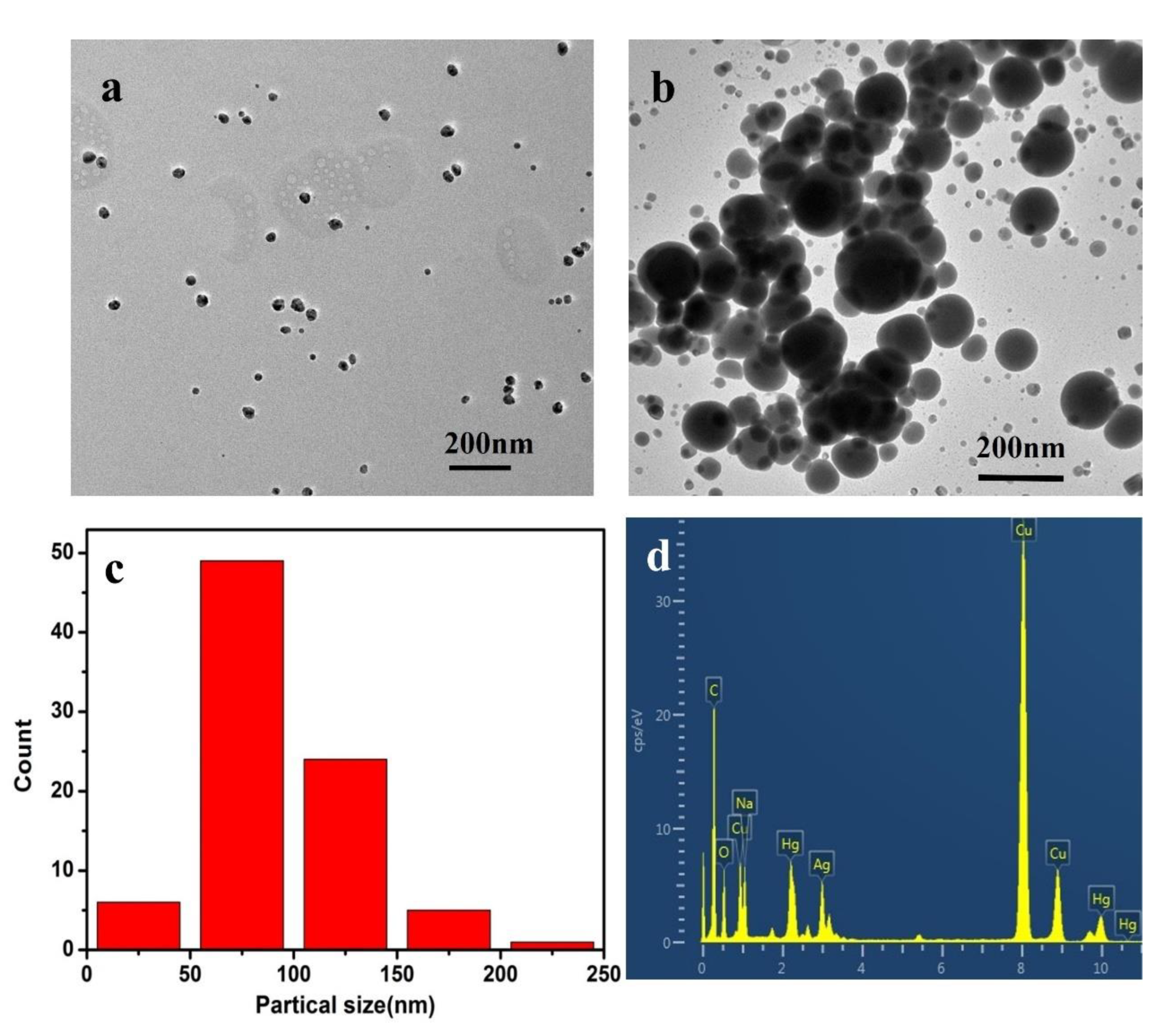
3.3. Mechanism Study for the CMS/PVP-Ag NPs Colloidal Solution and the Detection of Hg2+ and Fe3+

Figure 8 exhibits the reaction scheme and mechanism of the CMS/PVP-Ag NPs colloid for the detection of Hg2+ and Fe3+. The synthesis processes involve adsorption, in situ reduction and a complexation/oxidation process. As previously mentioned, CMS and PVP were self-assembled to form the stabilized skeleton and the silver ions were uniformly anchored on the surface of the CMS molecules via a non-covalent bond. Subsequently, the glucose molecules containing aldehyde groups act as the reducing agent to form the Ag NPs through a silver mirror reaction. The larger steric hindrance effect caused the Ag NPs to be uniformly dispersed and not easy to coalesce, as shown in Figure 8b. It was observed that when Hg2+ was added into the CMS/PVP-Ag NPs colloidal solution, it became colorless and the SPR band intensity diminished with a blue shift (see Figure 4a), which can also be demonstrated by the morphology changes shown in Figure 8a. The size of Ag NPs was changed from about 25 nm to 50–150 nm in Figure 8b and induced the aggregation of Ag NPs. Furthermore, the detective absorption of Hg2+ was examined by a redox reaction with Ag0 and Hg2+. The generated Hg0 adhered to the surface of Ag NPs, forming the Ag–Hg amalgam to enlarge the aggregation of Ag NPs (Figure 8b) [45]. When Fe3+ was added to the CMS/PVP-Ag NPs colloidal solution, it became colorless and the SPR band intensity diminished without any red or blue shift in Figure 5a. It indicated that the concentration of Ag NPs in the solution decreases, but the size of Ag NPs remained unchanged, which can be attributed to the oxidation of the Ag0, leading to the decomposition of Ag NPs, reducing the SPR band strength and making the solution colorless (Figure 8b) [56,57].

3.4. Detection of Hg2+ and Fe3+ in Tap Water and Seawater

To test the feasibility and practicability of composites, the prepared product was applied to monitor tap water and seawater. The tap water and seawater were, respectively, taken from our laboratory and China’s Yellow Sea. Firstly, the tested water samples were filtered with a 0.22 μm membrane to remove insoluble matter. Secondly, three known concentrations of Hg2+ and Fe3+ were added to CMS/PVP-Ag NPs colloidal solution with constant stirring, maintained for 1 min and we observed the color change with the naked eye. Lastly, the water samples with different concentrations of ions were measured with UV-vis spectrophotometer and the recoveries of Hg2+ and Fe3+ were calculated in real samples. We measured the recoveries of Hg2+ in real samples with a range of 98.33~104.2% (Table 1) and the recoveries of Fe3+ in water samples ranged from 98.85% to 104.80% (Table 2). The results show that this method can provide accurate and reliable results for the determination of Hg2+ and Fe3+ in tap water and seawater. Our colloidal solution could be used to detect Hg2+ and Fe3+ in water samples for practical applications.

4. Conclusions

In summary, we have developed an efficient and effective colorimetric chemosensor CMS/PVP-Ag NPs colloid through a facile strategy, which had superior detection selectivity for Hg2+ and Fe3+. At the addition of Hg2+, Ag NPs nanoparticles were induced to enlarge the size of the nanoparticles. Benefiting from the existence of an obvious color change for metal ions and localized SPR absorption of Ag NPs, the colloidal solution exhibited prominent colorimetric responses from yellow to white in the presence of Hg2+ and from yellow to buff in the presence of Fe3+; the achromatic mechanism may be that Ag–Hg alloy was formed by an oxidation-reduction reaction between Hg2+ and nano silver ions, and Fe2+ was formed by Fe3+. CMS/PVP-Ag NPs exhibited excellent selectivity for Hg2+ and Fe3+ with low limits of detection of 1.4 μg L−1 (7.1 nM) and 0.2 μg L−1 (3.6 nM), respectively. Additionally, a simple method for detecting the concentration of Hg2+ and Fe3+ in tap water and seawater was used, proving their bright prospects to be practically applied in real analysis.

Supplementary Materials

The following supporting information can be downloaded at: https://www.mdpi.com/article/10.3390/polym14183745/s1, Table S1: Comparison of the proposed Hg2+ detection method with other reported methods; Table S2: Comparison of the proposed Fe3+ detection with other reported methods.

Author Contributions

Conceptualization, Y.Y.; methodology, Y.Y.; Software, Y.Y.; Validation, Y.X.; Investigation, R.D.; Data curation, Y.Y. and Y.W.; Writing—original draft preparation, Y.Y.; Writing—review and editing, Y.X.; Visualization, X.Z.; Supervision, Z.L., Y.X. and Q.L.; Funding acquisition, Q.L. All authors have read and agreed to the published version of the manuscript.

Funding

This work was funded by the National Natural Science Foundation of China (grant number 51773102, 51503110), and Natural Science Foundation of Shandong Province (grant number ZR2014JL010).

Institutional Review Board Statement

Not applicable.

Informed Consent Statement

Not applicable.

Data Availability Statement

The data presented in this study are available on request from the corresponding author.

Acknowledgments

The authors gratefully acknowledge the National Natural Science Foundation of China (grant number 51773102, 51503110), and Natural Science Foundation of Shandong Province (grant number ZR2014JL010) for their financial support.

Conflicts of Interest

The authors declare no conflict of interest.

References

  1. Li, L.; Gui, L.; Li, W. A colorimetric silver nanoparticle-based assay for Hg(II) using lysine as a particle-linking reagent, Microchim. Acta 2015, 182, 1977–1981. [Google Scholar]
  2. Ahmed, F.; Ali, I.; Ali, H.S.; Yasmeen, S.; Ullah, S.; Burki, S.; Adil, M.; Nisar, J.; Shah, M.R. Synthesis and characterization of a plant growth regulator based silver nanoparticles for the ultrasensitive detection of environmentally toxic Hg2+ ions in tap water. New J. Chem. 2021, 45, 18039–18047. [Google Scholar] [CrossRef]
  3. Mohammadi, A.; Yaghoubi, S. A new dual colorimetric chemosensor based on quinazolinone for CN, AcO and Cu2+ ions. Sens. Actuators B Chem. 2017, 241, 1069–1075. [Google Scholar] [CrossRef]
  4. Wang, Y.; Dong, X.; Zhao, L.; Xue, Y.; Zhao, X.; Li, Q.; Xia, Y. Facile and Green Fabrication of Carrageenan-Silver Nanoparticles for Colorimetric Determination of Cu2+ and S2-. Nanomaterials 2020, 10, 83. [Google Scholar] [CrossRef] [PubMed]
  5. Tang, T.; Wang, J.; Xu, D. Synthesis of a novel hyperbranched polymer and its application in multi-channel sensing Fe3+. Res. Chem. Intermed. 2019, 46, 1425–1435. [Google Scholar] [CrossRef]
  6. Chanajaree, R.; Ratanatawanate, C.; Ruangchaithaweesuk, S.; Lee, V.S.; Wittayanarakul, K. Colorimetric detection of Pb2+ ions using curcumin silver nanoparticles. J. Mol. Liq. 2021, 343, 117629. [Google Scholar] [CrossRef]
  7. Mohammadi, A.; Yaghoubi, S. Development of a highly selective and colorimetric probe for simultaneous detection of Cu2+ and CN− based on an azo chromophore. Sens. Actuators B Chem. 2017, 251, 264–271. [Google Scholar] [CrossRef]
  8. Alshawi, J.M.S.; Mohammed, M.Q.; Alesary, H.F.; Ismail, H.K.; Barton, S. Voltammetric Determination of Hg(2+), Zn(2+), and Pb(2+) Ions Using a PEDOT/NTA-Modified Electrode. ACS Omega 2022, 7, 20405–20419. [Google Scholar] [CrossRef] [PubMed]
  9. Mohammed, M.Q.; Ismail, H.K.; Alesary, H.F.; Barton, S. Use of a Schiff base-modified conducting polymer electrode for electrochemical assay of Cd(II) and Pb(II) ions by square wave voltammetry. Chem. Pap. 2021, 76, 715–729. [Google Scholar] [CrossRef]
  10. Wang, Y.; Liu, Y.; Zhao, L.; Sun, L.; Zhao, X.; Xia, Y. κ-Carrageenan-derived carbon dots for highly selective and sensitive detection of Fe3+ and oxytetracycline. J. Mater. Sci. 2020, 56, 1272–1285. [Google Scholar] [CrossRef]
  11. Ismail, M.; Khan, M.I.; Akhtar, K.; Seo, J.; Khan, M.A.; Asiri, A.M.; Khan, S.B. Phytosynthesis of silver nanoparticles; naked eye cellulose filter paper dual mechanism sensor for mercury ions and ammonia in aqueous solution. J. Mater. Sci. Mater. Electron. 2019, 30, 7367–7383. [Google Scholar] [CrossRef]
  12. Xu, J.; Wang, Y.; Sun, L.; Qi, Q.; Zhao, X. Chitosan and kappa-carrageenan-derived nitrogen and sulfur co-doped carbon dots "on-off-on" fluorescent probe for sequential detection of Fe3+ and ascorbic acid. Int. J. Biol. Macromol. 2021, 191, 1221–1227. [Google Scholar] [CrossRef] [PubMed]
  13. Gao, X.; Zhou, X.; Ma, Y.; Qian, T.; Wang, C.; Chu, F. Facile and cost-effective preparation of carbon quantum dots for Fe3+ ion and ascorbic acid detection in living cells based on the “on-off-on” fluorescence principle. Appl. Surf. Sci. 2019, 469, 911–916. [Google Scholar] [CrossRef]
  14. Zhu, H.; Guo, W.; Wang, J.; He, H.; Hou, X.; Zhou, S.; Wang, S. Tuneable design of a pulp fibre-based colorimetric sensor and its visual recognition mechanism for ppb levels of Ag+. Cellulose 2019, 26, 9149–9161. [Google Scholar] [CrossRef]
  15. Jain, A.; Wadhawan, S.; Kumar, V.; Mehta, S.K. Colorimetric sensing of Fe3+ ions in aqueous solution using magnesium oxide nanoparticles synthesized using green approach. Chem. Physic Lett. 2018, 706, 53–61. [Google Scholar] [CrossRef]
  16. Separovic, L.; Lourenço, F.R. Measurement uncertainty evaluation of an analytical procedure for determination of terbinafine hydrochloride in creams by HPLC and optimization strategies using Analytical Quality by Design. Microchem. J. 2022, 178, 107386. [Google Scholar] [CrossRef]
  17. Naik, V.M.; Gunjal, D.B.; Gore, A.H.; Pawar, S.P.; Mahanwar, S.T.; Anbhule, P.V.; Kolekar, G.B. Quick and low cost synthesis of sulphur doped carbon dots by simple acidic carbonization of sucrose for the detection of Fe3+ ions in highly acidic environment. Diam. Relat. Mater. 2018, 88, 262–268. [Google Scholar] [CrossRef]
  18. Tan, Y.; Yang, C.; Qian, W.; Teng, C. Flower-like MnO2 on layered carbon derived from sisal hemp for asymmetric supercapacitor with enhanced energy density. J. Alloy. Compd. 2020, 826, 154133. [Google Scholar] [CrossRef]
  19. Zhou, M.; Zhou, Z.; Gong, A.; Zhang, Y.; Li, Q. Synthesis of highly photoluminescent carbon dots via citric acid and Tris for iron(III) ions sensors and bioimaging. Talanta 2015, 143, 107–113. [Google Scholar] [CrossRef] [PubMed]
  20. Kim, S.Y.; Lee, S.Y.; Jung, J.M.; Kim, M.S.; Kim, C. Selective detection of Cu2+ and S2− by a colorimetric chemosensor: Experimental and theoretical calculations. Inorg. Chim. Acta 2018, 471, 709–717. [Google Scholar] [CrossRef]
  21. Zhang, R.; Zhang, W.; Shi, M.; Li, H.; Ma, L.; Niu, H. Morphology controllable synthesis of heteroatoms-doped carbon materials for high-performance flexible supercapacitor. Dye. Pigment. 2022, 199, 109968. [Google Scholar] [CrossRef]
  22. Ni, M.; Chen, J.; Wang, C.; Wang, Y.; Huang, L.; Xiong, W.; Zhao, P.; Xie, Y.; Fei, J. A High-Sensitive Dopamine Electrochemical Sensor Based on Multilayer Ti3C2 MXene, Graphitized Multi-Walled Carbon Nanotubes and ZnO Nanospheres. Microchem. J. 2022, 178, 107410. [Google Scholar] [CrossRef]
  23. Pouzesh, M.; Nekouei, S.; Zadeh, M.A.F.; Keshtpour, F.; Wang, S.; Nekouei, F. Fabrication of stable copper nanoparticles embedded in nanocellulose film as a bionanocomposite plasmonic sensor and thereof for optical sensing of cyanide ion in water samples. Cellulose 2019, 26, 4945–4956. [Google Scholar] [CrossRef]
  24. Zhang, R.; Zhang, W.; Yang, Q.; Dong, J.; Ma, L.; Jiang, Z.; Huang, Y. 3D hierarchical oxygen-deficient AlCoNi-(oxy)hydroxides/N-doped carbon hybrids enable efficient battery-type asymmetric supercapacitor. J. Energy Chem. 2022, 72, 416–423. [Google Scholar] [CrossRef]
  25. Song, J.; Zhao, L.; Wang, Y.; Xue, Y.; Deng, Y.; Zhao, X.; Li, Q. Carbon Quantum Dots Prepared with Chitosan for Synthesis of CQDs/AuNPs for Iodine Ions Detection. Nanomaterials 2018, 8, 1043. [Google Scholar] [CrossRef]
  26. Sun, L.; Wei, W.; Zhang, H.; Xu, J.; Zhao, X. A simple colorimetric and fluorescent “on-off-on” dual-mode sensor based on cyan fluorescent carbon dots/AuNPs for the detection of L-cysteine and Zinc thiazole. Microchem. J. 2022, 174, 107079. [Google Scholar] [CrossRef]
  27. Sun, B.; Gou, Y.; Ma, Y.; Zheng, X.; Bai, R.; Abdelmoaty, A.A.A.; Hu, F. Investigate electrochemical immunosensor of cortisol based on gold nanoparticles/magnetic functionalized reduced graphene oxide. Biosens. Bioelectron. 2017, 88, 55–62. [Google Scholar] [CrossRef]
  28. Akhondi, M.; Jafari, A.H.; Jamalizadeh, E. Selective colorimetric detection of HgII using silver nanoparticles modified with Apple and Nigella Sativa seed extracts and β-Cyclodextrin. J. Environ. Chem. Eng. 2020, 8, 103566. [Google Scholar] [CrossRef]
  29. Joshi, P.; Nemiwal, M.; Al-Kahtani, A.A.; Ubaidullah, M.; Kumar, D. Biogenic AgNPs for the non-cross-linking detection of aluminum in aqueous systems. J. King Saud Univ. Sci. 2021, 33, 101527. [Google Scholar] [CrossRef]
  30. Liu, T.; Dong, J.X.; Liu, S.G.; Li, N.; Lin, S.M.; Fan, Y.Z.; Lei, J.L.; Luo, H.Q.; Li, N.B. Carbon quantum dots prepared with polyethyleneimine as both reducing agent and stabilizer for synthesis of Ag/CQDs composite for Hg(2+) ions detection. J. Hazard. Mater. 2017, 322, 430–436. [Google Scholar] [CrossRef]
  31. Cheon, J.Y.; Park, W.H. Green Synthesis of Silver Nanoparticles Stabilized with Mussel-Inspired Protein and Colorimetric Sensing of Lead(II) and Copper(II) Ions. Int. J. Mol. Sci. 2016, 17, 2006. [Google Scholar] [CrossRef] [PubMed] [Green Version]
  32. Yu, Z.; Hu, C.; Guan, L.; Zhang, W.; Gu, J. Green Synthesis of Cellulose Nanofibrils Decorated with Ag Nanoparticles and Their Application in Colorimetric Detection of l-Cysteine. ACS Sustain. Chem. Eng. 2020, 8, 12713–12721. [Google Scholar] [CrossRef]
  33. Sharma, N.; Selvam, S.P.; Yun, K. Electrochemical detection of amikacin sulphate using reduced graphene oxide and silver nanoparticles nanocomposite. Appl. Surf. Sci. 2020, 512, 145742. [Google Scholar] [CrossRef]
  34. Gan, T.; Lv, Z.; Sun, J.; Shi, Z.; Liu, Y. Preparation of graphene oxide-wrapped carbon sphere@silver spheres for high performance chlorinated phenols sensor. J. Hazard. Mater. 2016, 302, 188–197. [Google Scholar] [CrossRef] [PubMed]
  35. Ma, Z.-M. The Preparation of Ag Nanoparticles/Graphene Nanocomposites with Polydopamine as Coupling Agent for Enhanced Detection of H2O2. Int. J. Electrochem. Sci. 2019, 14, 6840–6854. [Google Scholar] [CrossRef]
  36. Ali, S.; Shah, M.R.; Hussain, S.; Khan, S.; Latif, A.; Ahmad, M.; Ali, M. A Facile Approach Based on Functionalized Silver Nanoparticles as a Chemosensor for the Detection of Paraquat. J. Clust. Sci. 2021, 33, 413–420. [Google Scholar] [CrossRef]
  37. Ma, L.; Bi, Z.; Zhang, W.; Zhang, Z.; Xiao, Y.; Niu, H.; Huang, Y. Synthesis of a Three-Dimensional Interconnected Oxygen-, Boron-, Nitrogen-, and Phosphorus Tetratomic-Doped Porous Carbon Network as Electrode Material for the Construction of a Superior Flexible Supercapacitor. ACS Appl. Mater. Interfaces 2020, 12, 46170–46180. [Google Scholar] [CrossRef]
  38. Ullah, A.; Ali, I.; Ahmed, F.; Khan, S.; Shah, M.R.; Shaheen, F. Synthesis and characterization of peptide-conjugated silver nanoparticle for selective detection of Hg2+ in human blood plasma and tap water. J. Mol. Liq. 2019, 296, 112095. [Google Scholar] [CrossRef]
  39. Hoang, V.-T.; Dinh, N.X.; Trang, N.L.N.; Khi, N.T.; Quy, N.V.; Tuan, P.A.; Tri, D.Q.; Thang, L.H.; Huy, T.Q.; Le, A.-T. Functionalized silver nanoparticles-based efficient colorimetric platform: Effects of surface capping agents on the sensing response of thiram pesticide in environmental water samples. Mater. Res. Bull. 2021, 139, 111278. [Google Scholar] [CrossRef]
  40. Eswaran, S.G.; Ashkar, M.A.; Mamat, M.H.; Sahila, S.; Mahalingam, V.; Koppisetti, H.V.S.R.M.; Vasimalai, N. Preparation of a portable calorimetry kit and one-step spectrophotometric nanomolar level detection of l-Histidine in serum and urine samples using sebacic acid capped silver nanoparticles. J. Sci. Adv. Mater. Dev. 2021, 6, 100–107. [Google Scholar]
  41. Rajamanikandan, R.; Ilanchelian, M. β-cyclodextrin functionalised silver nanoparticles as a duel colorimetric probe for ultrasensitive detection of Hg2+ and S2− ions in environmental water samples. Mater. Today Commun. 2018, 15, 61–69. [Google Scholar] [CrossRef]
  42. Zhao, Y.; Gui, L.; Chen, Z. Colorimetric detection of Hg2+ based on target-mediated growth of gold nanoparticles. Sens. Actuators B Chem. 2017, 241, 262–267. [Google Scholar] [CrossRef]
  43. Deng, L.; Ouyang, X.; Jin, J.; Ma, C.; Jiang, Y.; Zheng, J.; Li, J.; Li, Y.; Tan, W.; Yang, R. Exploiting the higher specificity of silver amalgamation: Selective detection of mercury(II) by forming Ag/Hg amalgam. Anal. Chem. 2013, 85, 8594–8600. [Google Scholar] [CrossRef] [PubMed]
  44. Shrivas, K.; Nirmalkar, N.; Deb, M.K.; Dewangan, K.; Nirmalkar, J.; Kumar, S. Application of functionalized silver nanoparticles as a biochemical sensor for selective detection of lysozyme protein in milk sample. Spectrochim. Acta. A Mol. Biomol. Spectrosc. 2019, 213, 127–133. [Google Scholar] [CrossRef] [PubMed]
  45. Diamai, S.; Negi, D.P.S. Cysteine-stabilized silver nanoparticles as a colorimetric probe for the selective detection of cysteamine. Spectrochim. Acta. A Mol. Biomol. Spectrosc. 2019, 215, 203–208. [Google Scholar] [CrossRef] [PubMed]
  46. Das, S.; Aktara, M.N.; Sahoo, N.K.; Jha, P.K.; Hossain, M. Sensitive and robust colorimetric assay of Hg2+ and S2− in aqueous solution directed by 5-sulfosalicylic acid-stabilized silver nanoparticles for wide range application in real samples. J. Environ. Chem. Eng. 2017, 5, 5645–5654. [Google Scholar] [CrossRef]
  47. Chen, J.L.; Yang, P.C.; Wu, T.; Lin, Y.W. Determination of mercury (II) ions based on silver-nanoparticles-assisted growth of gold nanostructures: UV-Vis and surface enhanced Raman scattering approaches. Spectrochim. Acta. A Mol. Biomol. Spectrosc. 2018, 199, 301–307. [Google Scholar] [CrossRef] [PubMed]
  48. Eksin, E.; Erdem, A.; Fafal, T.; Kıvçak, B. Eco-friendly Sensors Developed by Herbal Based Silver Nanoparticles for Electrochemical Detection of Mercury (II) Ion. Electroanalysis 2019, 31, 1075–1082. [Google Scholar] [CrossRef]
  49. Ertürk, A.S. Biosynthesis of Silver Nanoparticles Using Epilobium parviflorum Green Tea Extract: Analytical Applications to Colorimetric Detection of Hg2+ Ions and Reduction of Hazardous Organic Dyes. J. Clust. Sci. 2019, 30, 1363–1373. [Google Scholar] [CrossRef]
  50. Faghiri, F.; Ghorbani, F. Colorimetric and naked eye detection of trace Hg2+ ions in the environmental water samples based on plasmonic response of sodium alginate impregnated by silver nanoparticles. J. Hazard. Mater. 2019, 374, 329–340. [Google Scholar] [CrossRef]
  51. Guo, L.; Song, Y.; Cai, K.; Wang, L. “On-off” ratiometric fluorescent detection of Hg2+ based on N-doped carbon dots-rhodamine B@TAPT-DHTA-COF. Spectrochim. Acta. A Mol. Biomol. Spectrosc. 2020, 227, 117703. [Google Scholar] [CrossRef] [PubMed]
  52. Guo, Y.; Cao, F.; Li, Y. Solid phase synthesis of nitrogen and phosphor co-doped carbon quantum dots for sensing Fe3+ and the enhanced photocatalytic degradation of dyes. Sens. Actuators B Chem. 2018, 255, 1105–1111. [Google Scholar] [CrossRef]
  53. Atchudan, R.; Edison, T.; Aseer, K.R.; Perumal, S.; Karthik, N.; Lee, Y.R. Highly fluorescent nitrogen-doped carbon dots derived from Phyllanthus acidus utilized as a fluorescent probe for label-free selective detection of Fe3+ ions, live cell imaging and fluorescent ink. Biosens. Bioelectron. 2018, 99, 303–311. [Google Scholar] [CrossRef] [PubMed]
  54. Xu, Q.; Pu, P.; Zhao, J.; Dong, C.; Gao, C.; Chen, Y.; Chen, J.; Liu, Y.; Zhou, H. Preparation of highly photoluminescent sulfur-doped carbon dots for Fe(iii) detection. J. Mater. Chem. A 2015, 3, 542–546. [Google Scholar] [CrossRef]
  55. Xing, A.; Miao, X.; Liu, T.; Yang, H.; Meng, Y.; Li, X. An intrinsic white-light-emitting hyperbranched polyimide: Synthesis, structure–property and its application as a “turn-off” sensor for iron(iii) ions. J. Mater. Chem. C 2019, 7, 14320–14333. [Google Scholar] [CrossRef]
  56. Gao, X.; Lu, Y.; He, S.; Li, X.; Chen, W. Colorimetric detection of iron ions (III) based on the highly sensitive plasmonic response of the N-acetyl-L-cysteine-stabilized silver nanoparticles. Anal. Chim. Acta 2015, 879, 118–125. [Google Scholar] [CrossRef]
  57. Kumar, V.; Mohan, S.; Singh, D.K.; Verma, D.K.; Singh, V.K.; Hasan, S.H. Photo-mediated optimized synthesis of silver nanoparticles for the selective detection of Iron(III), antibacterial and antioxidant activity. Mater. Sci. Eng. C 2017, 71, 1004–1019. [Google Scholar] [CrossRef]
Scheme 1. Schematic illustration for the preparation of CMS/PVP-Ag NPs.
Polymers 14 03745 sch001
Figure 1. UV–VIS spectra of CMS/PVP-Ag NPs Colloidal Solution: (a) Different volumes of AgNO3 (0.1 M), (b) Different molar ratio of AgNO3 to glucose, and (c) The stability of the molar ratio of AgNO3 to glucose (1:3).
Polymers 14 03745 g001
Figure 2. (a) XRD pattern of Ag NPs, (b) TEM images (inset: SAED pattern), (c) Size distribution and (d) EDS spectrum of CMS/PVP-Ag NPs.
Polymers 14 03745 g002
Figure 3. (a) UV–vis absorption spectra; and (b) ΔA of the proposed sensing system in the presence of various metal ions. The concentrations of all metal ions were 50 μM.
Polymers 14 03745 g003
Figure 4. (a) UV–vis spectroscopy of the CMS/PVP-Ag NPs colloidal solution with different concentrations of Hg2+ (0–100 μM), (b) The linear relative absorption intensity ΔA; and (c) SPR band shift in Δλ over the concentration of Hg2+ within the range of 0~100 μM (R2 represents the linear regression relationship between ΔA and different concentrations of Hg2+).
Polymers 14 03745 g004
Figure 5. (a) UV–vis spectroscopy of the CMS/PVP-Ag NPs colloidal solution with different concentrations of Fe3+, and (b) the linear relative absorption intensity ΔA over the concentration of Fe3+ is within the range of 0~60 μM.
Polymers 14 03745 g005
Figure 6. (a) TEM of the CMS/PVP-Ag NPs colloidal solution in the presence of Hg2+ (30 μM), (b) TEM of the CMS/PVP-Ag NPs colloidal solution after the addition of Hg2+ can induce the formation of Ag–Hg amalgam (c) size distribution, and (d) EDS spectrum of CMS/PVP-Ag NPs with the addition of Hg2+.
Polymers 14 03745 g006
Figure 7. (a) Selective area TEM image of CMS/PVP-Ag NPs in the presence of Hg2+, (b) elemental mapping images of Hg, (c) Ag and (d) elements superimposed images.
Polymers 14 03745 g007
Figure 8. (a) Reaction equation of AgNO3 and Glucose; and (b) Reaction scheme of the synthesis of Ag NPs and the interaction between Ag NPs and Hg2+ ions or Fe3+ ions.
Polymers 14 03745 g008
Table 1. Determination of Hg2+ in tap water and sea water samples.
Samples Concerntration of Hg2+ (μM)Recovery (%)RSD (n = 3, %)
SpikedMeasured
Tap water155. 21104.204.35
21010.34103.404.67
31514.8598.333.14
Sea water154.9899.603.89
21010.22102.204.20
31515.35102.364.35
Table 2. Determination of Fe3+ in tap water and sea water samples.
Samples Concerntration of Fe3+ (μM)Recovery (%)RSD (n = 3, %)
SpikedMeasured
Tap water155.24104.805.15
21010.26102.604.56
32019.7798.853.97
Sea water154.9699.203.85
21010.21102.105.25
32020.32101.604.28
Publisher’s Note: MDPI stays neutral with regard to jurisdictional claims in published maps and institutional affiliations.

