Article

Study of the Influence of Magnetite Nanoparticles Supported on Thermally Reduced Graphene Oxide as Filler on the Mechanical and Magnetic Properties of Polypropylene and Polylactic Acid Nanocomposites

by
Benjamin Constant-Mandiola
1,
Héctor Aguilar-Bolados
2,
Julian Geshev
3 and
Raul Quíjada
1,*
1
Facultad de Ciencias Físicas y Matemáticas, Universidad de Chile, Beauchef 851, Santiago 8370456, Chile
2
Departamento de Polímeros, Facultad de Ciencias Químicas, Universidad de Concepción, Concepción 3349001, Chile
3
Instituto de Física, URFGS, Porto Alegre 91501-970, Brazil
*
Author to whom correspondence should be addressed.
Polymers 2021, 13(10), 1635; https://doi.org/10.3390/polym13101635
Submission received: 30 April 2021 / Revised: 11 May 2021 / Accepted: 11 May 2021 / Published: 18 May 2021
(This article belongs to the Section Polymer Composites and Nanocomposites)

Abstract

:
A study addressed to develop new recyclable and/or biodegradable magnetic polymeric materials is reported. The selected matrices were polypropylene (PP) and poly (lactic acid) (PLA). As known, PP corresponds to a non-polar homo-chain polymer and a commodity, while PLA is a biodegradable polar hetero-chain polymer. To obtain the magnetic nanocomposites, magnetite supported on thermally reduced graphene oxide (TrGO:Fe3O4 nanomaterial) to these polymer matrices was added. The TrGO:Fe3O4 nanomaterials were obtained by a co-precipitation method using two types of TrGO obtained by the reduction at 600 °C and 1000 °C of graphite oxide. Two ratios of 2.5:1 and 9.6:1 of the magnetite precursor (FeCl3) and TrGO were used to produce these nanomaterials. Consequently, four types of nanomaterials were obtained and characterized. Nanocomposites were obtained using these nanomaterials as filler by melt mixer method in polypropylene (PP) or polylactic acid (PLA) matrix, the filler contents were 3, 5, and 7 wt.%. Results showed that TrGO600-based nanomaterials presented higher coercivity (Hc = 8.5 Oe) at 9.6:1 ratio than TrGO1000-based nanomaterials (Hc = 4.2 Oe). PLA and PP nanocomposites containing 7 wt.% of filler presented coercivity of 3.7 and 5.3 Oe, respectively. Theoretical models were used to analyze some relevant experimental results of the nanocomposites such as mechanical and magnetic properties.

Graphical Abstract

1. Introduction

The preparation of polymeric nanocomposites based on carbon nanomaterials is an excellent alternative to obtain polymeric materials with enhanced mechanical or electrical properties [1,2]. This is because carbon nanomaterials impart mechanical resistance and electrical conductivity to the soft and electrically insulating polymeric matrix [3,4,5]. As known, one of the most widely used nanomaterials as fillers is graphene, due to its diversity of properties, such as its high surface area of 2630 m2·g−1 and high mechanical properties of 1300 GPa [6]. In this respect, the high surface area of graphene allows for use in a wide range of multiple purposes, such as elastomers, thermoplastic, sustainable glassy nanocomposites [7,8,9] as well as hydrogel-based nanocomposites for environmental remediation, heavy metal absorption, and waste treatment [10,11,12]. Moreover, the high surface area of graphene allows hosting on its surface different nanomaterials which specific properties, such as magnetic properties [13].
There are several approaches to obtain graphene materials, one of which considers the thermal, photo, or chemical reduction of graphene oxide (GO) [14,15,16,17,18,19]. Using a modified Hummers’ method, GO can be obtained from graphite. However, the residual presence of functional groups present in GO gives it some differences compared with pristine graphene [20]. The thermal reduction of GO is considered one of the highest efficiency and least polluting procedures for obtaining graphene materials [14]. The heat used to reduce GO not only favors total or partial elimination of oxygen-functional groups present in the GO but also allows the exfoliation of the graphene sheets to take place [21,22]. The exfoliation favors the drastic increase of the surface area of these types of materials. Consequently, the partial functionalization with oxygen moieties and the high surface area of these graphene materials opens an interesting approach to explore the synthesis and support of different types of nanoparticles on the surface of graphene.
On the other hand, magnetite is an iron oxide, Fe3O4, which stands out by presenting magnetic properties [23]. Various methods allow the synthesis of magnetite: electrochemistry [24], sol-gel [25], and chemical coprecipitation [26], the latter being more widely used due to its easy scaling up to the industrial level. The coprecipitation method of magnetite is carried out by using as precursors basic aqueous solutions of FeCl3 and FeCl2, as shown in Equation (1) [27].
Fe 2 + + 2 Fe 3 + + 8 OH H 2 O / NH 3 Fe 3 O 4 + H 2 O
The synthesis of magnetite can also be performed on nanomaterial surfaces that present a large surface area such as graphene materials. The presence of functional groups favors the nucleation of magnetite on their surface, obtaining a material with magnetic properties [28].
On the other hand, the pollution generated by the α-olefin-based thermoplastics polymers has promoted new research with more sustainable polymers such as polylactic acid (PLA), which can be degraded by external agents such as humidity, UV light, and composting, reducing the adverse impact on the environment [29]. However, it is also necessary to find alternatives to recycle nondegradable polymers such as PP and facilitate this process. Obtaining nanocomposites with magnetic properties allows separating and collecting these materials from disposal polymer batches faster using an external magnetic field, facilitating their recycling. The dispersion of the nanoparticles in the polymers depends as much on the nanoparticle-polymer interaction forces as well as on the nanoparticle-nanoparticle interaction forces [30]. Nanoparticles with a higher polarity due to the presence of functional groups or groups with unpaired electrons will have a higher affinity with a polar polymer matrix such as PLA and PA [31]. In the case of nonpolar nanoparticles, they will have a higher affinity with a nonpolar polymer matrix such as PP [5].
Herein, two thermally reduced graphene oxides at 600 and 1000 °C that have different contents of oxygen functional groups were used as supports of magnetite synthesized by a coprecipitation method. The structure, morphology, and magnetic properties of these supported-magnetite nanoparticles on TrGO were studied. Besides, the influence of these hybrid nanomaterials on the properties of the thermoplastic polymer matrices, namely polypropylene and polylactic acid were studied. These matrices were selected because they are polymers with importance in the industry of thermoplastic polymers. For instance, PP is extensively used in household and industrial applications, consequently it is considered a commodity [32]. Conversely, PLA is far less used for manufacturing low price goods, the PLA’s importance stems from its biodegradable character and possesses prospective applications [33]. It is important to mention that PP corresponds to a non-polar homo-chain polymer, while PLA is a polar hetero-chain polymer. As known, nowadays, there is a significant interest in using biodegradable polymers in the thermoplastic industry. However, in both cases, the study of their preparation and magnetic properties could provide information for understanding the behavior of these nanocomposites.

2. Materials and Methods

2.1. Materials

Graphite, sulfuric acid (H2SO4, 98%), potassium permanganate (KMnO4, ≥99.0%), hydrochloric acid (HCl, 37%), sodium nitrate (NaNO3, 99.0%), iron(III) chloride hexahydrate (FeCl3·6H2O, 97%), iron(II) chloride tetrahydrate (FeCl2·4H2O, 98%) and ammonia solution (NH3, 25%) were supplied by Merck (Kenilworth, NJ, USA). Polypropylene (PP) PH 2621, density = 905 kg·m−3, molecular weight = 195 Kg·mol−1, melt flow index = 27 g/10 min (2.16 kg, 230 °C) supplied by Petroquim S.A. (Santiago, Chile). Polylactic acid (PLA) 4032D, density = 1240 kg·m−3, molecular weight = 107.3 Kg·mol−1, melt flow index = 7 (2.16 kg, 230 °C), supplied by NatureWorks (Minnetonka, Minneapolis, MN, USA). All materials were used as received.

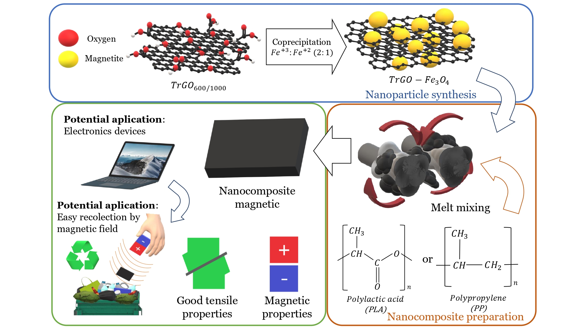
2.2. Synthesis of TrGO/Fe3O4 Nanoparticles

2.2.1. Graphene Oxide Synthesis and Thermally Reduction Process

The graphene oxide (GO) was obtained by a modified Hummers method, which considers the addition of 15 g of graphite and 7.5 g of NaNO3 dispersed in 375 mL of sulfuric acid under constant stirring at 0 °C. The suspension was stirred for 30 min, and then 45 g of KMnO4 was added slowly in small portions over a period of one hour. Then the suspension was mixed to room temperature and was left to react for 45 min. The mixture was poured carefully into 750 mL of distilled water, and 675 mL of H2O2 (5 vol%), added with the purpose of removing the excess of KMnO4. The reaction product was washed with HCl (26%) and was filtered several times until the supernatant achieved a pH ca. 7. Finally, the GO was dried at 80 °C for 12 h [34].
For the thermally reduced graphene oxide (TrGO) synthesis, GO was previously dried for 6 h at 80 °C under vacuum. A small portion of GO was introduced into a quartz reactor, which was sealed and purged using nitrogen gas. The reactor was slowly heated up 600 °C or 1000 °C and the reactor was left for 30 s at the working temperature. The reactor was then allowed to cool down to room temperature. The thermal shock is the prime requirement to achieve exfoliation and reduction of graphene sheet functional groups [14].

2.2.2. TrGO/Magnetite Nanomaterial Synthesis

The TrGO/magnetite nanomaterial was synthesized by coprecipitation of FeCl3·6H2O and FeCl2·4H2O in the presence of TrGO600 or TrGO1000. The aqueous solutions of ferric chloride and ferrous chloride were prepared in a 2:1 mole ratio. Two solutions with different concentrations were used in order to obtain materials with different FeCl3 and TrGO600 or TrGO1000 weight ratios of 2.5:1 and 9.6:1 FeCl3:TrGO, resulting in four samples of nanomaterials: (Mb600, Mb1000, Ma600, and Ma1000). The preparation of Mb600 and Mb1000 consisted of the addition of 50 mg of TrGO600 (for Mb600) or TrGO1000 (for Mb1000) to 50 mL of DI water. These suspensions were ultrasonicated for 1 h, and then 50 mL of FeCl3 (125 mg) and FeCl2 (49 mg) solution in DI water was added at 80 °C. Similarly, the preparation of Ma600 and Ma1000 considered the addition of 50 mg of TrGO600 (for Ma600) or TrGO600 (for Ma1000) in 50 mL of DI water was ultrasonicated for 1 h. Then 50 mL of FeCl3 (480 mg) and FeCl2 (188 mg) solutions were added at 80 °C [28,35,36].
After mixing the ferric chloride and ferrous chloride solutions with the TrGO solution, a 25% ammonia solution was added, increasing the pH to 10 at 80 °C. This process was carried out under a nitrogen gas flow. The resulting suspensions were stirred for 30 min. The suspensions were cooled at 0 °C in an ice bath, the nanomaterial was separated using a magnet and was washed three times with DI water, and finally vacuum-dried at 60 °C for 2 h.

2.3. Characterization of Nanoparticles

The different graphene materials were characterized by elemental analysis carried out using a Perkin Elmer MCHNSO/2400 analyzer (Waltham, MA, USA). These materials also were characterized by powder X-ray diffraction (XRD) and were recorded using a Siemens D-5000 wide-angle XRD spectrometer (München, Germany) with Cu Kα radiation, operating at 40 kV and 30 mA. The Raman spectroscopy was performed in an inVia Renishaw Raman spectrometer (Wotton-Under-Edge, UK) equipped with a 532 nm laser at a power of 10 mW. The specific surface areas were analyzed in a Nova Station A analyzer Quantachrome instruments, Anton Paar (Ashland, Wilmington, DE, USA), through the determination of the multi-point Brunauer-Emmett-Teller (BET) plot using nitrogen gas, all curves presented an R2 higher than 0.999 [37,38]. Scanning electron microscopy (SEM) studies were performed in an EVO MA10 Carl Zeiss microscope (Jena, Germany) operating at 13.4 kV.
The magnetic properties of TrGO:Fe3O4 (powder) were measured as a function of the applied magnetic field using an EZ29MicroSense vibrating magnetometer (VSM) with a maximum applied magnetic field of 20 KOe. The hysteresis of the magnetization was obtained by varying H between +20 KOe and −20 KOe at room temperature.

2.4. Preparation of Nanocomposites

The nanocomposites were prepared by melt mixing in a Brabender Plasticorder double screw mixer (Nordrhein-Westfalen, Germany). The nanocomposites based on PP and PLA nanocomposites were mixed at 190 and 200 °C for 10 min using a rotor speed of 110 RPM. The filler content in the nanocomposites varied over a range of 3–7% by weight. The PP, PLA, and TrGO:Fe3O4 nanomaterial were dried at 80 °C for 8 h prior to being mixed. Then, the nanocomposites were processed in an HP hydraulic press (model D-50) with a heating system, and a water-cooling system. The sample thickness was 1.0 nm for tensile and magnetic tests.

2.5. Characterization of Nanocomposites

The mechanical properties were determined by tensile-strain tests according to the ASTM D638 standard, at a rate of 25 mm·min−1 at room temperature using an Instron Universal Testing System model 3382, (Norwood, MA, USA). Three specimens were tested for each sample and the average was determined to obtain a representative data dispersion.
The magnetic properties of nanocomposite (films) were measured as a function of the applied magnetic field using an EZ29MicroSense vibrating magnetometer (VSM) (Lowell, MA, USA) with a maximum applied magnetic field of 20 KOe. The hysteresis of the magnetization was obtained by varying H between +20 KOe and −20 KOe at room temperature.

2.6. Theoretical Models

2.6.1. Halpin-Tsai Model

As known, one of the important aspects that influence the mechanical properties of nanocomposites (Young moduli) is the aspect ratio and filler/polymer matrix adhesion. Halpin Tsai model provides information about these features by considering the theoretical moduli of the filler, the matrix, and the experimental moduli of the different nanocomposites. Equations (2) and (3) present the de Halpin Tsai approach [39,40].
E = EM∗(1 + ƞ2αVf)/(1 − ƞVf)
Ƞ = (Ef/EM − 1)/(Ef/EM + 2α)
where EM is the young modulus of the polymer matrix, Ef corresponds to the Young modulus of the filler, α is the aspect ratios of the filler nanoparticles, which also considers their geometry and distribution, and Vf corresponds to the filler volume fraction.

2.6.2. Langevin Model

In order to understand the influence of the magnetic properties of the filler on the matrix polymerics, the Langevin model was used for determining the magnetic moment and nanoparticle size [41]. The Langevin equation, as shown in Equation (4), considers that each monodomain has magnetic moments randomly oriented, which interact with the external magnetic field.
M = Nµ∗(coth(µµoH/kbT) − kbT/µµoH)
where µ is the magnetic moment, µo is the vacuum permittivity, H the external magnetic field, kb corresponds to the Boltzman constant, T is the temperature and N is the Avogadro number.

3. Results and Discussion

3.1. Characterization of Thermally Reduced Graphene Oxide

Figure 1a shows the normalized XRD patterns of graphite, GO, TrGO600, and TrGO1000. Graphite presents an intense diffraction peak at 2θ = 26.3°, corresponding to the (002) plane which is associated with the interlayer spacing between graphene layers (0.338 nm). Once the graphite was oxidized, this peak shifted to 2θ = 12.6°, which indicates an increase in the interlayer distance of 0.702 nm. The increase of the interlayer distance is associated with the incorporation of oxygenated functional groups to the graphitic structure. The thermal reduction process of GO at 600 or 1000 °C produced a shift of this peak to 2θ = 25.1° and 2θ = 26.02°, corresponding to the interlayer spacing of 0.35 nm and 0.34 nm, respectively, as seen in Table 1. Table 1 also presents the chemical composition and the BET surface area of GO, TrGO600, and TrGO1000. In fact, the oxygen contents of TrGO600 and TrGO1000 are lower compared to that of GO (43.6%), and they are 15.7 and 8.7%, respectively. The lowest content achieved in TrGO1000 is because a higher temperature favors a more complete reduction. Concerning the BET surface area results of the nanomaterials (Table 1), it is seen that the surface area increases from 69 m2·g−1 for GO to 304 and 267 m2·g−1 for TrGO600 and TrGO1000, respectively. The increase of the surface area proves the effectiveness of the exfoliation of GO by a thermal reduction process.

3.2. Characterization of TrGO/Fe3O4 Nanomaterials

Figure 1b shows the XRD patterns of Mb600, Mb1000, Ma600, and Ma1000 magnetite (Described in Methodology 2.1.3), all of them showing six peaks at the 2θ angles of 30.2°, 35.4°, 43.3°, 53.8°, 57.2°, and 62.7°. The peak seen at 35.4° presents the highest intensity, which is characteristic of the magnetite-based structures. This peak probably corresponds to the (311) magnetite plane with a face-centered cubic arrangement. The diffraction pattern of the TrGO:Fe3O4 showed the characteristic peaks of Fe3O4, indicating the successful deposition of Fe3O4 on the TrGO surface. Moreover, the peak associated with the (002) plane (2θ = 26.4°) of TrGO was observed only in the Mb600 and Mb1000 samples as seen in Figure 1a, indicating that the TrGO surface probably has a lower magnetite content.
The crystal size was estimated using the Debye-Scherrer equation, and it was possible to determine that Fe3O4 had a 19.4 nm crystal size. Similar values of (311) crystal size of the Fe3O4-based structure deposited on TrGO are shown by samples Mb600 (16.5 nm), Mb1000 (12.2 nm), Ma1000 (16.2 nm), and Ma600 (19.2 nm). The crystal size of magnetite particles supported on TrGO tends to be comparable to the values reported by Baumgartner et al. [42], who used similar pH, temperature, and reaction time conditions. In addition, A. Zubir et al. demonstrated the formation of a mixed oxide of Fe(II) and Fe(III), namely as Fe3O4 supported on graphene oxide, using a similar method of coprecipitation. This was supported by using X-ray photoelectron spectroscopy for the characterization of these nanomaterials, wherein the disappearing of Fe 2p3/2 photoelectron line indicated the existence of the Fe(II) and Fe(III) species [43].
Figure 2a,b shows the Raman spectra of nanomaterials of Mb600, Mb1000, Ma600, and Ma1000. As expected, D and G bands characteristic of graphene materials are seen (Figure 2a). These bands appear at ca. 1350 cm−1 and 1580 cm−1, respectively [44,45]. It is seen that the sample corresponding to Ma1000 presents a Raman redshift, which is associated with the interaction of graphene layers with nanoparticles [46]. This suggests the occurrence of interaction between the Fe-based structure and the graphene oxygen moieties, favoring the nucleation structure on the graphene structure. On the other hand, Figure 2b shows the region of the Raman spectra between 150 cm−1 and 1000 cm−1, presented to study the contribution of magnetite absorption bands. Bands are seen at ca. 212 cm−1, 274 cm−1, and 385 cm−1 [47].
Regarding the BET analysis, samples Mb600, Mb1000, Ma600, and Ma1000 presented surface areas of 143.7, 166.8, 111.2, and 123.8 m2·g−1, respectively. It can therefore be inferred that these decreases depend on the type of graphene used. TrGO:Fe3O4 nanomaterials using TrGO600 as support present greater decreases of their surface areas compared to nanomaterials using TrGO1000 as support. TrGO600 presented more oxygen functional groups on their surface than TrGO1000, probably favoring the synthesis of magnetite on the surface of TrGO600. In samples with many magnetite nanoparticles, graphene layers are hindered, and their large available surface decreases.
Figure 3 shows SEM images of TrGO:Fe3O4 nanomaterials. In the case of nanomaterials of Mb600 and Mb1000 (Figure 3a,b), small magnetite particles are seen on the surface of the graphene flakes and tend to present an amorphous structure. In the case of Ma600 and Ma1000 nanomaterials (Figure 3c,d), they present a highly compacted morphology and heterogeneous distribution of nanoparticle sizes on the surface. The coprecipitation method does not produce homogeneous magnetite particle sizes, so it is possible to find broad size dispersion of magnetite particles on the surface of TrGO [42]. The appearance of the TrGO:Fe3O4 nanomaterials indicates that the Fe3O4 was deposited on the TrGO surfaces, hiding the flakes of this carbon-based nanomaterial. Consequently, the nanomaterials have heterogeneous character; namely, flakes of TrGO, covered by magnetite (Fe3O4).

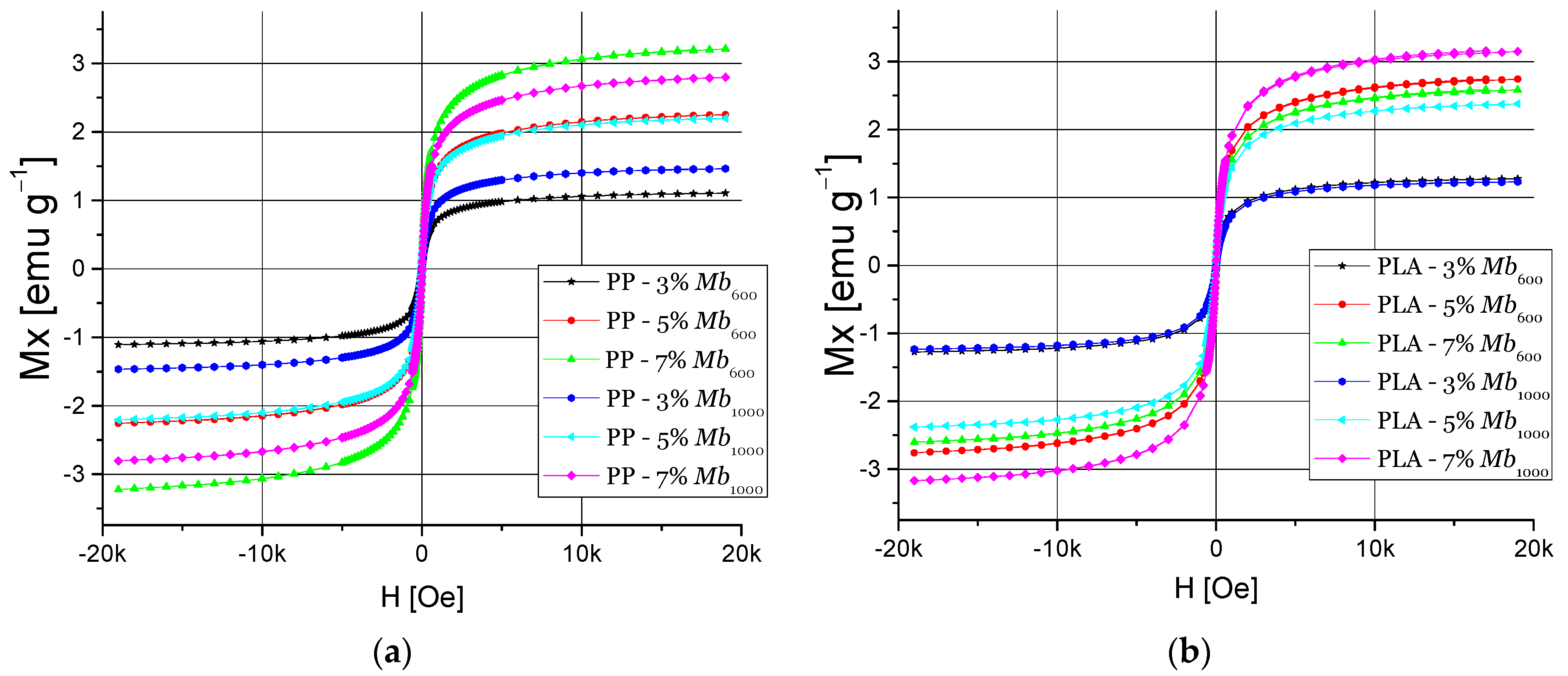
3.3. Magnetic Properties of Nanomaterials

Figure 4 shows the cycles of magnetic hysteresis curves of TrGO:Fe3O4 nanomaterials, where the superparamagnetic behavior of these samples is seen. Nanomaterials Mb600 and Mb1000 presented magnetic saturation (Ms) of 37.5 and 39.9 emu·g−1, and coercivity (Hc) of 0.373 Oe and 0.023 Oe, respectively. On the other hand, nanomaterials Ma600 and Ma1000 presented Ms values of 55.2 and 63.01 emu·g−1, respectively, and the Hc were 8.509 and 4.219 Oe, respectively. The tendency to increase Ms and coercivity Hc, which can be attributed to the higher Fe amount used to synthesize the Ma600 and Ma1000 nanomaterials. This can be inferred from the results of X-ray diffraction analysis and Raman spectroscopy. TrGO600-based nanomaterials have lower magnetic susceptibility but greater coercivity compared to TrGO1000-based nanomaterials. These results suggest that the coercivity and magnetic susceptibility depend on the crystal size of Fe3O4.

3.4. Properties of PP and PLA of Nanocomposites

3.4.1. Mechanical Properties

Figure 5 shows the Young’s modulus of the nanocomposites. In the case of PP-TrGO:Fe3O4 nanocomposites (Figure 5a), it is seen that their Young’s moduli have different behaviors depending on the type of filler used and their concentrations. PP nanocomposites containing 5% Ma600 as filler presented a 21% increase of Young’s modulus. When the filler content was increased to 7%, the Ma1000-based nanocomposites presented a 20% increase, while the Ma600-based nanocomposites had a decrease of their Young’s modulus. The different behaviors of the nanomaterials’ Young’s modulus are attributed to the affinity that nanomaterials have with the PP matrix. A nonpolar nanomaterial has a better affinity with the nonpolar polymer matrix, favoring the obtaining of homogeneous filler dispersion on the polymer matrix. Conversely, nanomaterials with a polar nature have less affinity with a polar polymer matrix such as PP; consequently, filler agglomerations in the polymer matrix take place. Mb1000 presented a higher affinity with the PP matrix, generating increased mechanical properties. This can be attributed to the fact that Mb nanocomposites have a lower content of magnetite, which has a polar nature. This probably favors the achievement of a homogeneous dispersion of filler in the PP matrix and an improved filler/polymer interfacial adhesion. Furthermore, the nanoparticle size favored the adhesion of the nanomaterial with the PP matrix. This is reflected in an increase of Young’s modulus [48]. However, when the filler content increased to 7%, a drastic reduction of mechanical properties took place.
In the case of PLA-TrGO:Fe3O4 nanocomposites (Figure 5b), it is seen that Young’s moduli depend on the filler content and its nature. PLA composites containing 5% filler of Ma1000 exhibited an 11% increase in Young’s modulus. Furthermore, Young’s modulus decreases 28% using a 7% filler of the same nanomaterials. There are two factors that likely can explain the behavior of the mechanical properties of PLA-TrGO:Fe3O4, namely reprocessing of nanomaterials and agglomeration in the polymer matrix. Probably, the processing of PLA at temperatures close to the melting point, 160–180 °C, generates a partial degradation of the polymer chains, as it was reported in the study of Carrasco et al. [49]. The PLA polymer chains start degrading by releasing methyl groups, causing a decrease in molecular weight, and consequently generating changes in the crystallinity of the polymer. This causes a decrease in the mechanical properties, which are linked to the polymer’s intermolecular forces [49,50]. In addition, the unexpected decrease of Young’s moduli of the different nanocomposites also can be related to the heterogeneous nature of the fillers. Magnetite and TrGO likely present weak interactions, and this fact imparts defects in the nanocomposites. As a result, the stiffness of the nanocomposites will decrease in the presence of TrGO:Fe3O4, and the brittleness of the nanocomposites will be higher than the neat polymer matrices. This suggests that the use of a binding agent or organic surface modification can be a strategy to increase the interaction between fillers and polymer matrix and to avoid the decrease of the mechanical properties [51,52,53,54,55].
Figure 6 shows the Halpin Tsai-determined theoretical and experimental young moduli as a function of the filler content of nanocomposites based on PP and PLA, respectively. In Figure 6a It is possible to observe the nanocomposites containing Mb1000 and Ma1000 as filler. As seen in this figure, the Halpin Tsai model tends to converge with experimental data, when the aspect ratio is α = 20 for PPMa1000. On the other hand, the estimated aspect ratio of the nanocomposite PPMb1000 is α = 5. However, the experimental results show a poor correlation with the theoretical data, where nanocomposite filled with 7% of Mb1000. This may be due to the fact that there are interactions between the nanoparticles that can favor or decrease the adhesion of the nanoparticle with the polymer matrix [56].
On the other hand, in Figure 6b, the data of Young’s modulus of PLA with filling of Mb600 and Ma600 with the Halpin Tsai model are shown as a function of the filler content. It is possible to observe that the theoretical data do not fit with the experimental results when α = 1 is used. This suggests that the adhesion of the PLA with nanofiller is low. Consequently, the experimental Young moduli of nanocomposite are lower than the expected values [57].

3.4.2. Magnetic Properties

Figure 7 shows the magnetic hysteresis cycles of PP and PLA nanocomposites with nanomaterials of Mb600 and Mb1000 as a filler with a content of 3, 5, and 7 wt.%. In the case of PP nanocomposites (Figure 7a), it was found Ms value of 3.21 emu·g−1 and a Hc of 0.24 Oe using Mb600 with 7 wt.% filler. These data agree with the mechanical properties found for this nanocomposite with 7 wt.% filler of where Mb600 presents a decrease of these properties due to the agglomerations of the nanomaterials, favoring the contact between the nanoparticles, allowing that a higher magnetic saturation can be achieved in the nanocomposite.
In the case of PLA nanocomposites, they present a high Ms of 3.2 emu·g−1 using Mb1000 with 7% filler (Figure 7b). PLA had a weak interaction with the Mb1000 nanoparticles when a 7% by weight filler was used, increasing the formation of agglomerations due to the interactions between nanomaterials, increasing the magnetic nanocomposite’s susceptibility. On the other hand, the coercivity of the PLA nanocomposites is higher than that of the nanoparticles, reaching 0.810 Oe with Mb600 at 7% filler, more than 200% of coercivity of the nanoparticle.
Figure 8 shows magnetic hysteresis cycles of PP and PLA nanocomposites with nanomaterials Ma600 and Ma1000 filler. PP nanocomposite magnetic properties showed a high Ms of 4.71 emu·g−1 using Ma1000 at 7% filler content and a high Hc of 5.35 using Ma600 at 7% filler (Figure 8a). Ma1000 presents a greater magnetic susceptibility than Ma600, so the PP nanocomposites based on this nanomaterial presented a higher magnetic susceptibility, and the contact between the nanoparticles with the polymer matrix likely favors the increase of Ms. In the case of PLA nanocomposites, they present a similar behavior as PP nanocomposites, while when using a 7 wt.% filler of Ma1000 a Ms of 4.8 emu·g−1 and a high Hc of 3.97 Oe using 5 wt.% Ma1000 as a filler (Figure 8b). Compared to PP nanocomposites, a difference in magnetic properties is seen due to interactions between the PLA matrix and the magnetite nuclei of the nanomaterials.
Figure 9 shows results of Langevin model for the nanocomposite, where is observed that the PP-7% Ma600 nanocomposites curve (Figure 9a) fits with the Langevin model and the estimated particle size is 68 nm and a magnetic moment of 2747 µb. On the other hand, it is observed that the experimental curve of the nanocomposite based on 7% Ma1000 also adjusts with the Langevin model. The calculated magnetic moment is 3113 µb and the particle size is 72 nm. It is possible to note that in both cases of PLA nanocomposites presents higher magnetic saturation than those of PP-based nanocomposites, which indicates a higher magnetic moment [58]. On the other hand, there is a correlation of particle size obtained in this theoretical approach with the results presented in the XRD section.

4. Conclusions

PP and PLA nanocomposites with TrGO:Fe3O4 as filler were prepared by melt mixing. TrGO600 has a greater number of functional groups, a larger surface area, and lower content of structural defects than TrGO1000, favoring the nucleation of ferric oxides (magnetite) in its surface, obtaining an average particle size of 18.1 nm and presenting a higher coercivity of 8.5 Oe. Nanomaterials of Ma600 and Ma1000 (high concentration of Fe) have higher magnetic susceptibility and coercivity than Mb600 and Mb1000. This was attributed to increased magnetite nucleation in graphene structures, covering their entire surface. The highest coercivity (5.3 Oe) observed was shown for PP nanocomposite using a 7% filler content of Ma600. In the case of PLA nanocomposite, the highest coercivity value was 3.9 Oe using 7% filler content of Ma1000. Langevin model was used in magnetic properties to estimate the momentum magnetic and nanoparticle size. All models fit magnetic properties, where PLA with 7% of Ma600 presented high momentum magnetic of 3113 µb and the particle size of 72 nm. On the other hand, the Halpin Tsai model was used to predict the aspect ratio of the nanocomposite by using the mechanical properties, only for PP with Ma1000 showed a good fit. While PLA nanocomposite did not fit properly. The decrease of the mechanical properties of matrices as a result of the addition of the filler can be related to the heterogeneous nature of the fillers based on magnetite and TrGO. The weak interaction between the magnetite and TrGO imparts defects in the nanocomposites. The results of this research provide evidence about the magnetic properties of nanocomposites based on recyclable or biodegradable thermoplastic polymers and shed light on the challenges to obtain magnetic polymer nanocomposites with proper mechanical properties performance.

Author Contributions

Conceptualization, B.C.-M., H.A.-B. and R.Q.; Methodology, B.C.-M. and J.G.; Investigation, B.C.-M. and J.G.; Writing—Original Draft, B.C.-M. and H.A.-B.; Writing—Review & Editing, B.C.-M., H.A.-B. and R.Q.; Resources, R.Q. All authors have read and agreed to the published version of the manuscript.

Funding

This research was supported by the Chilean Agency for Research and Development (ANID) under the FONDECYT project N° 1191642. Also, we acknowledge Professor Carlos Bergmann, from the Departamento de Engenharia dos Materiais, for providing some characterization of samples.

Institutional Review Board Statement

Not applicable.

Informed Consent Statement

Not applicable.

Data Availability Statement

Data are contained within the article.

Conflicts of Interest

The authors declare no conflict of interest.

References

  1. Garzón, C.; Palza, H. Electrical behavior of polypropylene composites melt mixed with carbon-based particles: Effect of the kind of particle and annealing process. Compos. Sci. Technol. 2014, 99, 117–123. [Google Scholar] [CrossRef]
  2. Méndez, R.; Constant, B.; Garzon, C.; Nisar, M.; Nachtigall, S.M.B.; Quijada, R. Barrier, mechanical and conductive properties of polycaprolactam nanocomposites containing carbon-based particles: Effect of the kind of particle. Polymer (Guildford) 2017, 130, 10–16. [Google Scholar] [CrossRef]
  3. Aguilar-Bolados, H.; Lopez-Manchado, M.A.; Brasero, J.; Avilés, F.; Yazdani-Pedram, M. Effect of the morphology of thermally reduced graphite oxide on the mechanical and electrical properties of natural rubber nanocomposites. Compos. Part B Eng. 2016, 87, 350–356. [Google Scholar] [CrossRef]
  4. Gao, Y.; Picot, O.T.; Bilotti, E.; Peijs, T. Influence of filler size on the properties of poly(lactic acid) (PLA)/graphene nanoplatelet (GNP) nanocomposites. Eur. Polym. J. 2017, 86, 117–131. [Google Scholar] [CrossRef]
  5. Gómez, M.; Díaz, A.; Reyes, P.; Yazdani-Pedram, M.; Bohrz Nachtigall, S.M.; Palza, H.; Quijada, R. Effect of thermally reduced graphene oxides obtained at different temperatures on the barrier and mechanical properties of polypropylene/TRGO and polyamide-6/TRGO nanocomposites. Polym. Compos. 2019, 40, E1746–E1756. [Google Scholar] [CrossRef]
  6. Kim, H.; Abdala, A.A.; Macosko, C.W. Graphene/Polymer Nanocomposites. Macromolecules 2010, 43, 6515–6530. [Google Scholar] [CrossRef]
  7. Aguilar-Bolados, H.; Brasero, J.; Lopez-Manchado, M.A.; Yazdani-Pedram, M. High performance natural rubber/thermally reduced graphite oxide nanocomposites by latex technology. Compos. Part B Eng. 2014, 67, 449–454. [Google Scholar] [CrossRef] [Green Version]
  8. Maldonado-Magnere, S.; Yazdani-Pedram, M.; Aguilar-Bolados, H.; Quijada, R. Thermally Reduced Graphene Oxide/Thermoplastic Polyurethane Nanocomposites: Mechanical and Barrier Properties. Polymers 2021, 13, 85. [Google Scholar] [CrossRef] [PubMed]
  9. Cuenca-Bracamonte, Q.; Yazdani-Pedram, M.; Hernández Santana, M.; Aguilar-Bolados, H. Electrical Properties of Poly(Monomethyl Itaconate)/Few-Layer Functionalized Graphene Oxide/Lithium Ion Nanocomposites. Polymers 2020, 12, 2673. [Google Scholar] [CrossRef] [PubMed]
  10. Alammar, A.; Park, S.-H.; Ibrahim, I.; Arun, D.; Holtzl, T.; Dumée, L.F.; Lim, H.N.; Szekely, G. Architecting neonicotinoid-scavenging nanocomposite hydrogels for environmental remediation. Appl. Mater. Today 2020, 21, 100878. [Google Scholar] [CrossRef]
  11. Yap, P.L.; Auyoong, Y.L.; Hassan, K.; Farivar, F.; Tran, D.N.H.; Ma, J.; Losic, D. Multithiol functionalized graphene bio-sponge via photoinitiated thiol-ene click chemistry for efficient heavy metal ions adsorption. Chem. Eng. J. 2020, 395, 124965. [Google Scholar] [CrossRef]
  12. Das, L.; Das, P.; Bhowal, A.; Bhattachariee, C. Synthesis of hybrid hydrogel nano-polymer composite using Graphene oxide, Chitosan and PVA and its application in waste water treatment. Environ. Technol. Innov. 2020, 18, 100664. [Google Scholar] [CrossRef]
  13. Sharif, F.; Arjmand, M.; Moud, A.A.; Sundararaj, U.; Roberts, E.P.L. Segregated Hybrid Poly(methyl methacrylate)/Graphene/Magnetite Nanocomposites for Electromagnetic Interference Shielding. ACS Appl. Mater. Interfaces 2017, 9, 14171–14179. [Google Scholar] [CrossRef] [PubMed]
  14. Blanco, C.; Santamarı, R.; Botas, C.; Patricia, A.; Rodrı, F.; Granda, M.; Gutie, M.D.; Mene, R. Critical temperatures in the synthesis of graphene-like materials by thermal exfoliation—Reduction of graphite oxide. Carbon 2013, 52, 476–485. [Google Scholar] [CrossRef] [Green Version]
  15. Botas, C.; Álvarez, P.; Blanco, C.; Gutiérrez, M.D.; Ares, P.; Zamani, R.; Arbiol, J.; Morante, J.R.; Menéndez, R. Tailored graphene materials by chemical reduction of graphene oxides of different atomic structure. RSC Adv. 2012, 2, 9643–9650. [Google Scholar] [CrossRef] [Green Version]
  16. Matsumoto, Y.; Koinuma, M.; Kim, S.Y.; Watanabe, Y.; Taniguchi, T.; Hatakeyama, K.; Tateishi, H.; Ida, S. Simple photoreduction of graphene oxide nanosheet under mild conditions. ACS Appl. Mater. Interfaces 2010, 2, 3461–3466. [Google Scholar] [CrossRef]
  17. Aguilar-Bolados, H.; Vargas-Astudillo, D.; Yazdani-Pedram, M.; Acosta-Villavicencio, G.; Fuentealba, P.; Contreras-Cid, A.; Verdejo, R.; López-Manchado, M.A. Facile and Scalable One-Step Method for Amination of Graphene Using Leuckart Reaction. Chem. Mater. 2017, 29, 6698–6705. [Google Scholar] [CrossRef] [Green Version]
  18. Aguilar-Bolados, H.; Contreras-Cid, A.; Yazdani-Pedram, M.; Acosta-Villavicencio, G.; Flores, M.; Fuentealba, P.; Neira-Carrillo, A.; Verdejo, R.; López-Manchado, M.A. Synthesis of fluorinated graphene oxide by using an easy one-pot deoxyfluorination reaction. J. Colloid Interface Sci. 2018, 524, 219–226. [Google Scholar] [CrossRef] [PubMed]
  19. Aguilar-Bolados, H.; Yazdani-Pedram, M.; Quinteros-Jara, E.; Cuenca-Bracamonte, Q.; Quijada, R.; Carretero-González, J.; Avilés, F.; Lopez-Manchado, M.A.; Verdejo, R. Synthesis of sustainable, lightweight and electrically conductive polymer brushes grafted multi-layer graphene oxide. Polym. Test. 2021, 93, 106986. [Google Scholar] [CrossRef]
  20. Huang, W. Graphene Oxide Nanopapers. In Nanopapers: From Nanochemistry and Nanomanufacturing to Advanced Applications; Elsevier Inc.: Amsterdam, The Netherlands, 2018; pp. 1–26. ISBN 9780323480208. [Google Scholar]
  21. McAllister, M.J.; Li, J.L.; Adamson, D.H.; Schniepp, H.C.; Abdala, A.A.; Liu, J.; Herrera-Alonso, M.; Milius, D.L.; Car, R.; Prud’homme, R.K.; et al. Single sheet functionalized graphene by oxidation and thermal expansion of graphite. Chem. Mater. 2007, 19, 4396–4404. [Google Scholar] [CrossRef]
  22. Schniepp, H.C.; Li, J.L.; McAllister, M.J.; Sai, H.; Herrera-Alonson, M.; Adamson, D.H.; Prud’homme, R.K.; Car, R.; Seville, D.A.; Aksay, I.A. Functionalized single graphene sheets derived from splitting graphite oxide. J. Phys. Chem. B 2006, 110, 8535–8539. [Google Scholar] [CrossRef] [PubMed] [Green Version]
  23. Yang, C.; Wu, J.; Hou, Y. Fe3O4 nanostructures: Synthesis, growth mechanism, properties and applications. Chem. Commun. 2011, 47, 5130–5141. [Google Scholar] [CrossRef] [PubMed]
  24. Cabrera, L.; Gutierrez, S.; Menendez, N.; Morales, M.P.; Herrasti, P. Magnetite nanoparticles: Electrochemical synthesis and characterization. Electrochim. Acta 2008, 53, 3436–3441. [Google Scholar] [CrossRef]
  25. Xu, J.; Yang, H.; Fu, W.; Du, K.; Sui, Y.; Chen, J.; Zeng, Y.; Li, M.; Zou, G. Preparation and magnetic properties of magnetite nanoparticles by sol-gel method. J. Magn. Magn. Mater. 2007, 309, 307–311. [Google Scholar] [CrossRef]
  26. Valenzuela, R.; Fuentes, M.C.; Parra, C.; Baeza, J.; Duran, N.; Sharma, S.K.; Knobel, M.; Freer, J. Influence of stirring velocity on the synthesis of magnetite nanoparticles (Fe3O4) by the co-precipitation method. J. Alloys Compd. 2009, 488, 227–231. [Google Scholar] [CrossRef]
  27. Petcharoen, K.; Sirivat, A. Synthesis and characterization of magnetite nanoparticles via the chemical co-precipitation method. Mater. Sci. Eng. B Solid State Mater. Adv. Technol. 2012, 177, 421–427. [Google Scholar] [CrossRef]
  28. Hatel, R.; Goumri, M.; Ratier, B.; Baitoul, M. Graphene derivatives/Fe3O4/polymer nanocomposite films: Optical and electrical properties. Mater. Chem. Phys. 2017, 193, 156–163. [Google Scholar] [CrossRef]
  29. Abdelwahab, M.A.; Flynn, A.; Chiou, B.S.; Imam, S.; Orts, W.; Chiellini, E. Thermal, mechanical and morphological characterization of plasticized PLA-PHB blends. Polym. Degrad. Stab. 2012, 97, 1822–1828. [Google Scholar] [CrossRef]
  30. Lamkin-Kennard, K.A.; Popovic, M.B. Molecular and Cellular Level—Applications in Biotechnology and Medicine Addressing Molecular and Cellular Level. In Biomechatronics; Elsevier: Amsterdam, The Netherlands, 2019; pp. 201–233. [Google Scholar]
  31. Hu, C.; Li, Z.; Wang, Y.; Gao, J.; Dai, K.; Zheng, G.; Liu, C.; Shen, C.; Song, H.; Guo, Z. Comparative assessment of the strain-sensing behaviors of polylactic acid nanocomposites: Reduced graphene oxide or carbon nanotubes. J. Mater. Chem. C 2017, 5, 2318–2328. [Google Scholar] [CrossRef]
  32. Maier, C.; Calafut, T. 11—Applications. In Plastics Design Library; Maier, C., Calafut, T.B.T.-P., Eds.; William Andrew Publishing: Norwich, NY, USA, 1998; pp. 87–107. ISBN 978-1-884207-58-7. [Google Scholar]
  33. Zhong, Y.; Godwin, P.; Jin, Y.; Xiao, H. Biodegradable polymers and green-based antimicrobial packaging materials: A mini-review. Adv. Ind. Eng. Polym. Res. 2020, 3, 27–35. [Google Scholar] [CrossRef]
  34. Zaaba, N.I.; Foo, K.L.; Hashim, U.; Tan, S.J.; Liu, W.W.; Voon, C.H. Synthesis of Graphene Oxide using Modified Hummers Method: Solvent Influence. Procedia Eng. 2017, 184, 469–477. [Google Scholar] [CrossRef]
  35. Moztahida, M.; Nawaz, M.; Kim, J.; Shahzad, A.; Kim, S.; Jang, J.; Lee, D.S. Reduced graphene oxide-loaded-magnetite: A Fenton-like heterogeneous catalyst for photocatalytic degradation of 2-methylisoborneol. Chem. Eng. J. 2019, 370, 855–865. [Google Scholar] [CrossRef]
  36. Ahn, T.; Kim, J.H.; Yang, H.M.; Lee, J.W.; Kim, J.D. Formation pathways of magnetite nanoparticles by coprecipitation method. J. Phys. Chem. C 2012, 116, 6069–6076. [Google Scholar] [CrossRef]
  37. Brunauer, S.; Emmett, P.H.; Teller, E. Adsorption of Gases in Multimolecular Layers. J. Am. Chem. Soc. 1938, 60, 309–319. [Google Scholar] [CrossRef]
  38. Walton, K.S.; Snurr, R.Q. Applicability of the BET Method for Determining Surface Areas of Microporous Metal–Organic Frameworks. J. Am. Chem. Soc. 2007, 129, 8552–8556. [Google Scholar] [CrossRef] [PubMed]
  39. Affdl, J.C.H.; Kardos, J.L. The Halpin-Tsai equations: A review. Polym. Eng. Sci. 1976, 16, 344–352. [Google Scholar] [CrossRef]
  40. Zare, Y. Development of Halpin-Tsai model for polymer nanocomposites assuming interphase properties and nanofiller size. Polym. Test. 2016, 51, 69–73. [Google Scholar] [CrossRef]
  41. García-Palacios, J.L.; Lázaro, F.J. Langevin-dynamics study of the dynamical properties of small magnetic particles. Phys. Rev. B Condens. Matter Mater. Phys. 1998, 58, 14937–14958. [Google Scholar] [CrossRef] [Green Version]
  42. Baumgartner, J.; Dey, A.; Bomans, P.H.H.; Le Coadou, C.; Fratzl, P.; Sommerdijk, N.A.J.M.; Faivre, D. Nucleation and growth of magnetite from solution. Nat. Mater. 2013, 12, 310–314. [Google Scholar] [CrossRef]
  43. Zubir, N.A.; Yacou, C.; Motuzas, J.; Zhang, X.; Diniz da Costa, J.C. Structural and functional investigation of graphene oxide–Fe3O4 nanocomposites for the heterogeneous Fenton-like reaction. Sci. Rep. 2014, 4, 4594. [Google Scholar] [CrossRef] [Green Version]
  44. Beams, R.; Gustavo Cançado, L.; Novotny, L. Raman characterization of defects and dopants in graphene. J. Phys. Condens. Matter 2015, 27, 083002. [Google Scholar] [CrossRef]
  45. Ferrari, A.C.; Basko, D.M. Raman spectroscopy as a versatile tool for studying the properties of graphene. Nat. Nanotechnol. 2013, 8, 235–246. [Google Scholar] [CrossRef] [Green Version]
  46. Zheng, X.; Chen, W.; Wang, G.; Yu, Y.; Qin, S.; Fang, J.; Wang, F.; Zhang, X.A. The Raman redshift of graphene impacted by gold nanoparticles. AIP Adv. 2015, 5, 057133. [Google Scholar] [CrossRef]
  47. Chicot, D.; Roudet, F.; Lepingle, V.; Louis, G. Strain gradient plasticity to study hardness behavior of magnetite (Fe3O4) under multicyclic indentation. J. Mater. Res. 2009, 24, 749–759. [Google Scholar] [CrossRef]
  48. Weidenfeller, B.; Riehemann, W.; Lei, Q. Mechanical spectroscopy of polymer-magnetite composites. Mater. Sci. Eng. A 2004, 370, 278–283. [Google Scholar] [CrossRef]
  49. Carrasco, F.; Pagès, P.; Gámez-Pérez, J.; Santana, O.O.; Maspoch, M.L. Processing of poly(lactic acid): Characterization of chemical structure, thermal stability and mechanical properties. Polym. Degrad. Stab. 2010, 95, 116–125. [Google Scholar] [CrossRef]
  50. Marin, P.; Tschan, M.J.-L.; Isnard, F.; Robert, C.; Haquette, P.; Trivelli, X.; Chamoreau, L.; Guérineau, V.; del Rosal, I.; Maron, L.; et al. Polymerization of rac-Lactide Using Achiral Iron Complexes: Access to Thermally Stable Stereocomplexes. Angew. Chem. Int. Ed. 2019, 58, 12585–12589. [Google Scholar] [CrossRef] [PubMed]
  51. Lu, H.; Liang, F.; Gou, J. Nanopaper enabled shape-memory nanocomposite with vertically aligned nickel nanostrand: Controlled synthesis and electrical actuation. Soft Matter 2011, 7, 7416–7423. [Google Scholar] [CrossRef]
  52. Lu, H.; Min Huang, W. Synergistic effect of self-assembled carboxylic acid-functionalized carbon nanotubes and carbon fiber for improved electro-activated polymeric shape-memory nanocomposite. Appl. Phys. Lett. 2013, 102, 231910. [Google Scholar] [CrossRef]
  53. Aguilar, H.; Yazdani-Pedram, M.; Toro, P.; Quijada, R.; López-Manchado, M.Á. Synergic effect of two inorganic fillers on the mechanical and thermal properties of hybrid polypropylene composites. J. Chil. Chem. Soc. 2014, 59, 2468–2473. [Google Scholar] [CrossRef]
  54. Carosio, F.; Kochumalayil, J.; Cuttica, F.; Camino, G.; Berglund, L. Oriented Clay Nanopaper from Biobased Components—Mechanisms for Superior Fire Protection Properties. ACS Appl. Mater. Interfaces 2015, 7, 5847–5856. [Google Scholar] [CrossRef]
  55. Aguilar-Bolados, H.; Quijada, R.; Yazdani-Pedram, M.; Maldonado-Magnere, S.; Verdejo, R.; Lopez-Manchado, M.A. SEBS-Grafted Itaconic Acid as Compatibilizer for Elastomer Nanocomposites Based on BaTiO3 Particles. Polymers 2020, 12, 643. [Google Scholar] [CrossRef] [PubMed] [Green Version]
  56. Kalaitzidou, K.; Fukushima, H.; Miyagawa, H.; Drzal, L.T. Flexural and tensile moduli of polypropylene nanocomposites and comparison of experimental data to Halpin-Tsai and Tandon-Weng models. Polym. Eng. Sci. 2007, 47, 1796–1803. [Google Scholar] [CrossRef]
  57. Lai, S.M.; Wu, S.H.; Lin, G.G.; Don, T.M. Unusual mechanical properties of melt-blended poly(lactic acid) (PLA)/clay nanocomposites. Eur. Polym. J. 2014, 52, 193–206. [Google Scholar] [CrossRef]
  58. Rosensweig, R.E. Heating magnetic fluid with alternating magnetic field. J. Magn. Magn. Mater. 2002, 252, 370–374. [Google Scholar] [CrossRef]
Figure 1. X-ray diffraction patterns of: (a) graphite, GO, TrGO600, and TrGO1000 and (b) Fe3O4, Mb600, Mb1000, Ma600, and Ma1000.
Polymers 13 01635 g001
Figure 2. Raman spectra of TrGO: (a) and (b) TrGO/Fe3O4.
Polymers 13 01635 g002
Figure 3. SEM images of TrGO:Fe3O4 nanomaterials: (a) Mb600, (b) Mb1000, (c) Ma600, and (d) Ma1000.
Polymers 13 01635 g003aPolymers 13 01635 g003b
Figure 4. Cycles of Hysteresis magnetic curve of TrGO:Fe3O4 powder: Mb600, Mb1000, Ma600, and Ma1000.
Polymers 13 01635 g004
Figure 5. Young’s modulus of nanocomposites: (a) PP-TrGO:Fe3O4; (b) PLA-TrGO:Fe3O4.
Polymers 13 01635 g005
Figure 6. Halpin Tsai model for nanocomposites of PP-TrGO:Fe3O4 (a) and PLA-TrGO:Fe3O4 (b).
Polymers 13 01635 g006
Figure 7. Magnetic hysteresis cycles: (a) PP:Mb600 and Mb1000 and (b) PLA:Mb600 and Mb1000 nanocomposites.
Polymers 13 01635 g007
Figure 8. Magnetic hysteresis cycles: (a) PP:Ma600 and Ma1000 and (b) PLA:Ma600 and Ma1000 nanocomposites.
Polymers 13 01635 g008
Figure 9. Langevin model for magnetic properties of PP-TrGO:Fe3O4 (a) and PLA-TrGO:Fe3O4 (b).
Polymers 13 01635 g009
Table 1. Interlayer spacing (d002 nm) and crystal size (Lc nm), elemental analysis, and BET Surface area of GO, TrGO600, and TrGO1000.
SampleXRDElemental AnalysisBET Surface Area
d002
nm
Lc
nm
%C%H%N%Om2·g−1
GO0.7024.0854.41.90.143.669.7
TrGO6000.3503.4283.90.30.115.7304
TrGO10000.3404.0190.80.40.18.70267
Publisher’s Note: MDPI stays neutral with regard to jurisdictional claims in published maps and institutional affiliations.

