Article

Structural Characterization of Glycerophosphorylated and Succinylated Cyclic β-(1→2)-d-Glucan Produced by Sinorhizobium mliloti 1021

1
Department of Bioscience and Biotechnology, Microbial Carbohydrate Resource Bank (MCRB), Center for Biotechnology Research in UBITA (CBRU), Konkuk University, Seoul 05029, Korea
2
Department of Systems Biotechnology & Institute for Ubiquitous Information Technology and Applications (UBITA), Center for Biotechnology Research in UBITA (CBRU), Konkuk University, Seoul 05029, Korea
*
Author to whom correspondence should be addressed.
Polymers 2020, 12(9), 2073; https://doi.org/10.3390/polym12092073
Submission received: 28 August 2020 / Revised: 9 September 2020 / Accepted: 10 September 2020 / Published: 12 September 2020
(This article belongs to the Special Issue Structural Modification of Polysaccharides)

Abstract

:
Rhizobia produces different types of surface polysaccharides. Among them, cyclic β-(1→2)-d-glucan is located in the periplasmic space of rhizobia and plays an important role in the adaptation of bacteria to osmotic adaptation. Cyclic β-(1→2)-d-glucan (CG), synthesized from Sinorhiozbbium meliloti 1021, has a neutral and anionic form. In the present study, we characterized the exact chemical structures of anionic CG after purification using size exclusion s (Bio-Gel P-6 and P-2) chromatography, and DEAE-Sephadex anion exchange chromatography. The exact structure of each isolated anionic CG was characterized using various analytical methods such as nuclear magnetic resonance (NMR), attenuated total reflection Fourier transform infrared (ATR-FTIR) spectroscopy and matrix associated laser desorption ionization-time of Flight (MALDI-TOF) mass spectrometry. The precise chemical structures of novel anionic CG molecules were elucidated by various NMR spectroscopic analyses, including 1H, 13C, 31P, and 2D HSQC NMR spectroscopy. As a result, we discovered that anionic CG molecules have either glycerophosphoryl or succinyl residues at C6 positions of a neutral CG. In addition, the results of MALDI-TOF mass spectrometric analysis confirmed that there are two types of patterns for anionic CG peaks, where one type of peak was the succinylated CG (SCG) and the other was glycerophospholated CG (GCG). In addition, it was revealed that each anionic CG has one to four substituents of the succinyl group of SCG and glycerophosphoryl group of GCG, respectively. Anionic CG could have potential as a cyclic polysaccharide for drug delivery systems and a chiral separator based on the complexation with basic target molecules.

Graphical Abstract

1. Introduction

Rhizobia are Gram-negative soil bacteria, which make Bacteroides in plants during root infection for nitrogen fixation [1]. The nodule development requires synthesis of signal molecules such as Nod factors for derivation of nodule development. Rhizobium spp. produces eight different types of surface polysaccharides for nodule development such as extracellular polysaccharide (EPS), lipopolysaccharide (LPS), capsular polysaccharide (CPS), cyclic β-(1→2)-d-glucan (CG), K-antigen poly-saccharide (KPS), neutral polysaccharide (NP), gel-forming-polysaccharide (GPS) and cellulose fibrils during root nodulation [2]. The cyclic β-(1→2)-d-glucan from Rhizobium spp. exists at the cytoplasmic or inner membrane in free-living conditions, which assists rhizobium strains in adapting to a hypoosmotic environment [3].
Cyclic β-(1→2)-d-glucan (CG) is found in the members of Rhizobiaceae, which are fast growing soil bacteria. These molecules are a class of unbranched cyclic oligosaccharides composed of β-(1→2)-d-glucans varying in size from 17 to 26 as a major neutral or anionic form [4]. The CG located in the periplasmic space of the bacteria plays important roles on the osmotic adaption as well as on the successful root-nodulation of Sinorhizobium species at the initial stage of the nitrogen fixation [3,5,6]. Recently, many studies have been conducted on inclusion complexes that use neutral CG. Since CG is very soluble, many studies have been conducted to improve insoluble compounds or bioactivity through an inclusion complex with various compounds such as ergosterol, fluorosine, indomethacin, paclitaxel, and atrazine [7,8,9,10,11]. CG molecules are larger and more diverse than cyclodextrin (CD), and have a flexible and twisted ring, so we can expect a completely different binding complex from CD, which has a smaller size and a hard ring [9]. Cyclodextrin (CD) is a cyclic polysaccharide composed of 6 (α-), 7(β-) or 8(γ-) glucopyranose units with α-1,4 glycosidic linkage. Donut shaped CDs with narrow and broad boundaries are known to make inclusion complexes through non-covalent interactions with various guest molecules that fit into the hydrophobic inner cavity of CDs [12,13,14].
Anionic CG are also located in the periplasmic space and play a role in osmotic regulation when the root nodulation for Sinorhizobium species to nitrogen fixation [6]. Various anionic substituents attached to CG are known to exist such as phosphoglycerol in A. tumefaciens, succinic acid in B. abortus, and both methyl malonic acid and succinic acid in R. radiobcter [15,16,17,18,19]. In the case of S. meliloti, it has been reported that the membrane derived phosphoglycerol group forms anionic CG, and its function is related to osmotic pressure regulation and vascularity [5,17]. It has been suggested that the anionic substituents of the surrounding cytoplasmic glucan significantly affect the movement potential across the outer membrane due to the net negative charge. Therefore, it will make an important contribution to maintaining the osmolarity of the surrounding vagina to the low osmotic concentration of the growth medium [20]. However, the exact chemical structures of all the substituents and the molecular weight distributions of anionic CG produced by S. meliloti have not been reported.
In this study, we characterized the chemical structures of anionic CG isolated from Sinorhizobium meliloti 1021 using matrix-assisted laser desorption/ionization time-of-flight mass spectrometry (MALDI-TOF MS) and various analytic methods, including nuclear magnetic resonance (NMR) Spectroscopy, attenuated total reflection Fourier transform infrared (ATR-FTIR) Spectroscopy and field emission scanning electron microscopy (FE-SEM).

2. Materials and Methods

2.1. Bacterial Strain and Culture Condition

Sinorhizobium meliloti 1021 was supplied by the Microbial Carbohydrate Resource Bank (MCRB) at Konkuk University, Korea. The bacterial strain used, Sinorhizobium meliloti 1021, was grown at 150 rpm on a rotary shaker at 30 °C in GMS medium (G: l-glutamic acid, M: d-mannitol, S: salts) [21]. For large amount isolation of cellular glucans, 30 mL pre-culture was inoculated into 500 mL GMS medium.

2.2. Preparation of Neutral and Anionic Cyclic β-(1→2)-d-Glucan

The Sinorhizobium meliloti 1021 was grown in GMS medium at 30 °C for 2 days. Cells were obtained by centrifugation (8000 rpm for 15 min at 4 °C), washed once with a 0.9% NaCl solution, then hot ethanol extraction was applied. Cells were extracted with 30 mL of 75% (v/v) ethanol at 70 °C for 30 min. After centrifugation, the remaining culture supernatant was precipitated by adding seven volumes of ethanol [6]. After centrifugation, the precipitate was separated on a Bio-Gel P-6 column for desalting. The fractions were assayed for carbohydrates using the phenol-sulfuric acid method. The desalted sample was applied to a DEAE-sephadex column for separation of neutral and anionic CG. The fractions containing anionic cyclic β-(1→2)-d-glucans were desalted by a Bio-Gel P-2 column.

2.3. Nuclear Magnetic Resonance (NMR) Spectroscopy

NMR spectroscopy was performed using Avance III-500 (Bruker, German). It was prepared by dissolving anionic CG in D2O 99.96%. The spectra of 1H NMR and 13C NMR and 31P NMR and 1H-13C HSQC(Heteronuclear Single Quantum Coherence) NMR were measured (500 MHz for 1H, 125 MHz for 13C, 202 MHz for 31P). 13C NMR analysis was performed using a general proton-decoupled method, and 31P NMR analysis was performed using two methods, proton-decoupled and proton-coupled, to obtain structural information of the glycerophosphoryl substituent. The rest of the parameters were taken by referring to the studies by K. MILLER et al. [17] and M. Batley et al. [22].

2.4. Matrix Assisted Laser Desorption/Ionization Time-of-Flight (MALDI-TOF) Mass Spectrometry

MALDI-TOF MS was obtained using the Voyager DE-STR (Applied Biosystems, Foster City, CA, USA) to obtain the MS spectrum, and 2,5-dihydroxybenzoic acid (DHB; Sigma-Aldrich, St. Louis, MO, USA) was used as a matrix in positive ion mode.

2.5. Attenuated Total Reflection-Fourier Transform Infrared (ATR-FTIR) Spectroscopy

The attenuated total reflectance (ATR) accessory was attached to an FTIR spectrometer (Spectrum Two FT-IR, Perkin Elmer, Waltham, MA, USA) to obtain a FTIR spectrum of the sample in the range of 4000–600 cm−1 at a resolution of 1 cm−1.

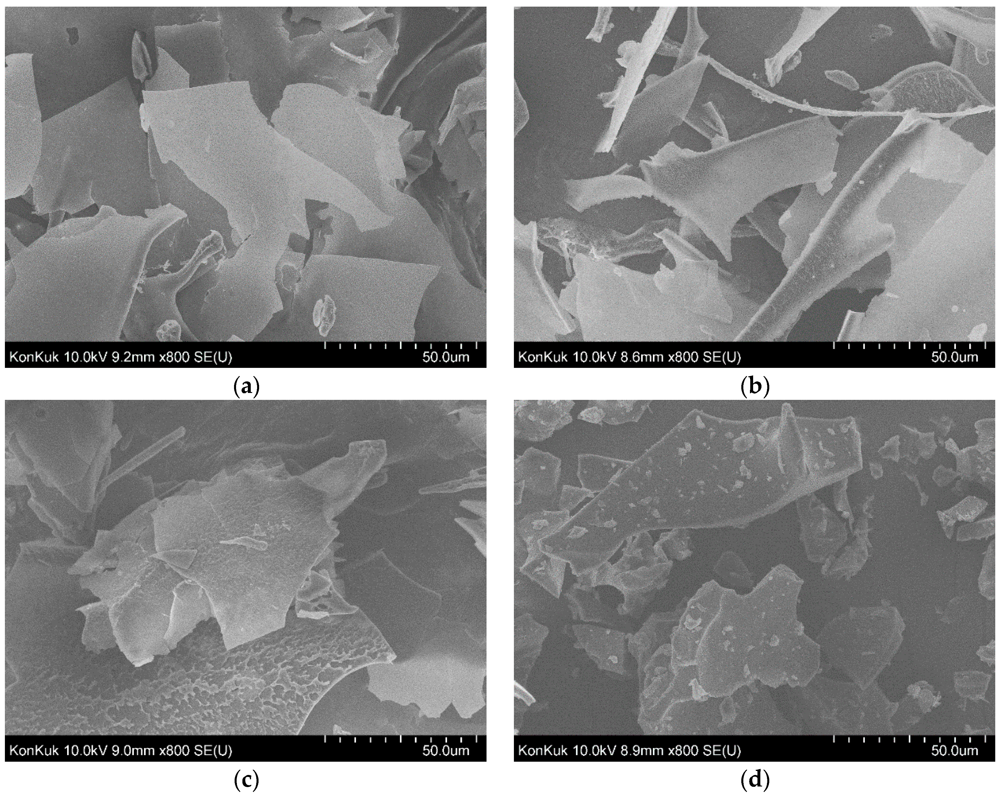
2.6. Field Emission Scanning Electron Microscopy (FE-SEM)
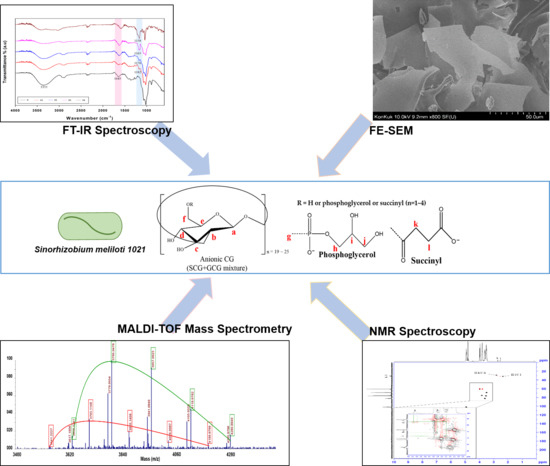
The Field Emission Scanning Electron Microscopy (FE-SEM) images of anionic CG were observed using FE-SEM (Hitachi S-4700, Tokyo, Japan). Samples were prepared in powder form after lyophilization. Carbon tape was attached on the brass stud and the sample was fixed. The powder samples were coated on the surface of a platinum layer prior to the FE-SEM analysis.

3. Results

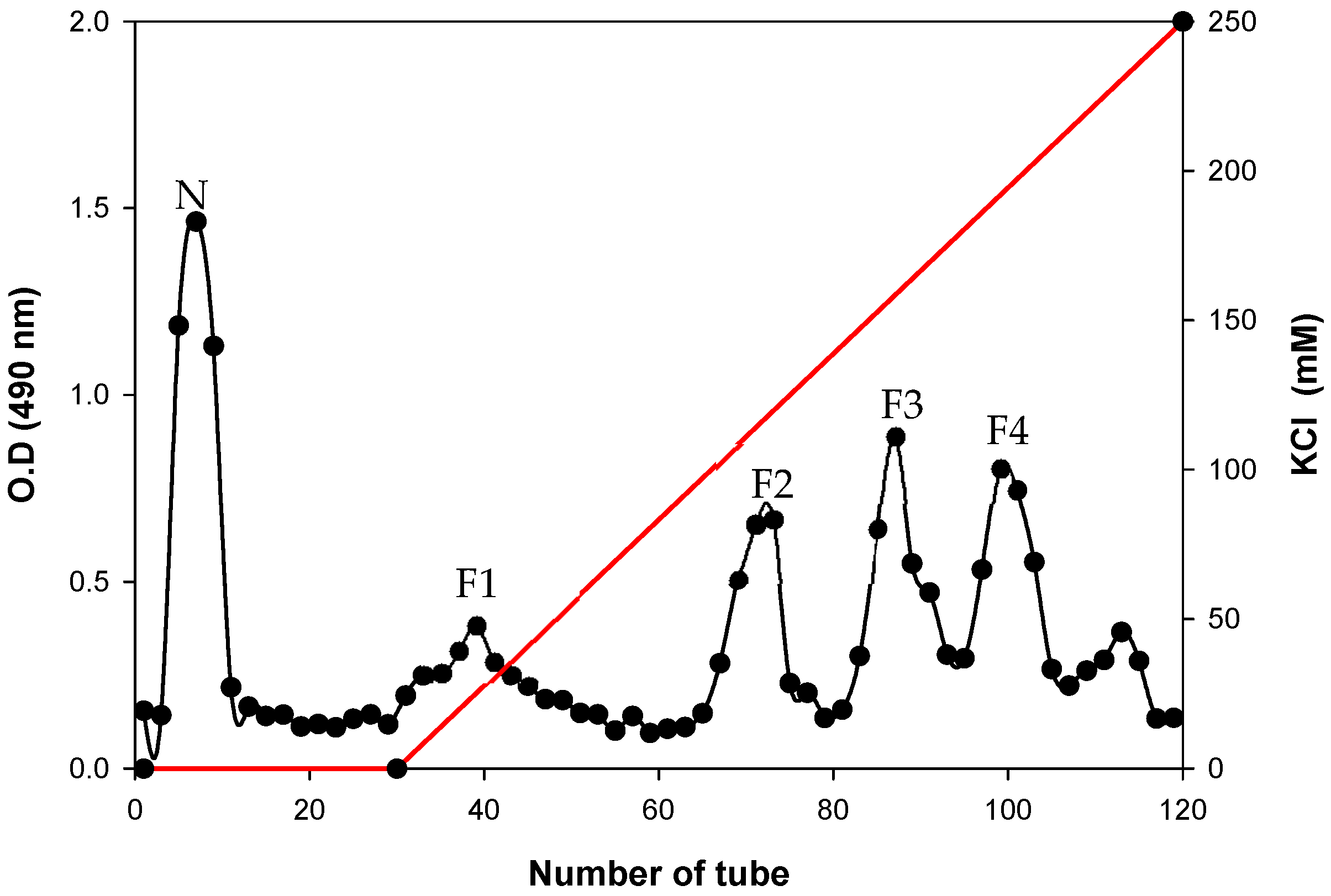
3.1. DEAE-Sephadex Chromatography of CG

When ethanol extracts of Sinorhizobium meliloti 1021 were chromatographed on Bio-Gel P-6 chromatography, one major peak of oligosaccharide material was detected (data not shown). After further fractionation of this extract on DEAE-Sephadex, these oligosaccharides were found to be anionic in character, with neutral oligosaccharides. The column size was 3 cm × 23.5 cm, which was eluted first with 150-mL Mops buffer with no KCl and then with a 450-mL linear gradient beginning with 0 mM and ending with 250 mM KCl in the same buffer. Fractions (5 mL) were collected and assayed for total carbohydrates using phenol-sulfuric acid. The one neutral fraction (N) and four anionic fractions (designated F1, F2, F3, and F4) were present (Figure 1). Figure 2 shows the approximate structure of anionic CG containing phosphoglycerol and succinyl groups. The fraction N indicates neutral form with no charge. Each of anionic fractions were subjected to various analyses performed as described below.

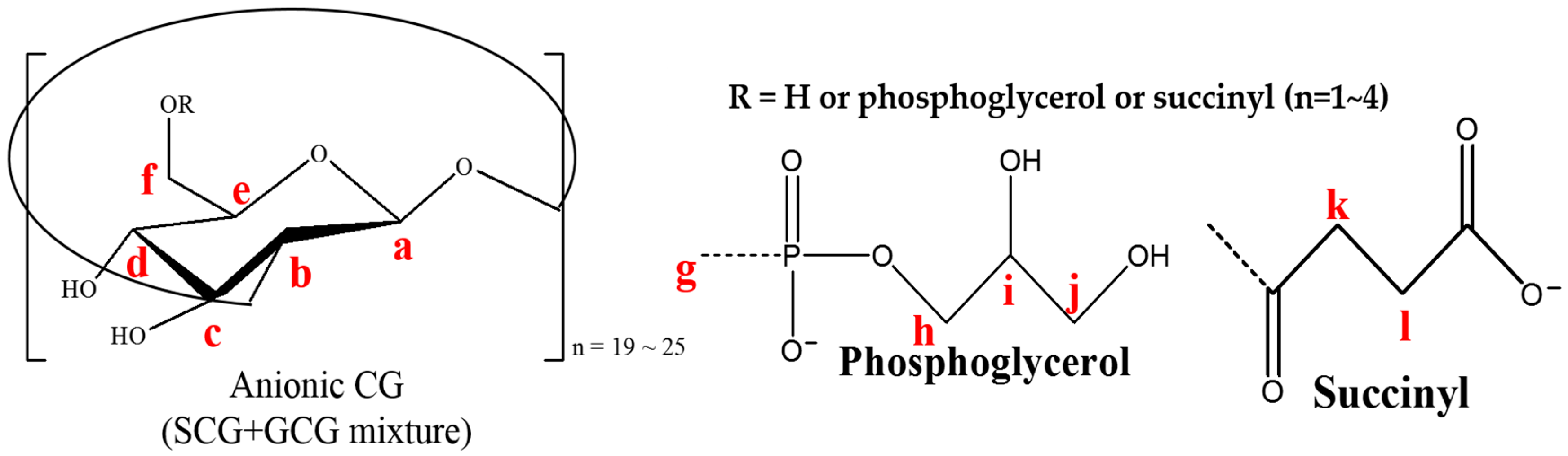
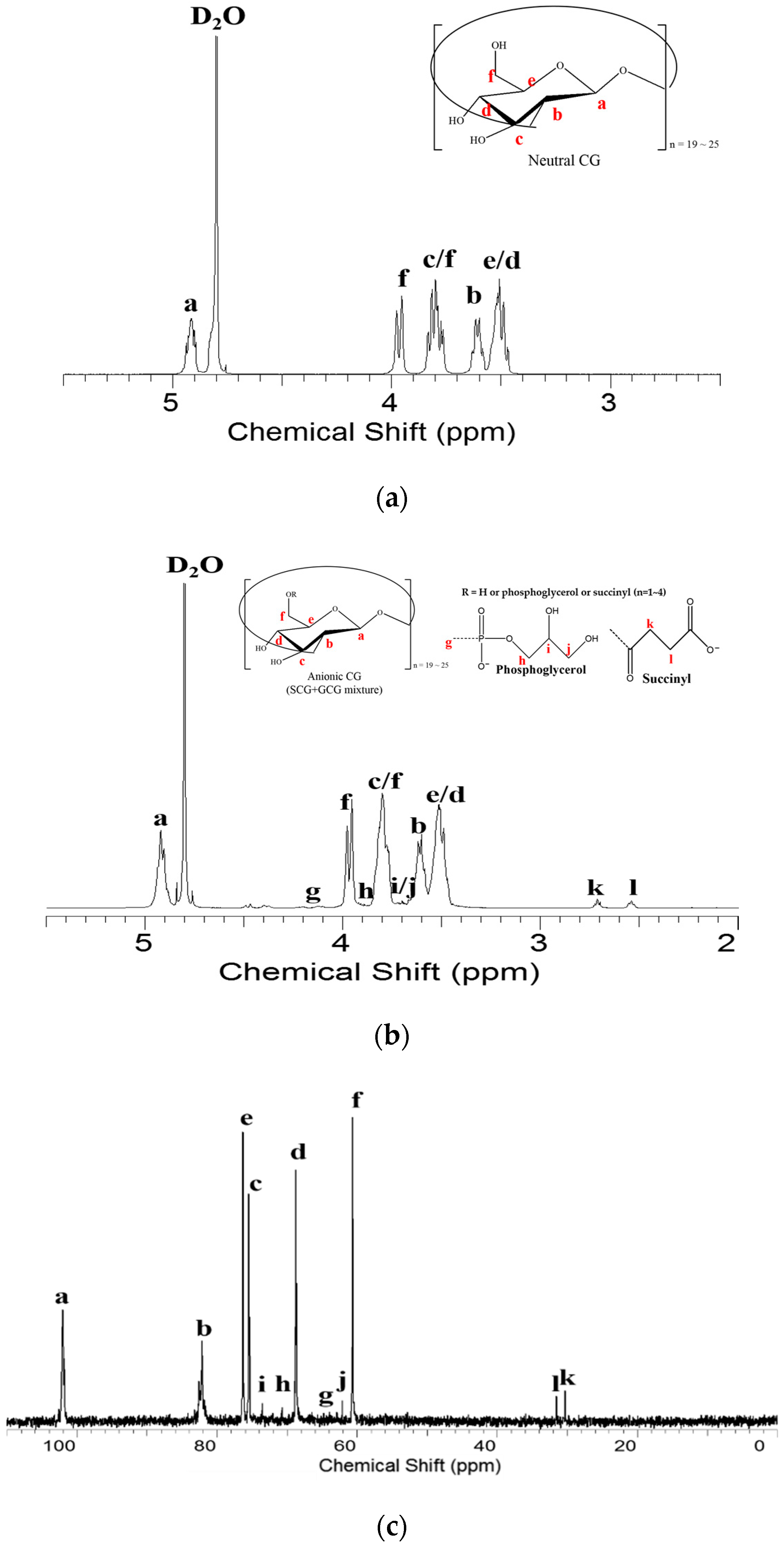
3.2. Nuclear Magnetic Resonance (NMR) Spectroscopy Analysis

We analyzed anionic fractions isolated by anion-exchange chromatography. The chromatogram displayed five different fractions, indicating one neutral (N) and four anionic (F1 to F4) fractions (Figure 1). Each of these anionic fractions were subjected to nuclear magnetic resonance (NMR) spectroscopy. The N fraction showed a 1H NMR spectrum similar to that reported previously (Figure 3a) [19]. The NMR data for the remaining fractions F2, F3 and F4 are in Figure S1, Figure S2 and Figure S3 in the supplementary materials. The F1 fraction showed the peaks at 2.71 and 2.54 ppm assigned to the H-k and H-l resonances of the succinyl residues of these molecules (Figure 3b). As shown in Figure 3b, each of the three peaks at 3.85, 3.77 and 3.64 ppm was assigned to the H-h, H-i and H-j resonances of the phosphoglycerol residues of these molecules, respectively. In addition, two doublet signals at 4.48 and 4.38 ppm indicated H-g resonances of the glucosyl residues connected by phosphoglycerol substituents [17,19]. Further 13C NMR analyses were conducted to determine the structures of substituents. As shown in Figure 3c, each of three peaks appearing at 73.54, 70.71 and 62.13 ppm was assigned to the C-i, C-h and C-j resonances of the phosphoglycerol residues of these molecules, respectively. In addition, two more peaks were detected at 31.54 and 30.35 ppm indicating the succinyl residues. There was one other peak at 65 ppm assigned to H-g resonances of the glucosyl residues connected by phosphoglycerol substituents. The presence of phosphorus was also confirmed by 31P NMR spectroscopy, and resonances were a singlet at 1.4 ppm. The proton-coupled signals are also shown to be a quintet consistent with coupling to four protons (Figure 3e). These characteristic peaks suggest that anionic CG was substituted with phosphoglycerol or succinyl residue substituents.

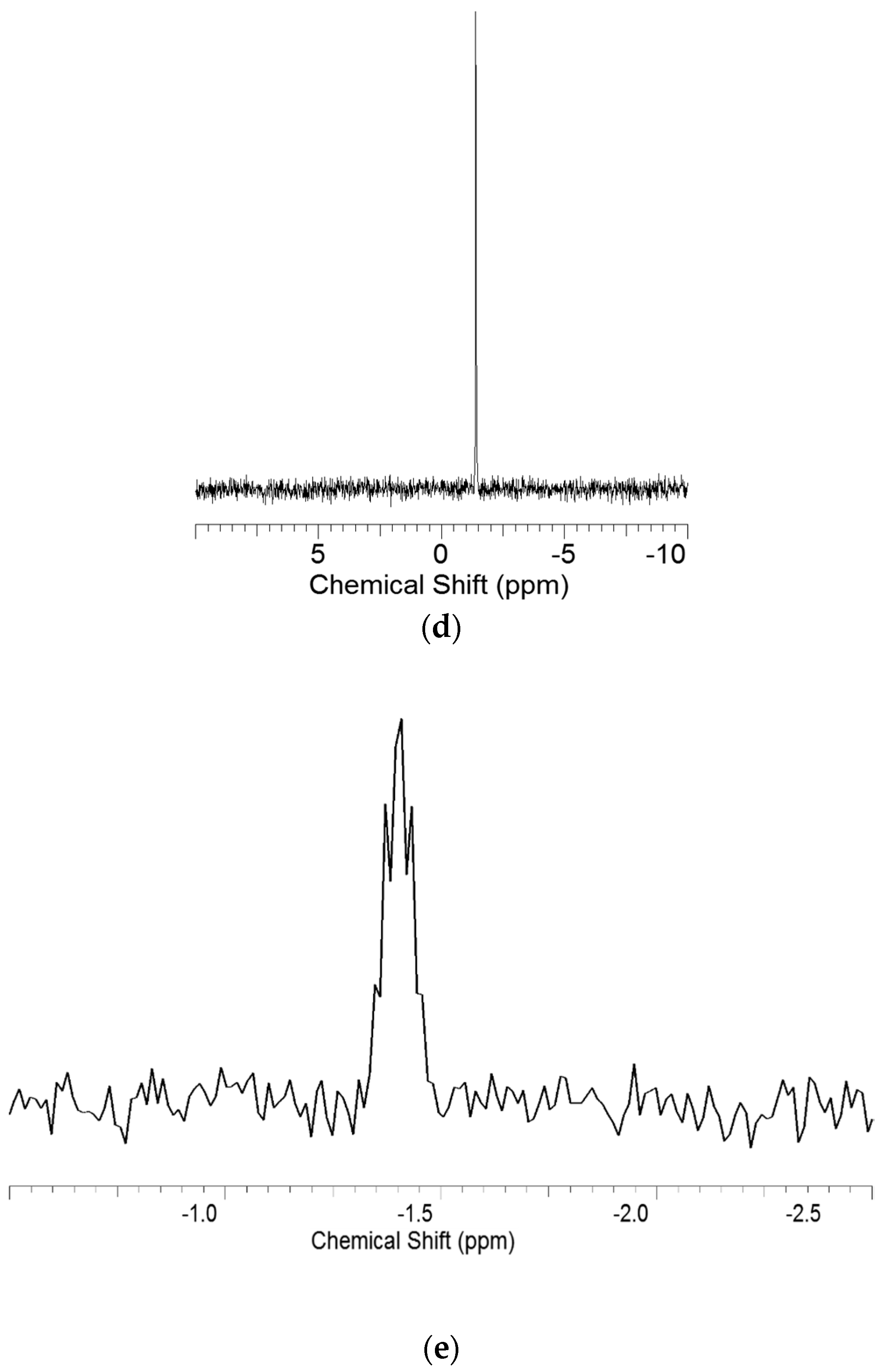
3.3. Heteronuclear Single Quantum Coherence Spectroscopy (HSQC) Spectra Analysis

The heteronuclear single quantum coherence spectroscopy (HSQC) experiment was further conducted (Figure 4 and Figure S4 in the supplementary materials). We confirmed the correlation between 13C NMR and 1H NMR through HSQC experiments. The NMR data for the remaining fractions F2, F3 and F4 are in Figure S3. In the HSQC experiments, the H-2′(S) was correlated with C-2′(S) and the H-3′(S) was correlated with C-3′(S), which represented the presence of succinyl substituents. In Figure 4, cross-peaks can be seen among the phosphoglycerol residue protons at 3.85, 3.77 and 3.64 ppm, and the two H-g protons are shown at 4.48 and 4.38 ppm. These results suggest that the anionic CG has glycerophosphoryl or succinyl residues at the C6 position.

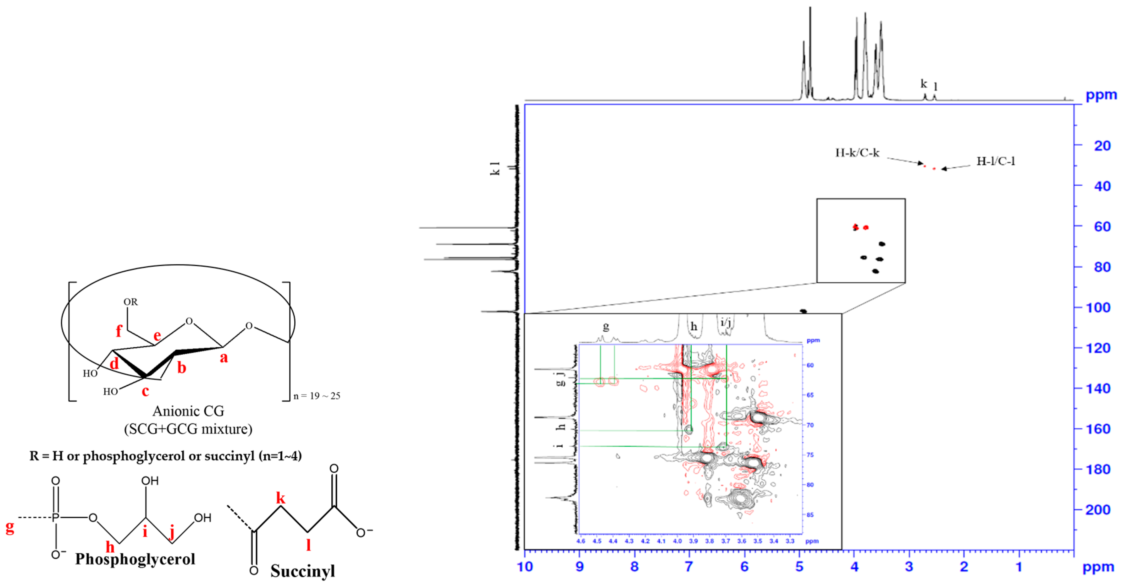
3.4. Attenuated Total Reflection (ATR)-Fourier Transform Infrared (FTIR) Spectra Analysis

Fourier transform infrared spectroscopy was used to characterize the structure of anionic CG fraction ATR-FTIR spectra of anionic CG and neutral CG, shown in Figure 5. The broad absorption peak at 3355 cm−1 indicates the stretching vibration of the hydroxyl group [11]. Neutral CG had an absorption peak at 1645 cm−1 that was an O–H bending vibration. Anionic CG including F1, F2, F3 and F4 peaks had shifted O-H bending vibration peaks since intramolecular hydrogen bonding with esters of the succinate and hydroxyl group from the sugar backbone. The succinyl groups overlapped with the O–H bending vibration peak. However, the presence of succinyl substituents could be confirmed through NMR spectroscopic analysis as shown by Figure 3b and Figure S1. The phosphates group (P=O peak) showed the strong bands at 1158, 1165, 1176 and 1182 cm−1, respectively. In the neutral CG spectrum, the phosphate peak did not appear (highlighted in the blue box in Figure 5).

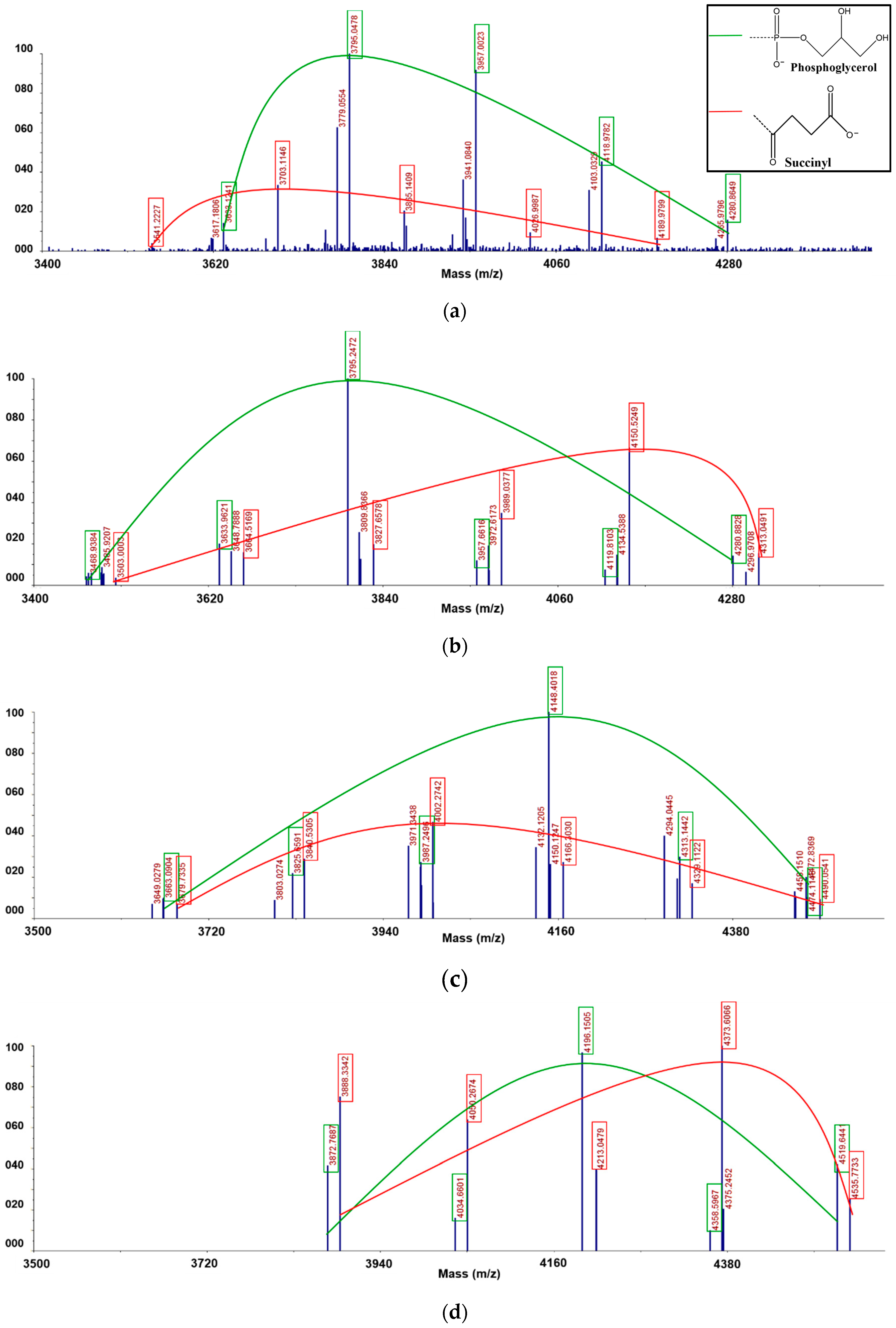
3.5. Matrix Assisted Laser Desorption/Ionization Time-of-Flight (MALDI-TOF) Mass Spectrometric Analysis

We analyzed anionic CG from Sinorhizobium meliloti 1021, with each of the separated fractions by matrix-assisted laser desorption/ionization time-of-flight (MALDI-TOF) mass spectrometry in a positive-ion mode (Figure 6 and Table 1). In the case of linear glucans, all the glucose monomers are linked through β-1, 2-glycosidic bonds, and the glucose at the end becomes the reducing end. However, compared to linear glucans, as they form cyclic glucans (CG) the reducing end disappears, resulting in a molecular weight without a single water molecule [23,24]. Reflecting this, the m/z values of anionic CG were also determined in MALDI-TOF analysis. Furthermore, it was also confirmed that both phosphoglycerol (PG) and succinyl (SU) groups were substituents for anionic CG. The spectrum from the F1 fraction showed each series of three ion species (Figure 6b and Table 1). The first series was a potassium cationized anionic CG with only succinyl substituents, which had [M + SU + K]+ ion species (m/z 3541.2, 3703.1, 3865.1, 4027.0, 4190.0). These ions series showed a molecular weight series in which only succinyl groups were substituted and composed of 21 to 24 cyclic-β-(1→2)-D-glucans. The difference of molecular weight of each unit was about 162, indicating one glucosyl linkage. The second series had [M + PG + 3Na − 2H]+ ion species (m/z 3617.2, 3779.1, 3941.1, 4103.0, 4266.0). These ion series showed a molecular weight series in which only phosphoglycerol groups were substituted and composed of 21 to 24 cyclic-β-(1→2)-D- glucans. The third series had [M + PG + K + 2Na - 2H]+ ion species (m/z 3633.1, 3795.0, 3957.0, 4119.0, 4280.9). As in the second of these ion series, only the phosphoglycerol group was substituted to the CG. The spectra of the F2, F3 and F4 fractions showed a series of molecular ions parallel to the F1 fraction, but each ionic species was different (Figure 6c,d and Table 1). The first series from the F2 fraction had two phosphoglycerol substituents and [M + 2PG + 4Na - 3H]+ ion species (m/z 3469.0, 3634.1, 3795.2, 3957.7, 4119.8, 4280.9) and composed 19 to 24 cyclic-β-(1→2)-d-glucans (Figure 6c). The second series also had only two phosphoglycerols without succinyl substituents, which had [M + 2PG + K + 3Na - 3H]+ ion species (m/z 3486.0, 3648.8, 3809.8, 3972.6, 4134.5, 4297.0). The third series had two succinyl substituents without phosphoglycerol substituents, which had [M + 2SU + K + Na - H]+ ion species (m/z 3503.0, 3664.5, 3827.7, 3989.0, 4150.5, 4313.0), and composed 20 to 24 cyclic-β-(1→2)-d-glucans. The F3 fraction showed a spectrum pattern similar to that of the F2 fraction (Figure 6d and Table 1). It had three types of ion spectrum series—two types of ion species had three phosphoglycerol substituents and the other type of ion species had three succinyl substituents. The first series had three phosphoglycerol substituents, [M + 3PG + 5Na - 4H]+ ion species (m/z 3649.0, 3803.0, 3971.3, 4132.1, 4294.0, 4458.2) and composed 19 to 24 cyclic-β-(1→2)-d-glucans. The second series also had only three phosphoglycerols without succinyl substituents, which had [M + 3PG + K + 4Na - 4H]+ ion species (m/z 3663.1, 3825.7, 3987.2, 4148.4, 4313.1, 4474.1). The third series had three succinyl substituents without phosphoglycerol substituents, which had [M + 3SU + K + Na - H]+ ion species (m/z 3679.7, 3840.5, 4002.3, 4166.3, 4329.1, 4490.1), and composed 21 to 25 cyclic-β-(1→2)-d-glucans. The F4 fraction consisted of two series of ion species (Figure 6e and Table 1). The first series had four phosphoglycerol substituents and [M + 4PG + Na]+ ion species (m/z 3872.8, 4034.7, 4196.2, 4358.6, 4519.6) and composed 20 to 24 cyclic-β-(1→2)-d-glucans. The second series also had only four succinyl without phosphoglycerol substituents, which had [M + 4SU + K]+ ion species (m/z 3888.3, 4050.3, 4213.0, 4373.6, 4535.8). As a result of the MALDI-TOF spectral analysis, each of phosphoglycerol and succinyl substituents had a separate molecular weight distribution. Thus, we confirmed that the two substituents were not present in one molecule at the same time, but existed separately in each molecule.

3.6. Field Emission Scannintg Electron Microscopy (FE-SEM) Analysis

The surface morphology of anionic CG was observed by FE-SEM, as shown in Figure 7a–d. The figure demonstrates the morphology of F1, F2, F3 and F4 fractions. The increase in the anionic property changed the surface and morphology of anionic CG depending on the anionic characteristics. All fractions of anionic CG took on a rough-plate shape (Figure 7). The F1 fraction showed a relatively smooth surface (Figure 7a) and the F2 fraction started to become a smooth and rough surface (Figure 7b). Thus, the F4 fraction formed a rougher and coarser surface than any other fractions (Figure 7d). The fact that the surface differed from fraction to fraction might indicate that different morphology characteristics could be induced by increasing the anionic property of CG.

4. Conclusions

There have been some reports that phosphoglycerol substituents of anionic CG produced in Sinorhizobium meliloti 1021 were derived from cell membrane lipids and that this negative CG is involved in the regulation of osmotic pressure [5,17]. However, studies to determine the exact position or number of substituents, and how anionic substituents are attached to the CG structure, have not been clearly conducted. In this study, we were able to find exact chemical structures of succinyl or glycerophosphoryl substituents constituting anionic CG using MALDI-TOF mass spectrometry and NMR spectroscopy. It was also confirmed that the anionic substituent of CG exists only in two types of succinyl groups or glycerophosphoryl groups. In other words, it was found that anionic CG that has two different substituents simultaneously was not present. That anionic CG would have potential in the fields of biotechnological applications like drug delivery systems and chirotechnology based on inclusion complexation with various guest molecules to increase solubility and bioavailability like CDs [25,26].

Supplementary Materials

The following are available online at https://www.mdpi.com/2073-4360/12/9/2073/s1, Figure S1: 1H NMR spectra of (a) F2, (b) F3 and (c) F4 fraction, Figure S2: 13C NMR spectra of (a) F2, (b) F3 and (c) F4 fraction, Figure S3: 31P NMR spectra of (a) F2, (b) F3 and (c) F4 fraction, Figure S4: 1H-13C heteronuclear single quantum coherence (HSQC) NMR spectra of (a) F2, (b) F3 and (c) F4 fraction.

Author Contributions

H.L. performed the experiments and wrote the paper; S.K. and Y.K. performed the methodology and validation; S.J. conceived and designed the experiments. All authors have read and agreed to the published version of the manuscript.

Funding

This paper was written as part of Konkuk University’s research support program for its faculty on sabbatical leave in 2019 and partially supported by the Basic Science Research Program through the National Research Foundation of Korea (NRF) funded by the Ministry of Education (2018R1D1A1B07050830).

Conflicts of Interest

The authors declare no conflict of interest.

References

  1. Dylan, T.; Ielpi, L.; Stanfield, S.; Kashyap, L.; Douglas, C.; Yanofsky, M.; Nester, E.; Helinski, D.R.; Ditta, G. Rhizobium meliloti genes required for nodule development are related to chromosomal virulence genes in Agrobacterium tumefaciens. Proc. Natl. Acad. Sci. USA 1986, 83, 4403–4407. [Google Scholar] [CrossRef] [PubMed] [Green Version]
  2. Ghosh, P.K.; Maiti, T.K. Structure of Extracellular Polysaccharides (EPS) produced by rhizobia and their functions in legume–bacteria symbiosis—A review. Achiev. Life Sci. 2016, 10, 136–143. [Google Scholar] [CrossRef] [Green Version]
  3. Miller, K.; Kennedy, E.; Reinhold, V. Osmotic adaptation by gram-negative bacteria: Possible role for periplasmic oligosaccharides. Science 1986, 231, 48–51. [Google Scholar] [CrossRef] [PubMed]
  4. Breedveld, M.W.; Miller, K.J. Cyclic beta-glucans of members of the family Rhizobiaceae. Microbiol. Rev. 1994, 58, 145–161. [Google Scholar] [CrossRef] [PubMed] [Green Version]
  5. Breedveld, M.W.; Hadley, J.A.; Miller, K.J. A novel cyclic beta-1,2-glucan mutant of Rhizobium meliloti. J. Bacteriol. 1995, 177, 6346–6351. [Google Scholar] [CrossRef] [PubMed] [Green Version]
  6. Seo, D.-H.; Lee, S.-H.; Park, H.-L.; Lee, D.-H.; Ji, E.-J.; Shin, D.-H.; Jung, S.-H. Structural analysis of total anionic cyclosophoraoses synthesized by Rhizobium meliloti 2011. Bull. Korean Chem. Soc. 2002, 23, 899–903. [Google Scholar]
  7. Kwon, C.; Choi, Y.-H.; Kim, N.; Yoo, J.S.; Yang, C.-H.; Kim, H.-W.; Jung, S. Complex forming ability of a family of isolated cyclosophoraoses with ergosterol and its monte carlo docking computational analysis. J. Incl. Phenom. Macrocycl. Chem. 2000, 36, 55–64. [Google Scholar] [CrossRef]
  8. Koizumi, K.; Okada, Y.; Horiyama, S.; Utamura, T.; Higashiura, T.; Ikeda, M. Preparation of cyclosophoraose-A and its complex-forming ability. J. Incl. Phenom. Macrocycl. Chem. 1984, 2, 891–899. [Google Scholar] [CrossRef]
  9. Lee, S.-H.; Kwon, C.-H.; Choi, Y.-J.; Seo, D.-H.; Kim, H.-W.; Jung, S.-H. Inclusion complexation of a family of cyclsohoraoses with indomethacin. J. Microbiol. Biotechnol. 2001, 11, 463–468. [Google Scholar]
  10. Cho, E.; Jung, S. Biotinylated cyclooligosaccharides for paclitaxel solubilization. Molecules 2018, 23, 90. [Google Scholar] [CrossRef] [Green Version]
  11. Kim, Y.; Shinde, V.V.; Jeong, D.; Choi, Y.; Jung, S. Solubility enhancement of atrazine by complexation with cyclosophoraose isolated from Rhizobium leguminosarum biovar trifolii TA-1. Polymers 2019, 11, 474. [Google Scholar] [CrossRef] [PubMed] [Green Version]
  12. Williams, R.O.; Mahaguna, V.; Sriwongjanya, M. Characterization of an inclusion complex of cholesterol and hydroxypropyl-β-cyclodextrin. Eur. J. Pharm. Biopharm. 1998, 46, 355–360. [Google Scholar] [CrossRef]
  13. Erden, N.; Çelebi, N. A study of the inclusion complex of naproxen with β-cyclodextrin. Int. J. Pharm. 1988, 48, 83–89. [Google Scholar] [CrossRef]
  14. Wen, X.; Tan, F.; Jing, Z.; Liu, Z. Preparation and study the 1:2 inclusion complex of carvedilol with β-cyclodextrin. J. Pharm. Biomed. Anal. 2004, 34, 517–523. [Google Scholar] [CrossRef]
  15. Miller, K.J.; Reinhold, V.N.; Weissborn, A.C.; Kennedy, E.P. Cyclic glucans produced by Agrobacterium tumefaciens are substituted with sn-1-phosphoglycerol residues. Biochim. Biophys. Acta (BBA) Biomembr. 1987, 901, 112–118. [Google Scholar] [CrossRef]
  16. Roset, M.S.; Ciocchini, A.E.; Ugalde, R.A.; De Iannino, N.I. The brucella abortus cyclic β-1,2-glucan virulence factor is substituted with o-ester-linked succinyl residues. J. Bacteriol. 2006, 188, 5003–5013. [Google Scholar] [CrossRef] [Green Version]
  17. Miller, K.J.; Gore, R.S.; Benesi, A.J. Phosphoglycerol substituents present on the cyclic beta-1,2-glucans of Rhizobium meliloti 1021 are derived from phosphatidylglycerol. J. Bacteriol. 1988, 170, 4569–4575. [Google Scholar] [CrossRef] [Green Version]
  18. Hisamatsu, M.; Yamada, T.; Higashiura, T.; Ikeda, M. The production of acidic, O-acylated cyclosophorans [cyclic (1→2)-β-d-glucans] by Agrobacterium and Rhizobium species. Carbohydr. Res. 1987, 163, 115–122. [Google Scholar] [CrossRef]
  19. Kawaharada, Y.; Kiyota, H.; Eda, S.; Minamisawa, K.; Mitsui, H. Structural characterization of neutral and anionic glucans from Mesorhizobium loti. Carbohydr. Res. 2008, 343, 2422–2427. [Google Scholar] [CrossRef]
  20. Kennedy, E. Membrane-derived oligosaccharides (periplasmic beta-D-glucans) of Escherichia coli. In Escherichia Coli and Salmonella. Cellular and Molecular Biology, 2nd ed.; Neidhardt, F.C., Curtiss, R., III, Ingraham, J.L., Lin, E.C.C., Low, K.B., Maganasik, B., Reznikoff, W.S., Riley, M., Schaechter, M., Umbarger, H.E., Eds.; American Society for Microbiology: Washington, DC, USA, 1996; pp. 1064–1074. [Google Scholar]
  21. Breedveld, M.W.; Zevenhuizen, L.P.; Zehnder, A.J. Excessive excretion of cyclic beta-(1,2)-glucan by Rhizobium trifolii TA-1. Appl. Environ. Microbiol. 1990, 56, 2080–2086. [Google Scholar] [CrossRef] [Green Version]
  22. Batley, M.; Redmond, J.; Djordjevic, S.; Rolfe, B. Characterisation of glycerophosphorylated cyclic β-1,2-glucans from a fast-growing Rhizobium species. Biochim. Biophys. Acta (BBA) Biomembr. 1987, 901, 119–126. [Google Scholar] [CrossRef]
  23. Dell, A.; York, W.S.; McNeil, M.; Darvill, A.G.; Albersheim, P. The cyclic structure of β-d-(1→2)-linked d-glucans secreted by Rhizobia and Agrobacteria. Carbohydr. Res. 1983, 117, 185–200. [Google Scholar] [CrossRef]
  24. Garozzo, D.; Spina, E.; Sturiale, L.; Montaudo, G.; Rizzo, R.; Daolio, S. Quantitative determination of β (1–2) cyclic glucans by matrix-assisted laser desorption mass spectrometry. Rapid Commun. Mass Spectrom. 1994, 8, 358–360. [Google Scholar] [CrossRef]
  25. Kicuntod, J.; Khuntawee, W.; Wolschann, P.; Pongsawasdi, P.; Chavasiri, W.; Kungwan, N.; Rungrotmongkol, T. Inclusion complexation of pinostrobin with various cyclodextrin derivatives. J. Mol. Graph. Model. 2016, 63, 91–98. [Google Scholar] [CrossRef] [PubMed]
  26. Jullian, C.; Moyano, L.; Yáñez, C.; Olea-Azar, C. Complexation of quercetin with three kinds of cyclodextrins: An antioxidant study. Spectrochim. Acta Part A Mol. Biomol. Spectrosc. 2007, 67, 230–234. [Google Scholar] [CrossRef] [PubMed]
Figure 1. DEAE-Sephadex chromatography of neutral and anionic CG fractions from Sinorhizobium meliloti 1021.
Polymers 12 02073 g001
Figure 2. Structures of the anionic CG from Sinorhizobium meliloti 1021. Each of the two substituents, phosphoglycerol and succinyl, was attached to CG separately as SCG or GCG. No anionic CG containing both substituents was found.
Polymers 12 02073 g002
Figure 3. (a) 1H NMR spectra of N faction; (b) 1H NMR spectra of F1 faction; (c) 13C NMR spectra of F1 fraction; (d) The proton-decoupled 31P NMR spectrum of F1 fraction; (e) The proton-coupled 31P NMR spectrum of F1 fraction.
Polymers 12 02073 g003aPolymers 12 02073 g003b
Figure 4. HSQC spectrum of F1 fraction.
Polymers 12 02073 g004
Figure 5. ATR-FTIR spectra of N fraction and anionic CG fraction region from 4000 to 600 cm−1.
Polymers 12 02073 g005
Figure 6. MALDI-TOF mass spectra of anionic CG obtained from (a) F1 fraction; (b) F2 fraction; (c) F3 fraction; (d) F4 fraction; acquired in positive ion reflector mode. The mass spectrums resulted from peak deisotoping based on the generic formula [C6H12O6]n showing various sodium and potassium adduct peaks. Each green and red curve shows the molecular weight distribution of SCG and GCG, respectively.
Polymers 12 02073 g006
Figure 7. SEM images of (a) F1 fraction; (b) F2 fraction; (c) F3 fraction; (d) F4 fraction.
Polymers 12 02073 g007
Table 1. MALDI-TOF MS data for F1 fraction, F2 fraction, F3 fraction and F4 fraction.
FractionMeasured MassCompositionPhosphoglycerolSuccinic Acid
F13541.2227Glc21+SU+K01
3617.1806Glc21+PG+3Na-2H10
3633.1241Glc21+PG+K+2Na-2H10
3703.1146Glc22+Su+K01
3779.0554Glc22 +PG+3Na-2H10
3795.0478Glc22 +PG+K+2Na-2H10
3865.1409Glc23 +SU+K01
3941.0840Glc23+PG+3Na-2H10
3957.0023Glc23+PG+K+2Na-2H10
4026.9987Glc24+SU+K01
4103.0329Glc24+PG+3Na-2H10
4118.9782Glc24+PG+K+2Na-2H10
4189.9799Glc25+SU+K01
4265.9796Glc25+PG+3Na-2H10
4280.8649Glc25+PG+K+2Na-2H10
FractionMeasured MassCompositionPhosphoglycerolSuccinic Acid
F23468.9384Glc19+2PG+4Na-3H20
3485.9207Glc19+2PG+K+3Na-3H20
3503.0003Glc20+2SU+K+Na-H02
3633.9621Glc20+2PG+4Na-3H20
3648.7888Glc20+2PG+ K+3Na-3H20
3664.5169Glc21+2SU+K+Na-H02
3795.2472Glc20+2PG+4Na-3H20
3809.8366Glc20+2PG+ K+3Na-3H20
3827.6578Glc21+2SU+K+Na-H02
3957.6616Glc21+2PG+4Na-3H20
3972.6173Glc21+2PG+K+3Na-3H20
3989.0377Glc22+2SU+K+Na-H02
4119.8103Glc22+2PG+4Na-3H20
4134.5388Glc22+2PG+K+3Na-3H20
4150.5249Glc23+2SU+K+Na-H02
4280.8828Glc23 +2PG+4Na-3H20
4296.9708Glc23+2PG+K+3Na-3H20
4313.0491Glc24+2SU+K+Na-H02
FractionMeasured MassCompositionPhosphoglycerolSuccinic Acid
F33649.0279Glc19+3PG+5Na-4H30
3663.0904Glc19+3PG+K+4Na-4H30
3679.7335Glc20+3SU+3K+Na-3H03
3803.0274Glc20+3PG+5Na-4H30
3825.6591Glc20+3PG+K+4Na-4H30
3840.5305Glc21+3SU+3K+Na-3H03
3971.3438Glc21+3PG+5Na-4H30
3987.2496Glc21+3PG+K+4Na-4H30
4002.2742Glc22+3SU+3K+Na-3H03
4132.1205Glc22+3PG+5Na-4H30
4148.4018Glc22+3PG+K+4Na-4H30
4166.3030Glc23+3SU+3K+Na-3H03
4294.0445Glc23+3PG+5Na-4H30
4313.1442Glc23+3PG+K+4Na-4H30
4329.1122Glc24+3SU+3K+Na-3H03
4458.1510Glc24+3PG+5Na-4H30
4474.1148Glc24+3PG+K+4Na-4H30
4490.0541Glc25+3SU+3K+Na-3H03
FractionMeasured MassCompositionPhosphoglycerolSuccinic Acid
F43872.7687Glc20+4PG+Na40
3888.3342Glc20+4SU+K04
4034.6601Glc21+4PG+Na40
4050.2674Glc21+4SU+K04
4196.1505Glc22+4PG+Na40
4213.0479Glc22+4SU+K04
4358.5967Glc23+4PG+Na40
4373.6066Glc23+4SU+K04
4519.6441Glc24+4PG+Na40
4535.7733Glc24+4SU+K04

