Hybrid Antibacterial and Electro-Conductive Coating for Textiles Based on Cationic Conjugated Polymer

 ,
, 

Abstract

1. Introduction
2. Materials and Methods
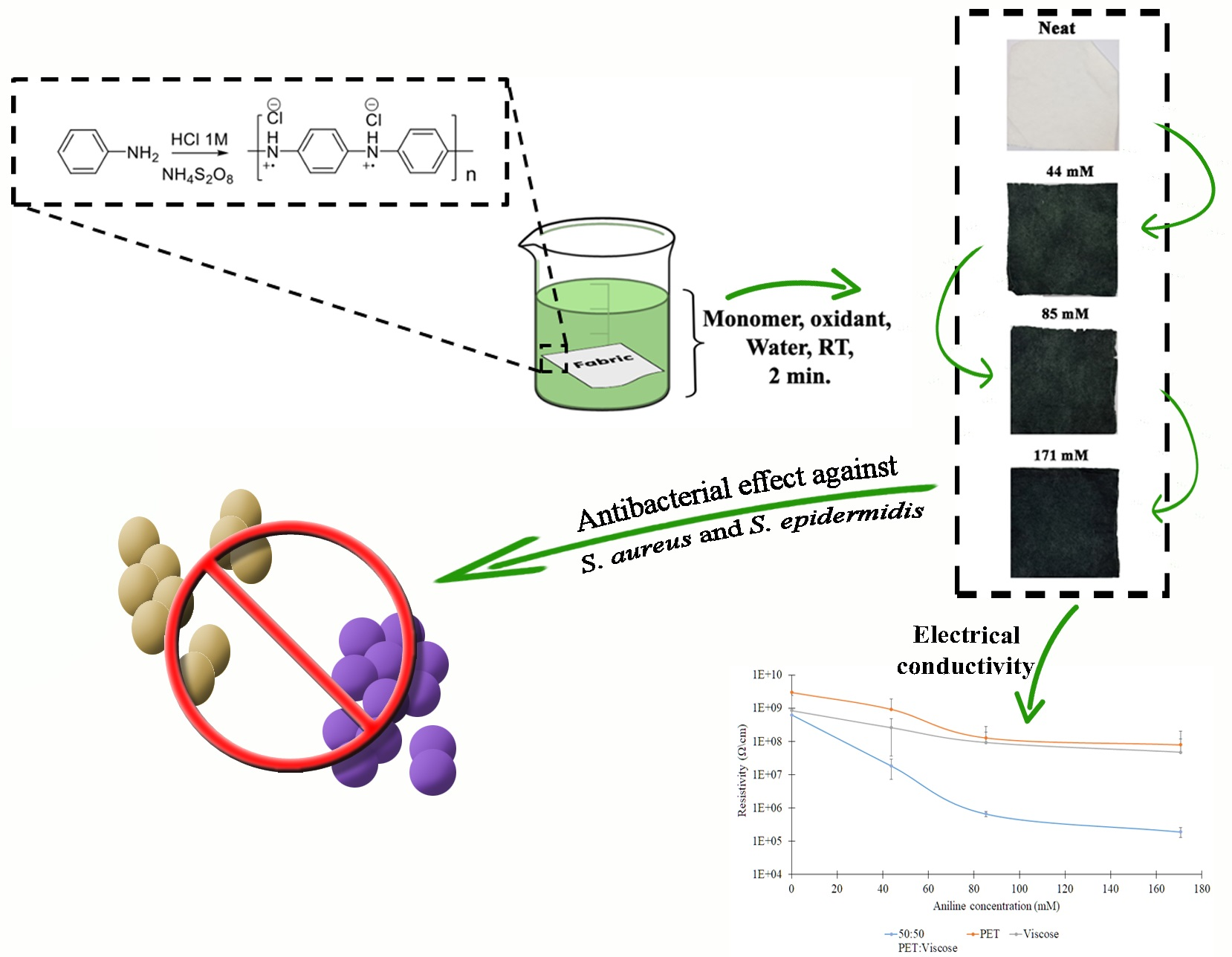
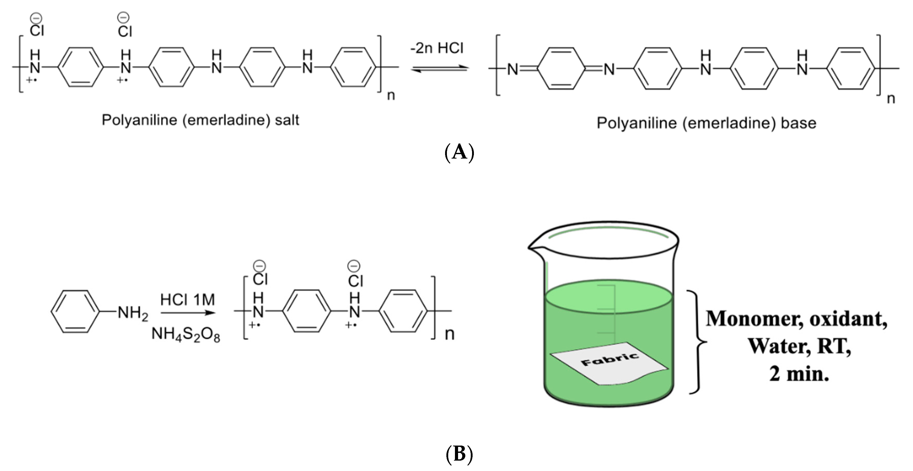
2.1. Coating Procedure
2.2. Antibacterial Studies
3. Results and Discussion
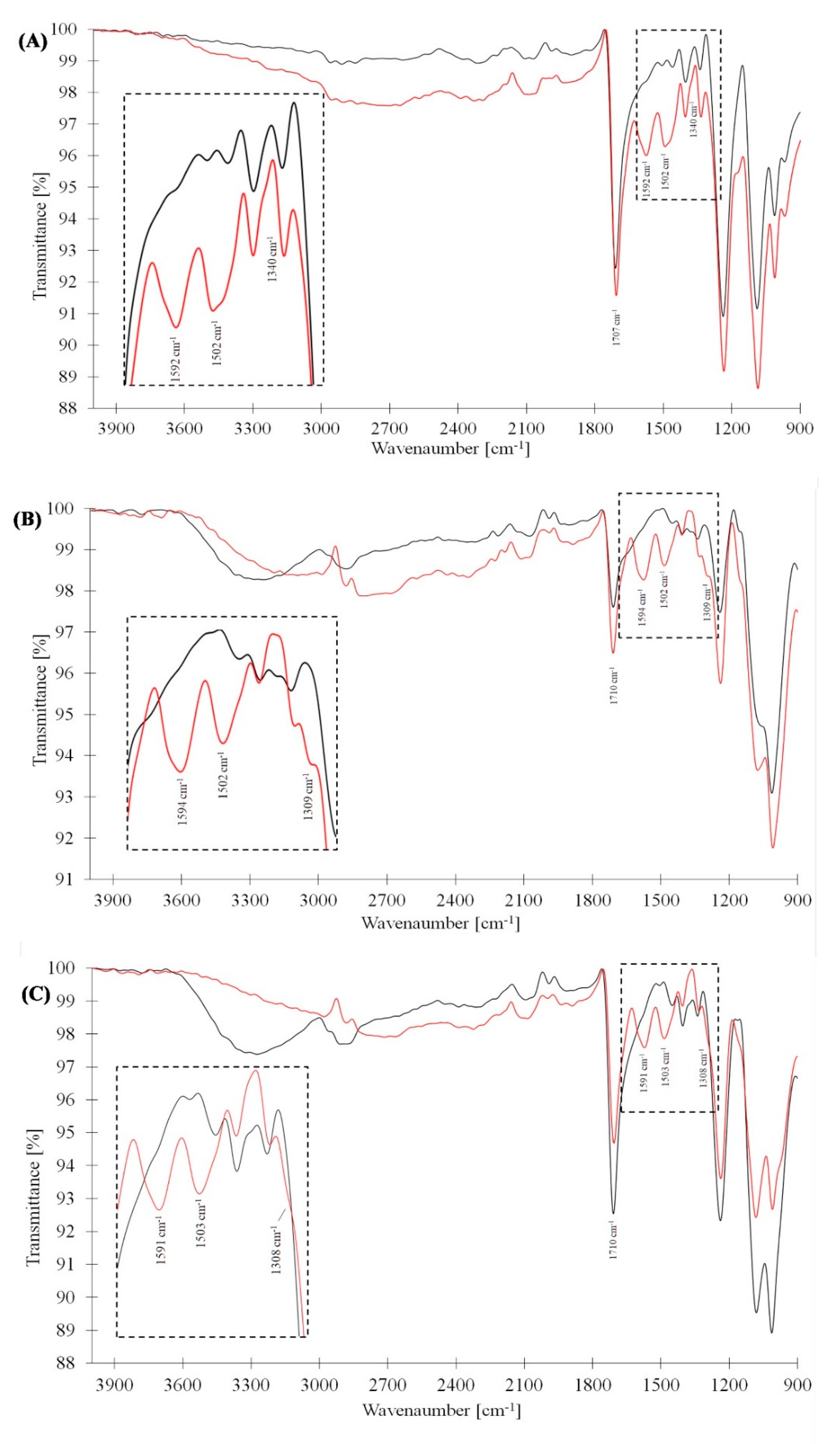
3.1. FTIR Analysis
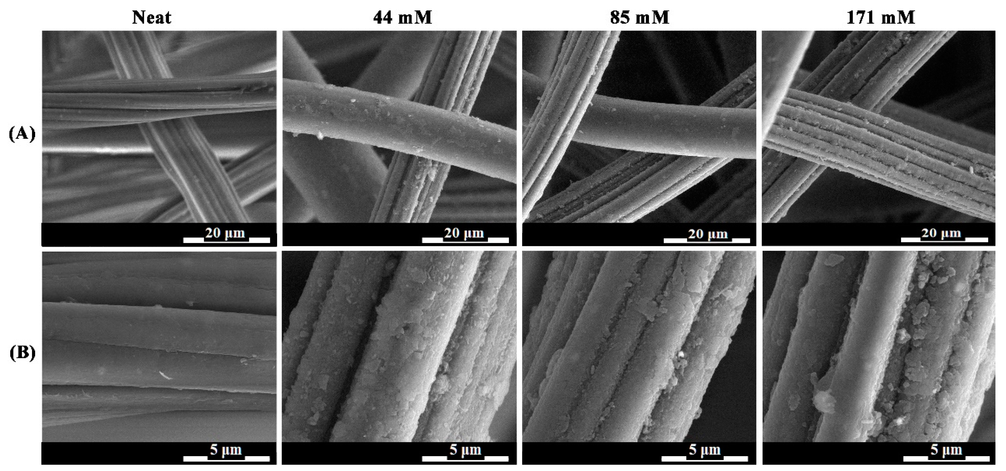
3.2. SEM Analysis
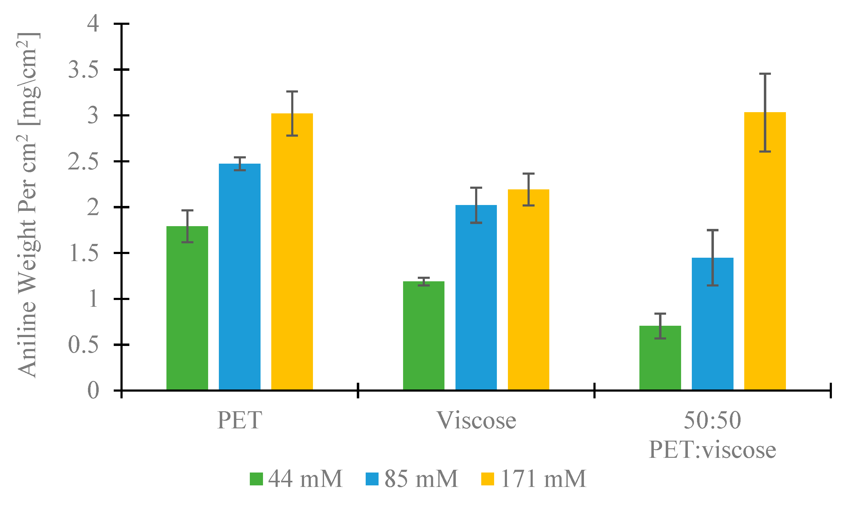
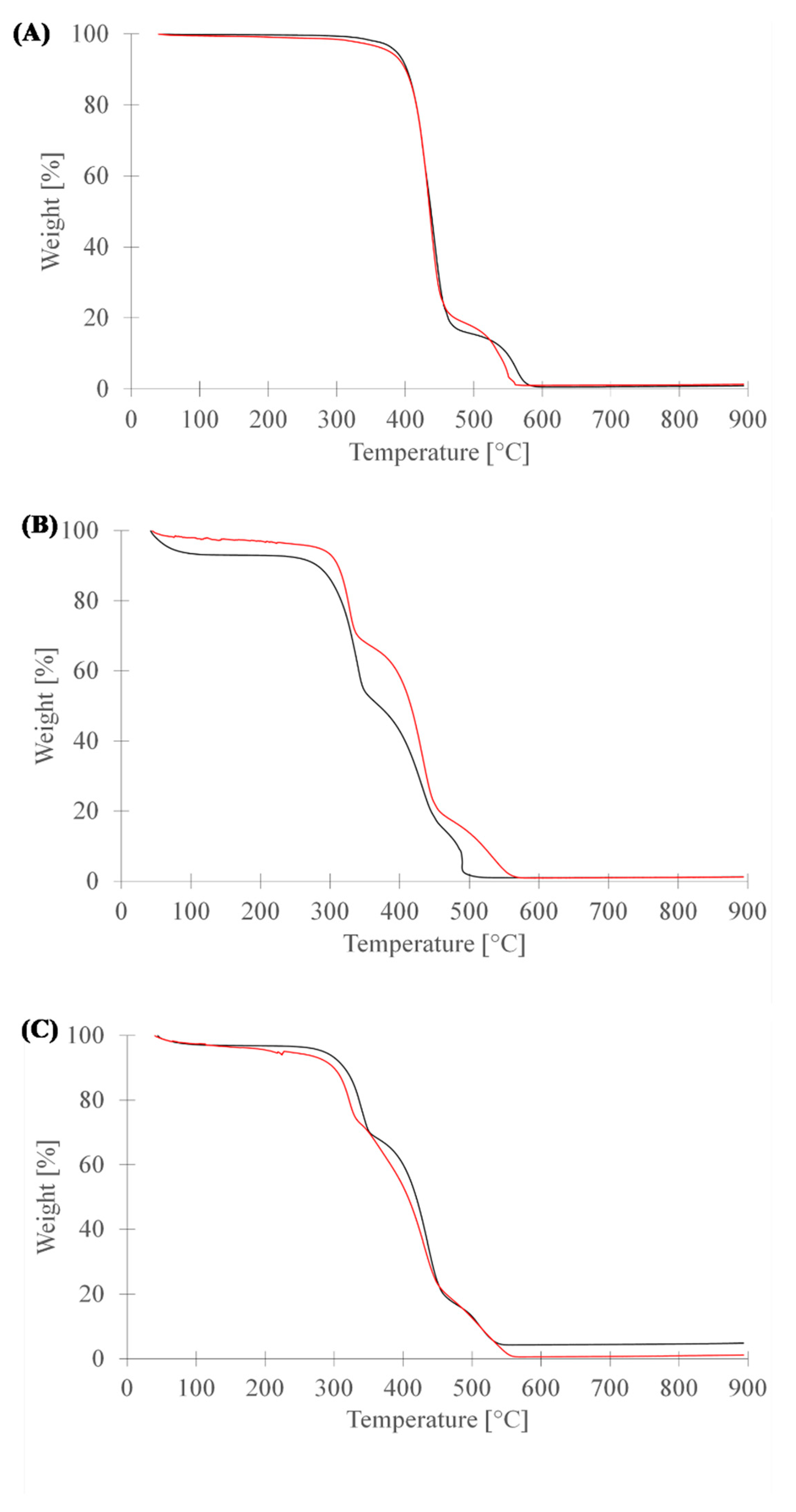
3.3. Thermogravimetric Analysis (TGA)
3.4. Resistivity Measurements
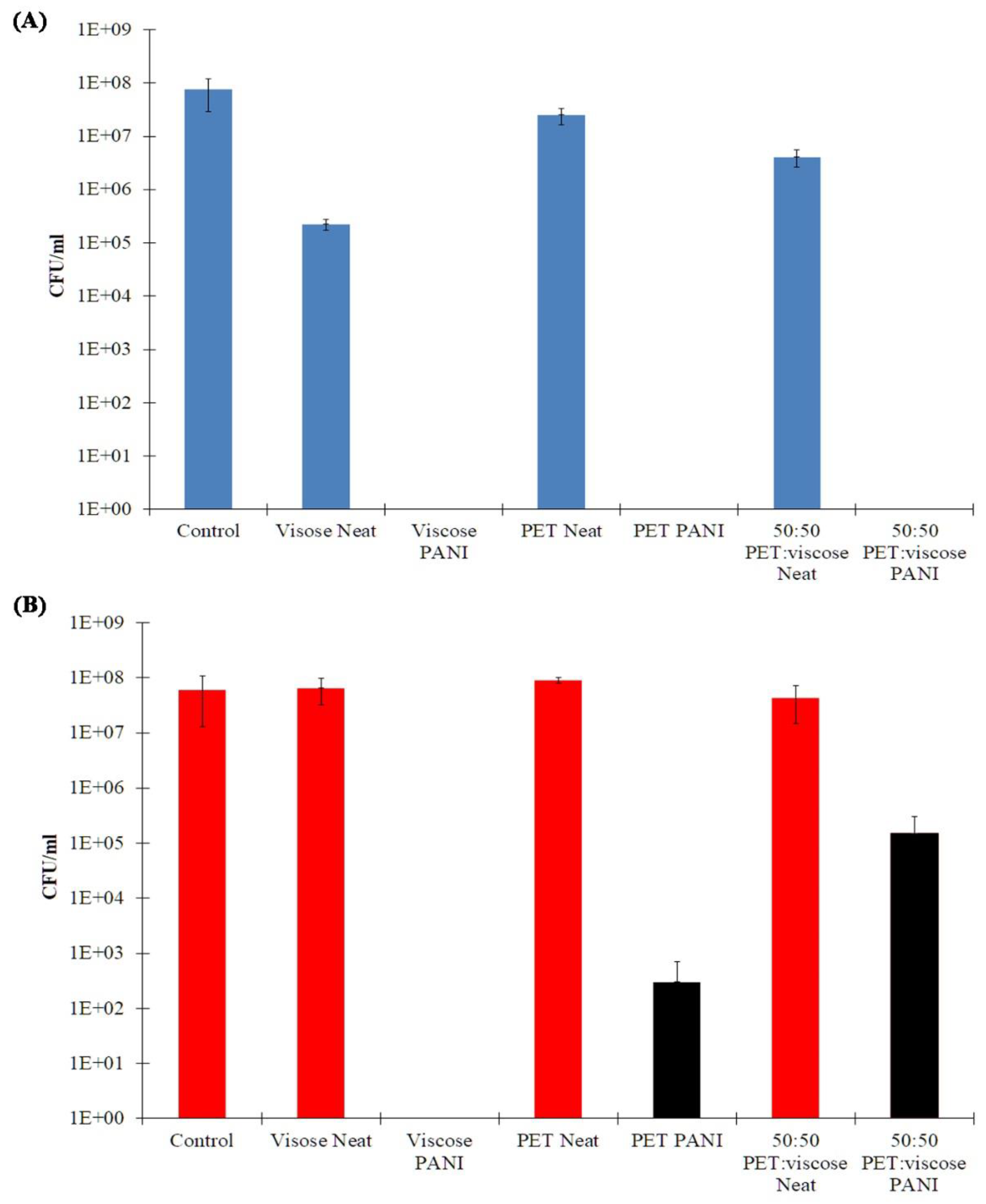
3.5. Antibacterial Studies
4. Conclusions
Supplementary Materials
Author Contributions
Funding
Conflicts of Interest
References
- Subbiah, D.K.; Babu, K.J.; Das, A.; Rayappan, J.B.B. NiOx Nanoflower Modified Cotton Fabric for UV Filter and Gas Sensing Applications. ACS Appl. Mater. Interfaces 2019, 11, 20045–20055. [Google Scholar] [CrossRef] [PubMed]
- David, N.C.; David, Y.; Katz, N.; Milanovich, M.; Anavi, D.; Buzhor, M.; Amir, E. Electro-conductive fabrics based on dip coating of cotton in poly(3-hexylthiophene). Polym. Adv. Technol. 2017, 28, 583–589. [Google Scholar] [CrossRef]
- Dan, Y.; Popowski, Y.; Buzhor, M.; Menashe, E.; Rachmani, O.; Amir, E. Covalent Surface Modification of Cellulose-Based Textiles for Oil–Water Separation Applications. Ind. Eng. Chem. Res. 2020. [Google Scholar] [CrossRef]
- Anavi, D.; Popowski, Y.; Slor, G.; Segal, M.; Frid, L.; Amir, R.J.; Amirav, A.; Amir, E. Covalent functionalization of solid cellulose by divergent synthesis of chemically active dendrons. J. Polym. Sci. Part A Polym. Chem. 2018, 56, 2103–2114. [Google Scholar] [CrossRef]
- Wu, Y.; Yang, Y.; Liu, H.; Yao, X.; Leng, F.; Chen, Y.; Tian, W. Long-term antibacterial protected cotton fabric coating by controlled release of chlorhexidine gluconate from halloysite nanotubes. RSC Adv. 2017, 7, 18917–18925. [Google Scholar] [CrossRef]
- Hartman, C.; Popowski, Y.; Raichman, D.; Amir, E. Biodegradable polymer coating for controlled release of hydrophobic functional molecules from cotton fabrics. J. Coat. Technol. Res. 2019. [Google Scholar] [CrossRef]
- Hu, J. Controlled release of hydrogel modified textile products. J. Control. Release 2011, 152, e31–e33. [Google Scholar] [CrossRef]
- Hashemikia, S.; Hemmatinejad, N.; Ahmadi, E.; Montazer, M. A novel cotton fabric with anti-bacterial and drug delivery properties using SBA-15-NH2/polysiloxane hybrid containing tetracycline. Mater. Sci. Eng. C 2016, 59, 429–437. [Google Scholar] [CrossRef]
- Gupt, P. Retracted: Antimicrobial action of chemically modified cotton fabric with cyclodextrin. Adv. Polym. Technol. 2013, 32. [Google Scholar] [CrossRef]
- Dhar, N. Novel Cellulose Nanoparticles for Potential Cosmetic and Pharmaceutical Applications; University of Waterloo: Waterloo, ON, Canada, 2010. [Google Scholar]
- Lund, E. The significance of oxidation in chemical inactivation of poliovirus. Arch. Gesamte Virusforsch. 1963, 12, 648–660. [Google Scholar] [CrossRef]
- Samuni, A.; Aronovitch, J.; Godinger, D.; Chevion, M.; Czapski, G. On the cytotoxicity of vitamin C and metal ions: A site-specific Fenton mechanism. Eur. J. Biochem. 1983, 137, 119–124. [Google Scholar] [CrossRef]
- Samuni, A.; Chevion, M.; Czapski, G. Roles of copper and superoxide anion radicals in the radiation- induced inactivation of T7 bacteriophage. Radiat. Res. 1984, 99, 562. [Google Scholar] [CrossRef] [PubMed]
- Tilton, R.C.; Rosenberg, B. Reversal of the Silver Inhibition of Microorganisms by Agar. Appl. Environ. Microbiol. 1978, 35, 1116–1120. [Google Scholar] [CrossRef] [PubMed]
- Khandelwal, N.; Kaur, G.; Kumar, N.; Tiwari, A. Application of silver nanoparticles in viral inhibition: A new hope for antivirals. Dig. J. Nanomater. Biostruct. 2014, 9, 175–186. [Google Scholar]
- Petering, H.G. Pharmacology and toxicology of heavy metals: Silver. Pharmacol. Ther. Part A Chemother. Toxicol. 1976, 1, 127–130. [Google Scholar] [CrossRef]
- Sun, L.Z.; Heng, X.; Chen, S.J. Theory meets experiment: Metal ion effects in HCV genomic RNA kissing complex formation. Front. Mol. Biosci. 2017, 4, 1–7. [Google Scholar] [CrossRef]
- Mönttinen, H.A.M.; Ravantti, J.J.; Poranen, M.M. Evidence for a non-catalytic ion-binding site in multiple RNA-dependent RNA polymerases. PLoS ONE 2012, 7. [Google Scholar] [CrossRef]
- Thurman, R.B.; Gerba, C.P. The molecular mechanisms of copper and silver ion disinfection of bacteria and viruses. Crit. Rev. Environ. Control 1989, 18, 295–315. [Google Scholar] [CrossRef]
- Sunada, K.; Minoshima, M.; Hashimoto, K. Highly efficient antiviral and antibacterial activities of solid-state cuprous compounds. J. Hazard. Mater. 2012, 235–236, 265–270. [Google Scholar] [CrossRef]
- Hassabo, A.G.; El-Naggar, M.E.; Mohamed, A.L.; Hebeish, A.A. Development of multifunctional modified cotton fabric with tri-component nanoparticles of silver, copper and zinc oxide. Carbohydr. Polym. 2019, 210, 144–156. [Google Scholar] [CrossRef]
- Gabbay, J. Antimicrobial and Antiviral Polymeric Materials. US 7,169,402B2, 30 January 2007. [Google Scholar]
- Ishida, T. Antiviral activities of Zn2+ ions for viral prevention, replication, Capsid protein in intracellular proliferation of viruses. World Sci. News 2018, 97, 28–50. [Google Scholar]
- Suara, R.O.; Crowe, J.E. Effect of zinc salts on respiratory syncytial virus replication. Antimicrob. Agents Chemother. 2004, 48, 783–790. [Google Scholar] [CrossRef] [PubMed]
- Chang, E.L.; Simmers, C.; Knight, D.A. Cobalt complexes as antiviral and antibacterial agents. Pharmaceuticals 2010, 3, 1711–1728. [Google Scholar] [CrossRef] [PubMed]
- Benzaghou, I.; Bougie, I.; Bisaillon, M. Effect of metal ion binding on the structural stability of the hepatitis C virus RNA polymerase. J. Biol. Chem. 2004, 279, 49755–49761. [Google Scholar] [CrossRef]
- Shigeta, S.; Mori, S.; Kodama, E.; Kodama, J.; Takahashi, K.; Yamase, T. Broad spectrum anti-RNA virus activities of titanium and vanadium substituted polyoxotungstates. Antivir. Res. 2003, 58, 265–271. [Google Scholar] [CrossRef]
- Dan, K.; Katoh, N.; Matsuoka, T.; Fujinami, K. In vitro Antimicrobial effects of virus block, which contains multiple polyoxometalate compounds, and hygienic effects of virus block-supplemented moist hand towels. Pharmacology 2019, 104, 98–112. [Google Scholar] [CrossRef] [PubMed]
- Abbas, M.; Naeem, N.; Iftikhar, H.; Latif, U. Synthesis, Characterization and antimicrobial properties of silver nanocomposites. In Silver Nanoparticles—Fabrication, Characterization and Applications; IntechOpen Limited: London, UK, 2018; pp. 71–91. [Google Scholar]
- Galdiero, S.; Falanga, A.; Vitiello, M.; Cantisani, M.; Marra, V.; Galdiero, M. Silver nanoparticles as potential antiviral agents. Molecules 2011, 16, 8894–8918. [Google Scholar] [CrossRef]
- Ng, L.Y.; Mohammad, A.W.; Leo, C.P.; Hilal, N. Polymeric membranes incorporated with metal/metal oxide nanoparticles: A comprehensive review. Desalination 2013, 308, 15–33. [Google Scholar] [CrossRef]
- Bignozzi, C.A.; Dissette, V.; Corallini, A.; Carra, G.; della Valle, R. Functional Nanomaterials with Antibacterial and Antiviral Activity. US 8,158,137 B2, 17 April 2012. [Google Scholar]
- Si, Y.; Zhang, Z.; Wu, W.; Fu, Q.; Huang, K.; Nitin, N.; Ding, B.; Sun, G. Daylight-driven rechargeable antibacterial and antiviral nanofibrous membranes for bioprotective applications. Sci. Adv. 2018, 4. [Google Scholar] [CrossRef]
- Malmsten, M. Antimicrobial and antiviral hydrogels. Soft Matter 2011, 7, 8725–8736. [Google Scholar] [CrossRef]
- Bromberg, L.; Bromberg, D.J.; Hatton, T.A.; Bandín, I.; Concheiro, A.; Alvarez-Lorenzo, C. Antiviral properties of polymeric aziridine- and biguanide-modified core-shell magnetic nanoparticles. Langmuir 2012, 28, 4548–4558. [Google Scholar] [CrossRef] [PubMed]
- Botequim, D.; Maia, J.; Lino, M.M.F.; Lopes, L.M.F.; Simões, P.N.; Ilharco, L.M.; Ferreira, L. Nanoparticles and surfaces presenting antifungal, antibacterial and antiviral properties. Langmuir 2012, 28, 7646–7656. [Google Scholar] [CrossRef] [PubMed]
- Haufe, H.; Muschter, K.; Siegert, J.; Böttcher, H. Bioactive textiles by sol-gel immobilised natural active agents. J. Sol-Gel Sci. Technol. 2008, 45, 97–101. [Google Scholar] [CrossRef]
- Chirkov, S.N. The antiviral activity of chitosan (review). Prikl. Biokhim. Mikrobiol. 2002, 38, 12–13. [Google Scholar] [CrossRef]
- Shamshina, J.L.; Kelly, A.; Oldham, T.; Rogers, R.D. Agricultural uses of chitin polymers. Environ. Chem. Lett. 2020, 18, 53–60. [Google Scholar] [CrossRef]
- Pasquier, N.; Keul, H.; Heine, E.; Moeller, M. From multifunctionalized poly(ethylene imine)s toward antimicrobial coatings. Biomacromolecules 2007, 8, 2874–2882. [Google Scholar] [CrossRef]
- Sinclair, T.R.; Robles, D.; Raza, B.; van den Hengel, S.; Rutjes, S.A.; de Roda Husman, A.M.; de Grooth, J.; de Vos, W.M.; Roesink, H.D.W. Virus reduction through microfiltration membranes modified with a cationic polymer for drinking water applications. Colloids Surf. A Physicochem. Eng. Asp. 2018, 551, 33–41. [Google Scholar] [CrossRef]
- Ichiyama, K.; Yang, C.; Chandrasekaran, L.; Liu, S.; Rong, L.; Zhao, Y.; Gao, S.; Lee, A.; Ohba, K.; Suzuki, Y.; et al. Cooperative orthogonal macromolecular assemblies with broad spectrum antiviral activity, high selectivity, and resistance mitigation. Macromolecules 2016, 49, 2618–2629. [Google Scholar] [CrossRef]
- Larson, A.M.; Hsu, B.B.; Rautaray, D.; Haldar, J.; Chen, J.; Klibanov, A.M. Hydrophobic polycationic coatings disinfect poliovirus and rotavirus solutions. Biotechnol. Bioeng. 2011, 108, 720–723. [Google Scholar] [CrossRef]
- Tuladhar, E.; de Koning, M.C.; Fundeanu, I.; Beumer, R.; Duizer, E. Different virucidal activities of hyperbranched quaternary ammonium coatings on poliovirus and influenza virus. Appl. Environ. Microbiol. 2012, 78, 2456–2458. [Google Scholar] [CrossRef]
- Haldar, J.; An, D.; de Cienfuegos, L.Á.; Chen, J.; Klibanov, A.M. Polymeric coatings that inactivate both influenza virus and pathogenic bacteria. Proc. Natl. Acad. Sci. USA 2006, 103, 17667–17671. [Google Scholar] [CrossRef] [PubMed]
- Wang, Y.; Canady, T.D.; Zhou, Z.; Tang, Y.; Price, D.N.; Bear, D.G.; Chi, E.Y.; Schanze, K.S.; Whitten, D.G. Cationic phenylene ethynylene polymers and oligomers exhibit efficient antiviral activity. ACS Appl. Mater. Interfaces 2011, 3, 2209–2214. [Google Scholar] [CrossRef] [PubMed]
- Park, D.; Larson, A.M.; Klibanov, A.M.; Wang, Y. Antiviral and antibacterial polyurethanes of various modalities. Appl. Biochem. Biotechnol. 2013, 169, 1134–1146. [Google Scholar] [CrossRef] [PubMed]
- Xue, Y.; Xiao, H. Antibacterial/antiviral property and mechanism of dual-functional quaternized pyridinium-type copolymer. Polymers 2015, 7, 2290–2303. [Google Scholar] [CrossRef]
- Xue, Y.; Pan, Y.; Xiao, H.; Zhao, Y. Novel quaternary phosphonium-type cationic polyacrylamide and elucidation of dual-functional antibacterial/antiviral activity. RSC Adv. 2014, 4, 46887–46895. [Google Scholar] [CrossRef]
- Yu, J.; Pang, Z.; Zhang, J.; Zhou, H.; Wei, Q. Conductivity and antibacterial properties of wool fabrics finished by polyaniline/chitosan. Colloids Surf. A Physicochem. Eng. Asp. 2018, 548, 117–124. [Google Scholar] [CrossRef]
- Ibrahim, N.A.; Gouda, M.; Husseiny, S.M.; El-Gamal, A.R.; Mahrous, F. UV-protecting and antibacterial finishing of cotton knits. J. Appl. Polym. Sci. 2009, 112, 3589–3596. [Google Scholar] [CrossRef]
- Shigita, S.; Tsurumi, H.; Naka, H. Anti-Viral Fiber, Process for Producing the Fiber, and Textile Product Comprising the Fiber. US 2007/0169278A1, 26 July 2007. [Google Scholar]
- Parthasarathi, V.; Thilagavathi, G. Development of tri-laminate antiviral surgical gown for liquid barrier protection. J. Text. Inst. 2015, 106, 1095–1105. [Google Scholar] [CrossRef]
- Perelshtein, I.; Applerot, G.; Perkas, N.; Guibert, G.; Mikhailov, S.; Gedanken, A. Sonochemical coating of silver nanoparticles on textile fabrics (nylon, polyester and cotton) and their antibacterial activity. Nanotechnology 2008, 19, 245705. [Google Scholar] [CrossRef]
- Harifi, T.; Montazer, M. A review on textile sonoprocessing: A special focus on sonosynthesis of nanomaterials on textile substrates. Ultrason. Sonochem. 2015, 23, 1–10. [Google Scholar] [CrossRef]
- Zafar, M.; Shah, T.; Rawal, A.; Siores, E. Preparation and characterisation of thermoresponsive nanogels for smart antibacterial fabrics. Mater. Sci. Eng. C 2014, 40, 135–141. [Google Scholar] [CrossRef] [PubMed]
- Erdem, R.; Rajendran, S. Influence of Silver Loaded Antibacterial Agent on Knitted and Nonwoven Fabrics and Some Fabric Properties. J. Eng. Fiber Fabr. 2016, 11, 38–46. [Google Scholar] [CrossRef]
- Seino, S.; Imoto, Y.; Kosaka, T.; Nishida, T.; Nakagawa, T.; Yamamoto, T.A. Antiviral Activity of Silver Nanoparticles Immobilized onto Textile Fabrics Synthesized by Radiochemical Process. MRS Adv. 2016, 1, 705–710. [Google Scholar] [CrossRef]
- Iyigundogdu, Z.U.; Demir, O.; Asutay, A.B.; Sahin, F. Developing novel antimicrobial and antiviral textile products. Appl. Biochem. Biotechnol. 2016, 181, 1155–1166. [Google Scholar] [CrossRef]
- Yamamoto, I.; Takayama, K.; Honma, K.; Gonda, T.; Matsuzaki, K.; Hatanaka, K.; Uryu, T.; Yoshida, O.; Nakashima, H.; Yamamoto, N.; et al. Synthesis, structure and antiviral activity of sulfates of curdlan and its branched derivatives. Br. Polym. J. 1990, 23, 245–250. [Google Scholar] [CrossRef]
- Neurath, A.R.; Strick, N.; Li, Y.Y. Anti-HIV-1 activity of anionic polymers: A comparative study of candidate microbicides. BMC Infect. Dis. 2002, 2, 1–11. [Google Scholar] [CrossRef] [PubMed]
- Vaillant, A. Nucleic acid polymers: Broad spectrum antiviral activity, antiviral mechanisms and optimization for the treatment of hepatitis B and hepatitis D infection. Antivir. Res. 2016, 133, 32–40. [Google Scholar] [CrossRef]
- Wang, C.C.; Wu, W.Y.; Chen, C.C. Antibacterial and swelling properties of N-isopropyl acrylamide grafted and collagen/chitosan-immobilized polypropylene nonwoven fabrics. J. Biomed. Mater. Res. Part B Appl. Biomater. 2011, 96, 16–24. [Google Scholar] [CrossRef]
- Wang, C.; Yang, F.; Liu, L.F.; Fu, Z.; Xue, Y. Hydrophilic and antibacterial properties of polyvinyl alcohol/4-vinylpyridine graft polymer modified polypropylene non-woven fabric membranes. J. Memb. Sci. 2009, 345, 223–232. [Google Scholar] [CrossRef]
- Nohr, R.S.; Macdonald, J.G. New biomaterials through surface segregation phenomenon: New quaternary ammonium compounds as antibacterial agents. J. Biomater. Sci. Polym. Ed. 1994, 5, 607–619. [Google Scholar] [CrossRef]
- Chen, J.P.; Kuo, C.Y.; Lee, W.L. Thermo-responsive wound dressings by grafting chitosan and poly(N-isopropylacrylamide) to plasma-induced graft polymerization modified non-woven fabrics. Appl. Surf. Sci. 2012, 262, 95–101. [Google Scholar] [CrossRef]
- Asanović, K.; Mihailović, T.; Škundrić, P.; Simović, L. Some properties of sntimicrobial coated knitted textile material evaluation. Text. Res. J. 2010, 80, 1665–1674. [Google Scholar] [CrossRef]
- Ye, W.; Leung, M.F.; Xin, J.; Kwong, T.L.; Lee, D.K.L.; Li, P. Novel core-shell particles with poly(n-butyl acrylate) cores and chitosan shells as an antibacterial coating for textiles. Polymer (Guildf.) 2005, 46, 10538–10543. [Google Scholar] [CrossRef]
- Ramirez, D.O.S.; Varesano, A.; Carletto, R.A.; Vineis, C.; Perelshtein, I.; Natan, M.; Perkas, N.; Banin, E.; Gedanken, A. Antibacterial properties of polypyrrole-treated fabrics by ultrasound deposition. Mater. Sci. Eng. C 2019, 102, 164–170. [Google Scholar] [CrossRef]
- Song, J.; Chen, P.; Liu, W. A superhydrophobic and antibacterial surface coated on cotton fabrics by polydopamine. Fibers Polym. 2019, 20, 1380–1386. [Google Scholar] [CrossRef]
- Gregory, R.V.; Kimbrell, W.C.; Kuhn, H.H. Conductive textiles. Synth. Met. 1989, 28, 823–835. [Google Scholar] [CrossRef]
- Hong, K.H.; Oh, K.W.; Kang, T.J. Polyaniline-nylon 6 composite fabric for ammonia gas sensor. J. Appl. Polym. Sci. 2004, 92, 37–42. [Google Scholar] [CrossRef]
- Dhawan, S.K.; Singh, N.; Venkatachalam, S. Shielding behaviour of conducting polymer-coated fabrics in X-band, W-band and radio frequency range. Synth. Met. 2002, 129, 261–267. [Google Scholar] [CrossRef]
- Neelakandan, R.; Madhusoothanan, M. Electrical resistivity studies on polyaniline coated polyester fabrics. J. Eng. Fiber Fabr. 2010, 5. [Google Scholar] [CrossRef]
- Nouri, M.; Kish, M.H.; Entezami, A.A.; Edrisi, M. Conductivity of textile fibers treated with aniline. Iran. Polym. J. (Engl. Ed.) 2000, 9, 49–58. [Google Scholar]
- Simal, A.L.; de Souza, A.M.C. Composites based on poly(ethylene terephthalate) fibers with polyaniline. I. Effect of the aniline monomer in the morphology of the PET substrate. J. Appl. Polym. Sci. 2000, 77, 2126–2138. [Google Scholar] [CrossRef]
- Simal, A.L.; de Souza, A.M.C. Composites based on poly(ethylene terephthalate) fibers with polyaniline. II. The effect of the growth of the polyaniline molecules during the polymerization in the morphology of the PET substrate. J. Appl. Polym. Sci. 2000, 77, 2346–2362. [Google Scholar] [CrossRef]
- Huang, H.; Liu, W. Polyaniline/poly(ethylene terephthalate) conducting composite fabric with improved fastness to washing. J. Appl. Polym. Sci. 2006, 102, 5775–5780. [Google Scholar] [CrossRef]
- Dhawan, S.K.; Singh, N.; Venkatachalam, S. Shielding effectiveness of conducting polyaniline coated fabrics at 101 GHz. Synth. Met. 2001, 125, 389–393. [Google Scholar] [CrossRef]
- Akşit, A.C.; Onar, N.; Ebeoglugil, M.F.; Birlik, I.; Celik, E.; Ozdemir, I. Electromagnetic and electrical properties of coated cotton fabric with barium ferrite doped polyaniline film. J. Appl. Polym. Sci. 2009, 113, 358–366. [Google Scholar] [CrossRef]
- Kim, B.; Koncar, V.; Dufour, C. Polyaniline-coated PET conductive yarns: Study of electrical, mechanical, and electro-mechanical properties. J. Appl. Polym. Sci. 2006, 101, 1252–1256. [Google Scholar] [CrossRef]
- Tsekouras, G.; Ralph, S.F.; Price, W.E.; Wallace, G.G. Gold recovery using inherently conducting polymer coated textiles. Fibers Polym. 2004, 5, 1–5. [Google Scholar] [CrossRef]
- Molina, J.; del Río, A.I.; Bonastre, J.; Cases, F. Electrochemical polymerisation of aniline on conducting textiles of polyester covered with polypyrrole/AQSA. Eur. Polym. J. 2009, 45, 1302–1315. [Google Scholar] [CrossRef]
- Hirase, R.; Shikata, T.; Shirai, M. Selective formation of polyaniline on wool by chemical polymerization, using potassium iodate. Synth. Met. 2004, 146, 73–77. [Google Scholar] [CrossRef]
- Fryczkowski, R.; Rom, M.; Fryczkowska, B. Polyester fibres finished with polyaniline. Fibres Text. East. Eur. 2005, 13, 141–143. [Google Scholar]
- Kutanis, S.; Karakişla, M.; Akbulut, U.; Saçak, M. The conductive polyaniline/poly(ethylene terephthalate) composite fabrics. Compos. Part A Appl. Sci. Manuf. 2007, 38, 609–614. [Google Scholar] [CrossRef]
- Seshadri, D.T.; Bhat, N.V. Use of polyaniline as an antimicrobial agent in textiles. Indian J. Fibre Text. Res. 2005, 30, 204–206. [Google Scholar]
- Wheeler, E. The Manufacture of Artificial Silk, with Special Reference to the Viscose Process; D. Van Nostrand Company: New York, NY, USA, 1931; p. xviii. 177p. [Google Scholar]
- Butoi, B.; Groza, A.; Dinca, P.; Balan, A.; Barna, V. Morphological and structural analysis of polyaniline and poly(o-anisidine) layers generated in a DC glow discharge plasma by using an oblique angle electrode deposition configuration. Polymers 2017, 9, 732. [Google Scholar] [CrossRef] [PubMed]
- Singu, B.S.; Srinivasan, P.; Pabba, S. Benzoyl peroxide oxidation route to nano form polyaniline salt containing dual dopants for pseudocapacitor. J. Electrochem. Soc. 2011, 159, A6–A13. [Google Scholar] [CrossRef]
- Bashir, T.; Ali, M.; Persson, N.K.; Ramamoorthy, S.K.; Skrifvars, M. Stretch sensing properties of conductive knitted structures of PEDOT-coated viscose and polyester yarns. Text. Res. J. 2014, 84, 323–334. [Google Scholar] [CrossRef]
- Sharaf, S.; Farouk, A.; El-Hady, M.M.A. Novel conductive textile fabric based on polyaniline and CuO nanoparticles. Int. J. Pharm Tech Res. 2016, 9, 461–472. [Google Scholar]
- Wang, N.; Li, G.; Zhang, X.; Qi, X. Chemical synthesis and characterization of dodecylbenzene sulfonic acid-doped polyaniline/viscose fiber. RSC Adv. 2015, 5, 44687–44695. [Google Scholar] [CrossRef]
- Li, G.; Yu, Z.; Han, N.; Zhang, X.; Wang, N.; Qi, X. Preparation and Properties of Polyaniline/Viscose Fiber Adducts. Polym. Compos. 2015, 38, 782–788. [Google Scholar] [CrossRef]
- Yildiz, Z.; Gungor, A.; Onen, A.; Usta, I. Synthesis and characterization of dual-curable epoxyacrylates for polyester cord/rubber applications. J. Ind. Text. 2016, 46, 596–610. [Google Scholar] [CrossRef]
- Jaffe, M.; Menczel, J.D. Thermal Analysis of Textiles and Fibers; Woodhead Publishing: Sawston, UK, 2020; ISBN 9780081005729. [Google Scholar]
- Rohani, R.; Hyland, M.; Patterson, D. Effects of process parameters on polyaniline nanofiltration membranes synthesis via phase inversion-immersion precipitation method. J. Eng. Sci. Technol. 2016, 11, 16–35. [Google Scholar]
- Gericke, A.; Van der Pol (née De Bruin), J. A comparative study of regenerated bamboo, cotton and viscose rayon fabrics. Part 2: Antimicrobial properties. J. Fam. Ecol. Consum. Sci. Tydskrif Vir Gesinsekologie En Verbruikerswetenskappe 2011, 39, 368. [Google Scholar] [CrossRef][Green Version]


| (A) | ||||
| Neat | 44 mM | 85 mM | 171 mM | |
| 50:50 PET:Viscose | 8.8 ± 0.9 | 7.3 ± 0.7 | 5.8 ± 0.5 | 5.3 ± 0.5 |
| PET | 9.5 ± 0.8 | 9.0 ± 0.9 | 8.1 ± 0.8 | 7.9 ± 0.8 |
| Viscose | 8.9 ± 0.7 | 8.4 ± 0.8 | 8.0 ± 0.8 | 7.7 ± 0.8 |
| (B) | ||||
| Neat | 44 mM | 85 mM | 171 mM | |
| 50:50 PET:Viscose | 9.9 ± 1.0 | 7.0 ± 0.7 | 5.8 ± 0.5 | 5.2 ± 0.5 |
| PES | 8.5 ± 0.7 | 7.9 ± 0.8 | 7.6 ± 0.8 | 7.0 ± 0.7 |
| Viscose | 9.7 ± 0.9 | 8.1 ± 0.8 | 7.9 ± 0.8 | 7.7 ± 0.8 |
© 2020 by the authors. Licensee MDPI, Basel, Switzerland. This article is an open access article distributed under the terms and conditions of the Creative Commons Attribution (CC BY) license (http://creativecommons.org/licenses/by/4.0/).
Share and Cite
Jarach, N.; Meridor, D.; Buzhor, M.; Raichman, D.; Dodiuk, H.; Kenig, S.; Amir, E. Hybrid Antibacterial and Electro-Conductive Coating for Textiles Based on Cationic Conjugated Polymer. Polymers 2020, 12, 1517. https://doi.org/10.3390/polym12071517
Jarach N, Meridor D, Buzhor M, Raichman D, Dodiuk H, Kenig S, Amir E. Hybrid Antibacterial and Electro-Conductive Coating for Textiles Based on Cationic Conjugated Polymer. Polymers. 2020; 12(7):1517. https://doi.org/10.3390/polym12071517
Chicago/Turabian StyleJarach, Natanel, David Meridor, Marina Buzhor, Daniel Raichman, Hanna Dodiuk, Shmuel Kenig, and Elizabeth Amir. 2020. "Hybrid Antibacterial and Electro-Conductive Coating for Textiles Based on Cationic Conjugated Polymer" Polymers 12, no. 7: 1517. https://doi.org/10.3390/polym12071517
APA StyleJarach, N., Meridor, D., Buzhor, M., Raichman, D., Dodiuk, H., Kenig, S., & Amir, E. (2020). Hybrid Antibacterial and Electro-Conductive Coating for Textiles Based on Cationic Conjugated Polymer. Polymers, 12(7), 1517. https://doi.org/10.3390/polym12071517