Review

Progress in Polymeric Nano-Medicines for Theranostic Cancer Treatment

1
Department of Chemistry, College of Sciences, Taibah University, Al-Medina Al-Munawara 41477, Saudi Arabia
2
Department of Chemistry, Jamia Millia Islamia (Central University), New Delhi 110025, India
3
Cheminformatics Laboratory—Postgraduate Program in Natural Products and Synthetic Bioactive, Federal University of Paraíba-Campus I, João Pessoa 58051-970, PB, Brazil
4
Department of Biomedical Engineering, Chung Yuan Christian University, 200 Chung Pei Road, Chung Li District, Taoyuan 32023, Taiwan
5
Center for Minimally-Invasive Medical Devices and Technologies, Chung Yuan Christian University, 200 Chung Pei Road, Chung Li District, Taoyuan 32023, Taiwan
6
Department of Pharmacy, Kaohsiung Armed Forces General Hospital, No.2, Zhongzheng 1st Rd., Lingya Dist., Kaohsiung 80284, Taiwan
7
Institute of Biomedical Engineering, National Chiao Tung University, 1001 University Rd., Hsinchu 300, Taiwan
*
Author to whom correspondence should be addressed.
Co-Correspondence authors.
Polymers 2020, 12(3), 598; https://doi.org/10.3390/polym12030598
Submission received: 28 November 2019 / Revised: 31 December 2019 / Accepted: 1 January 2020 / Published: 6 March 2020
(This article belongs to the Special Issue Polymeric Colloidal Materials for Biomedical Applications)

Abstract

:
Cancer is a life-threatening disease killing millions of people globally. Among various medical treatments, nano-medicines are gaining importance continuously. Many nanocarriers have been developed for treatment, but polymerically-based ones are acquiring importance due to their targeting capabilities, biodegradability, biocompatibility, capacity for drug loading and long blood circulation time. The present article describes progress in polymeric nano-medicines for theranostic cancer treatment, which includes cancer diagnosis and treatment in a single dosage form. The article covers the applications of natural and synthetic polymers in cancer diagnosis and treatment. Efforts were also made to discuss the merits and demerits of such polymers; the status of approved nano-medicines; and future perspectives.

Graphical Abstract

1. Introduction

The cancer is a complex, heterogeneous, aggressive and horrible disease, killing millions of people globally. As per GLOBOCAN estimation, 18.1 million new cases and 9.6 million deaths occurred worldwide in 2018 due to cancer alone [1]. Therefore, there is a great need to combat this deadly disease. Nowadays, theranostic management of various diseases is gaining importance, including horrible cancer ailments. The term theranostic was given by John Funkhouser (US consultant) in August, 1998 in a press release from the company Cardiovascular Diagnostics. It was used to combine investigative and treatment approaches into a single unit. It involves a set of diagnoses, a drug carrier and the observation of treatment reaction. This approach provides treatment protocols that are more precise to individuals, and hence, are more likely to deliver better predictions by diagnosis and treatment. It delivers exact, safe and targeted treatment via the right drug, in the right dose and at the right place in the human body. Besides, theranostic management also contributes to the economy of a country by reduced cost strategies with specific and efficient medicine procedures. It is a targeted treatment protocol, and hence, has very little side effects. This approach is represented in Figure 1.
The number of publications in polymer therapeutics is increasing continuously. During our research of more than 30 years and available literature on cancer treatment [2,3,4,5,6,7,8,9,10,11,12,13,14,15], it was observed that chemotherapy is considered the best approach to curb different types of cancers [16,17,18,19,20,21,22,23,24,25,26,27,28]. But this has severe toxicity to normal cells. It is a challenge to researchers, academicians and oncologists. Therefore, a theranostic approach may be useful for treating cancer, and of course, gaining efficacy for patients.
In cancer treatment and management, the presently used techniques are not capable of diagnosing cancers in early stages and are also not very specific. However, nanotechnology is providing a bias for the diagnosis, treatment and management of different cancers [29,30]. The nanotechnology is offering a chance for diagnosis and therapy for cancer. Nanoparticle-centered imaging and therapy are being investigated continuously. The nanotechnology is proficient in diagnosis, drug delivery and observation of therapeutic response. It is likely to perform an important role in personalized medicine and treatment. The polymeric nanoparticles, metallic nanoparticles, liposomes, dendrimers, carbon nanotubes quantum dots, etc., are being used as nano-formulations for cancer theranostics. Among these nanoparticles, polymeric ones are gaining importance due to their ease of preparation, high drug-loading capacities, their biocompatibility and biodegradable nature. Much work has been carried out in this direction and some reviews are available on the subject [31,32,33,34,35,36,37,38], but there is no review specially dedicated to polymeric nana-particles (NPs) in the diagnoses and treatments of various cancers. Therefore, this is the time to review the progress in polymeric nano-medicines for theranostic cancer management. The present article describes a state-of-the-art of polymeric nano-medicines for theranostic cancer management. Besides, effort was made to highlight future challenges and perspectives in this area of research.

2. Nanoparticles

Nano theranostic is a growing field for instantaneous diagnosis, drug delivery and curing cancers [39,40,41]. It is well known that nanoparticles have diameters ranging from 1.0 to 100.0 nm, which are about a thousand times smaller than the normal human cells. Besides, these nanoparticles have great chances of cellular interactions with cellular receptors, enzymes and antibodies [42]. The facility of nanoparticle modification makes them ideal candidates for the exact diagnosis and treatment [43,44]. The literature indicates the formulations of several theranostic nanoparticle agents. These are made of several materials, including ions of gold, silica carbon, etc. [45,46]. These were evaluated in many animal models, and have found promising identities in early detection of cancer via several imaging probes. But the drawbacks associated with them are immunogenicity, toxicity and slow excretion rate from the body [47,48]. Accordingly, some macromolecules have been tested for these purposes. The most important molecules exploited for such platforms are polylactic acid (PLA), poly(ε-caprolactone), poly(lactide-co-glycolide) (PLGA), poly(alkylcyanoacrylate) and polyglycolic acid. Contrarily, the natural polymers used are peptides, proteins, nucleic acids, dextran ester and chitosan. Of course, these molecules are excellent but suffer from short half-lives, non-specific distribution rates and limited applications because of their interactions with drug molecules. Consequently, synthetic, polymeric, biodegradable nanoparticles were explored [49,
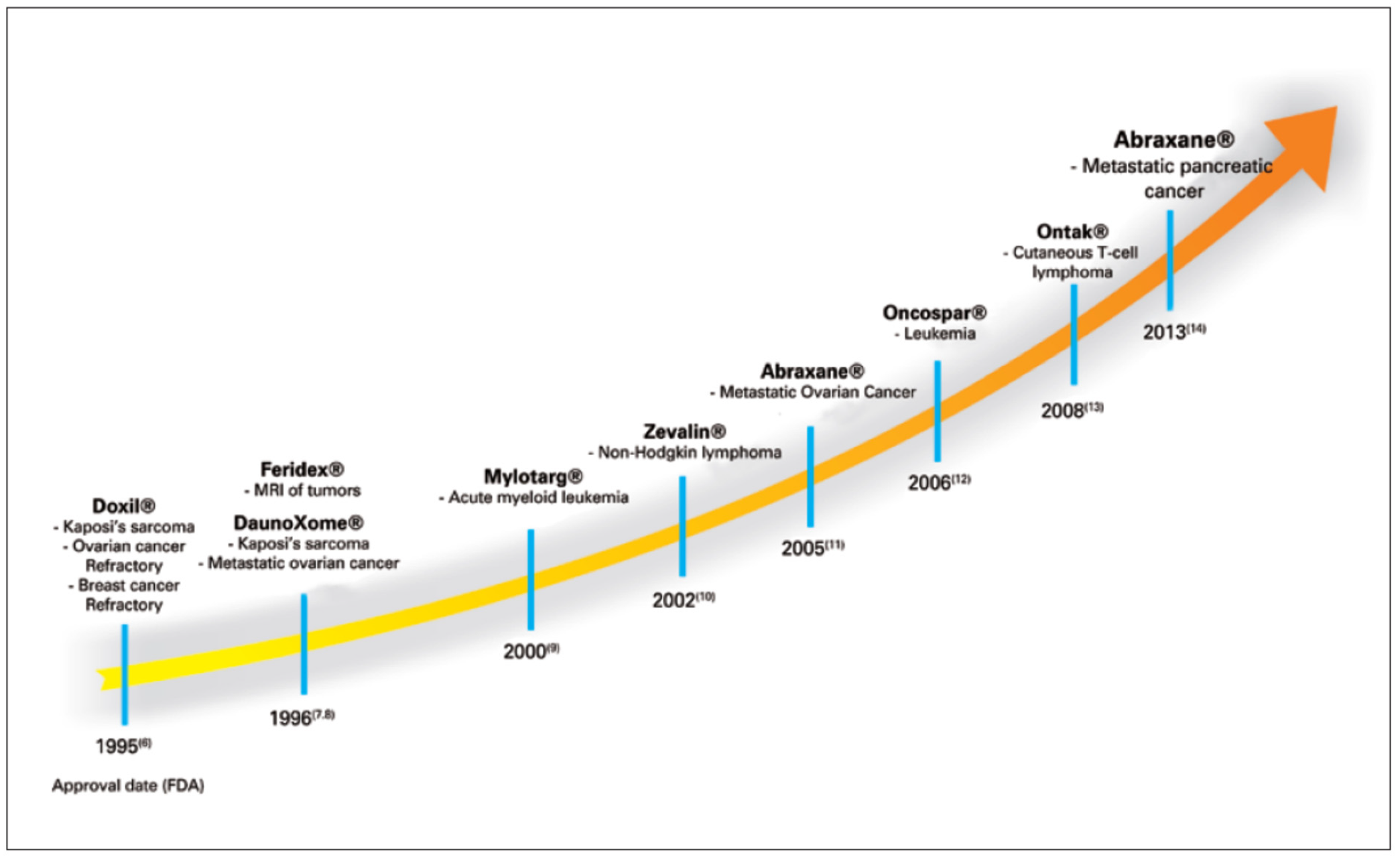
50]. These polymeric nanoparticles were developed with much effort through simulation studies and synthetic chemistry. The various synthetic biodegradable polymeric nanocarriers are poly(2-hydroxyethyl-L-aspartamide), poly(L-aspartate), poly(D,L-lactic acid-co-glycolic acid), poly(ε-caprolactone), poly(ethylene glycol) (PEG), poly(N-vinyl pyrrolidone) (PVP), poly(N-isopropyl acrylamide) (PNIPAM), poly(hydroxypropyl methacrylamide) (PHPMA), poly(methyl methacrylate), poly(ethylene glycol), poly-(chloromethyl-styrene) (PCMS), etc. The theranostic cancer management is based on diagnosis and treatment, and hence, this review is divided into two parts; i.e., diagnosis and treatment of cancer. The different types of polymeric NPs for drugs delivery are shown in Figure 2. The timeline of the development of polymeric NPs for cancer treatment is shown in Figure 3. The biocompatible polymer-based cancer theranostics are given in Table 1, while polymeric NPs in clinical trials are given in Table 2.

3. Diagnosis of Cancer

The early detection of cancer is a very important issue in order to treat this disease properly. Cancers can be treated and managed if diagnosed in an early stage. In this way, the early diagnosis of cancer is one of the most important approaches to manage cancers. Unluckily, only 16% of cancer cases are diagnosed prior to malignancy and most of them are noticed in advanced or metastatic or late stages [80]. Generally, the diagnoses of cancers depend on the type of cancer, and sizes and positions of tumors. The commonly used ways of cancer diagnosis are biopsy, positron emission tomography scanning, magnetic resonance imaging, computed tomography, biosensors, X-rays, radionuclide, fluorescent imaging, etc. The nanotechnology approach has been coupled to the above modalities to detect cancers in the early stage. This section deals with the diagnosis of cancer using polymeric NPs only. Many NPs have been used to detect cancers but polymeric-based ones are considered the best one due to biodegradable nature and capacity to carry many lesion-detecting molecules. Besides, polymeric NPs have targeting moieties that show a therapeutic drug with an imaging agent to be expected to have an excellent theranostic platform. Additionally, polymeric NPs may directly transport the imaging agent to the lesion, and can be simply administrated by inhalation, oral or intravenous ways, respectively. The most common identities used for cancer diagnosis are cancer antigen 125 (CA125), α-fetoprotein (AFP), cancer antigen 19-9 (CA19-9), cancer antigen 153 (CA15-3), carcinoembryonic antigen (CEA), epidermal growth factor receptor (EGFR), breast cancer (BRCA), prostate-specific antigen (PSA), interleukins (ILs), mucin 1 (MUC 1), tumor necrosis factor-alpha (TNF-α), human epidermal growth factor receptor 2 (HER2), vascular endothelial growth factor (VEGF) and squamous cell carcinoma antigen (SCC-Ag). These are identified by means of nanotechnology. Generally, these are used to precisely transport imaging and therapeutic molecules (theranostic agents) into the sites of cancerous tissues.
The following are the essential requirements for NPs for cancer diagnosis:
  • Being biocompatible and biodegradable in nature;
  • Low toxicity to normal cells;
  • Small size;
  • High stability in physiological conditions;
  • Capacity to carry the imaging agents;
  • Controlled release of therapeutic agents.
Gadolinium NPs have been used to detect tumors in magnetic resonance tomography (MRI) because of their good contrast. Boyes et al. [81] reported a review addressing the chemical aspects in the development of molecular probes based upon gadolinium NPs and their potential role in translational clinical imaging and therapy. Similarly, Cao et al. [82] reviewed the applications of prepared gadolinium NPs for cancer detection in MRI. The authors described these NPs to have the promising capability of improved longitudinal relaxivity of individual gadolinium ions. Further, the authors highlighted the developments and applications with future perspectives for their future development. Mi et al. [83] reported gadolinium (Gd) chelates-loaded nanocarriers for achieving magnetic resonance imaging (MRI)-guided Gd neutron capture therapy (GdNCT) of tumors. The authors developed calcium phosphate micelles hybridized with PEG-polyanion block copolymers, and united them with MRI contrast agent Gd-diethylenetriaminepentaacetic acid (Gd-DTPA/CaP). The Gd-DTPA/CaP displayed an increased distribution of Gd-DTPA in tumors, leading to a sufficient MRI contrast improvement of tumor location. Vuu et al. [84] used gadolinium−rhodamine NPs, prepared from lipid monomers for cancer detection in MRI. As per the authors, these NPs might be used to track cells in vivo.
Some other important works in the direction of cancer diagnosis are described in the following sub-sections. Mitra et al. [85] reviewed the applications of liposomes, micelles, dendrimers and water-soluble polymers for cancer diagnosis. The authors used these NPs to carry large payloads, improving cancer detection. The authors used nuclear imaging techniques such as dual-modality imaging with positron emission tomography/computed tomography (PET/CT) to detect cancer in various models. As per the authors, furthermore pre-clinical, clinical and long-term toxicity studies are needed before applying in real-life problems. Janib et al. [86] and Park et al. [87] reviewed the theranostic NPs, including drug conjugates, drug complexes, micelles, dendrimers, core-shell particles and carbon nanotubes. A lot of these formulations were applied for carriers of drugs or contrast agents. To observe the interactions of these formulations with cancer disease, contrast agents, including metals, metal oxides, optically active molecules, radionuclides and ultrasonic contrast mediums, were encapsulated. Wang et al. [88] used poly(ethylene glycol) and ATP aptamer to detect breast cancer by estimating ATP levels in the biological samples. The authors reported detection limit 0.1 pM with a linear range of 0.1–1000 pM. Lipid-polymer hybrid (LPH) NPs have been utilized to diagnose cancers. These acts as imaging agents, which are used in tomography or magnetic imaging resonance (MRI). Aryal et al. [89] described the use of LPHs in MRI, the former having a shell of gadolinium ions chelated with lipid PEG and DOTA. These were used to test in-vitro uptake by J-774 cells while the murine xenograft (B16-F10) model indicated an accumulation of the LPNs in the tumor within a time of 3 h.
Kandel et al. [90] used LPHs having a fluorescent cores; e.g., poly-(fluorene-alt-benzothidiazole)-PFBT enclosed by a lipid-PEG (DMPE-PEG) layer. The prepared LPHs, having particle sizes of 20–30 nm and quantum yield of ≈50%, indicated bright fluorescence because of the insertion of lipid tail in polymeric core and resulted in an augment of space and intrachain quenching. Sun et al. [91] used gold NPs and SPION-loaded micelles for in vitro detection of glioblastoma multiforme (brain cancer). These NPs were used as a contrast agent in computed tomography and MRI. These results were used to determine tumor size. As per the authors, the reported NPs were excellent agents in MRI, even after few days. It was realized that these NPs may be used as MRI agents for early detection of brain cancer. Mottaghitalab et al. [92] prepared silk fibroin NPs (SFNPs) beset with SP5-52 peptide for supplying Gem into lung cancer cells. The NPs were found to accumulate in cancer parts. This confirmed the efficiency of the synthesized NPs in cancer detection. Guo et al. [93] prepared polymeric nanosturucture (PHEMA-star-PLLA-PEG) with poly(hydroxyethyl methacrylate) core and PLLA-PEG as arms for cancer diagnosis. 1,4,7-triazacyclononane-1,4,7-triacetic acid and TRC105 and were altered (grafted) on the end groups for CD105 facilitated target, as well as 64Cu labelling and PET imaging. In vivo imaging and assessments showed that targeted ligand awarded this particular polymer with long dispersed half-time, improved tumor increase and a decent image comparison. As per the authors, quick imaging might be obtained via this imaging policy, with tumors noticeable at 0.5 h post-injection.
Magnetic NPs are the best ones in MRI, as these provide good contrast. In this series, Li et al. [94] prepared aqueous dispersive polyethyleneimine (PEI) coated Fe3O4 nanoparticles following modification by fluorescein isothiocyanate (FI) and PEGylated folic acid (FA) through PEI facilitated conjugation. The residual PEI surface amines were exposed to acetylation to make the colloidally stable FA functionalized Fe3O4 NPs for MRI. The authors claimed that the developed NPs may be used in vivo and in vitro for the diagnosis of different tumors. Wang et al. [95] prepared optical activated NPs for photoacoustic/fluorescent imaging in cancer diagnosis. The NPs consisted of perfluorohexane liquid and gold nanoparticles (GNPs) in the core stabilized by a poly(lactide-co-glycolic acid) (PLGA) polymer. The authors claimed that the reported NPs were evaluated in vitro experiments, showing the potential of photoacoustic imaging for future clinical investigations in cancer therapy. Huang et al. [96] designed polymeric substrate nanoparticles for cancer detection by the dissipative particle dynamics simulation. It was observed that the cancer cells and the normal cells were differentiated by different uptakes of nanoparticles. The antagonism between nanoparticle cell-specific and nanoparticle polymer non-specific interactions was the chief issue for dissimilar uptake performances. The authors enhanced cancer detection by investigating the effects of the polymer and ligand types, and density. As per the authors, this study may deliver valuable visions into the proposal of the functionalized substrate-based nanomaterials in biomedicine.
Ali et al. [97] used gold and polymeric superparamagnetic iron oxide nanoparticles (SPIONs) loaded micelles (GSMs) coated by polyethylene glycol-polycaprolactone (PEG-PCL) polymers for detecting brain tumors. GSMs were used as a contrast agent for both computed tomography (CT) and magnetic resonance imaging (MRI) studies of stereotactically implanted GBM tumors in a mouse model. These results suggested that with further expansion and testing, GSMs may possibly be combined into both imaging and treatment of brain tumors, helping a theranostic determination as both an MRI-based contrast agent and a radiosensitizer. The polymeric micelles have been used in nuclear imaging, X-rays, MRIs, and CT scans for cancer diagnosis [98]. Utilization of micelles conjugated with the gamma emitters (99mTc and 111In) includes non-invasive biodistribution [99,100]. The addition to iron oxide particles in micelles or the use of chelators for the blending of paramagnetic metals to the hydrophilic wedge of micelle making block copolymers are two methods used for preparing micelle-based MRI contrast means [101,102,103,104,105]. The multifunctional polymeric micelles with imaging contrast means and bunches of magnetic NPs for effective MRI and chemotherapeutic agents targeting ligands have been described [105]. Qiao and Shi [106] and Yang et al. [107] described the syntheses of iron oxide NPs conjugated with Arg-Gly-Asp modified dendrimers for targeted MRI of C6 glioma cells.

4. Treatment of Cancer

Nanotechnology is gaining importance in treating and managing cancer. Different types of nanoparticles are used to control cancer diseases. Among many NPs, polymer-based NPs are gaining importance in cancer treatment. It is because these NPs are biocompatible, biodegradable, non-toxic and non-immunogenic. Different types of polymeric NPs, including composite NPs, have been used in cancer treatment, and these are discussed in the following sub-sections.
The salient requirements of a NP to treat and control a cancer are that it:
  • Should be biocompatible, biodegradable, non-toxic and non-immunogenic.
  • Should be able to carry the required drugs.
  • Should be able to discharge the medications at the location of the tumor.
  • NPs–drug complex should be stable under physiological conditions.
  • NPs–drug complex should be able to control either by receptor-facilitated interfaces or by electroparamegnetic resonance spectroscopy (EPR) effect.
As discussed above, polymeric nanoparticles (PNPs) are excellent carriers of the required drugs in cancer treatment. Many synthetic and natural precursors are being used for the preparation of polymeric NPS. The synthetic ones include poly(lactic-co-glycolic acid), poly(lactic acid) and polyethyleneimine, whereas the natural ones are albumin, collagen, chitosan and gelatin. First of all, in 1980, Couvreur et al. [108] used polymeric nanoparticles for cancer treatment. The authors developed PNPs by using poly(alkyl cyanoacrylate). After this research, many PNPs were prepared and used in curing cancer disease, especially the delivery of anti-cancer drugs. The applications of PNPs in cancer treatment are discussed in the following sub-sections.

4.1. Biodegradable Nanoparticles

The biodegradable polymeric nanoparticles are submicron-sized particles with drug molecules adsorbed, diffused, entangled, linked or encapsulated into the NPs or a gene of interest encapsulated inside a polymeric matrix. There are two methods for the preparation of nanoparticles: (i) by distributing polymers that are performed, and (ii) polymerizing monomers [109]. These NPs are easy to manufacture, relatively low-cost, biocompatible, non-immunogenic, non-toxic and water-soluble. These are also of great worth as an effective method for dispensing pharmaceuticals to particular tissues/organs, as a means of DNA gene therapy, and for their competency with distributing proteins, peptides and genes by oral administration [110]. The anticancer drugs, cisplatin, doxorubicin, paclitaxel, 5-fluorouracil, triptorelin, 9-nitrocamptothecin, xanthone, dexamethasone, etc., have been effectively related with poly(glycolic acid) (PLGA) [111]. The nanospheres having naturally degradable hydrophobic poly(ε-caprolactone) (PCL) and hydrophilic methoxy poly(ethylene glycol) (MePEG) have been used for taxol delivery [112].

4.2. Polymeric Superparamagnetic NPs

The magnetic NPs stay in superparamagnetic states, but have magnetic responses and super-paramagnetism. These NPs are used to enhance the therapeutic presentation of drugs and decrease the side effects related to the straight treatment of cancer. These may be guided towards the tumors with the help of outside magnetism. By binding these NPs with synthetic and natural polymers, they may be used as drug carriers. The polymeric superparamagnetic iron oxide nanoparticles (SPIONs) were coupled with polymersomes and micelles for drug delivery. This is due to the fact that these have good drug loading with biocompatibility, stability, fine particle size distribution, prolonged blood circulation and controlled drug release [43]. Huang et al. [113] described SPIONs coupled with folic acid for dual-targeted drug delivery. The doxorubicin (DOX) was laden onto folic acid (FA)-SPIONs nanoparticles covered with PEI (polyethylenimine) and PEG poly(ethylene glycol) polymers. As per the authors, the drug release was magnetically targeted and pH-sensitive. The prepared formulation was applied to the mice model with MCF-7 breast cancer. The authors reported an enhanced anticancer activity. Guthi et al. [114] used multifunctional micelle superparamagnetic iron oxide NPs for carrying doxorubicin and tested for H2009 lung cancer cells. The authors reported successful doxorubicin release. Maeng et al. [115] used superparamagnetic iron oxide to grow a drug delivery method with improved efficacy and minimized adverse effects by coating poly(ethylene oxide)-trimellitic anhydride chloride-folate and doxorubicin. The efficiency of the nanoparticles was assessed in rats and rabbits with liver cancer, in comparison with free-DOX (FD) and a commercial liposome drug, doxorubicin (DOXIL). As per the authors, these results indicated that the reported NPs may be good agents for curing liver cancer.
Ling et al. [116] used PLGA to encapsulate SPIONs and docetaxel for releasing the drugs to tumor cells. The multifunctional polymer vesicles were prepared by carboxy-terminated poly(lactide-co-glycolidelactide) utilizing a single emulsion evaporation method. The authors reported a 147 nm diameter with drug encapsulation efficacy of 6.02%–23%. The NPs showed a triphasic drug release pattern in vitro in 30 days. Improved cellular uptake ability and antiproliferative effect in prostate cancer PC3 cells were observed. Therefore, these stable and tumor targeting polymer NPs could be promising multifunctional vesicles for simultaneous targeting imaging, drug delivery and real-time monitoring of therapeutic effect. Yang et al. [117] used coated SPIONs for targeted drug delivery. The anticancer drug was conjugated onto the PEGylated SPIONs by pH-sensitive bonds. As per the authors, coated SPIONs indicated higher tumor accumulation than Arginylglycylaspartic acid (cRGD)-free ones. Thus, these coated SPIONs showed encouraging properties for combined targeted anticancer drug delivery. Yallapu et al. [118] synthesized superparamagnetic iron oxide nanoparticles by precipitation of iron salts in the presence of ammonia and β-cyclodextrin and pluronic polymer coatings. The curcumin-loaded formulation showed a good effect on ovarian, breast and prostate cancers. The better therapeutic effects were confirmed by molecular effects using western blotting and transmission electron microscopic (TEM) studies. Chen et al. [119] used copolymer of reducible polyamidoamine with polyethylene glycol/dodecyl amine for delivery of doxorubicin to targeted cells. Xu et al. [120] prepared multifunctional nanoparticles by using a superparamagnetic iron oxide core with PEG/PEI/polysorbate 80 (Ps 80), and that was utilized to encapsulate DOX. As per the authors, the NPs were capable of delivering drugs effectively to gliomas. Because of the magnetic targeting and Ps 80 arbitrated endocytosis; DOX@Ps 80-SPIONs in the occurrence of a magnetic field, led to the whole destruction of glioma growth in vivo at 28 days after treatment. The therapeutic mechanism of DOX@Ps 80-SPIONs acted by tempting apoptosis through the caspase-3 pathway.

4.3. Polymeric Gadolinium NPs

The polymeric gadolinium NPs have great potential in cancer diagnosis in MRI due to their good thermodynamic association constants and low dissociation kinetic rates. Besides, these NPs have been used as drug carriers for cancer treatment. Some of the important research works are summarized in this section. Lio et al. [121] used galodinium paramagnetic metal ions to prepare NPs with poly(DL-lactide-coglycolide; PLGA) and a hydrophilic paramagnetic folate coated PEGylated lipid shell (PFPL) having a core-shell structure. The NPs had good properties to load and release the hydrophobic drugs. As per the authors, the reported NPs had a good perspective on cancer treatment. Lee et al. [122] used Cd(III) NPs for the delivery of gemcitabine drugs. The Gd(III) conjugates indicated good drug uptake and control. Cheng et al. [123] reported polymeric (Gd2O3) nanoparticles with high longitudinal relaxation rate. The polymer used was polypyrrole (PPy) modified with hyaluronic acid (HA) and laden aluminum phthalocyanine (AlPc). The authors described the use of these NPS in MIR cancer detection and remote-controlled, PTT/PDT combined anti-tumor therapy. The nanoparticles showed a photothermal effect that may have triggered the release and de-quenching of AlPc.
Rowe et al. [124] prepared a multifunctional polymeric gadolinium (Gd) metal-organic framework (MOF) nanoparticles for cancer diagnosis and treatment. The polymer used was poly(N-isopropylacrylamide)-co-poly(N-acryloxysuccinimide)-co-poly(fluorescein O-methacrylate). Cao et al. [125] described diagnosis and therapy for cancer by developing magnetic nanoparticle (Gd@SiO2-DOX/ICG-PDC) integrating doxorubicin (DOX), indocyanine green (ICG) and gadolinium(III)-chelated silica nanospheres (Gd@SiO2) with a poly(diallyldimethylammonium chloride) (PDC) coating. The DOX release from Gd@SiO2-DOX/ICG-PDC depended on temperature and pH. As per the authors, the reported NPs are safe in both diagnosis (MRI scanning) and treatment. Roy et al. [126] reported the preparation of stimuli-responsive, polymer-adapted, gadolinium-doped iron oxide nanoparticles (poly@Gd-MNPs) and the use as cancer theranostic NPs. The NPs showed an excellent loading capacity for anti-cancer drug methotrexate along with stimuli dependent release by changing pH and temperature of the cancer cells. The poly@Gd-MNPs showed 86% drug loading capacity with 70% drug release in the first 2 h. The cytotoxic assay (MTT) established that the NPs did not disturb the feasibility of normal cells but capably killed the MCF7 cancer cells in the attendance of an outside magnetic field.

4.4. Polymer Gold NPs

Gold metal has been used for long to treat epilepsy and tuberculosis, and the properties of gold NPs make them ideal candidates. The chemical, surface, electronic and optical properties of gold NPs are unique for cancer treatment. These NPs may be delivered to cancer tissues by passive or active targeting plans. The various shapes of gold NPS are nanorods, nanoshells, nanospheres, nanostars, nanoboxes, nanocrystal, nanocages, nanoclusters, nanocubes and triangular bipyramaids. The modifications of these NPs with polymeric materials make them ideal candidates for drug delivery with increased circulation time, improved drugs-loading ability and stability and reduced cytotoxicity.
Gold NPs coagulated with polymers are ideal candidates for drug release due to the ease of surface modification. The surface modifications augment circulation time, drug targeting and the progressive stability of NPs, and decreases cell cytotoxicity. The chemotherapeutic anticancer drugs are loaded on these NPs via covalent and ionic bonds and physical sorption. The drugs loaded with these NPs are doxorubicin, methotrexate, paclitaxel, docetaxel, tamoxifen, oxaliplatin and 3-mercaptopropionic [127]. Ding et al. [128] conjugated paclitaxel with mercapto functionalized AuNPs using oligoethylene glycol spacer via carbodiimide based esterification for improved cytotoxicity toward paclitaxel sensitive and resistant cell lines. Yang et al. [129] reported doxorubicin-loaded AuNCgs for enhancing doxorubicin loading. The authors used a thermal effect on the release of the reported drug. As per the authors, in vitro studies showed the feasibility and advantage of the reported NPs for remote-controlled drug release systems. Sun et al. [130] developed doxorubicin-loaded AuNPs nano-conjugates for drug delivery. The drug release by NPs was better than free drug in MCF-ADR cells. As per the authors, well-designed drug delivery system and conventional chemotherapeutic agents may be promising for cancer stem cell therapy.
Li et al. [131] reported AuNPs PEGylated Polyamidoamine (PAMAM) dendrimers loaded with doxorubicin to improve therapeutic efficacy in in vitro and in vivo situations. The NPs showed uniform sizes and good colloidal stability. The drug release was pH controlled, and hence this may be used for controlled drug releases as per the requirement. The authors also demonstrated the combined photothermal-chemo treatment of cancer cells using PEG-DOX-PAMAM-AuNR for synergistic hyperthermia ablation. The chemotherapy was showed in both in vitro and in vivo conditions. Larson et al. [132] described AuNPs with aminohexylgeldanamycin (AHGDM), docetaxel and cisplatin drugs. The NPs were assessed in vitro for the ability to synergistically induce cytotoxicity in combination with moderate hyperthermia. The targeted docetaxel conjugates showed high potency against DU145 cells with an IC50 of 2.4 nM. Topete et al. [133] described the preparation of PLGA/DOXO-core Au-branched shell nanostructures (BGNSHs) functionalized with a human serum albumin/indocyanine green/folic acid complex (HSA-ICG-FA) to provide a multifunctional nanotheranostic platform. The prepared NPs were loaded with doxorubicin (topoisomerase II inhibitor and DNA intercalating agent). The reported formulation was tested by determining the biodistribution of the drug in a tumor bearing mice model. Hwu et al. [134] developed paclitaxel-PEG-SH coupled AuNPs for anticancer activities. Some other authors reported folic-acid-loaded PEGylated AuNPs for targeting purposes to cancerous cells [135,136,137]. The polymeric gold NPs used for drug delivery are given in Table 3.

4.5. Carbon Nanotubes (CNTs)

It is a well-known fact that the carbon nanotubes are the miracle identities in the world of nanoscience. The oxidation and grafting polymers on side walls of carbon nanotubes are useful to load the polymeric materials. In this series, polymeric materials have been conjugated with carbon nanotubes (CNTs). Feazell et al. [150] loaded DOX onto branched PL-PEG functionalized CNTs by π-π stacking to the sidewall of carbon nanotubes. In vivo studies in a mouse model of breast cancer indicated increased therapeutic efficacy and a marked reduction in toxicity as compared to free DOX. The same group [151] used paclitaxel to load on polymeric CNTs for testing cancer. Gao et al. [152] prepared nano/microbubbles of biodegradable block copolymers. These were found to be suitable for dramatical release of drugs. The authors reported suitable chemotherapy for MDA MB231 breast cancer. Chen et al. [153] prepared long polymer nanotubes with cross linked poly(glycidyl methacrylate) and a pendant poly(N-isopropyl acrylamide) for delivery of anti-cancer drugs. The authors used doxorubicin as a model drug for the study. The developed polymer nanotubes were found to have good biocompatibility in the test with KB-3-1 cancer cells. The toxicity assay confirmed IC50 value as 1.4 μmol L1. It was observed that the described polymer nanotubes had a good potential for effective anti-cancer drug delivery.
Zhang et al. [154] developed multifunctional targeted poly(lactic-co-glycolic acid) (PLGA) nanobubbles (NBs) for use as targeted anticancer drug carriers and a synergistic agent for increasing therapeutic efficiency of High-intensity focused ultrasound (HIFU) ablation. The authors used methotrexate (MTX)-loaded NBs with active tumor-targeting monoclonal anti-HLA-G antibodies (mAbHLA-G) coated onto the surfaces of nanobubbles. These tubules were tested both in vitro and in vivo. The authors reported the augmented efficiency of HIFU ablation. It was observed that the reported tubules constituted an efficient and non-invasive HIFU synergistic treatment of cancer with the additional tasks of massacring remaining tumor cells and checking tumor reappearance/metastasis. Yang et al. [155] designed and developed triple stimuli-responsive, biodegradable nanocapsules having perfluorohexane and doxorubicin-loaded poly(methacrylic acid (PMAA) of 300 nm. It was observed that the drug loading efficiency was about 93.5%. It was observed that the reported nanocapsules may be biodegradable with a good therapeutic effect. Pistone et al. [156] reported two pH and temperature-precise drug delivery systems for cancer therapy by using hydrophilic polyethylene glycol (PEG) functionalized multi-walled carbon nanotubes. The NPs were loaded with doxorubicin and covered with the biocompatible polylactide and measured for drug release performance on three different human cancer cell lines. The authors evaluated and compared, stressing the opportunity to adjust the quantity of drug released by regulating the functionalization grade of the carbon nanotube material. The biological tests emphasized the high biocompatibility of polymeric carbon nanotubes to deliver DOX to cancer cells.

4.6. Polymeric Micelles

A micelle is an accumulation of surfactant molecules with both hydrophobic and hydrophilic parts, which undergo self-assembly in an aqueous media to form arrangements with a hydrophobic focus with a stable hydrophilic encapsulation. The beneficial features of polymeric micelles are their drug-carrying abilities, including solubilization of not-very-soluble drugs, and their abilities for tumor direction and precise drug discharge. The ease of micelle preparation by self-assembly of amphiphilic block copolymer moieties and drug encapsulation by physical entrapment rather than chemical conjugation are very pretty properties of polymeric micelles. These are collected by different means (outward from the core), including preparation of the monomer (G) in the main core [157] or convergent (inner core) methods [158]. Micelles are good identities in chemotherapy as colloidal transporters for hydrophobic drugs [97,159]. The small size of micelles is a good feature for penetrating cancer tissue for drug release. The micelles have been used to deliver low water solubility anticancer drugs to the target in animal models [160,161]. First of all, the polymeric micelles were introduced as drug delivery carriers in the early 1980s by Helmut Ringsdorf [162]. Besides, Genexol-PM formulation has got Food and Drugs Administration (FDA) consent for use in breast cancer treatment [95]. Some reviews have appeared on the use of micelles for drugs delivery [33,35].
Valenzuela-Oses et al. [163] developed a formulation of miltefosine-loaded polymeric micelles of the copolymer Pluronic-F127. The formulation was tested against HeLa carcinoma cells, which showed promising results. As per the thermal analyses, miltefosine was molecularly spread within PM. Pluronic-F127 polymeric micelles with miltefosine (80 μM) offered a noteworthy decrease of hemolytic effect (80%, p < 0.05) in contrast to free drugs. It was observed that miltefosine-loaded Pluronic-F127 polymeric micelles may be a hopeful nanocarrier in cancer therapy. Yang et al. [164] developed a polymeric on polyethylene glycol derivatized GA (PEG-Fmoc-GA) for DOX delivery. It was observed that this formulation had more effective synergistic action on cell proliferation inhibition and apoptosis. Besides, the formulation had long blood circulation time, drug concentration area under the curve and low distribution and clearance of DOX. As per biodistribution studies, the formulation was found to be accumulated at the tumor site. As per the authors, the so formed-polymeric prodrug micelles may be a promising dual-function co-delivery system to attain synergistic anti-cancer activity of DOX and GA.
Wang et al. [165] reported the improved blood retention and tumor accumulation of gambogic acid (GA) in vitro by encapsulating it in poly(ethylene glycol)-poly(ε-caprolactone)-poly(trimethylene carbonate) [MPEG-P(CL-ran-TMC)]. It was observed that the reported NPs indicated better dispersion in water, prolonged release behavior in vitro and enhanced tumor cellular uptake, showing superior antitumor efficacy and better apoptosis. Volsi et al. [166] reported the synthesis of polymeric micelles encapsulating both doxorubicin and gold core-shell quantum dots nanoparticles (Au-SiO2/QDs). The formulation was found to have the cytotoxicity of the breast cancer cells. The micellar NPs in clinical trials are shown in Table 4.

4.7. Dendrimers

The dendrimers are extremely branched synthetic polymers with symmetric centers and spherical 3-dimesional micro-molecules [176]. The dendrimers have five parts; i.e., a core, branches (called generations) emanating from the core, repeating units, at least one branch junction and numerous terminal functional groups. Generally, the center is made of diaminobutyl (DAB), ethylene diamine (EDA), polypropylimine (PPI) and polyamidoamine (PAMAM), with numerous exterior residues, such as carboxyl, amine and alcoholic functional groups, determining surface properties. Generally, a dendrimer is prepared with various materials depending on the properties required, such as dimension, shape, thermal stability, solubility, etc., using a convergent or divergent approach. These are changed as water-soluble my surface modifications. These modifications enhanced the capabilities of dendrimers in chemotherapy. These are classified by types of polymers (hyper branched and brush polymers) and their molecular weights (low and high molecular weight). Generally, dendrimer drug conjugations are made of covalent bonding between dendrimers and anticancer drugs. The melanin conjugated dendrimers were used to increase the solvability of anti-cancer drugs such as 6-mercaptopurine and methotrexate [177]. Highly branched dendrimer augmented aptamer probes may be simply reconstructed and have great appeal and specificity for a wide range of objectives. These are capable of reaching numerous targets with such great reliability, sensitivity and selectivity due to their good magnetic, optical, electric, biological and chemical properties [106]. The gold NP-based dendrimers have been used for the photothermal treatment of cancers [178,179]. Some reviews have been appeared on dendrimers drug delivery [36,38,180,181]. Malik et al. [182] reported PEGylated PAMAM dendrimer loaded cisplatin against B16F10 solid melanoma tumors. On the other hand, Kaminskas et al. [183] described PEGylated poly-L-lysine (PLL) dendrimers (G5, PEG1100) loaded with methotrexate as accumulated in HT1080 fibrosarcoma tumors in rats and mice.
Al-Jamal et al. [184] described doxorubicin-loaded cationic PLL dendrimer Gly-Lys63 (NH2)64 as anti-angiogenic active in tumor-bearing mice. Amreddy et al. [185] developed a folic acid (FA)-conjugated polyamidoamine dendrimer (Den)-based nanoparticle for co-delivery of siRNA against HuR mRNA (HuR siRNA) and cis-diamine platinum (CDDP) to folate receptor-α (FRA)-overexpressing H1299 lung cancer cells. The co-delivery of HuR siRNA and CDDP using the FRA-targeted NP had a meaningfully greater therapeutic effect than individual therapeutics. Besides, FRA-targeted NP showed improved cytotoxicity compared to non-targeted nanoparticles with lung cancer cells. The dendrimers and drugs interactions on cancer therapy are given in Table 5.

4.8. NPs Prepared by Electrospray

Boda et al. [192] reviewed the electrospraying as a versatile technology for manufacturing nanoparticles of various compositions, morphologies and sizes, which can be applied for imaging for diagnostics, drug delivery and sensing. Astolfi et al. [193] mentioned that cisplatin is a famous chemotherapeutic anticancer drug; however, the side effects, such as alopecia, nausea, nephrotoxicity and cardiotoxicity, limited the usage of this drug. Bai et al. [194] reported a simple method of preparing PEG-PLGA encapsulated cisplatin bioconjugated with CD44 antibody using electrospray. The anti-proliferative ability of the cis-encapsulated CD44-PEG-PLGA nanoparticles is 10%–14% greater against SKOV-03 and CP70 cells, which is useful in cancer therapy of targeting ovarian cancer cells in vivo or in vitro. Guo et al. [195] prepared core-shell polymer microparticles for theranostics by coaxial electrospray. In the microparticles, gold nanoparticles encapsulated folic acid conjugated chitosan as Au@CS-FA, and the shell was loaded with doxorubicin (DOX). The microparticles can generate strong surface-enhanced Raman scattering (SERS) signals for cancer detection due to highly branched gold nanoparticles inside. Therefore, as a laser-irradiated the microparticles, the locations of cancer cells could be imaged, and the DOX could be released due to the heat generated by the irradiating of Au@CS-FA particles simultaneously. Wu Y. [196] synthesized theranostic lipoplexes in one step by static micromixer-coaxial electrospray (MCE) technology with high reproducibility. QD605/Cy5-G3139-loaded lipoplexes were produced from the model imaging reagent quantum dots (QD605) and the anti-cancer drug Cy5-labelled antisense oligonucleotides (Cy5-G3139) by MCE. They were highly uniform with a polydispersity of 0.024 ± 0.006 and mean diameter by volume of 194 ± 15 nm. The lipoplex particles were efficiently delivered to A549 cells, a non-small cell lung cancer cell line, and the Bcl-2 gene expression could be down-regulated by 48% ± 6% simultaneously.

4.9. Nanoparticles Prepared by Supercritical Fluids

Taboada et al. [197] synthesized monodisperse iron oxide/microporous silica core/shell composite nanoparticles, core(g-Fe2O3)/shell(SiO2), in a one-step process with a high magnetization by combining sol-gel chemistry and a supercritical fluid method. These nanoparticles show superparamagnetic behavior at room temperature, are proven to be a suitable T2 MRI imaging agent and have the potential to be used for theranostic drug delivery. Chattopadhyay et al. [198] generated magnetically responsive poly(methyl methacrylate) using supercritical antisolvent (SAS) technology. These magnetite-encapsulated polymer nanoparticles can be used as contrast agents for magnetic resonance imaging scans for controlled-release drug formulations. The sizes of the nanoparticles are controllable via the SAS-EM technique developed at Auburn University, and the morphologies and drug-release kinetics of these nanoparticles were also studied. Chen et al. [199] prepared 5-fluorouracil-poly(L-lactide) (5-Fu-PLLA) microparticles by a solution-enhanced dispersion by supercritical CO2 (SEDS) process; the 5-fluorouracil is an anti-metabolite with activity against colon, rectal and prostate cancers. Therefore, the therapeutic efficacy of the 5-Fu drugs is increased by encapsulating the drugs in biodegradable polymers as sustained delivery systems. Chen et al. [200] synthesized indocyanine green (ICG)-encapsulated silk fibroin (SF) (ICG-SF) nanoparticles by supercritical fluid (SCF) technology. The encapsulated nanoparticles have excellent photothermal stability and can be observed with near-infrared (NIR) light at 808 nm. The ICG can be released from SF specifically due to this process being pH-responsive in the tumor acidic environment, and these nanoparticles can destruct cancer cells barely under light-induced hyperthermia in vitro and in vivo. These results showed that the biocompatible ICG-SF nanoparticles synthesized by SCF technology had high photothermal therapy efficiency, and showed the great potential of theranostic nanomedicine for cancer.

4.10. Miscellaneous Polymeric NPs

Besides the above-discussed polymeric NPs, some other biodegradable materials have been conjugated or prepared for loading and delivering drugs in cancer treatment. These include, chitosan, polystyrene, etc. Kim et al. [201] described chitosan-based nanoparticles (CNPs) and used them for control drug release. The authors described their unique features like deformability, stability and quick uptake by tumor cells. The loaded drug was paclitaxel and the reported NPs showed pointedly augmented tumor homing ability with low non-specific uptake by other cells tumor having mice. The authors reported that these NPs may be future anti-cancer agents. Park et al. [202] modified three glycol chitosan (GC—20 kDa, GC—100 kDa and GC—250 kDa) derivatives with cholanic acid to increase the in vivo tumor targeting features of polymeric nanoparticles. These amphiphilic glycol chitosan cholanic acid molecules conjugated self-assembled into glycol chitosan nanoparticles (GC—20 kDa NP, GC—100 kDa NP and GC—250 kDa NP). The distribution, tumor accumulation and time-dependent excretion of these NPs were determined in SCC7 tumor having mice using NIR fluorescence imaging systems. These NPs showed the most decreased time-dependent excretion and blood circulation time and raised tumor accumulation with growing polymer molecular weight. It was observed that high molecular weight glycol chitosan NPs remained for a longer time in the blood circulation, leading to an augmented gathering at the tumor site. The authors proposed that improved tumor targeting by high molecular weight NPS was related to better in vivo stability; centered on a pharmacokinetic enhancement in blood circulation period. Gao et al. [203] encapsulated doxorubicin in polymeric micelles. The materials used were pluronic P-105, PEG2000-diacylphospholipid and poly(ethylene glycol)-co-poly(beta-benzyl-L-aspartate) micelles as drug carriers. The authors compared drug bio-distribution with a molecularly dissolved one in ovarian carcinoma tumor model in mice. The authors reported good and controlled bio-distribution in the encapsulated formulation. Lammers et al. [204] used radionuclide (131I) with N-(2-hydroxypropyl)-methacrylamide (HPMA) polymer loaded with doxorubicin and gemicitabine for therapeutic efficiency. Dunning AT1 rat prostate carcinoma was utilized as a model for study. It was observed that the radioactive polymeric transferor indicated choosy gathering and lengthy movement period. The synergistic effect increased the therapeutic effects on cancer cells.

5. Polymeric NPs in Clinical Trials

During the last few years, some polymeric NPs have been tested in clinical trials while some others have been approved by FDA. The most important formulations are Genexol®-PM, NK105, NC-6004, NC-4016, NK012, NK911 and SP1049C. Doxil (pegylated liposomal doxorubicin) is the first nano-drug approved by FDA in 1995 to treat some cancers: AIDS-related Kaposi’s sarcoma, ovarian cancer, etc. The formulation contains doxorubicin (adriamycin) in about 80 nm size liposomes covered with PEG. This formulation allows the drug molecules to remain in the bloodstream for a long time for drug distribution to cancer cells to be prolonged [205,206]. For the approval of generic version of Doxil by FDA in 2003, Sun Pharma in India started its production at a large scale [207]. Another formulation is Lipodox made of doxorubicin and pegylated liposomes with a surface coating of PEG [208]. Some studies were carried out for clinical trial phases of DOX-loaded NPs [209].
Albumin nanoparticles (Abraxane®) have been approved by FDA for chemotherapy transportation for different types of cancer treatment [210,211]. Genexol-PM is approved by FDA for breast cancer treatment [98]. The polymeric micellar based drug (NK911) was tested under clinical trial in Japan in 2001. The formulation was having DOX encapsulated in PEO5000-b-P(Asp)4000-DOX micelles. After that, the formulation entered clinical trial phase II at National Cancer Center (NCC) Hospital in Japan. The proposed formulation was found to be effective in animal models but with some side effects [212]. Danson, Alakhov et al. [213] reported pluronic formulation with DOX (SP1049C) in an animal model, which was later entered in a clinical trial in Canada in 1999. In 2004. Kim et al. [214] reported PTX-loaded PEO2000-PDLLA1750 micelles (Genexol s-PM) in animal models. As per the authors, the maximum tolerated dose (MTD) of micellar PTX Genexols-PM was three times higher than of non-micellar formulation Taxols. PTX was loaded onto the polymeric micelles of PEG–P(Asp) modified with 4-phenyl-1-butanoate. This formulation was named NK105 [215], showing about 90 and 25 times higher plasma and tumor Area under the curve (AUCs), respectively, as compared to free PTX. This formulation NK105 is under phase I clinical trial at the National Cancer Center in Japan. Many patents have been filed by different workers globally. A comparison of patent filings is shown in Figure 4. It is clear from this figure that a maximum of 340 patents were filed on polymeric NPs, indicating the utility of polymer-based NPs. The liposomal NPs in clinical trials are given in Table 6.
Regarding the market status, about 30 nano-anticancer drugs are available in the market, with 17.5 billion USD made in 2011, 21.6 billion USD in 2012 and 53.7 billion USD in 2017; the figure is expected to reach 136 billion USD in 2021. This increase is due to the development of diagnostic instruments, detail patient information and the approval of several drugs by the FDA and other drugs controlling agencies globally. Some famous companies working on the production of nano drugs are Access Pharmaceuticals, ProLindac, Leonardo Bio Systems, Abraxis Biosciences, Immunolight LLC, Abbot and Sun Pharma. The approved polymeric nanomedicines for cancer therapy are given in Table 7.

6. Merits and Demerits of Polymeric NPs

The most important advantages of polymeric NPs are their small sizes, biocompatibility, biodegradability, high drug payload, low or no toxicity to normal (healthy) cells, high stability in physiological conditions, capacity to carry the imaging agents, controlled release of drugs, NPs-drug complex stability at biological buffered pH, NPs-drug complex receptor facilitated interfaces, etc. Besides them, other advantages include ease of their preparation and good control over size and distribution. The polymeric NPs have good protection capabilities for the encapsulated drugs regarding environmental factors. The good drug retention and long blood circulation times are other assets of polymeric NPs. There are no serious demerits of polymeric NPs. However, the limited targeting abilities and the problem of discontinuation of the therapy may be considered the major drawbacks. Sometimes, the autonomic imbalance disturbance may occur via polymeric NPs. Occasionally, polymeric NPs show effects on vascular and heart functions.

7. Future Perspectives

Of course, polymeric NPs are successful candidates for cancer detection, treatment, and management. Not to limit them to cancer chemotherapy, these NPs have been also exploited for AIDS, radiation, and gene therapies. The future is quite bright for these NPs in delivering antibiotics, proteins, vaccines, and virostatics to various cells. Despite good progress in cancer diagnosis, still, the techniques are not advanced enough to detect cancer at early stages. Moreover, the sensitivity and selectivity of conventional diagnostic techniques are not quite enough. Additionally, some toxicity has been reported due to the use of polymeric NPs. However, these NPs are the hope for a wide range of platforms for a varied array of biomedical applications in the future.
It is important to mention here that polymeric NPs are not fully developed and need more advances. The complex structures of the polymeric NPs used are difficult to handle during preparation. The imaging strategies need to be improved to avoid toxicity and advancement in anatomical to molecular imaging. Besides, it is essential to modify the physical, chemical, and biological properties to increase biocompatibility, biodegradability, and drug loadability; reduce toxicity to low levels or none for normal cells; and extend circulation time. The identification of the requirements of the specific combinations of NPs carriers and target molecules is very important. Certainly, efficiency enhancements will lead to improved therapeutic results and low toxicity. Also, there is a need to find out the pharmacotherapy with special emphasis on absorption, distribution, degradation, and excretion mechanisms in blood. There are certain issues to be tackled by PNPs, such as low drug solubility, the differences between loading and tracking of the drugs; non-selective tissue penetration, and the drug binding interferences. Finally, the pre-clinical trials, clinical trials, and the approval time period should be curtailed to provide cancer medication to the patients.

8. Conclusions

Nano-theranostics is a hopeful approach to cancer diagnosis and treatment under one umbrella. The polymeric nanoparticles have been synthesized and used for diagnosis and drugs delivery. It is only possible to provide all the three aspects of cancer cure (diagnosis, treatment, and management) by polymeric NPs. Theranostics also provide a holistic transition from trial and error medication to personalized medication. The polymeric NPs are the only safe chemotherapeutic anticancer drugs carriers. Some polymeric nanoparticle-based nanodrugs are approved by the sanction agencies while some others are in clinical trials. It is expected that the polymeric nano drugs are future medications to eradicate cancer from its root. Certainly, this area of research promises to augment the excellence of clinical care and treatments. Besides, the use of polymeric nanodrugs will ultimately save the lives of millions and billions of USD in the future. But still, there is a great need to develop these drugs collectively at the global level.

Author Contributions

The preparation of the different parts of the original manuscript, I.A., M.A., L.S., M.T.S., S.-T.T., M.F.H.; Review and editing, R.-S.Y., J.-C.C.; funding acquisition, M.F.H. All authors have read and agreed to the published version of the manuscript.

Funding

This research was funded by Ministry of Science and Technology, Taiwan (Grant number: MOST 107-2119-M-033-003).

Acknowledgments

The authors are thankful to Ministry of Science and Technology, Taiwan for financial assistance through Grant number: MOST 107-2119-M-033-003.

Conflicts of Interest

There is no conflict of interest for this manuscript.

References

  1. Bray, F.; Ferlay, J.; Soerjomataram, I.; Siegel, R.L.; Torre, L.A.; Jemal, A. Global cancer statistics 2018: GLOBOCAN estimates of incidence and mortality worldwide for 36 cancers in 185 countries. CA Cancer J. Clin. 2018, 68, 394–424. [Google Scholar] [CrossRef] [PubMed] [Green Version]
  2. Aboul-Enein, H.Y.; Ali, I. Determination of tadalafil in pharmaceutical preparation by HPLC using monolithic silica column. Talanta 2004, 65, 276–280. [Google Scholar] [CrossRef] [PubMed]
  3. Ali, I.; Gaitonde, V.D.; Aboul-Enein, H.Y.; Hussain, A. Chiral separation of β-adrenergic blockers on cellucoat column by HPLC. Talanta 2009, 78, 458–463. [Google Scholar] [CrossRef] [PubMed]
  4. Ali, I.; Saleem, K.; Hussain, I.; Gaitonde, V.D.; Aboul-Enein, H.Y. Polysaccharides chiral stationary phases in liquid chromatography. Sep. Purif. Rev. 2009, 38, 97–147. [Google Scholar] [CrossRef]
  5. Ali, I.; Gupta, V.K.; Aboul-Enein, H.Y.; Prashant, S.; Bhavtosh, S. Role of racemization in optically active drug development. Chirality 2007, 19, 453–463. [Google Scholar] [CrossRef]
  6. Ali, I.; Aboul-Enein, H.Y. Impact of immobilized polysaccharide chiral stationary phases on enantiomeric separations. J. Sep. Sci. 2006, 29, 762–769. [Google Scholar] [CrossRef]
  7. Ali, I.; Haque, A.; Saleem, K.; Hsieh, M.F. Curcumin-I Knoevenagel’s condensates and their Schiff’s bases as anticancer agents: Synthesis, pharmacological and simulation studies. Bioorg. Med. Chem. 2013, 21, 3808–3820. [Google Scholar] [CrossRef]
  8. Aboul-Enein, H.Y.; Ali, I. Optimization strategies for HPLC enantioseparation of racemic drugs using polysaccharides and macrocyclic glycopeptide antibiotic chiral stationary phases. IL Farm. 2002, 57, 513–529. [Google Scholar] [CrossRef]
  9. Aboul-Enein, H.Y.; Ali, I. Studies on the effect of alcohols on the chiral discrimination mechanisms of amylose stationary phase on the enantioseparation of nebivolol by HPLC. J. Biochem. Biophys. Methods 2001, 48, 175–188. [Google Scholar] [CrossRef]
  10. Ali, I.; Suhail, M.; AL-Othman, Z.A.; Alwarthan, A.; Aboul-Enein, H.Y. Enantiomeric resolution of multiple chiral centres racemates by capillary electrophoresis. Biomed. Chromatogr. 2016, 30, 683–694. [Google Scholar] [CrossRef]
  11. Aboul-Enein, H.Y.; Ali, I. A comparative study of the enantiomeric resolution of econazole, miconazole and sulconazole by HPLC on various cellulose chiral columns in normal phase mode. J. Pharm. Biomed. Anal. 2002, 27, 441–446. [Google Scholar] [CrossRef]
  12. Ali, I.; Naim, L.; Ghanem, A.; Aboul-Enein, Y.H. Chiral separations of piperidine-2,6-dione analogues on Chiralpak IA and Chiralpak IB columns by using HPLC. Talanta 2006, 69, 1013–1017. [Google Scholar] [CrossRef] [PubMed]
  13. Ali, I.; Lone, M.N.; Suhail, M.; AL-Othman, Z.A.; Alwarthan, A. Enantiomeric resolution and simulation studies of four enantiomers of 5-bromo-3-ethyl-3-(4-nitrophenyl)-piperidine-2,6-dione on a Chiralpak IA column. RSC Adv. 2016, 6, 14372–14380. [Google Scholar] [CrossRef]
  14. Aboul-Enein, H.Y.; Ali, I. HPLC Enantiomeric resolution of nebivolol on normal and reversed amylose based chiral phases. Pharmazie 2001, 56, 214–216. [Google Scholar] [PubMed]
  15. Ali, I.; Gupta, V.K.; Aboul-Enein, H.Y. Chirality: A challenge to the environmental scientists. Curr. Sci. 2003, 84, 152–156. [Google Scholar]
  16. Ali, I.; Wani, W.A.; Saleem, K.; Hseih, M.F. Design and synthesis of thalidomide based dithiocarbamate Cu(II), Ni(II) and Ru(III) complexes as anticancer agents. Polyhedron 2013, 56, 134–143. [Google Scholar] [CrossRef]
  17. Ali, I.; Aboul-Enein, H.Y.; Ashraf, G. Enantioselective toxicities and carcinogenesis. Curr. Pharm. Anal. 2005, 1, 109–125. [Google Scholar] [CrossRef]
  18. Al-Othman, Z.A.; Al-Warthan, A.; Ali, I. Advances in enantiomeric resolution on chiral monolithic phases in liquid chromatography and electrochromatography. J. Sep. Sci. 2014, 37, 1033–1057. [Google Scholar] [CrossRef]
  19. Ali, I. Nano anti-cancer drugs: Pros and cons and future perspectives. Curr. Can. Drug Targ. 2011, 11, 131–134. [Google Scholar] [CrossRef]
  20. Ali, I.; Saleem, K.; Wesselinova, D.; Haque, A. Synthesis, DNA binding, hemolytic and anticancer assays of curcumin I based ligands and their ruthenium (III) complexes. Med. Chem. Res. 2013, 22, 1386–1398. [Google Scholar] [CrossRef]
  21. Ali, I.; Rahis-ud-din Saleem, K.; Aboul-Enein, H.Y.; Rather, A. Social aspects of cancer genesis. Can. Ther. 2011, 8, 6–14. [Google Scholar]
  22. Ali, I.; Lone, M.N.; Al-Othman, Z.A.; Al-Warthan, A. Heterocyclic scaffolds: Centrality in anticancer drug development. Curr. Drug Targ. 2015, 16, 711–734. [Google Scholar] [CrossRef] [PubMed]
  23. Ali, I.; Al-Othman, Z.A.; Al-Warthan, A.; Leonid, A.; Alexander, C. Advances in chiral separations of small peptides by capillary electrophoresis and chromatography. J. Sep. Sci. 2014, 37, 2447–2466. [Google Scholar] [CrossRef] [PubMed]
  24. Saleem, K.; Wani, W.A.; Haque, A.; Lone, M.N.; Hsieh, M.F.; Jairajpuri, M.A.; Ali, I. Synthesis, DNA binding, hemolysis assays and anticancer studies of copper(II), nickel(II) and iron(III) complexes of a pyrazoline based ligand. Future Med. Chem. 2013, 5, 135–146. [Google Scholar] [CrossRef]
  25. Ali, I.; Wani, W.A.; Khan, A.; Haque, A.; Ahmad, A.; Saleem, K.; Manzoor, N. Synthesis and synergistic antifungal activities of a pyrazoline based ligand and its Copper (II) and Nickel (II) complexes with conventional antifungals. Microb. Pathog. 2012, 53, 66–73. [Google Scholar] [CrossRef]
  26. Ali, I.; Suhail, M.; Asnin, L. Chiral separation of quinolones by liquid chromatography and capillary electrophoresis. J. Sep. Sci. 2017, 40, 2863–2882. [Google Scholar] [CrossRef]
  27. Ali, I.; Wani, W.A.; Saleem, K.; Wesselinova, D. Syntheses, DNA binding and anticancer profiles of L-Glutamic acid ligand and its copper(II) and ruthenium(III) complexes. Med. Chem. 2013, 9, 11–21. [Google Scholar] [CrossRef]
  28. Alharbi, O.M.; Khattab, R.A.; Ali, I. Health and environmental effects of persistent organic pollutants. J. Mol. Liq. 2018, 263, 442–453. [Google Scholar] [CrossRef]
  29. Duncan, R.; Vicent, M.J. Polymer therapeutics-prospects for 21st century: The end of the beginning. Adv. Drug Deliv. Revs. 2013, 65, 60–70. [Google Scholar] [CrossRef]
  30. Peer, D.; Karp, J.M.; Hong, S.; Farokhzad, O.C.; Margalit, R.; Langer, R. Nanocarriers as an emerging platform for cancer therapy. Nat. Nanotechnol. 2007, 2, 751–760. [Google Scholar] [CrossRef]
  31. Avnesh, S.; Thakor, S.; Gambhir, S. Nanooncology: The Future of Cancer Diagnosis and Therapy. CA Can. J. Clin. 2013, 63, 395–418. [Google Scholar]
  32. Rashmi, H.P.; Vandana, B.P.; Medha, D.J. Polymeric nanoparticles for targeted treatment in oncology: Current insights. Intern. J. Nanomed. 2015, 10, 1001–1018. [Google Scholar]
  33. Biswas, S.; Kumari, P.; Lakhani, P.M.; Ghosh, B. Recent advances in polymeric micelles for anti-cancer drug delivery. Eur. J. Pharm. Sci. 2016, 83, 184–202. [Google Scholar] [CrossRef] [PubMed]
  34. Thakur, S.; Pramod, K.S.; Malviya, R. Utilization of Polymeric Nanoparticle in Cancer Treatment: A Review. J. Pharm. Care Health Syst. 2017, 4, 1–12. [Google Scholar]
  35. Cagel, M.; Tesan, F.C.; Bernabeu, E.; Salgueiro, M.J.; Zubillaga, M.B.; Moretton, M.A.; Chiappetta, D.A. Polymeric mixed micelles as nanomedicines: Achievements and Perspectives. Eur. J. Pharm. Biopharm. 2017, 113, 211–228. [Google Scholar] [CrossRef] [PubMed]
  36. Atul, P.S.; Mrunal, J.; Bhushan, R.D.; Darshana, K. Dendrimers: A versatile nanocarrier for drug delivery and targeting. Intern. J. Pharm. 2018, 548, 707–720. [Google Scholar]
  37. Abedi-Gaballu, F.; Dehghan, G.; Ghaffari, M.; Yekta, R.; Abbaspour-Ravasjani, S.; Baradaran, B.; Dolatabadi, J.E.N.; Hamblin, M.R. PAMAM dendrimers as efficient drug and gene delivery nanosystems for cancertherapy. Appl. Mater. Today 2018, 12, 177–190. [Google Scholar] [CrossRef]
  38. Kesharwani, P.; Choudhury, H.; Meher, J.G.; Pandey, M.; Gorain, B. Dendrimer-entrapped gold nanoparticles as promising nanocarriers for anticancer therapeutics and imaging. Prog. Mater. Sci. 2019, 103, 484–508. [Google Scholar] [CrossRef]
  39. Zhen, L.I.; Shirui, T.A.N.; Shuan, L.I.; Qiang, S.; Kunhua, W. Cancer drug delivery in the nano era: An overview and perspectives (Review). Oncol. Rep. 2017, 38, 611–624. [Google Scholar]
  40. Mohammadi, M.; Ramezani, M.; Abnous, K.; Alibolandi, M. Biocompatible polymersomes-based cancer theranostics: Towards multifunctional nanomedicine. Intern. J. Pharm. 2017, 519, 287–303. [Google Scholar] [CrossRef]
  41. Abid, S.; Hussain, T.; Raza, Z.A.; Nazir, A. Current applications of electrospun polymeric nanofibers in cancer therapy. Mater. Sci. Eng. C 2019, 97, 966–977. [Google Scholar] [CrossRef] [PubMed]
  42. Fan, Z.; Fu, P.F.; Yu, H.; Ray, P.C. Theranostic nanomedicine for cancer detection and treatment. J. Food Drug Anal. 2014, 22, 3–17. [Google Scholar] [CrossRef] [PubMed] [Green Version]
  43. Ulbrich, K.; Hola, K.; Subr, V.; Bakandritsos, A.; Tucek, J.; Zboril, R. Targeted drug delivery with polymers and magnetic nanoparticles: Covalent and noncovalent approaches, release control, and clinical studies. Chem. Rev. 2016, 116, 5338–5431. [Google Scholar] [CrossRef] [PubMed]
  44. Kamaly, N.; Yameen, B.; Wu, J.; Farokhzad, O.C. Degradable controlled-release polymers and polymeric nanoparticles: Mechanisms of controlling drug release. Chem. Rev. 2016, 116, 2602–2663. [Google Scholar] [CrossRef] [Green Version]
  45. Yong, K.T.; Wang, Y.C.; Roy, I.; Rui, H.; Swihart, M.T.; Law, W.C.; Kwak, S.K.; Ye, L.; Liu, J.W.; Mahajan, S.D.; et al. Preparation of Quantum Dot/Drug Nanoparticle Formulations for Traceable Targeted Delivery and Therapy. Theranostics 2012, 2, 681–694. [Google Scholar] [CrossRef] [Green Version]
  46. Webb, J.A.; Bardhan, R. Emerging advances in nanomedicine with engineered gold nanostructures. Nanoscale 2014, 6, 2502–2530. [Google Scholar] [CrossRef]
  47. Ahmed, N.; Fessi, H.; Elaissari, A. Theranostic applications of nanoparticles in cancer. Drug Discov. Today 2012, 17, 929–934. [Google Scholar] [CrossRef]
  48. Luk, B.T.; Zhang, L. Current advances in polymer-based nanotheranostics for cancer treatment and diagnosis. ACS Appl. Mat. Interfaces 2014, 6, 21859–21873. [Google Scholar] [CrossRef] [Green Version]
  49. Hsieh, M.F.; Lin, T.Y.; Gau, R.J.; Chang, H.T.; Lo, Y.L.; Lai, C.H. Biodegradable polymeric nanoparticles bearing stealth peg shell and lipophilic polyester core. J. Chin. Inst. Chem. Eng. 2005, 36, 609–615. [Google Scholar]
  50. Cuong, N.V.; Jiang, J.L.; Lsi, Y.L.; Chen, J.R.; Jwo, S.C.; Hsieh, M.F. Doxorubicin-loaded PEG-PCL-PEG micelle using xenograft model of nude mice: Effect of multiple administration of micelle on the suppression of human breast cancer. Cancers 2011, 3, 61–78. [Google Scholar] [CrossRef]
  51. Sanson, C.; Diou, O.; Thevenot, J.; Ibarboure, E.; Soum, A.; Brulet, A.; Miraux, S.; Thiaudiere, E.; Tan, S.; Brisson, A. Doxorubicin loaded magnetic polymersomes: Theranostic nanocarriers for MR imaging and magneto-chemotherapy. ACS Nano 2011, 5, 1122–1140. [Google Scholar] [CrossRef] [PubMed] [Green Version]
  52. Pourtau, L.; Oliveira, H.; Thevenot, J.; Wan, Y.; Brisson, A.R.; Sandre, O.; Miraux, S.; Thiaudiere, E.; Lecommandoux, S. Antibody-functionalized magnetic polymersomes: In vivo targeting and imaging of bone metastases using high resolution MRI. Adv. Healthc. Mater. 2013, 2, 1420–1424. [Google Scholar] [CrossRef] [PubMed] [Green Version]
  53. Chiang, W.H.; Huang, W.C.; Chang, C.W.; Shen, M.Y.; Shih, Z.F.; Huang, Y.F.; Lin, S.C.; Chiu, H.C. Functionalized polymersomes with outlayered polyelectrolyte gels for potential tumor-targeted delivery of multimodal therapies and MR imaging. J. Control. Release 2013, 168, 280–288. [Google Scholar] [CrossRef] [PubMed]
  54. Bleul, R.; Thiermann, R.; Marten, G.U.; House, M.J.; Pierre, T.G.S.; Hafeli, U.O.; Maskos, M. Continuously manufactured magnetic polymersomes—a versatile tool (not only) for targeted cancer therapy. Nanoscale 2013, 5, 11385–11393. [Google Scholar] [CrossRef]
  55. Oliveira, H.; Pérez-Andrés, E.; Thevenot, J.; Sandre, O.; Berra, E.; Lecommandoux, S. Magnetic field triggered drug release from polymersomes for cancer therapeutics. J. Control. Release 2013, 169, 165–170. [Google Scholar] [CrossRef] [Green Version]
  56. Yang, X.; Grailer, J.J.; Rowland, I.J.; Javadi, A.; Hurley, S.A.; Steeber, D.A.; Gong, S. Multifunctional SPIO/DOX-loaded wormlike polymer vesicles for cancer therapy and MR imaging. Biomaterials 2010, 31, 9065–9073. [Google Scholar] [CrossRef]
  57. Yang, X.; Grailer, J.J.; Rowland, I.J.; Javadi, A.; Hurley, S.A.; Matson, V.Z.; Steeber, D.A.; Gong, S. Multifunctional stable and pH-responsive polymer vesicles formed by heterofunctional triblock copolymer for targeted anticancer drug delivery and ultrasensitive MR imaging. ACS Nano 2010, 4, 6805–6817. [Google Scholar] [CrossRef]
  58. Andhariya, N.; Chudasama, B.; Mehta, R.; Upadhyay, R. Biodegradable thermoresponsive polymeric magnetic nanoparticles: A new drug delivery platform for doxorubicin. J. Nanopart. Res. 2011, 13, 1677–1688. [Google Scholar] [CrossRef]
  59. Li, W.P.; Su, C.H.; Chang, Y.C.; Lin, Y.J.; Yeh, C.S. Ultrasound-induced reactive oxygen species-mediated therapy and imaging using a fenton reaction activable polymersome. ACS Nano 2016, 10, 2017–2027. [Google Scholar] [CrossRef]
  60. Min, K.H.; Min, H.S.; Lee, H.J.; Park, D.J.; Yhee, J.Y.; Kim, K.; Kwon, I.C.; Jeong, S.Y.; Silvestre, O.F.; Chen, X. pH-controlled gas-generating mineralized nanoparticles: A theranostic agent for ultrasound imaging and therapy of cancers. ACS Nano 2015, 9, 134–145. [Google Scholar] [CrossRef]
  61. Alibolandi, M. Synthesis of AS1411-aptamer-conjugated CdTe quantum dots with high fluorescence strength for probe labeling tumor cells. J. Fluoresc. 2014, 24, 1519–1529. [Google Scholar] [CrossRef] [PubMed]
  62. Alibolandi, M. The chemotherapeutic potential of doxorubicin-loaded PEG-b-PLGA nanopolymersomes in mouse breast cancer model. Eur. J. Pharm. Biopharm. 2015, 94, 521–531. [Google Scholar] [CrossRef] [PubMed]
  63. Lai, M.H.; Lee, S.; Smith, C.E.; Kim, K.; Kong, H. Tailoring polymersome bilayer permeability improves enhanced permeability and retention effect for bioimaging. ACS Appl. Mater. Interfaces 2014, 6, 10821–10829. [Google Scholar] [CrossRef] [PubMed] [Green Version]
  64. Hofmann, C.; Qi, W.; Landon, C.; Therien, M.; Dewhirst, M.; Palmer, G. In vitro drug release and in vivo tumor delivery of near-infrared emissive biodegradable polymersomes containing poly (ethylene glycol) and randomized poly (trimethylene carbonate-co-caprolactone). In Proceedings of the Controlled Release Society Annual Meeting, Honolulu, HI, USA, 21–24 July 2013. [Google Scholar]
  65. Liu, X.; Miller, A.L., II; Yaszemski, M.J.; Lu, L. Biodegradable and crosslinkable PPF–PLGA–PEG self-assembled nanoparticles dual-decorated with folic acid ligands and Rhodamine B fluorescent probes for targeted cancer imaging. RSC Adv. 2015, 5, 33275–33282. [Google Scholar] [CrossRef]
  66. Liu, X.; Miller, A.L., II; Waletzki, B.E.; Mamo, T.K.; Yaszemski, M.J.; Lu, L. Hydrolysable core crosslinked particles for receptor-mediated pH-sensitive anticancer drug delivery. New J. Chem. 2015, 39, 8840–8847. [Google Scholar] [CrossRef] [Green Version]
  67. Gu, X.; Wei, Y.; Fan, Q.; Sun, H.; Cheng, R.; Zhong, Z.; Deng, C. cRGD-decorated biodegradable polytyrosine nanoparticles for robust encapsulation and targeted delivery of doxorubicin to colorectal cancer in vivo. J. Control. Release 2019, 301, 110–118. [Google Scholar] [CrossRef]
  68. Li, G.; Yuan, S.; Deng, D.; Ou, T.; Li, Y.; Sun, R.; Lei, Q.; Wang, X.; Shen, W.; Cheng, Y.; et al. Fluorinated Polyethylenimine to Enable Transmucosal Delivery of Photosensitizer-Conjugated Catalase for Photodynamic Therapy of Orthotopic Bladder Tumors Postintravesical Instillation. Adv. Funct. Mater. 2019, 29, 1901932. [Google Scholar] [CrossRef]
  69. Miele, E.; Spinelli, G.P.; Miele, E.; Tomao, F.; Tomao, S. Albumin bound formulation of paclitaxel (Abraxane ABI-007) in the treatment of breast cancer. Int. J. Nanomed. 2009, 4, 99–105. [Google Scholar]
  70. Cirstea, D.; Hideshima, T.; Rodig, S.; Santo, L.; Pozzi, S.; Vallet, S.; Ikeda, H.; Perrone, G.; Gorgun, G.; Patel, K. Dual inhibition of akt/mammalian target of rapamycin pathway by nanoparticle albumin-bound-rapamycin and perifosine induces antitumor activity in multiple myeloma. Mol. Cancer 2010, 9, 963–975. [Google Scholar] [CrossRef] [Green Version]
  71. Zheng, Y.R.; Suntharalingam, K.; Johnstone, T.C.; Yoo, H.; Lin, W.; Brooks, J.G.; Lippard, S.J. Pt (IV) prodrugs designed to bindnon-covalently to human serum albumin for drug delivery. J. Am. Chem. Soc. 2014, 136, 8790–8798. [Google Scholar] [CrossRef] [Green Version]
  72. Barraud, L.; Merle, P.; Soma, E.; Lefrançois, L.; Guerret, S.; Chevallier, M.; Dubernet, C.; Couvreur, P.; Trépo, C.; Vitvitski, L. Increase of doxorubicin sensitivity by doxorubicin-loading into nanoparticles for hepatocellular carcinoma cells in vitro and in vivo. J. Hepatol. 2005, 42, 736–743. [Google Scholar] [CrossRef] [PubMed]
  73. VonHoff, D.D.; Mita, M.M.; Ramanathan, R.K.; Weiss, G.J.; Mita, A.C.; LoRusso, P.M.; Burris, H.A.; Hart, L.L.; Low, S.C.; Parsons, D.M. Phase I study of PSMA-targeted docetaxel-containing nanoparticle BIND-014 in patients with advanced solid tumors. Clin. Cancer Res. 2016, 22, 3157–3163. [Google Scholar] [CrossRef] [PubMed] [Green Version]
  74. Zuckerman, J.E.; Gritli, I.; Tolcher, A.; Heidel, J.D.; Lim, D.; Morgan, R.; Chmielowski, B.; Ribas, A.; Davis, M.E.; Yen, Y. Correlating animal and human phase Ia/Ib clinical data with CALAA-01, a targeted, polymer-based nanoparticle containing siRNA. Proc. Natl. Acad. Sci. USA 2014, 111, 11449–11454. [Google Scholar] [CrossRef] [Green Version]
  75. Zhou, Q.; Sun, X.; Zeng, L.; Liu, J.; Zhang, Z. A randomized multicenter phase II clinical trial of mitoxantrone-loaded nanoparticles in the treatment of 108 patients with unresected hepatocellular carcinoma. Nanomed. Nanotech. Biol. Med. 2009, 5, 419–423. [Google Scholar] [CrossRef] [PubMed]
  76. Svenson, S. Clinical translation of nanomedicines. Curr. Opin. Solid State Mater. Sci. 2012, 16, 287–294. [Google Scholar] [CrossRef]
  77. Williamson, S.K.; Johnson, G.A.; Maulhardt, H.A.; Moore, K.M.; McMeekin, D.S.; Schulz, T.K.; Reed, G.A.; Roby, K.F.; Mackay, C.B.; Smith, H.J. A phase I study of intraperitoneal nanoparticulate paclitaxel (Nanotax®) in patients with peritoneal malignancies.Cancer Chemother. Cancer Chemother. Pharmacol. 2015, 75, 1075–1087. [Google Scholar] [CrossRef] [Green Version]
  78. Pang, X.; Du, H.; Zhang, H.Q.; Zhai, Y.J.; Zhai, G.X. Polymerdrug conjugates: Present state of play and future perspectives.Drug Discov. Drug Discov. Today 2013, 18, 1316–1322. [Google Scholar] [CrossRef]
  79. Vieira, D.B.; Gamarra, L.F. Avanços nautilização de nanocarreadores no tratamento e no diagnóstico de cancer. Einstein 2016, 14, 99–103. [Google Scholar] [CrossRef] [Green Version]
  80. Barash, O.; Peled, N.; Hirsch, F.R.; Haick, H. Sniffing the Unique “Odor Print” of Non-Small-Cell Lung Cancer with Gold Nanoparticles. Small 2009, 5, 2618–2624. [Google Scholar] [CrossRef]
  81. Boyes, S.G.; Rowe, M.D.; Serkova, N.J.; Kim, F.J.; Lambert, J.R.; Werahera, P.N. Polymer-modified gadolinium nanoparticles for targeted magnetic resonance imaging and therapy. Nano Life 2010, 1, 263–275. [Google Scholar] [CrossRef]
  82. Cao, Y.; Xu, L.; Kuang, Y.; Xiong, D.; Pei, R. Gadolinium-based nanoscale MRI contrast agents for tumor imaging. J. Mater. Chem. B 2017, 5, 3431–3461. [Google Scholar] [CrossRef]
  83. Mi, P.; Dewi, N.; Yanagie, H.; Kokuryo, D.; Suzuki, M.; Sakurai, Y.; Li, Y.; Aoki, I.; Ono, K.; Takahashi, H.; et al. Kazunori. ACS Nano 2015, 9, 5913–5921. [Google Scholar] [CrossRef] [PubMed] [Green Version]
  84. Vuu, K.; Xie, J.; McDonald, M.A.; Bernardo, M.; Hunter, F.; Zhang, Y.; Li, K.; Bednarski, M.; Guccione, S. Gadolinium-Rhodamine Nanoparticles for Cell Labeling and Tracking via Magnetic Resonance and Optical Imaging. Bioconjug. Chem. 2005, 16, 995–999. [Google Scholar] [CrossRef] [PubMed]
  85. Mitra, A.; Nan, A.; Line, B.R.; Ghandehari, H. Nanocarriers for nuclear imaging and radiotherapy of cancer. Curr. Pharm. Des. 2006, 12, 4729–4749. [Google Scholar] [CrossRef] [PubMed]
  86. Janib, S.M.; Moses, A.S.; MacKay, J.A. Imaging and Drug Delivery Using Theranostic Nanoparticles Adv. Drug Deliv. Rev. 2010, 62, 1052–1063. [Google Scholar] [CrossRef] [Green Version]
  87. Park, K.; Lee, S.; Kang, E.; Kim, K.; Choi, K.; Kwon, I.C. New Generation of Multifunctional Nanoparticles for Cancer Imaging and therapy. Adv. Funct. Mater. 2009, 19, 1553–1566. [Google Scholar] [CrossRef]
  88. Wang, G.; Xu, Q.; Liu, L.; Su, X.; Lin, J.; Xu, G.; Luo, X. Mixed Self-Assembly of Polyethylene Glycol and Aptamer on Polydopamine Surface for Highly Sensitive and Low-Fouling Detection of Adenosine Triphosphate in Complex Media. Interfaces 2017, 9, 31153–31160. [Google Scholar] [CrossRef]
  89. Aryal, S.; Key, J.; Stigliano, C.; Ananta, J.S.; Zhong, M.; Decuzzi, P. Engineered magnetic hybrid nanoparticles with enhanced relaxivity for tumor imaging. Biomaterials 2013, 34, 7725–7732. [Google Scholar] [CrossRef]
  90. Kandel, P.K.; Fernando, L.P.; Ackroyd, P.C.; Christensen, K.A. Incorporating functionalized polyethylene glycol lipids into reprecipitated conjugated polymer nanoparticles for bioconjugation and targeted labeling of cells. Nano 2011, 3, 1037–1045. [Google Scholar]
  91. Sun, L.; Joh, D.Y.; Al-Zaki, A.; Stangl, M.; Murty, S.; Davis, J.J.; Baumann, B.C.; Alonso-Basanta, M.; Kao, G.D.; Tsourkas, A.; et al. Theranostic application of mixed gold and superparamagnetic iron oxide nanoparticle micelles in glioblastoma multiforme. J. Biomed. Nanotech. 2016, 12, 347–356. [Google Scholar] [CrossRef] [Green Version]
  92. Mottaghitalab, F.; Kiani, M.; Farokhi, M.; Kundu, S.C.; Reis, R.L.; Gholami, M.; Bardania, H.; Dinarvand, R.; Geramifar, P.; Beiki, D. Targeted delivery system based on gemcitabine-loaded silk fibroin nanoparticles for lung cancer therapy. ACS Appl. Mater. Interfaces 2017, 9, 31600–31611. [Google Scholar] [CrossRef] [PubMed]
  93. Guo, J.; Hong, H.; Chen, G.; Shi, S.; Nayak, T.R.; Theuer, C.P.; Barnhart, T.E.; Cai, W.; Gong, S. Theranostic unimolecular micelles based on brush-shaped amphiphilic block copolymers for tumor-targeted drug delivery and positron emission tomography imaging. ACS Appl. Mater. Interfaces 2014, 6, 21769–21779. [Google Scholar] [CrossRef] [PubMed]
  94. Li, J.; Hu, Y.; Yang, J.; Sun, W.; Cai, H.; Wei, P.; Sun, Y.; Zhang, G.; Shi, X.; Shen, M. Facile synthesis of folic acid-functionalized iron oxide nanoparticles with ultrahigh relaxivity for targeted tumor MR imaging. J. Mater. Chem. B 2015, 3, 5720–5730. [Google Scholar] [CrossRef]
  95. Wang, Y.; Strohm, E.; Sun, Y.; Wang, Z.; Zheng, Y.; Wang, Z.; Kolios, M. Biodegradable polymeric nanoparticles containing gold nanoparticles and Paclitaxel for cancer imaging and drug delivery using photoacoustic methods. Biomed. Opt. Exp. 2016, 10, 4125–4138. [Google Scholar] [CrossRef] [Green Version]
  96. Huang, L.; Yu, Y.; Lu, X.; Ding, H.; Ma, Y. Designing a nanoparticle-containing polymeric substrate for detecting cancer cells by computer, simulations. Nanoscale 2019, 11, 2170–2178. [Google Scholar] [CrossRef]
  97. Ali, I.; Suhail, M.; Alothman, Z.A.; Alwarthan, A. Recent advances in syntheses, properties and applications of TiO2. RSC Adv. 2018, 8, 30125–30147. [Google Scholar] [CrossRef] [Green Version]
  98. Oerlemans, C.; Bult, W.; Bos, M.; Storm, G.; Nijsen, J.F.; Hennink, W.E. Polymeric micelles in anticancer therapy: Targeting, imaging and triggered release. Pharm. Res. 2010, 27, 2569–2589. [Google Scholar] [CrossRef] [Green Version]
  99. Lee, H.; Hoang, B.; Fonge, H.; Reilly, R.M.; Allen, C. In vivo distribution of polymeric nanoparticles at the whole-body, tumor, and cellular levels. Pharm. Res. 2010, 27, 2343–2355. [Google Scholar] [CrossRef]
  100. Hoang, B.; Lee, H.; Reilly, R.M.; Allen, C. Noninvasive monitoring of the fate of 111In-labeled block copolymer micelles by high resolution and high sensitivity microSPECT/CT imaging. Mol. Pharm. 2009, 6, 581–592. [Google Scholar] [CrossRef]
  101. Shiraishi, K.; Kawano, K.; Minowa, T.; Maitani, Y.; Yokoyama, M. Preparation and in vivo imaging of PEG-poly (L-lysine)-based polymeric micelle MRI contrast agents. J. Control. Release 2009, 136, 14–20. [Google Scholar] [CrossRef]
  102. Talelli, M.; Rijcken, C.J.; Lammers, T.; Seevinck, P.R.; Storm, G.; van Nostrum, C.F.; Hennink, W.E. Superparamagnetic iron oxide nanoparticles encapsulated in biodegradable thermosensitive polymeric micelles: Toward a targeted nanomedicine suitable for image-guided drug delivery. Langmuir 2009, 25, 2060–2067. [Google Scholar] [CrossRef] [PubMed]
  103. Khemtong, C.; Kessinger, C.W.; Togao, O.; Ren, J.; Takahashi, M.; Sherry, A.D.; Gao, J. Off-resonance saturation magnetic resonance imaging of superparamagnetic polymeric micelles. In Proceedings of the 2009 Annual International Conference of the IEEE Engineering in Medicine and Biology Society, Minneapolis, MN, USA, 3–6 September 2009; pp. 4095–4097. [Google Scholar]
  104. Lu, J.; Ma, S.; Sun, J.; Xia, C.; Liu, C.; Wang, Z.; Shuai, X. Manganese ferrite nanoparticle micellar nanocomposites as MRI contrast agent for liver imaging. Biomaterials 2009, 30, 2919–2928. [Google Scholar] [CrossRef] [PubMed]
  105. Nasongkla, N.; Bey, E.; Ren, J.; Ai, H.; Khemtong, C.; Guthi, J.S.; Gao, J. Multifunctional polymeric micelles as cancer-targeted, MRI ultrasensitive drug delivery systems. Nano Lett. 2006, 6, 2427–2430. [Google Scholar] [CrossRef] [PubMed]
  106. Qiao, Z.; Shi, X. Dendrimer-based molecular imaging contrast agents. Prog. Polym. Sci. 2015, 44, 1–27. [Google Scholar] [CrossRef]
  107. Yang, J.; Luo, Y.; Xu, Y.; Li, J.; Zhang, Z.; Wang, H.; Shen, M.; Shi, X.; Zhang, G. Conjugation of iron oxide nanoparticles with RGD-modified dendrimers for targeted tumor MR imaging. ACS Appl. Mater. Interfaces 2015, 7, 5420–5428. [Google Scholar] [CrossRef] [PubMed]
  108. Couvreur, P.; Kante, B.; Lenaerts, V.; Scailteur, V.; Roland, M.; Speiser, P. Tissue distribution of anti-tumor drugs associated with polyalkylcyanoacrylate nanoparticles. J. Pharm. Sci. 1980, 69, 199–202. [Google Scholar] [CrossRef]
  109. Soppimathm, K.S.; Aminabhavi, T.M.; Kulkarni, A.R.; Rudzinski, W.E. Biodegradable polymeric nanoparticles as drug delivery devices. J. Control. Release 2001, 70, 1–20. [Google Scholar] [CrossRef]
  110. Panyam, J.; Labhasetwar, V. Biodegradable nanoparticles for drug and gene delivery to cells and tissue. Adv. Drug Deliv. Rev. 2003, 55, 329–347. [Google Scholar] [CrossRef]
  111. Kumari, A.; Yadav, S.K.; Yadav, S.C. Biodegradable polymeric nanoparticles based drug delivery systems. Colloids Surf. B Biointerfaces 2010, 75, 1–18. [Google Scholar] [CrossRef]
  112. Kim, S.Y.; Lee, Y.M. Taxol-loaded block copolymer nanospheres composed of methoxy poly (ethylene glycol) and poly (epsiloncaprolactone) as novel anticancer drug carriers. Biomaterials 2001, 22, 1697–1704. [Google Scholar] [CrossRef]
  113. Huang, Y.; Mao, K.; Zhang, B.; Zhao, Y. Superparamagnetic iron oxide nanoparticles conjugated with folic acid for dual target-specific drug delivery and MRI in cancer theranostics. Mater. Sci. Eng. 2017, 70, 763–771. [Google Scholar] [CrossRef] [PubMed]
  114. Guthi, J.S.; Yang, S.G.; Huang, G.; Li, S.; Khemtong, C.; Kessinger, C.W.; Peyton, M.; Minna, J.D.; Brown, K.C.; Gao, J. MRI-Visible Micellar Nanomedicine for Targeted Drug Delivery to Lung Cancer Cells. Mol. Pharmac. 2009, 7, 32–40. [Google Scholar] [CrossRef] [PubMed] [Green Version]
  115. Maeng, J.H.; Lee, D.-H.; Jung, K.H.; Bae, Y.-H.; Park, I.-S.; Jeong, S.; Jeon, Y.-S.; Shim, C.-K.; Kim, W.; Kim, J.; et al. Multifunctional doxorubicin loaded superparamagnetic iron oxide nanoparticles for chemotherapy and magnetic resonance imaging in liver cancer. Biomaterials 2010, 31, 4995–5006. [Google Scholar] [CrossRef] [PubMed]
  116. Ling, Y.; Wei, K.; Luo, Y.; Gao, X.; Zhong, S.Z. Dual Docetaxel/Superparamagnetic Iron Oxide Loaded Nanoparticles for Both Targeting Magnetic Resonance Imaging and Cancer Therapy. Biomaterials 2011, 32, 7139–7150. [Google Scholar] [CrossRef] [PubMed]
  117. Yang, X.; Hong, H.; Grailer, J.J.; Rowland, I.J.; Javadi, A.; Hurley, S.A.; Xiao, Y.; Yang, Y.; Zhang, Y.; Nickles, R.J.; et al. cRGD-functionalized, DOX-conjugated, and 64Cu-labeled superparamagnetic iron oxide nanoparticles for targeted anticancer drug delivery and PET/MR imaging. Biomaterials 2011, 32, 4151–4160. [Google Scholar] [CrossRef] [Green Version]
  118. Yallapu, M.M.; Othman, S.F.; Curtis, E.T.; Gupta, B.K.; Jaggi, M.; Chauhan, S.C. Multi-functional magnetic nanoparticles for magnetic resonance imaging and cancer therapy. Biomaterials 2011, 32, 1890–1905. [Google Scholar] [CrossRef] [Green Version]
  119. Chen, J.; Shi, M.; Liu, P.; Ko, A.; Zhong, W.; Liao, W.; Xing, M.M. Reducible polyamidoamine-magnetic iron oxide self-assembled nanoparticles for doxorubicin delivery. Biomaterials 2014, 35, 1240–1248. [Google Scholar] [CrossRef]
  120. Xu, H.-L.; Mao, K.-L.; Huang, Y.-P.; Yang, J.-J.; Xu, J.; Chen, P.-P.; Fan, Z.-L.; Zou, S.; Gao, Z.-Z.; Yin, J.-Y.; et al. Glioma-targeted superparamagnetic iron oxide nanoparticles as drug-carrying vehicles for theranostic effects. Nanoscale 2016, 8, 14222–14236. [Google Scholar] [CrossRef]
  121. Liao, Z.Y.; Wang, H.J.; Wang, X.D.; Zhao, P.Q.; Wang, S.; Su, W.Y.; Chang, J. Multifunctional Nanoparticles Composed of a poly (DL-lactide-co-glycolide) core and a paramagnetic liposome shell for simultaneous magnetic resonance imaging and targeted. Adv. Funct. Mater. 2011, 21, 1179–1186. [Google Scholar] [CrossRef]
  122. Lee, S.M.; Song, Y.; Hong, B.J.; MacRenaris, K.W.; Mastarone, D.J.; OHalloran, T.V.; Meade, T.J.; Nguyen, S.T. Modular polymer-caged nanobins as a theranostic platform with enhanced magnetic resonance relaxivity and pH-responsive drug release. Angew. Chem. Intern. Ed. 2010, 49, 9960–9964. [Google Scholar] [CrossRef]
  123. Cheng, Y.; Tan, X.; Wang, J.; Wang, Y.; Song, Y.; You, Q.; Sun, Q.; Liu, L.; Wang, S.; Tan, F.; et al. Polymer-based gadolinium oxide nanocomposites for FL/MR/PA imaging guided and photothermal/photodynamic combined anti-tumor therapy. J. Control. Release 2018, 277, 77–88. [Google Scholar] [CrossRef] [PubMed]
  124. Rowe, M.D.; Thamm, D.H.; Kraft, S.L.; Boyes, S.G. Polymer-modified gadolinium metal-organic framework nanoparticles used as multifunctional nanomedicines for the targeted imaging and treatment of cancer. Biomacromolecules 2009, 10, 983–993. [Google Scholar] [CrossRef] [PubMed]
  125. Cao, M.; Wang, P.; Kou, Y.; Wang, J.; Liu, J.; Li, Y.; Li, J.; Wang, L.; Chen, C. Gadolinium(III)-Chelated Silica Nanospheres Integrating Chemotherapy and Photothermal Therapy for Cancer Treatment and Magnetic Resonance Imaging. ACS Appl. Mater. Interfaces 2015, 7, 25014–25023. [Google Scholar] [CrossRef] [PubMed]
  126. Roy, E.; Patra, S.; Madhuri, R.; Sharma, P.K. Stimuli-responsive poly (N-isopropyl acrylamide)-co-tyrosine@gadolinium: Iron oxide nanoparticle-based nanotheranostic for cancer diagnosis and treatment. Colloids Surf. B Biointerfaces 2016, 142, 248–258. [Google Scholar] [CrossRef]
  127. Muddineti, O.S.; Ghosh, B.; Biswas, S. Current trends in using polymer coated gold nanoparticles for cancer Therapy. Intern. J. Pharm. 2015, 484, 252–267. [Google Scholar] [CrossRef]
  128. Ding, Y.; Zhou, Y.Y.; Chen, H.; Geng, D.D.; Wu, D.Y.; Hong, J.; Shen, W.B.; Hang, T.J.; Zhang, C. The performance of thiol-terminated PEG-paclitaxelconjugated gold nanoparticles. Biomaterials 2013, 34, 10217–10227. [Google Scholar] [CrossRef]
  129. Yang, J.; Shen, D.; Zhou, L.; Li, W.; Li, X.; Yao, C.; Wang, R.; El-Toni, A.M.; Zhang, F.; Zhao, D. Spatially confined fabrication of core–shell gold nanocages@ mesoporous silica for near-infrared controlled photothermal drug release. Chem. Mater. 2013, 25, 3030–3037. [Google Scholar] [CrossRef]
  130. Sun, T.M.; Wang, Y.C.; Wang, F.; Du, J.Z.; Mao, C.Q.; Sun, C.Y.; Tang, R.Z.; Liu, Y.; Zhu, J.; Zhu, Y.H.; et al. Cancer stem cell therapy using doxorubicin conjugated to gold nanoparticles via hydrazone bonds. Biomaterials 2014, 35, 836–845. [Google Scholar] [CrossRef]
  131. Li, X.; Takashima, M.; Yuba, E.; Harada, A.; Kono, K. PEGylated PAMAM dendrimer-doxorubicin conjugate-hybridized gold nanorod for combined photothermal-chemotherapy. Biomaterials 2014, 35, 6576–6584. [Google Scholar] [CrossRef]
  132. Larson, N.; Gormley, A.; Frazier, N.; Ghandehari, H. Synergistic enhancement of cancer therapy using a combination of heat shock protein targeted HPMA copolymer-drug conjugates and gold nanorod induced hyperthermia. J. Control. Release 2013, 170, 41–50. [Google Scholar] [CrossRef] [Green Version]
  133. Topete, A.; Alatorre-Meda, M.; Iglesias, P.; Villar-Alvarez, E.M.; Barbosa, S.; Costoya, J.A.; Taboada, P.; Mosquera, V. Fluorescent drug-loaded, polymeric-based, branched gold nanoshells for localized multimodal therapy and imaging of tumoral cells. ACS Nano 2014, 8, 2725–2738. [Google Scholar] [CrossRef] [PubMed]
  134. Hwu, J.R.; Lin, Y.S.; Josephrajan, T.; Hsu, M.H.; Cheng, F.Y.; Yeh, C.S.; Su, W.C.; Shieh, D.B. Targeted paclitaxel by conjugation to iron oxide and gold nanoparticles. J. Am. Chem. Soc. 2009, 131, 66–68. [Google Scholar] [CrossRef] [PubMed]
  135. Bhattacharya, R.; Patra, C.R.; Earl, A.; Wang, S.; Katarya, A.; Lu, L.; Kizhakkedathu, J.N.; Yaszemski, M.J.; Greipp, P.R.; Mukhopadhyay, D.; et al. Attaching folic acid on gold nanoparticles using noncovalent interaction via different polyethylene glycol backbones and targeting of cancer cells. Nanomedicine 2007, 3, 224–238. [Google Scholar] [CrossRef]
  136. Arvand, M.; Dehsaraei, M. A simple and efficient electrochemical sensor for folic acid determination in human blood plasma based on gold nanoparticles modified carbon paste electrode. Mater. Sci. Eng. C 2013, 33, 3474–3480. [Google Scholar] [CrossRef] [PubMed]
  137. Chen, Q.; Li, K.; Wen, S.; Liu, H.; Peng, C.; Cai, H.; Shen, M.; Zhang, G.; Shi, X. Targeted CT/MR dual mode imaging of tumors using multifunctional dendrimer-entrapped gold nanoparticles. Biomaterials 2013, 34, 5200–5209. [Google Scholar] [CrossRef]
  138. Jang, H.; Ryoo, S.R.; Kostarelos, K.; Han, S.W.; Min, D.H. The effective nuclear delivery of doxorubicin from dextran-coated gold nanoparticles larger than nuclear pores. Biomaterials 2013, 34, 3503–3510. [Google Scholar] [CrossRef]
  139. De-Oliveira, R.; Zhao, P.; Li, N.; Maria, L.C.D.S.; Vergnaud, J.; Ruiz, J.; Astruc, D.; Barratt, G. Synthesis and in vitro studies of gold nanoparticles loaded with docetaxel. Int. J. Pharm. 2013, 454, 703–711. [Google Scholar] [CrossRef]
  140. Asadishad, B.; Vossoughi, M.; Alemzadeh, I. Folate-receptor-targeted delivery of doxorubicin using polyethylene glycol-functionalized gold nanoparticles. Ind. Eng. Chem. Res. 2010, 49, 1958–1963. [Google Scholar] [CrossRef]
  141. Kudgus, A.R.; Bhattacharya, R.; Mukherjee, P. Cancer nanotechnology: Emerging role of gold nanoconjugates. Anti-Cancer Agents Med. Chem. 2011, 11, 965–973. [Google Scholar] [CrossRef] [Green Version]
  142. Lu, S.; Neoh, K.G.; Huang, C.; Shi, Z.; Kang, E.T. Polyacrylamide hybrid nanogels for targeted cancer chemotherapy via co-delivery of gold nanoparticles and MTX. J. Colloid Interface Sci. 2013, 412, 46–55. [Google Scholar] [CrossRef]
  143. Manju, S.; Sreenivasan, K. Gold nanoparticles generated and stabilized by water soluble curcumin-polymer conjugate: Blood compatibility evaluation and targeted drug delivery onto cancer cells. J. Colloid Interface Sci. 2012, 368, 144–151. [Google Scholar] [CrossRef]
  144. Brown, S.D.; Nativo, P.; Smith, J.A.; Stirling, D.; Edwards, P.R.; Venugopal, B.; Flint, D.J.; Plumb, J.A.; Graham, D.; Wheate, N.J. Gold nanoparticles for the improved anticancer drug delivery of the active component of oxaliplatin. J. Am. Chem. Soc. 2010, 132, 4678–4684. [Google Scholar] [CrossRef] [PubMed]
  145. Dreaden, E.C.; Mwakwari, S.C.; Sodji, Q.H.; Oyelere, A.K.; El-Sayed, M.A. Tamoxifen—Poly (ethylene glycol)—Thiol gold nanoparticle conjugates: Enhanced potency and selective delivery for breast cancer treatment. Bioconjug. Chem. 2009, 20, 2247–2253. [Google Scholar] [CrossRef] [PubMed] [Green Version]
  146. Gu, Y.J.; Cheng, J.; Man, C.W.; Wong, W.T.; Cheng, S.H. Gold-doxorubicin nanoconjugates for overcoming multidrug resistance. Nanomedicine 2012, 8, 204–211. [Google Scholar] [CrossRef] [PubMed]
  147. Chen, R.; Wang, X.; Yao, X.; Zheng, X.; Wang, J.; Jiang, X. Near-IR-triggered photothermal/photodynamic dual-modality therapy system via chitosan hybrid nanospheres. Biomaterials 2013, 34, 8314–8322. [Google Scholar] [CrossRef]
  148. Aryal, S.; Bahadur, R.K.C.; Bhattarai, N.; Lee, B.M.; Kim, H.Y. Stabilization of gold nanoparticles by thiol functionalized poly (e-caprolactone) for the labeling of PCL biocarrier. Mater. Chem. Phys. 2006, 98, 463–469. [Google Scholar] [CrossRef]
  149. Paciotti, G.F.; Kingston, D.G.; Tamarkin, L. Colloidal gold nanoparticles: A novel nanoparticle platform for developing multifunctional tumor-targeted drug delivery vectors. Drug Dev. Res. 2006, 67, 47–54. [Google Scholar] [CrossRef]
  150. Feazell, R.P.; Nakayama-Ratchford, N.; Dai, H.; Lippard, S.J. Soluble single-walled carbon nanotubes as longboat delivery systems for platinum(IV) anticancer drug design. J. Am. Chem. Soc. 2007, 129, 8438–8439. [Google Scholar] [CrossRef] [Green Version]
  151. Wu, W.; Li, R.; Bian, X.; Zhu, Z.; Ding, D.; Li, X.; Jia, Z.; Jiang, X.; Hu, Y. Covalently combining carbon nanotubes with anticancer agent: Preparation and antitumor activity. ACS Nano 2009, 3, 2740–2750. [Google Scholar] [CrossRef]
  152. Gao, Z.; Kennedy, A.M.; Christensen, D.A.; Rapoport, N.Y. Drug-loaded nano/microbubbles for combining ultrasonography and targeted chemotherapy. Ultrasonics 2008, 48, 260–270. [Google Scholar] [CrossRef] [Green Version]
  153. Chen, G.; Chen, R.; Zou, C.; Yang, D.; Chen, Z.-S. Fragmented polymer nanotubes from sonication induced scission with a thermo-responsive gating system for anti-cancer drug delivery. J. Mat. Chem. B 2014, 2, 1327–1334. [Google Scholar] [CrossRef] [Green Version]
  154. Zhang, X.; Zheng, Y.; Wang, Z.; Huang, S.; Chen, Y.; Jiang, W.; Zhang, H.; Ding, M.; Li, Q.; Xiao, X.; et al. Methotrexate-loaded PLGA nanobubbles for ultrasound imaging and Synergistic Targeted therapy of residual tumor during HIFU ablation. Biomaterials 2014, 35, 5148–5161. [Google Scholar] [CrossRef] [PubMed]
  155. Yang, P.; Li, D.; Jin, S.; Ding, J.; Guo, J.; Shi, W.; Wang, C. Stimuli-responsive biodegradable poly (methacrylic acid) based nanocapsules for ultasound traced and triggered drug delivery system. Biomaterials 2014, 35, 2079–2088. [Google Scholar] [CrossRef] [PubMed]
  156. Pistone, A.; Iannazzo, D.; Ansari, S.; Milone, C.; Salamò, M.; Galvagno, S.; Cirmi, S.; Navarra, M. Tunable doxorubicin release from polymer-gated multiwalled carbon nanotubes. Intern. J. Pharm. 2016, 515, 30–36. [Google Scholar] [CrossRef]
  157. Hawker, C.J.; Frechet, J.M.J. Preparation of polymers with controlled molecular architecture. A new convergent approach to dendritic macromolecules. J. Am. Chem. Soc. 1990, 112, 7638–7647. [Google Scholar] [CrossRef]
  158. Svenson, S.; Tomalia, A. Dendrimers in biomedical applications-reflections on the field. Adv. Drug Deliv. Rev. 2005, 57, 2106–2129. [Google Scholar] [CrossRef]
  159. Rawat, M.; Singh, D.; Saraf, S.; Saraf, S. Nanocarriers: Promising vehicle for bioactive drugs. Biol. Pharm. Bull. 2006, 29, 1790–1798. [Google Scholar] [CrossRef] [Green Version]
  160. Torchilin, V.P.; Lukyanov, A.N.; Gao, Z.; Papahadjopoulos-Sternberg, B. Immunomicelles: Targeted pharmaceutical carriers for poorly soluble drugs. Proc. Natl. Acad. Sci. USA 2003, 100, 6039–6044. [Google Scholar] [CrossRef] [Green Version]
  161. Kataoka, K.; Matsumoto, T.; Yokoyama, M.; Okano, T.; Sakurai, Y.; Fukushima, S.; Kwon, G.S. Doxorubicin-loaded poly (ethylene glycol)-poly (beta-benzyl-Laspartate) copolymer micelles: Their pharmaceutical characteristics and biological significance. J. Control. Release 2000, 64, 143–153. [Google Scholar] [CrossRef]
  162. Kalal, J.; Drobnik, J.; Kopecek, J.; Exner, J. Water soluble polymers for medicine. Br. Polym. J. 1978, 10, 111–114. [Google Scholar] [CrossRef]
  163. Valenzuela-Oses, J.K.; García, M.C.; Feitosa, V.A.; Pachioni-Vasconcelos, J.A.; Gomes-Filho, S.M.; Lourenço, F.R.; Cerize, N.N.P.; Bassères, D.S.; Rangel-Yagui, C.O. Development and characterization of miltefosine-loaded polymeric micelles for cancer treatment. Mater. Sci. Eng. C 2017, 81, 327–333. [Google Scholar] [CrossRef] [PubMed]
  164. Yang, T.; Lan, Y.; Cao, M.; Ma, X.; Cao, A.; Sun, Y.; Yang, J.; Li, L.; Liu, Y. Glycyrrhetinic acid-conjugated polymeric prodrug micelles co-delivered with doxorubicin as combination therapy treatment for liver cancer. Colloids Surf. B Biointerfaces 2019, 175, 106–115. [Google Scholar] [CrossRef] [PubMed]
  165. Wang, Y.; Wang, X.; Zhang, J.; Wang, L.; Ou, C.; Shu, Y.; Wu, Q.; Ma, G.; Gong, C. Gambogic acid-encapsulated polymeric micelles improved therapeutic effects on pancreatic cancer. Chin. Chem. Lett. 2019, in press. [Google Scholar] [CrossRef]
  166. Volsi, A.L.; Fiorica, C.; D’Amico, M.; Scialabba, C.; Palumbo, F.S.; Giammona, G.; Licciardi, M. Hybrid Gold/Silica/Quantum-Dots supramolecular-nanostructures encapsulated in polymeric micelles as potential theranostic tool for targeted cancer therapy. Eur. Polym. J. 2018, 105, 38–47. [Google Scholar] [CrossRef]
  167. Ahn, H.; Jung, M.; Sym, S.J.; Shin, D.B.; Kang, S.M.; Kyung, S.Y.; Park, J.W.; Jeong, S.H.; Cho, E.K. A phase II trial of Cremorphor EL-free paclitaxel (Genexol-PM) and gemcitabine in patients with advanced non-small cell lung cancer. Cancer Chemother. Pharm. 2014, 74, 277–282. [Google Scholar] [CrossRef] [Green Version]
  168. Ghamande, S.; Lin, C.C.; Cho, D.C.; Coleman, T.; Chaudhary, I.; Shapiro, G.I.; Silverman, M.; Kuo, M.W.; Mach, W.B.; Tseng, Y. Abstract A89: A phase I study of the novel DNA topoisomerase-1 inhibitor, TLC388 (Lipotecan®), administered intravenously to patients with advanced solid tumors. Mol. Cancer Ther. 2011, 10 (Suppl. S1), 89. [Google Scholar]
  169. Endo, K.; Ueno, T.; Kondo, S.; Wakisaka, N.; Murono, S.; Ito, M.; Kataoka, K.; Kato, Y.; Yoshizaki, T. Tumor-targeted chemotherapy with the nanopolymer-based drug NC-6004 for oral squamous cell carcinoma. Cancer Sci. 2013, 104, 369–374. [Google Scholar] [CrossRef] [Green Version]
  170. Kato, K.; Chin, K.; Yoshikawa, T.; Yamaguchi, K.; Tsuji, Y.; Esaki, T.; Sakai, K.; Kimura, M.; Hamaguchi, T.; Shimada, Y. Phase II study of NK105, a paclitaxel-incorporating micellar nanoparticle, for previously treated advanced or recurrent gastric cancer. Investig. New Drugs 2012, 30, 1621–1627. [Google Scholar] [CrossRef]
  171. Takahashi, A.; Yamamoto, Y.; Yasunaga, M.; Koga, Y.; Kuroda, J.; Takigahira, M.; Harada, M.; Saito, H.; Hayashi, T.; Kato, Y. NC-6300, an epirubicin-incorporating micelle, extends the antitumor effect and reduces the cardiotoxicity of epirubicin. Cancer Sci. 2013, 104, 920–925. [Google Scholar] [CrossRef]
  172. Ueno, T.; Endo, K.; Hori, K.; Ozaki, N.; Tsuji, A.; Kondo, S.; Wakisaka, N.; Murono, S.; Kataoka, K.; Kato, Y. Assessment of antitumor activity and acute peripheral neuropathy of 1,2-diaminocyclohexane platinum (II)-incorporating micelles (NC-4016). Int. J. Nanomed. 2014, 9, 3005–3012. [Google Scholar] [CrossRef] [Green Version]
  173. Vergote, I.; Brize, A.; Lisyanskaya, A.S.; Lichinitser, M. Randomized phase III study comparing paclical-carboplatin with paclitaxel-carboplatin in patients with recurrent platinum sensitive epithelial ovarian cancer. J. Clin. Oncol. 2015, 33, 5517. [Google Scholar] [CrossRef]
  174. Owen, S.C.; Chan, D.P.; Shoichet, M.S. Polymeric micelle stability. Nano Today 2012, 7, 53–65. [Google Scholar] [CrossRef]
  175. Pitto-Barry, A.; Barry, N.P. Pluronic® block-copolymers in medicine: From chemical and biological versatility to rationalization and clinical advances. Polym. Chem. 2014, 5, 3291–3297. [Google Scholar] [CrossRef] [Green Version]
  176. Tomalia, D.A.; Baker, H.; Dewald, J.; Hall, M.; Kallos, G. A New Class of Polymers: Starburst-Dendritic Macromolecules. Polym. J. 1985, 17, 117–132. [Google Scholar] [CrossRef] [Green Version]
  177. Neerman, M.F.; Chen, H.T.; Parrish, A.R.; Simanek, E.E. Reduction of drug toxicity using dendrimers based on melamine. Mol. Pharm. 2004, 1, 390–393. [Google Scholar] [CrossRef]
  178. Shi, X.; Wang, S.; Meshinchi, S.; Van-Antwerp, M.E.; Bi, X. Dendrimer-entrapped gold nanoparticles as a platform for cancer-cell targeting and imaging. Small 2007, 3, 1245–1252. [Google Scholar] [CrossRef] [Green Version]
  179. Wolinsky, J.B.; Grinstaff, M.W. Therapeutic and diagnostic applications of dendrimers for cancer treatment. Adv. Drug Deliv. Rev. 2008, 60, 1037–1055. [Google Scholar] [CrossRef]
  180. Sharma, A.K.; Gothwal, A.; Kesharwani, P.; Alsaab, H.; Iyer, A.K.; Gupta, U. Dendrimer nanoarchitectures for cancer diagnosis and anticancer drug delivery. Drug Discov. Today 2017, 22, 314–326. [Google Scholar] [CrossRef]
  181. Huang, D.; Wu, D. Biodegradable dendrimers for drug delivery. Mater. Sci. Eng. 2018, 90, 713–727. [Google Scholar] [CrossRef]
  182. Malik, N.; Evagorou, E.G.; Duncan, R. Dendrimer-platinate: A novel approach to cancer chemotherapy. Anticancer Drugs 1999, 10, 767–776. [Google Scholar] [CrossRef]
  183. Kaminskas, L.M.; Kelly, B.D.; McLeod, V.M.; Boyd, B.J.; Krippner, G.Y.; Williams, E.D.; Porter, C.J. Pharmacokinetics and tumor disposition of PEGylated, methotrexate conjugated poly-l-lysine dendrimers. Mol. Pharm. 2009, 6, 1190–1204. [Google Scholar] [CrossRef] [PubMed] [Green Version]
  184. Al-Jamal, K.T.; Al-Jamal, W.T.; Wang, J.T.W.; Rubio, N.; Buddle, J.; Gathercole, D.; Zloh, M.; Kostarelos, K. Cationic poly-L-lysine dendrimer complexes doxorubicin and delays tumor growth in vitro and in vivo. ACS Nano 2013, 7, 1905–1917. [Google Scholar] [CrossRef] [PubMed]
  185. Amreddy, N.; Babu, A.; Panneerselvam, J.; Srivastava, A.; Muralidharan, M.S.R.; Chen, M.S.A.; Zhao, Y.D.; Munshi, A.; Ramesh, R. Chemo-biologic combinatorial drug delivery using folate receptor-targeted dendrimer nanoparticles for lung cancer treatment, Nanomedicine: Nanotech. Biol. Med. 2018, 14, 373–384. [Google Scholar]
  186. Nguyen, H.; Nguyen, N.J.; Tran, N.Q.; Nguyen, C.K. Improved method for preparing Cisplatin-dendrimer nanocomplex and its behavior against NCI-H460 lung cancer cell. J. Nanosci. Nanotechnol. 2015, 15, 4106–4110. [Google Scholar] [CrossRef]
  187. He, X.; Alves, C.S.; Oliveira, N.; Rodrigues, J.; Zhu, J.; Banyai, I.; Tomas, H.; Shi, X. RGD peptide modified multifunctional dendrimer platform for drug encapsulation and targeted inhibition of cancer cells. Colloids Surf. B Biointerfaces 2015, 125, 82–89. [Google Scholar] [CrossRef] [Green Version]
  188. Kaminskas, L.M.; Mcleod, V.M.; Ryan, G.M.; Kelly, B.D.; Haynes, J.M.; Williamson, M.; Thienthong, N.; Owen, D.J.; Porter, C.J. Pulmonary administration of a doxorubicin-conjugated dendrimerenhances drug exposure to lung metastases and improves cancer therapy. J. Control. Release 2014, 183, 18–26. [Google Scholar] [CrossRef]
  189. Dichwalkar, T.; Bapat, S.; Pancholi, P.; Yellepeddi, V.K.; Sehdev, V. Abstract 2200: Omega-3 fatty acid conjugated paclitaxel dendrimers exhibit enhanced anticancer activity in various preclinical models of gastrointestinal cancers. Cancer Res. 2016, 76, 2200. [Google Scholar]
  190. Luo, T.; Loira-Pastoriza, C.; Patil, H.P.; Ucakar, B.; Muccioli, G.G.; Bosquillon, C.; Vanbever, R. PEGylation of paclitaxel largely improves its safety and anti-tumor efficacy following pulmonary delivery in a mouse model of lung carcinoma. J. Control. Release 2016, 239, 62–71. [Google Scholar] [CrossRef]
  191. Kesharwani, P.; Xie, L.; Banerjee, S.; Mao, G.; Padhye, S.; Sarkar, F.H.; Iyer, A.K. Hyaluronic acidconjugated polyamidoamine dendrimers for targeted delivery of 3,4-difluorobenzylidene curcumin to CD44 overexpressing pancreatic cancer cells. Colloids Surf. B 2015, 136, 413–423. [Google Scholar] [CrossRef]
  192. Boda, S.K.; Li, X.; Xie, J. Electrospraying an enabling technology for pharmaceutical and biomedical applications: A review. J. Aerosol Sci. 2018, 125, 164–181. [Google Scholar] [CrossRef]
  193. Astolfi, L.; Ghiselli, S.; Guaran, V.; Chicca, M.; Simoni, E.; Olivetto, E.; Lelli, G. Martini, Correlation of adverse effects of cisplatin administration in patients affected by solid tumours: A retrospective evaluation. Oncol. Rep. 2013, 29, 1285–1292. [Google Scholar] [CrossRef] [PubMed] [Green Version]
  194. Bai, M.Y.; Liu, S.Z. A simple and general method for preparing antibody-PEG-PLGA sub-micron particles using electrospray technique: An in vitro study of targeted delivery of cisplatin to ovarian cancer cells. Colloids Surf. B Biointerfaces 2014, 117, 346–353. [Google Scholar] [CrossRef] [PubMed]
  195. Guo, L.; Zhao, Q.; Wang, M. Triggered release of anticancer drug and theranostics from microspherical vehicles made by coaxial electrospray. In Proceedings of the Paper Presented at the 10th World Biomaterials Congress, Montreal, QC, Canada, 17–22 May 2016. [Google Scholar]
  196. Wu, Y.; Li, L.; Mao, Y.; Lee, L.J. Static micromixer–coaxial electrospray synthesis of theranostic lipoplexes. ACS Nano 2012, 6, 2245–2252. [Google Scholar] [CrossRef] [PubMed]
  197. Taboada, E.; Solanas, R.; Rodriguez, E.; Weissleder, R.; Roig, A. Supercritical-Fluid-Assisted One-Pot Synthesis of Biocompatible Core(g-Fe2O3)/Shell(SiO2) Nanoparticles as High Relaxivity T2-Contrast Agents for Magnetic Resonance Imaging. Adv. Funct. Mater. 2009, 19, 2319–2324. [Google Scholar] [CrossRef]
  198. Chattopadhyay, P.; Gupta, R.B. Supercritical CO2 Based Production of Magnetically Responsive Micro- and Nanoparticles for Drug Targeting. Ind. Eng. Chem. Res. 2002, 41, 6049–6058. [Google Scholar] [CrossRef]
  199. Chen, A.Z.; Pu, X.M.; Kang, Y.Q.; Liao, L.; Yao, Y.D.; Yin, G.F. Preparation of 5-Fluorouracil-Poly(L-lactide) Microparticles Using Solution-Enhanced Dispersion by Supercritical CO2. Macromol. Rapid Commun. 2006, 27, 1254–1259. [Google Scholar] [CrossRef]
  200. Chen, B.Q.; Kankala, R.K.; He, G.Y.; Yamg, D.Y.; Li, G.P.; Wang, P.; Wang, S.B.; Zhang, Y.S.; Chen, A.Z. Supercritical Fluid-Assisted Fabrication of Indocyanine Green-Encapsulated Silk Fibroin Nanoparticles for Dual-Triggered Cancer Therapy. ACS Biomater. Sci. Eng. 2018, 4, 3487–3497. [Google Scholar] [CrossRef]
  201. Kim, K.; Kim, J.H.; Park, H.; Kim, Y.S.; Park, K.; Nam, H.; Lee, S.; Park, J.H.; Park, R.W.; Kim, I.S.; et al. Tumor-homing multifunctional nanoparticles for cancer theragnosis: Simultaneous diagnosis, drug delivery, and therapeutic monitoring. J. Control. Release 2010, 146, 219–227. [Google Scholar] [CrossRef]
  202. Park, K.; Kim, J.H.; Nam, Y.S.; Lee, S.; Nam, H.Y.; Kim, K.; Park, J.H.; Kim, I.S.; Choi, K.; Kim, S.Y. Effect of polymer molecular weight on the tumor targeting characteristics of self-assembled glycol chitosan nanoparticles. J. Control. Release 2007, 122, 305–314. [Google Scholar] [CrossRef]
  203. Gao, Z.G.; Fain, H.D.; Rapoport, N. Controlled and targeted tumor chemotherapy by micellar-encapsulated drug and ultrasound. J. Control. Release 2005, 102, 203–222. [Google Scholar] [CrossRef]
  204. Lammers, T.; Subr, V.; Peschke, P.; Kuhnlein, R.; Hennink, W.E.; Ulbrich, K.; Kiessling, F.; Heilmann, M.; Debus, J.; Huber, P.E.; et al. Image-guided and passively tumour-targeted polymeric nanomedicines for radiochemotherapy. Br. J. Cancer 2008, 99, 900–910. [Google Scholar] [CrossRef] [PubMed]
  205. Gewirtz, D.A. A critical evaluation of the mechanisms of action proposed for the antitumor effects of the anthracycline antibiotics adriamycin and daunorubicin. Biochem. Pharm. 1999, 57, 727–741. [Google Scholar] [CrossRef]
  206. Park, J.W. Liposome-based drug delivery in breast cancer treatment. Breast Cancer Res. 2002, 4, 95–99. [Google Scholar] [CrossRef] [PubMed] [Green Version]
  207. Pillai, G.K.; Maria, L.C.; Coronel, M.L.C. Science and technology of the emerging nanomedicines in cancer therapy: A primer for physicians and pharmacists. SAGE Open Med. 2013, 1. [Google Scholar] [CrossRef] [PubMed] [Green Version]
  208. Johnston, S.R.; Core, M.E. Caelyx: Phase II studies in ovarian cancer. Eur. J. Cancer 2001, 37 (Suppl. S9), S8–S14. [Google Scholar] [CrossRef]
  209. Le-Droumaguet, B.; Nicolas, J.; Brambilla, D.; Mura, S.; Maksimenko, A.; De Kimpe, L. Versatile and efficient targeting using a single nanoparticulate platform: Application to cancer and Alzheimer’s disease. ACS Nano 2012, 6, 5866–5879. [Google Scholar] [CrossRef]
  210. Gradishar, W.J. Albumin-bound paclitaxel: A next-generation taxane. Expert Opin. Pharm. 2006, 7, 1041–1053. [Google Scholar] [CrossRef]
  211. Saif, M.W. U.S. Food and Drug Administration approves paclitaxel protein-bound particles (Abraxane®) in combination with gemcitabine as first-line treatment of patients with metastatic pancreatic cancer. JOP 2013, 14, 686–688. [Google Scholar]
  212. Matsumura, Y.; Hamaguchi, T.; Ura, T.; Muro, K.; Yamada, Y.; Shimada, Y. Phase I clinical trial and pharmacokinetic evaluation of NK911, micelle-encapsulated doxorubicin. Br. J. Cancer 2004, 91, 1775–1781. [Google Scholar] [CrossRef]
  213. Danson, S.; Ferry, D.; Alakhov, V.; Margison, J.; Kerr, D.; Jowle, D. PhaseI dose escalation and pharmacokinetic study of pluronic polymer-bound doxorubicin (sp1049c) in patients with advanced cancer. Br. J. Cancer 2004, 90, 2085–2091. [Google Scholar] [CrossRef]
  214. Kim, S.C.; Kim, D.W.; Shim, Y.H.; Bang, J.S.; Oh, H.S.; Wan, K.S. In vivo evaluation of polymeric micellar paclitaxel formulation: Toxicity and efficacy. J. Control. Release 2001, 72, 191–202. [Google Scholar] [CrossRef]
  215. Hamaguchi, T.; Matsumura, Y.; Suzuki, M. Nk105, a paclitaxel-incorporating micellar nanoparticle formulation, can extend in vivo antitumour activity and reduce the neurotoxicity of paclitaxel. Br. J. Cancer 2005, 92, 1240–1246. [Google Scholar] [CrossRef] [PubMed] [Green Version]
  216. Dragovich, T.; Mendelson, D.; Kurtin, S.; Richardson, K.; VonHoff, D.; Hoos, A. A Phase 2 trial of the liposomal DACH platinum L-NDDP in patients with therapy-refractory advanced colorectal cancer. Cancer Chemother. Pharm. 2006, 58, 759–764. [Google Scholar] [CrossRef] [PubMed]
  217. Chinsriwongkul, A.; Chareanputtakhun, P.; Ngawhirunpat, T.; Rojanarata, T.; Sila-on, W.; Ruktanonchai, U.; Opanasopit, P. Nanostructured lipid carriers (NLC) for parenteral delivery of an anticancer drug. AAPS Pharm. Sci. Technol. 2012, 13, 150–158. [Google Scholar] [CrossRef]
  218. Strumberg, D.; Schultheis, B.; Traugott, U.; Vank, C.; Santel, A.; Keil, O.; Giese, K.; Kaufmann, J.; Drevs, J. Phase I clinical development of Atu027, a siRNA formulation targeting PKN3 in patients with advanced solid tumors. Int. J. Clin. Pharm. Ther. 2012, 50, 76–78. [Google Scholar] [CrossRef]
  219. Stuart, R.K.; Strickland, S.A.; Hogge, D.; Solomon, S.R. Final results of a phase III randomized trial of CPX-351 versus 7+3 in older patients with newly diagnosed high risk (secondary) AM. J. Clin. Oncol. 2016, 34, 7000. [Google Scholar]
  220. Mamot, C.; Ritschard, R.; Wicki, A.; Stehle, G.; Dieterle, T.; Bubendorf, L.; Hilker, C.; Deuster, S.; Herrmann, R.; Rochlitz, C. Tolerability, safety, pharmacokinetics, and efficacy of doxorubicin-loaded anti EGFR immunoliposomes in advanced solid tumours: A phase 1 dose-escalation study. Lancet Oncol. 2012, 13, 1234–1241. [Google Scholar] [CrossRef]
  221. James, N.D.; Coker, R.J.; Tomlinson, D.; Harris, J.R.; Gompels, M.; Pinching, A.J.; Stewart, J.S. Liposomal doxorubicin (Doxil): An effective new treatment for Kaposi’s sarcoma in AIDS. Clin. Oncol. 1994, 6, 294–296. [Google Scholar] [CrossRef]
  222. Markman, M. Pegylated liposomal doxorubicin in the treatment of cancers of the breast and ovary. Expert Opin. Pharm. 2006, 7, 1469–1474. [Google Scholar] [CrossRef]
  223. Awada, A.; Bondarenko, I.N.; Bonneterre, J.; Nowara, E.; Ferrero, J.M.; Bakshi, A.V.; Wilke, C.; Piccart, M. CT4002 study group: A randomized controlled phase II trial of a novel composition of paclitaxel embedded into neutral and cationic lipids targeting tumor endothelial cells in advanced triple-negative breast cancer (TNBC). Ann. Oncol. 2014, 25, 824–831. [Google Scholar] [CrossRef]
  224. Semple, S.C.; Leone, R.; Wang, J.; Leng, E.C.; Klimuk, S.K.; Eisenhardt, M.L.; Yuan, Z.N.; Edwards, K.; Maurer, N.; Hope, M.J. Optimization and characterization of a sphingomyelin/cholesterol liposome formulation of vinorelbine with promising antitumor activity. J. Pharm. Sci. 2005, 94, 1024–1038. [Google Scholar] [CrossRef] [PubMed]
  225. Pattni, B.S.; Chupin, V.V.; Torchilin, V.P. New developments in liposomal drug delivery. Chem. Rev. 2015, 115, 10938–10966. [Google Scholar] [CrossRef]
  226. Rau, K.M.; Lin, Y.C.; Chen, Y.Y.; Chen, J.S.; Lee, K.D.; Wang, C.H.; Chang, H.K. Pegylated liposomal doxorubicin (Lipo-Dox®) combined with cyclophosphamide and 5-fluorouracil is effective and safe as salvage chemotherapy in taxane-treated metastatic breast cancer: An open-label, multi-center, non-comparative phase II study. BMC Cancer 2015, 15, 423. [Google Scholar] [CrossRef] [PubMed] [Green Version]
  227. Slingerland, M.; Guchelaar, H.J.; Rosing, H.; Scheulen, M.E.; Warmerdam, L.J.C.V.; Beijnen, J.H.; Gelderblom, H. Bioequivalence of Liposome-Entrapped Paclitaxel Easy-To-Use (LEP-ETU) formulation and paclitaxel in polyethoxylated castor oil: A randomized, two-period crossover study in patients with advanced cancer. Clin. Ther. 2013, 35, 1946–1954. [Google Scholar] [CrossRef] [PubMed]
  228. Bala, V.; Rao, S.; Boyd, B.J.; Prestidge, C.A. Prodrug and nanomedicine approaches for the delivery of the camptothecin analogue SN38. J. Control. Release 2013, 172, 48–61. [Google Scholar] [CrossRef]
  229. Stathopoulos, G.; Boulikas, T. Lipoplatin formulation review article. J. Drug Deliv. 2012. [Google Scholar] [CrossRef] [Green Version]
  230. Heffner, T.L.; Parekh, S.; Andreeff, M.; O’Brien, S.; Kantarjian, H.M. Phase I/II trial of nanomolecular liposomal annamycin in adult patients with relapsed/refractory acute lymphoblastic leukemia. Clin. Lymphoma Myeloma Leuk. 2013, 13, 430–434. [Google Scholar]
  231. McMurtry, V.; Nieves-Alicea, R.; Donato, N.; Tari, A. Liposome incorporated Grb2 antisense oligonucleotides as a novel therapy against drug resistant chronic myelogenous leukemia. Cancer Res. 2008, 68, 1503. [Google Scholar]
  232. Ugwu, S.; Zhang, A.; Parmar, M.; Miller, B.; Sardone, T.; Peikov, V.; Ahmad, I. Preparation, characterization, and stability of liposome-based formulations of mitoxantrone. Drug Dev. Ind. Pharm. 2005, 31, 223–229. [Google Scholar] [CrossRef]
  233. Stathopoulos, G.P.; Boulikas, T.; Kourvetaris, A.; Stathopoulos, J. Liposomal oxaliplatin in the treatment of advanced cancer: A phase I study. Anticancer Res. 2006, 262, 1489–1493. [Google Scholar]
  234. Batist, G.; Ramakrishnan, G.; Rao, C.S.; Chandrasekharan, A.; Gutheil, J.; Guthrie, T.; Shah, P.; Khojasteh, A.; Nair, M.K.; Hoelzer, K. Reduced cardiotoxicity and preserved antitumor efficacy of liposome-encapsulated doxorubicin and cyclophosphamide compared with conventional doxorubicin and cyclophosphamide in a randomized, multicenter trial of metastatic breast cancer. J. Clin. Oncol. 2001, 19, 1444–1454. [Google Scholar] [CrossRef]
  235. Silverman, J.A.; Deitcher, S.R. Marqibo® (vincristine sulfate liposome injection) improves the pharmacokinetics and pharmacodynamics of vincristine. Cancer Chemother. Pharm. 2013, 71, 555–564. [Google Scholar] [CrossRef] [Green Version]
  236. Senzer, N.N.; Matsuno, K.; Yamagata, N.; Fujisawa, T.; Wasserman, E.; Sutherland, W.; Sharma, S.; Phan, A. Abstract C36: MBP-426, a novel liposome-encapsulated oxaliplatin, in combination with 5-FU/leucovorin (LV): Phase I results of a Phase I/II study in gastro-esophageal adenocarcinoma, with pharmacokinetics. Mol. Cancer 2009, 8 (Suppl. S1), C36. [Google Scholar]
  237. DiGiulio, S. FDA approves onivyde combo regimen for advanced pancreatic cancer. Oncol. Times 2015, 37, 8. [Google Scholar]
  238. Seiden, M.V.; Muggia, F.; Astrow, A.; Matulonis, U.; Campos, S.; Roche, M.; Sivret, J.; Rusk, J.; Barrett, E. A phase II study of liposomal lurtotecan (OSI-211) in patients with topotecan resistant ovarian cancer. Gynecol. Oncol. 2004, 93, 229–232. [Google Scholar] [CrossRef] [PubMed]
  239. Hough, B.; Posner, M.; Chung, C. A phase II study of single agent OSI-7904L in patients with metastatic or recurrent squamous cell carcinoma of the head and neck (SCCHN). J. Clin. Oncol. 2016, 27, e17005. [Google Scholar]
  240. Ohyanagi, F.; Horai, T.; Sekine, I.; Yamamoto, N.; Nakagawa, K.; Nishio, M.; Senger, S.; Morsli, N.; Tamura, T. Safety of BLP25 liposome vaccine (L-BLP25) in Japanese patients with unresectable stage III NSCLC after primary chemoradiotherapy: Preliminary results from a Phase I/II study. Jpn. J. Clin. Oncol. 2011, 41, 718–722. [Google Scholar] [CrossRef] [PubMed] [Green Version]
  241. Noble, G.T.; Stefanick, J.F.; Ashley, J.D.; Kiziltepe, T.; Bilgicer, B. Ligand-targeted liposome design: Challenges and fundamental considerations. Trends Biotechnol. 2014, 32, 32–45. [Google Scholar] [CrossRef] [PubMed]
  242. Hwang, J.H.; Lim, M.C.; Seo, S.S.; Park, S.Y.; Kang, S. Phase II study of belotecan (CKD 602) as a single agent in patients with recurrent or progressive carcinoma of uterine cervix. Jpn. J. Clin. Oncol. 2011, 41, 624–629. [Google Scholar] [CrossRef] [Green Version]
  243. Dou, Y.; Hynynen, K.; Allen, C. To heat or not to heat: Challenges with clinical translation of thermosensitive liposomes. J. Control. Release 2017, 249, 63–73. [Google Scholar] [CrossRef]
Figure 1. The schematic representation of polymeric nano-partcles (NPs) theranostic approach.
Polymers 12 00598 g001
Figure 2. The different types of polymeric NPs as carriers for drugs delivery.
Polymers 12 00598 g002
Figure 3. The timeline of the development of polymeric NPs based drugs for cancer treatment [79].
Polymers 12 00598 g003
Figure 4. Numbers of the patents on the different types of NP-based drugs deliver systems.
Polymers 12 00598 g004
Table 1. The biocompatible polymer-based cancer theranostics.
Imaging ModalityPolymersImaging AgentsEncapsulated MoietyTargeting MoietyRefs
MRIPoly(trimethylene carbonate)–poly(L-glutamic acid) (PTMC-b-PGA copolymer)Ultrasmall superparamagnetic iron oxides (USPIOs)Doxorubicin[51]
MRIPTMC-b-PGA copolymerUSPIOsDoxorubicinHER-2[52]
MRIPoly(acrylic acid-co distearin acrylate)Superparamagnetic iron oxide nanoparticles (SPIONs)DoxorubicinFolic acid[53]
MRIPluronicR L-121 (EO5-PO68-EO5)SPIONsCamptothecinBombesin[54]
MRIPTMC b-PGA copolymerUSPIO’sDoxorubicin[55]
MRI(FA or methoxy)-PEG114–P (Glu-Hyd-DOX)-PEG–acrylate46 SPIOSuperparamagnetic iron oxides (SPIO)DoxorubicinFolic acid[56,57]
MRIPEO–PLGA–PEO (polyethylene oxide–poly D,L lactide-co-glycolide–polyethylene oxide)Iron oxide (Fe3O4)Doxorubicin[58]
MRImethoxy or folate (FA)-PEG 114–PLA-PEG-acrylate 46 SPIO NPsSPIO NPsDoxorubicinFolic acid[56,57]
UltrasoundPLGA (poly(D,L-lactide-coglycolide))Fe3O4H2O2[59]
Ultrasound Optical imagingPoly(ethylene glycol)-bpoly(L-aspartic acid) (PEG-PAsp)-Doxorubicin Calcium carbonateRabies virus glycoprotein (RVG) peptide[60]
Optical imagingPLGAMercaptosuccinic acid( MSA)-coated CdTe QDsDoxorubicin[61,62]
Optical imagingpoly(2-hydroxyethyl-cooctadecyl aspartamide) (PHEA-C18)Magnetic resonance tomography (MIR) fluorescent probe, FPR-675Folic acid[63]
Optical imagingpoly(ethylene glycol)-copoly(trimethylene carbonate-cocaprolactone)Multiporphyrin near-infrared (NIR) fluorophores[64]
Optical imagingPPF–PLGA–PEG polymerRhodamine BFolic acid[65]
Optical imagingPPF–PLGA–PEG copolymer-Doxorubicin[66]
Optical imagingPolytyrosine-Doxorubicin[67]
Optical imagingPolyethylenimineHematoporphyrin-[68]
Table 2. The polymer NP-based drugs in clinical trials.
ProductsDrugsPlatform (NPs)StatusApplicationsRefs
AbraxanePaclitaxelAlbuminApprovedLung, breast and pancreatic cancers[69]
ABI-009RapamycinAlbuminPhase I/IISolid tumors[70]
ABI-008DocetaxelAlbuminPhase I/IIProstrate & breast cancers[71]
BA-003DoxorubicinPolymericPhase IIIHepatocellular cancer[72]
BIND-014DocetaxelPEG-PLGA polymericPhase I/IILung cancer[73]
CALAA-01siRNA targetingCyclodextrinPhase ISolid tumors[74]
DHAD-PBCA-NPsMitoxantronePolymericPhase IIHepatocellular cancer[75]
Docetaxel-PNPDocetaxelPolymericPhase ISolid tumors[76]
NanotaxPaclitaxelPolymericPhase INeoplasms[77]
ProLindacDACHPtHPMA-polymericPhase II/IIIOvarian cancer[78]
Table 3. The polymeric gold NPs as carriers in drug delivery.
Au NP ModificationsDrugs LoadedFunctionalization ModesCell Lines and ModelsRefs
Aminated dextranDoxorubicinCovalent linkingHeLa cells[138]
Folate altered and PEG-SHDocetaxelPhysical sorptionLnCaP cells[139]
Folate adapted PEGDoxorubicinPhysical sorptionKB cells[140]
Folic acid & PEG-SHCisplatinPEG-SH linkingOVCAR-5, OV-167, HUVEC, OSE[141]
Folate & polyacrylamideMethotrexateCovalent linkingKB cells[142]
PEG-folateCurcuminHyaluronic acid (HA)-curcumin linkingHeLa cells, C6 glioma cells and caco 2 cells[143]
PEG-SHOxaliplatinPEG-SH linkingA549, HCT116, HCT15, HT29, RKO[144]
PEG-SHTamoxifenPEG-SH linkingMDA-MB-231, MCF-7, HSC-3[145]
PEG bis amine & mercaptopropionic acidDoxorubicinDOX-SH via disulfide linkingMDR cells[146]
PEGPaclitaxelPaclitaxel-PEG-SH linkingHepG2 cells[128]
PEG-SH, cyclodextrin as drug pocket, anti-EGFRB-LapachonPhysical sorptionOV-167, OVCAR-5, HUVEC, OSE MCF-7[137,147]
PEG-SH and methyl thioglycolateDoxorubicinPEG-SH linking4T1 cells[148]
TNF & PEG–SHPaclitaxelPaclitaxel-SH linkingMC-38, C57/BL6 mice implanted with B16-F10 melanoma cells[149]
Table 4. The micellar NPs based drugs in clinical trials.
ProductsDrugsPlatformStatusApplicationsRefs
Genexol-PMPaclitaxelmPEG-PLA polymeric micelleApprovedBreast cancer[167]
LipotecanCamptothecin analogPolymeric micellePhase I/IIRenal & liver cancers[168]
NC-6004CisplatinPEG-PGA polymeric micellePhase II/IIIGenitourinary & gastrointestinal cancers[169]
NK012SN-38PEG-PGA polymeric micellePhase IILung, colorectal & ovarian cancers[78]
NK105PaclitaxelPEG-PAA polymeric micellePhase II/IIIGastric & breast cancers[170]
NC-6300EpirubicinPEG-b-PAH polymeric micellePhase ISolid tumors[171]
NC-4016OxaliplatinPolymeric micellePhase ISolid tumors[172]
PaclicalPaclitaxelPolymeric micellePhase IIIOvarian cancer[173]
NK911DoxorubicinPEG-PAA polymeric micellePhase ISolid tumors[174]
SP1049CDoxorubicinPluronic L61 and F 127 polymeric micellePhase II/IIILung cancer[175]
Table 5. The dendrimers and drugs interactions in cancer therapy.
Dendrimers UsedDrugs UsedDrug-Dendrimer InteractionsFunctionsRefs
Carboxylated PAMAM Dendrimer G3.5CisplatinCovalent ConjugationGood loading efficiency & reduced cytotoxicity[186]
Cyclic arginine-glycineaspartic acid (RGD) peptide-conjugated generation 5 (G5) poly(amidoamine) dendrimersDoxorubicinEncapsulationDrugs released in a sustained way[187]
56 kDa PEGylated polylysine dendrimerDoxorubicinCovalent ConjugationInhalable drug delivery systems[188]
Omega-3 fatty acid [docosahexanoic acid (DHA)]-poly(amido)amine (PAMAM) dendrimerPaclitaxelCovalent ConjugationGood anticancer activity[189]
6 kDa and 20 kDa Polyethylene glycol dendrimerPaclitaxelCovalent ConjugationLong drug residential time & sustained-release with reduced toxicity[190]
Hialuronic acid-amine terminated fourth generation poly(amidoamine) (PAMAM) dendrimer3,4-Difluorobenzylidene CurcuminEncapsulationTargeted specificity and good cellular uptake[191]
Table 6. The liposomal NPs based drugs in clinical trials.
ProductsDrugsStatusApplicationsRefs
AroplatinCisplatinPhase IIColorectal cancer[216]
AtragenTretinoinPhase IILeukemia[217]
Atu027PKN3 siRNAPhase IISolid tumors[218]
CPX-351Cytarabine & daunorubicinPhase IIIMyeloid leukemia[219]
Anti-EGFR immuno-DoxorubicinliposomesDoxorubicinPhase ISolid tumors[220]
DoxilDoxorubicinApprovedOvarian and breast cancers & Kaposi sarcoma[221,222]
EndoTAG-1PaclitaxelPhase IIPancreatic & breast cancers[223]
INX-0125 VVinorelbinePhase ISolid tumors[224]
INX-0076TopotecanPhase ISolid tumors[225]
LipoDoxDoxorubicinApprovedOvarian and breast cancers[226]
LEP-ETUPaclitaxelPhase IIBreast, ovarian & lung cancers[227]
LE-SN38SN38Phase IIColorectal cancer[228]
LipoplatinCisplatinPhase IIIBreast, pancreatic, neck & head, gastric, and non-squamous non-small cell lung cancers[229]
Liposomal annamycinAnnamycinPhase I/IILymphocytic leukemia[230]
Liposomal Grb-2Grb2-antisenseoligodeoxynucleotidePhase IMyeloid leukemia, chronic myelogenous leukemia, & acute lymphoblastic leukemia[231]
LEM-ETUMitoxantronePhase IBreast, stomach, leukemia, breast, stomach, liver, and ovarian cancers[232]
LiPlaCisCisplatinPhase IRefractory tumors[233]
LipoxalOxaliplatinPhase IGastrointestinal cancer[234]
MyocetDoxorubicinApprovedBreast cancer[235]
MarqiboVincristineApprovedUveal cancer[236]
MBP-426OxaliplatinPhase IIGastroesophageal, gastric & esophageal adeno-carcinomas[237]
OnivydeIrinotecanApprovedPancreatic cancer[238]
OSI-211LurtotecanPhase IIHead, ovarian & head and neck cancers[239]
OSI-7904LThymidylate synthaseinhibitorPhase I/IIColorectal, head & neck, gastric, and gastroesophageal cancers[240]
StimuvaxBLP25 TecemotidePhase IIIMultiple myeloma developed encephalitis[241]
SPI-077CisplatinPhase IIOvarian and head and neck cancers[242]
ThermoDoxDoxorubicinPhase IIINon-resectable hepatocellular cancer[243]
Table 7. The approved polymeric nanomedicines for the cancer therapy.
Drugs UsedPolymersManufacturerCancersApproval Status
AbraxanePaclitaxel in combination with gemcitabine & albumin-bound Paclitaxel nanospheresAstrazeneca Celgene & Abraxis BioscienceMetastatic pancreatic cancerApproved in September 2013
AroplatinLiposomesAntigenics, Inc.Colorectal cancerIn clinical phase I/II
AtragenLiposomesAronexPharmaceuticalsPromyelocyticleukemiaIn clinical phase II
Aurimmune (CYT-6091)TNF-αCytimmune SciencesNeck & head cancersIn clinical phase II
CisplatinPEG-b-poly(L-glutamic acid)Nanocarrier, Co., JapanPancreatic cancerIn clinical trial phase II
Doxil (Caelyx)PegylatedOrthobiotech, Schering-PloughBreast & ovarian cancersApproved in 1995
DoxorubicinLiposomes-Ovary cancerIn clinical trial, Jan., 2008, Russia
DoxorubicinPEG-b-poly(α,β-aspartic acid)Nippon Kayaku, Co., JapanSolid tumorsIn clinical trial phase II
DepoCytLiposomesSkye Pharma, EnzonLymphomatous meningitisApproved in 1999
DaunoXomeLiposomesGilead ScienceHIV-related Kaposi sarcomaApproved in 1996
EpirubicinPEG-b-poly(aspartate-hydrazone)Nippon Kayaku, Co., JapanSolid tumorsIn clinical trial phase I
Genexol-PMPolymeric micelleSamyangLung & breast cancersMarketed in Europe, Korea
LEP-ETULiposomesNeopharmaBreast, lung & ovarian cancersIn clinical phase I/II
LipoplatinLiposomesRegulonHead, neck & pancreatic cancersIn clinical phase III
MyocetLiposomesElan Pharmaceuticals/Sopherion TherapeuticsBreast cancerApproved in 2000, Europe and Canada
Narekt-102PEGylated liposomeNektar TherapeuticsColorectal & breast cancersIn clinical Phase 3
Onco-TCSLiposomesInexNon-Hodgkin LymphomaIn clinical phase I/II
OSI-211LiposomesOSIOvarian & lung cancersIn clinical phase II
OncasparPEGasparaginaseEnzonLymphocytic LeukemiaApproved in 1994
OntakDiphtheria toxin andSeragen, Inc.Cutaneous T-cellApproved 1999
OxoplatinPEG-b-poly(L-glutamic acid)Nippon Kayaku, Co., JapanSolid tumorsIn clinical trial phase I
PaclicalPaclitaxel micellesOasmia Pharmaceutical ABOvarian cancerPhase III
PaclitaxelMicelles-Breast cancerIn clinical trial, 2009, South Korea
PaclitaxelPEG-b-poly(α,β-aspartic acid) PEG–poly(D,L-lactide)Nippon Kayaku, Co., JapanBresat & gastric cancersIn clinical trial phase III
SPI-77Stealth Liposomal CisplatinAlzaNeck, lung & head cancersIn clinical Phase III
ThermoDoxDoxorubicinCelsion CorporationHepatocellular carcinomaIn clinical phase III

