Article

Crystal Structure of Chaperonin GroEL from Xanthomonas oryzae pv. oryzae

1
Department of Biological Sciences, Konkuk University, 120 Neungdong-ro, Gwangjin-gu, Seoul 05029, Korea
2
Genomics Division, National Institute of Agricultural Sciences, Rural Development Administration (RDA), Jeonju 03016, Korea
3
Department of Biotechnology, Sangmyung University, 20 Hongji-dong 2-gil, Jongno-gu, Seoul 03016, Korea
*
Author to whom correspondence should be addressed.
These authors contributed equally to this paper.
Crystals 2019, 9(8), 399; https://doi.org/10.3390/cryst9080399
Submission received: 16 July 2019 / Revised: 28 July 2019 / Accepted: 28 July 2019 / Published: 2 August 2019
(This article belongs to the Special Issue Crystallographic Studies of Enzymes)

Abstract

:
Xanthomonas oryzae pv. oryzae (Xoo) is a plant pathogen that causes bacterial blight of rice, with outbreaks occurring in most rice-growing countries. Thus far, there is no effective pesticide against bacterial blight. Chaperones in bacterial pathogens are important for the stabilization and delivery of effectors into host cells to cause disease. In bacteria, GroEL/GroES complex mediates protein folding and protects proteins against misfolding and aggregation caused by environmental stress. We determined the crystal structure of GroEL from Xanthomonas oryzae pv. oryzae (XoGroEL) at 3.2 Å resolution, which showed the open form of two conserved homoheptameric rings stacked back-to-back. In the open form structure, the apical domain of XoGroEL had a higher B factor than the intermediate and equatorial domains, indicating that the apical domain had a flexible conformation before the binding of substrate unfolded protein and ATP. The XoGroEL structure will be helpful in understanding the function and catalytic mechanism of bacterial chaperonin GroELs.

Graphical Abstract

1. Introduction

Rice is the most cultivated food crop, feeding more than half of the world population [1]. Although a rapidly growing global population has led to an increase in demand for rice, severe environment stresses, such as climate change and disease pressures, add challenges to rice production [2]. Xanthomonas oryzae pv. oryzae (Xoo) causes bacterial blight, which is one of the most problematic diseases in rice and can cause crop losses of up to 50% [3].
Type III secretion system (T3SS) encoded by hrp genes is essential for the pathogenicity of Xoo, via which virulent T3 effector proteins are delivered to rice to modulate the host system for disease progress [4]. The T3SS consists of a narrow, needle-like structure such as pili, within which T3 effectors should be translocated to host rice [5]. Chaperones in Xoo play a role in stabilizing effector proteins before the delivery to host [6,7].
Most proteins need assistance to fold into their native three-dimensional state to achieve correct functions [8]. In the extremely crowded cellular environment, newly synthesized proteins are prone to aggregate, and partially folded or unfolded proteins are toxic to life. Chaperonins have large, double-ring complexes of 800–1000 kDa with 7–9 subunits per ring [9]. Chaperonins are classified into two structural groups: Group I and Group II. GroEL in bacteria and heat-shock protein 60 (Hsp60) in eukaryotic organelles belong to Group I, while thermosome in archaea and chaperonin, such as TCP1 or TCP1 ring complex in eukaryotic cytosol, belong to Group II [9]. In this study, we determined the crystal structure of XoGroEL at 3.2 Å resolution from the plant pathogen Xoo. The findings will be useful in understanding the allosteric protein folding machinery of GroEL.

2. Materials and Methods

2.1. Reagents

The expression vector, pET11a, was purchased from Novagen (San Diego, CA, USA). The expression host cell, Escherichia coli BL21 (DE3), and all restriction enzymes were purchased from New England Biolabs (Hertfordshire, UK). Luria-Bertani (LB) medium was purchased from BD Biosciences (San Jose, CA, USA). The prestained protein makers for SDS-PAGE were purchased from MBI Fermentas (Hanover, MD, USA).

2.2. Gene Cloning and Protein Expression and Purification

Gene cloning and protein expression and purification were performed as described in [10]. Basically, the XoGroEL (Xoo4288)-coding DNA sequence was amplified using the genomic DNA of Xanthomonas oryzae pv. oryzae (Xoo ATCC10331) as a template and cloned into a pET11a expression vector to generate the recombinant pET11a-His-TEV-XoGroEL plasmid, which harbors the XoGroEL gene with a 7×His tag at the N-terminus and a tobacco etch virus (TEV) protease cleavage site between the 7×His tag and XoGroEL gene.
The recombinant pET11a-His-TEV-XoGroEL plasmid was transformed into E. coli BL21(DE3) cells. Expression of XoGroEL was induced with 0.5 mM isopropyl β-d-1-thiogalactopyranoside (IPTG) at the optical density of 0.5 at 600 nm (OD600). The induced cells were cultured for an additional 20 h at 288 K and harvested. The cell pellet was resuspended in a lysis buffer containing 25 mM Tris-HCl (pH 7.5), 250 mM NaCl, and 10 mM imidazole and homogenized on ice using ultrasonication (Sonomasher, S&T Science, South Korea). The crude cell extract was centrifuged at 277 K for 30 min at 21,000 × g (Vision VS24-SMTi V508A rotor) to remove cell debris. The lysate was applied onto a Ni-NTA His·Bind® Resin (Novagen) to purify XoGroEL. XoGroEL protein was eluted by an elution buffer containing 25 mM Tris-HCl (pH 7.5), 250 mM NaCl, and 250 mM imidazole. Dialyzed XoGroEL protein was loaded onto a 5 mL Hi-Trap Q FF column (GE Healthcare) equilibrated in buffer A containing 20 mM Tris-HCl (pH 8.0), 15 mM NaCl, and 3 mM β-mercaptoethanol. XoGroEL was washed and eluted with a gradient of 0%–100% buffer B (buffer A with 1 M NaCl). Purified XoGroEL was dialyzed against the crystallization buffer containing 20 mM Tris-HCl (pH 8.0), 20 mM NaCl, and 3 mM β-mercaptoethanol and concentrated to a final protein concentration of 7.8 mg mL−1 with a Vivaspin20 concentrator (3000 MWCO, Satorius).

2.3. Crystallization and X-ray Data Collection

Initial crystallization was performed as described in [10]. Crystals of XoGroEL were obtained on a submicroliter scale at 287 K by the sitting-drop vapor-diffusion method in 96-well Intelli plates (Hampton Research) using a Hydra II e-drop automated pipetting system (Matrix). Thin crystals were observed in condition No. 1 of the Crystal Screen kit containing 0.02 M calcium chloride dihydrate, 0.1 M sodium acetate trihydrate (pH 4.6), and 30% (v/v) (+/−)-2-methyl-2,4 pentadiol. For the optimization of XoGroEL crystals, the hanging drops were manually set up with 0.7 μL of protein solution and 0.7 μL of reservoir solution in Nextal NCK-24 crystallization plates (Nextal Biotech, Canada) over 1 mL of reservoir solution. The initial crystallization reservoir solution was adjusted to the new solution containing 0.1 M sodium chloride dihydrate, 0.1 M sodium citrate (pH 4.0–4.5), and 25%–30% (v/v) (+/−)-2-methyl-2,4 pentadiol. In two weeks, single orthorhombic crystals (0.25 × 0.1 × 0.04 mm) appeared. From the cryo-cooled crystal, X-ray diffraction data were collected on beamline 5C at the Pohang Accelerator Laboratory (PAL), South Korea [11]. The crystal was diffracted at 3.2 Å resolution. Data were integrated using DENZO and scaled using SCALEPACK [12]. The statistics on data collection and processing are summarized in Table 1.

2.4. Structure Determination

Phases of XoGroEL were determined by molecular replacement (MR) with Phaser in the CCP4 software package (Oxon, UK) [13] using E. coli GroEL (PDB ID: 4PKN [14], 78% sequence identity) as the search model. Model building and electron density interpretations were carried out using the COOT program (Oxford, UK) [15]. Structure was refined using the CCP4 program Refmac5 [16]. Structure was validated using WHATIF [17] and SFCheck [18]. The determined XoGroEL structure was deposited in PDB (PDB ID: 6KFV). The refinement statistics are shown in Table 1. Graphical representations were created using PyMOL (New York, NY) [19].

3. Results

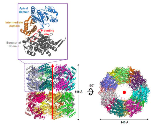
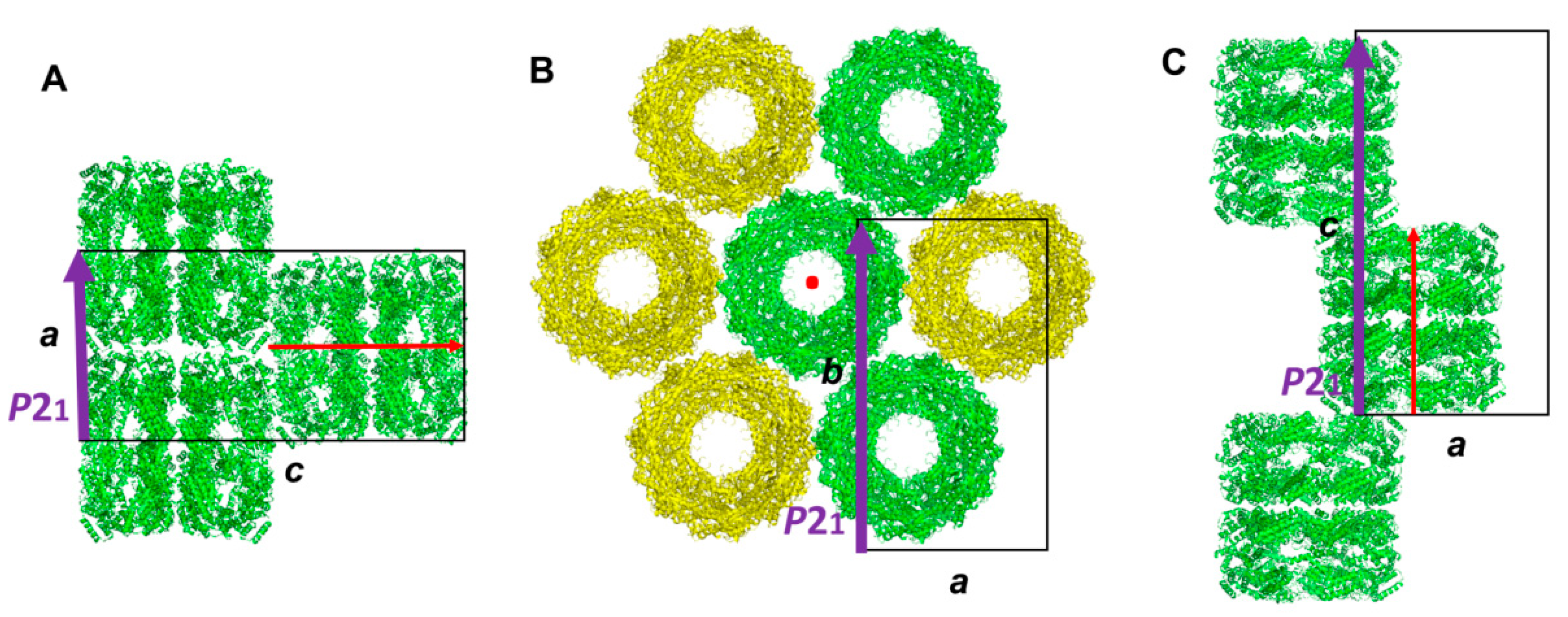
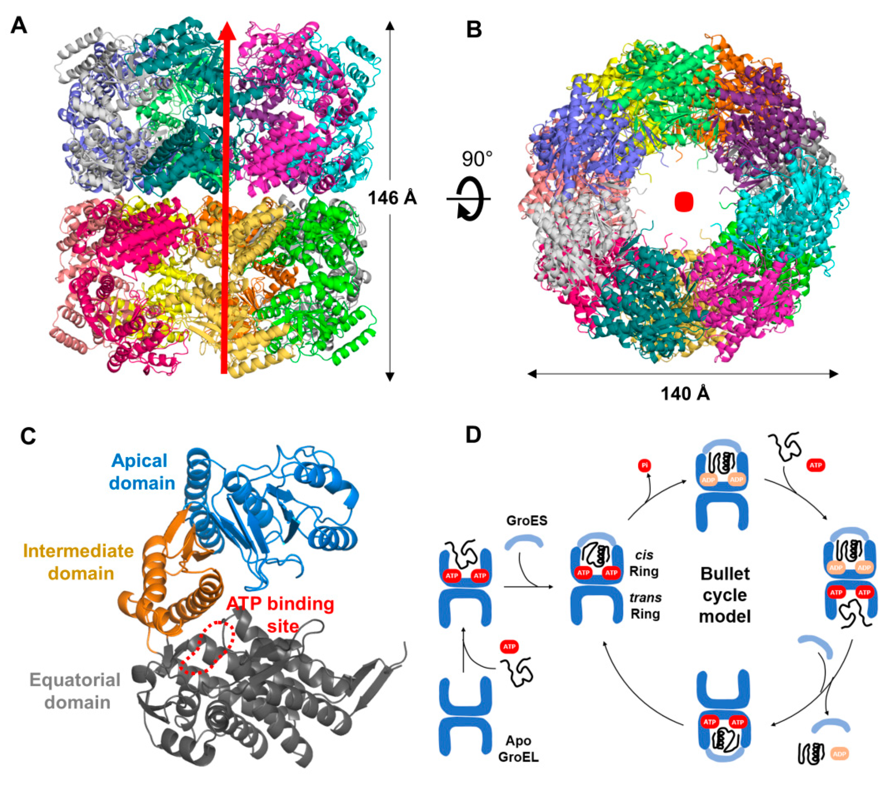
3.1. Molecular Packing of XoGroEL in Crystal

There were 14 protomers in the asymmetric unit of XoGroEL crystal. The space group was determined as P212121 as there were three two-fold screw crystallographic axes along the unit cell axes of a, b, and c (Figure 1). The 14 protomers existed as two rings; one ring consisted of seven protomers and was stacked back-to-back on the other ring. The stacking axis was parallel to the longest axis c. XoGroEL of 14 protomers was also stacked on other XoGroEL molecule in a staggered way. Each ring was in contact with six other symmetry-related rings parallelly in a × b plane like a flower-shape.

3.2. Overall Structure of XoGroEL

The XoGroEL consisted of 14 protomers, arranged in a symmetry as two back-to-back seven-member rings (Figure 2A,B). Each XoGroEL protomer was composed of three domains of an equatorial domain at the back-to-back interacting surface between rings, an apical domain at the terminal ends, and an intermediate domain connecting the equatorial and apical domains (Figure 2C, Figures S1 and S2). Two rings were in contact each other in a staggered way at equatorial domains. There was an ATP-binding pocket in each equatorial domain. ATP binding and hydrolysis is known to cause major movements during the reaction cycle of substrate protein folding (Figure 2D). Apical domain existed at the terminal end of the cylinder and exposed hydrophobic residues to solvent facing the internal cavity of substrate protein binding pocket.

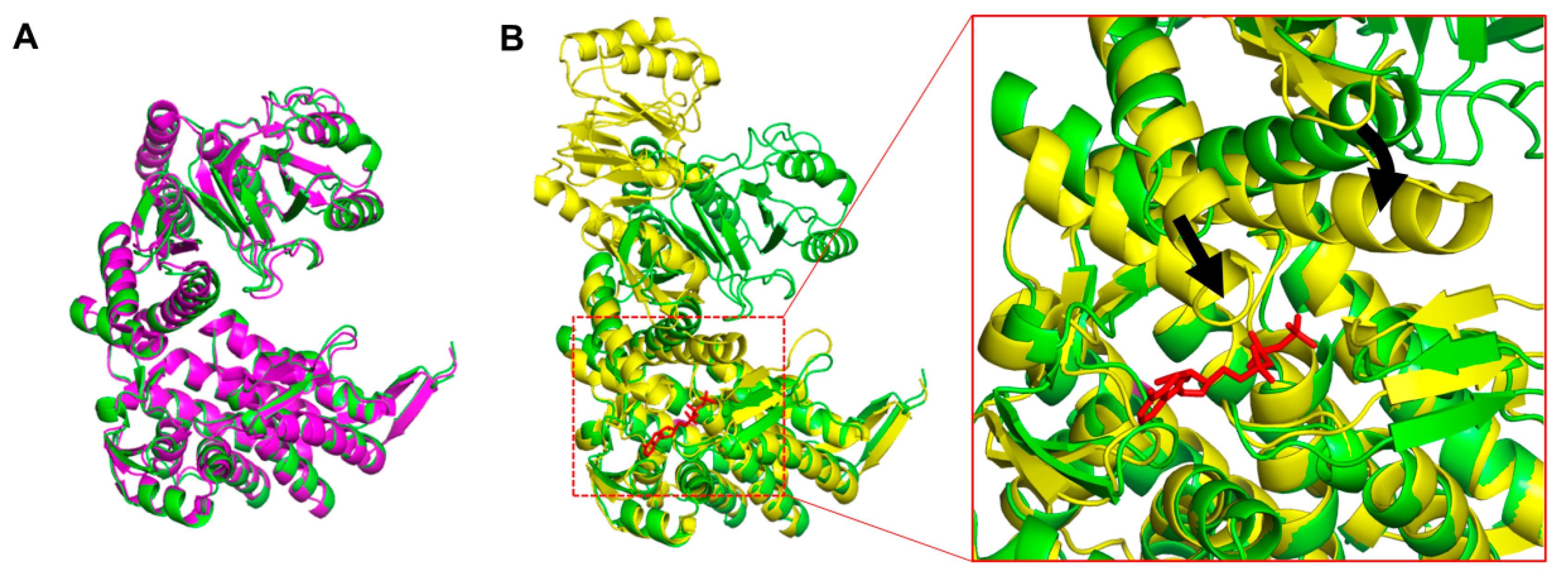
3.3. Conformation of XoGroEL Protomer

GroEL undergoes a series of conformational changes during the cycle of substrate protein folding with co-chaperone GroES and ATP [9] (Figure 2D). The traditional ‘‘bullet cycle’’ model proposes that the binding of ATP and substrate unfolded protein facilitates GroES binding to GroEL, which encapsulates a folding chamber of the cis ring. After the hydrolysis of bound ATP in the cis ring, ATP binding in the opposite trans ring releases folded protein from the cis ring and initiates a new cycle of unfolded protein of GroEL [20].
When the 14 protomers of XoGroEL were structurally compared to each other, the conformation of each protomer was well conserved. The root mean square deviation (RMSD) between XoGroEL protomers was less than 0.85 Å in more than 400 amino acids. XoGroEL structure was superimposed to the Apo structure (PDB ID: 1XCK [21]) and GroES and ATP-bound structure (PDB ID: 3WVL [22]) of E. coli GroEL (EcGroEL). XoGroEL was well superimposed to the open form of apo EcGroEL with RMSD of 0.77 Å in 481 amino acids (Figure 3A). In the open form of GroEL, protomers in the cis ring and in the trans ring had the same conformation. When GroEL bound to ATP and GroES, GroEL had an allosteric conformational change of both apical and intermediate domains (Figure 3B). The apical domain moved away from the equatorial domain, and the intermediate domain got closer to the equatorial domain to form a tight ATP-binding site.
Although we tried to determine XoGroES (GroES from Xoo) or ATP-bound XoGroEL structure with a soaking method, we could not determine the complex structure. In the XoGroEL crystal packing, there was no free space above the cis ring of XoGroEL for XoGroES binding. The tight packing between XoGroEL molecules also prohibited the conformational change of the apical and equatorial domains caused by ATP binding in the XoGroEL crystal. We could not get the cocrystallization condition for ATP-bound XoGroEL or XoGroES-bound XoGroEL.

4. Discussion

A protomer of XoGroEL consists of 546 amino acids (57 kDa). Fourteen protomers of XoGroEL constitute a large complex of more than 800 kDa. Although various approaches for the crystallization of a mega Dalton protein complex have been developed [23,24], the determination of the crystal structure of such a large complex at high quality is still challenging. The overall B factor (temperature factor) of XoGroEL is 82.3. Conformation of each protomer of GroEL changes allosterically upon the binding of substrate unfolded protein, ATP, and GroES during catalysis. We speculate the high B factor comes from the flexible conformation of XoGroEL in the open form before substrate binding. When we analyzed the B factor of the XoGroEL structure by domain, the equatorial domain, located at the interface between two homoheptameric rings, had the lower average B factor of 62.6, while the intermediate domain had 88.7 and the apical domain had 104.0 (Figure S1). With the current X-ray diffraction data, the electron density for the side chain of residues was weak, especially in the apical domain, which caused approximately 8% of the difference between R-work and R-free values. In the open form of XoGroEL without XoGroES and ATP binding, the apical domain to bind substrate unfolded protein seemed to be more flexible than the equatorial domain.
We used the previously published RNASeq data to study the time-resolved transcriptional expressions of XoGroEL/XoGroES genes in the pathogenicity activated (rice leaf extract-treated) and control (untreated) Xoo cells [25]. The RNASeq data were obtained from in vitro assay system to activate the pathogenicity of Xoo cells by treating rice leaf extract on the Xoo cell culture. The in vitro assay system enabled us to simultaneously initiate the pathogenicity of Xoo cells in the culture broth, which provided a high signal-to-noise transcriptome data compared to in vivo data. Both XoGroEL and XoGroES genes showed the U-shaped gene expression pattern in the first hour, i.e., the decreased expression in the initial 30 min (approximately 50%) and the recovered expression in an hour (Figure S3). Although transcriptional expression patterns and expression level fold changes of GroEL and GroES genes were similar, the GroEL gene had three times higher RPKM value than the GroES gene. The RNASeq data showed that, at the initial stage of Xoo and rice interaction, both XoGroEL and XoGroES genes were downregulated and recovered to the normal expression level in an hour. The crystal structure and pathogenicity-related expression of XoGroEL will provide better understanding of the chaperone function of Xoo in pathogenicity.

Supplementary Materials

The following are available online at https://www.mdpi.com/2073-4352/9/8/399/s1, Figure S1: The B factor of XoGroEL protomers in crystal and the 2FoFc map of XoGroEL protomer, Figure S2: The sequence alignment of chaperonin GroELs, Figure S3: Time-resolved transcriptional expression of XoGroEL (Xoo4288) and XoGroES genes (Xoo4289) of Xoo on rice leaf extract (RLX) treatment.

Author Contributions

T.H.T. and L.J. planned and preformed the experiments, analyzed the data, and wrote the manuscript. P.H. and A.Y.J. analyzed the data and wrote the manuscript. K.S. and K.J.G. preformed the RNASeq analysis.

Funding

This work was carried out with the support of the "Cooperative Research Program for Agriculture Science & Technology Development” (Project No. PJ013270012019), Rural Development Administration, Republic of Korea, the project” Development of biomedical materials based on marine proteins” (Project No. 20170305), funded by the Ministry of Oceans and Fisheries, Korea (J.-H.L.), and the Bio & Medical Technology Development Program of the National Research Foundation of Korea (NRF) funded by the Ministry of Science and ICT (NRF-2017M3A9E4078017).

Acknowledgments

We are grateful to the staff members of Beamline 5C at the Pohang Accelerator Laboratory (PAL), Korea.

Conflicts of Interest

The authors declare no conflict of interest.

References

  1. FAOSTAT. Available online: http://faostat.fao.org (accessed on 1 August 2019).
  2. Khush, G.S. What it will take to feed 5.0 billion rice consumers in 2030. Plant Mol. Biol. 2005, 59, 1–6. [Google Scholar] [CrossRef] [PubMed]
  3. Verdier, V.; Vera Cruz, C.; Leach, J.E. Controlling rice bacterial blight in Africa: needs and prospects. J. Biotechnol. 2012, 159, 320–328. [Google Scholar] [CrossRef] [PubMed]
  4. Alfano, J.R.; Collmer, A. Type III secretion system effector proteins: Double agents in bacterial disease and plant defense. Annu. Rev. Phytopathol. 2004, 42, 385–414. [Google Scholar] [CrossRef] [PubMed]
  5. Nans, A.; Kudryashev, M.; Saibil, H.R.; Hayward, R.D. Structure of a bacterial type III secretion system in contact with a host membrane in situ. Nat. Commun. 2015, 6, 10114. [Google Scholar] [CrossRef] [PubMed]
  6. Portaliou, A.G.; Tsolis, K.C.; Loos, M.S.; Zorzini, V.; Economou, A. Type III secretion: Building and operating a remarkable nanomachine. Trends Biochem. Sci. 2016, 41, 175–189. [Google Scholar] [CrossRef] [PubMed]
  7. Parsot, C.; Hamiaux, C.; Page, A.L. The various and varying roles of specific chaperones in type III secretion systems. Curr. Opin. Microbiol. 2003, 6, 7–14. [Google Scholar] [CrossRef]
  8. Hartl, F.U.; Bracher, A.; Hayer-Hartl, M. Molecular chaperones in protein folding and proteostasis. Nature 2011, 475, 324–332. [Google Scholar] [CrossRef]
  9. Skjaerven, L.; Cuellar, J.; Martinez, A.; Valpuesta, J.M. Dynamics, flexibility, and allostery in molecular chaperonins. FEBS Lett. 2015, 589, 2522–2532. [Google Scholar] [CrossRef]
  10. Tran, H.T.; Pham, T.V.; Ngo, H.P.; Hong, M.K.; Kim, J.G.; Lee, S.H.; Ahn, Y.J.; Kang, L.W. Crystallization and preliminary X-ray crystallographic analysis of the XoGroEL chaperonin from Xanthomonas oryzae pv. oryzae. Acta Crystallogr. F Struct. Biol. Commun. 2014, 70, 604–607. [Google Scholar] [CrossRef]
  11. Park, S.Y.; Ha, S.C.; Kim, Y.G. The protein crystallography beamlines at the pohang light source II. Biodesign 2017, 5, 30–34. [Google Scholar]
  12. Otwinowski, Z.; Minor, W. Processing of X-ray diffraction data collected in oscillation mode. Meth. Enzymol. 1997, 277, 307–326. [Google Scholar]
  13. McCoy, A.J.; Grosse-Kunstleve, R.W.; Adams, P.D.; Winn, M.D.; Storoni, L.C.; Read, R.J. Phaser crystallographic software. J. Appl. Crystallogr. 2007, 40, 658–674. [Google Scholar] [CrossRef] [PubMed]
  14. Fei, X.; Ye, X.; LaRonde, N.A.; Lorimer, G.H. Formation and structures of GroEL: GroES2 chaperonin footballs, the protein-folding functional form. Proc. Natl. Acad. Sci. USA 2014, 111, 12775–12780. [Google Scholar] [CrossRef] [PubMed]
  15. Moodie, E.E.; Richardson, T.S.; Stephens, D.A. Demystifying optimal dynamic treatment regimes. Biometrics 2007, 63, 447–455. [Google Scholar] [CrossRef] [PubMed]
  16. Mansell, W. An integrative formulation-based cognitive treatment of bipolar disorders: Application and illustration. J. Clin. Psychol. 2007, 63, 447–461. [Google Scholar] [CrossRef] [PubMed]
  17. DeFrank, J.J.; Cheng, T.C. Purification and properties of an organophosphorus acid anhydrase from a halophilic bacterial isolate. J. Bacteriol. 1991, 173, 1938–1943. [Google Scholar] [CrossRef] [Green Version]
  18. Steegborn, C.; Messerschmidt, A.; Laber, B.; Streber, W.; Huber, R.; Clausen, T. The crystal structure of cystathionine gamma-synthase from nicotiana tabacum reveals its substrate and reaction specificity. J. Mol. Biol. 1999, 290, 983–996. [Google Scholar] [CrossRef]
  19. DeLano, W.L. The PyMOL Molecular Graphics System; Version 1.3r1; Schrodinger: New York, NY, USA, 2010. [Google Scholar]
  20. Clare, D.K.; Vasishtan, D.; Stagg, S.; Quispe, J.; Farr, G.W.; Topf, M.; Horwich, A.L.; Saibil, H.R. ATP-triggered conformational changes delineate substrate-binding and -folding mechanics of the GroEL chaperonin. Cell 2012, 149, 113–123. [Google Scholar] [CrossRef]
  21. Bartolucci, C.; Lamba, D.; Grazulis, S.; Manakova, E.; Heumann, H. Crystal structure of wild-type chaperonin GroEL. J. Mol. Biol. 2005, 354, 940–951. [Google Scholar] [CrossRef]
  22. Koike-Takeshita, A.; Arakawa, T.; Taguchi, H.; Shimamura, T. Crystal structure of a symmetric football-shaped GroEL: GroES2-ATP14 complex determined at 3.8A reveals rearrangement between two GroEL rings. J. Mol. Biol. 2014, 426, 3634–3641. [Google Scholar] [CrossRef]
  23. Govada, L.; Chayen, N.E. Choosing the method of crystallization to obtain optimal results. Crystals 2019, 9, 106. [Google Scholar] [CrossRef]
  24. Blackburn, A.; Partowmah, S.H.; Brennan, H.M.; Mestizo, K.E.; Stivala, C.D.; Petreczky, J.; Perez, A.; Horn, A.; McSweeney, S.; Soares, A.S. A simple technique to improve microcrystals using gel exclusion of nucleation inducing elements. Crystals 2018, 8, 464. [Google Scholar] [CrossRef]
  25. Kim, S.; Cho, Y.J.; Song, E.S.; Lee, S.H.; Kim, J.G.; Kang, L.W. Time-resolved pathogenic gene expression analysis of the plant pathogen Xanthomonas oryzae pv. oryzae. BMC Genom. 2016, 17, 345. [Google Scholar] [CrossRef] [PubMed]
Figure 1. Symmetry-related XoGroEL molecules in unit cell. XoGroEL molecules in P21 symmetry along with a axis in a × c plane (A), along with b axis in a × b plane (B), and along with c axis in a × c plane (C). Two-fold screw axis of P21 is represented as purple arrows. Stacking axis of two homoheptameric rings in XoGroEL is represented as red arrows. A red arrow perpendicular to a × b plane is shown as red dot in (B).
Crystals 09 00399 g001
Figure 2. Crystal structure and mechanism of XoGroEL. XoGroEL structure with 14 protomers from side view (A) and top view (B). Red arrow and red dot represent the stacking axis of two homoheptameric rings in XoGroEL. (C) Apical (pale blue), intermediate (orange), and equatorial (grey) domains with ATP-binding site (red dotted line) in a XoGroEL protomer. (D) A bullet cycle model of chaperonin GroEL and co-chaperonin GroES (modified from [9]).
Crystals 09 00399 g002
Figure 3. Conformation of XoGroEL protomer. (A) Superimposed structures of an open form of XoGroEL (green) and EcGroEL (purple). (B) Superimposed structures of an open form of XoGroEL (green) and a closed form of EcGroEL (yellow) in complex with ATP (red) and GroES (not shown in this figure). The conformational change on ATP binding in the intermediate domain is shown by the black arrow.
Crystals 09 00399 g003
Table 1. Data collection and refinement statistics.
DataXoGroEL (PDB ID: 6KFV)
Data collection
Wavelength (Å)0.97949
Resolution range (Å)47.2–3.2 (3.3–3.2) *
Space groupP212121
Unit cell (Å)137.1 239.5 278.3 90 90 90
Total reflections683,467
Unique reflections136,180 (10475)
Multiplicity4.1 (3.0)
Completeness (%)91.7 (71.3)
Mean I/sigma (I)8.5 (2.1)
Wilson B-factor68.9
R-merge18.9 (59.0)
Refinement
Reflections used in refinement136,115 (10,470)
Reflections used for R-free6839 (542)
R-work0.21 (0.31)
R-free0.29 (0.37)
Number of non-hydrogen atoms
In macromolecules53,872
In ligands28
In solvent33
Protein residues7342
RMS (bonds)0.015
RMS (angles)1.87
Ramachandran favored (%)82.8
Ramachandran allowed (%)13.2
Ramachandran outliers (%)4.0
Rotamer outliers (%)16.5
Average B-factor
Macromolecules82.3
Ligands99.3
Solvent36.9
* Values in parentheses are for the shell with the highest resolution. Rmerge = hkl i   | ( I i ( h k l ) ) −〈 I ( h k l ) | / h k l i I i ( h k l ) , where I i ( h k l ) is the mean intensity of ith observation of symmetry-related reflections hkl. Rfree = hkl | | F o b s | | F c a l c | | / h k l | F o b s | , where F c a l c is the calculated protein structure factor from the atomic model (Rfree was calculated with a randomly selected 5% of the reflections).

