Novel Devices for Transporting Protein Crystals to the Synchrotron Facilities and Thermal Protection of Protein Crystals

 , ,
, , 
Abstract
1. Introduction
2. Materials and Methods
2.1. Protein Crystallization Conditions and Cryo-Protection
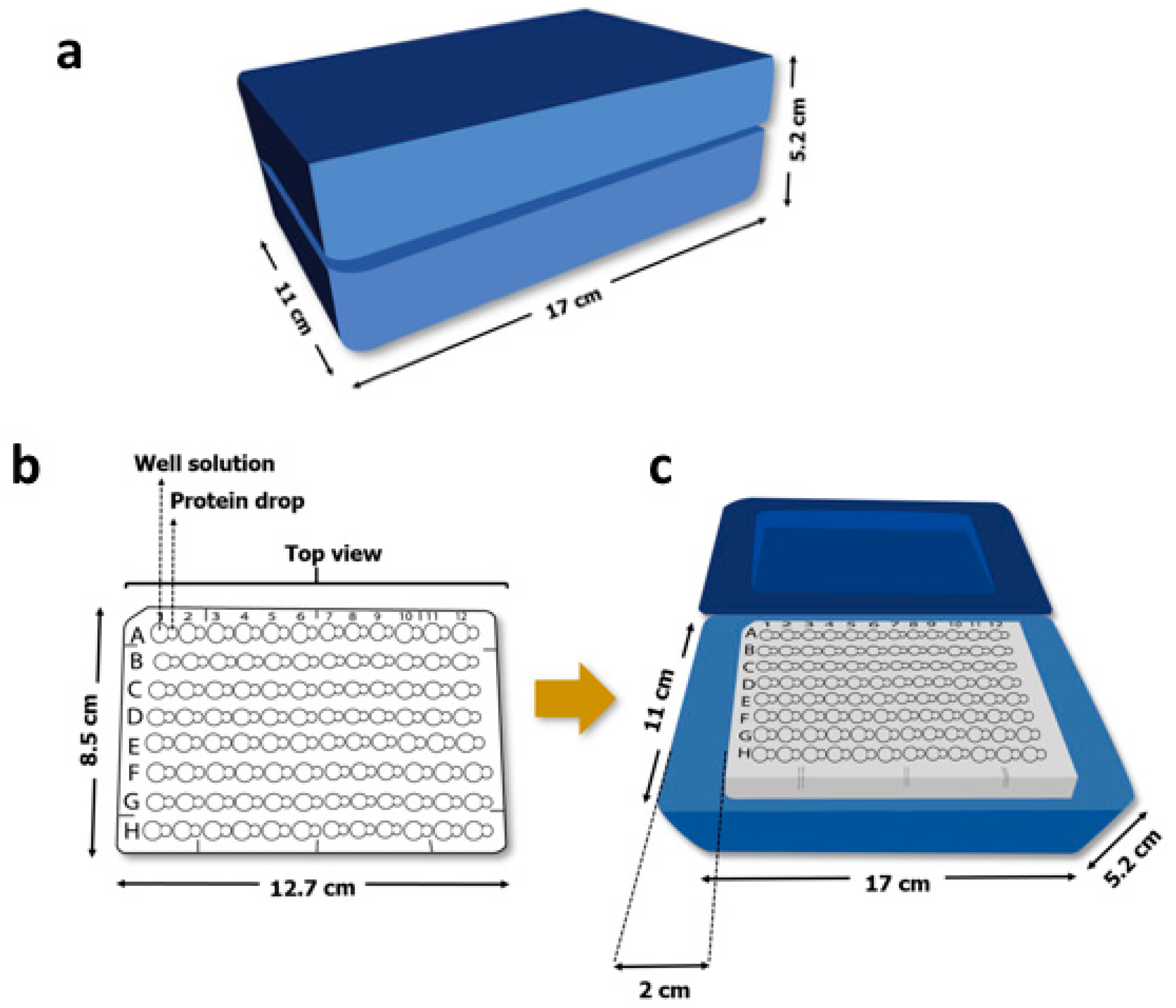
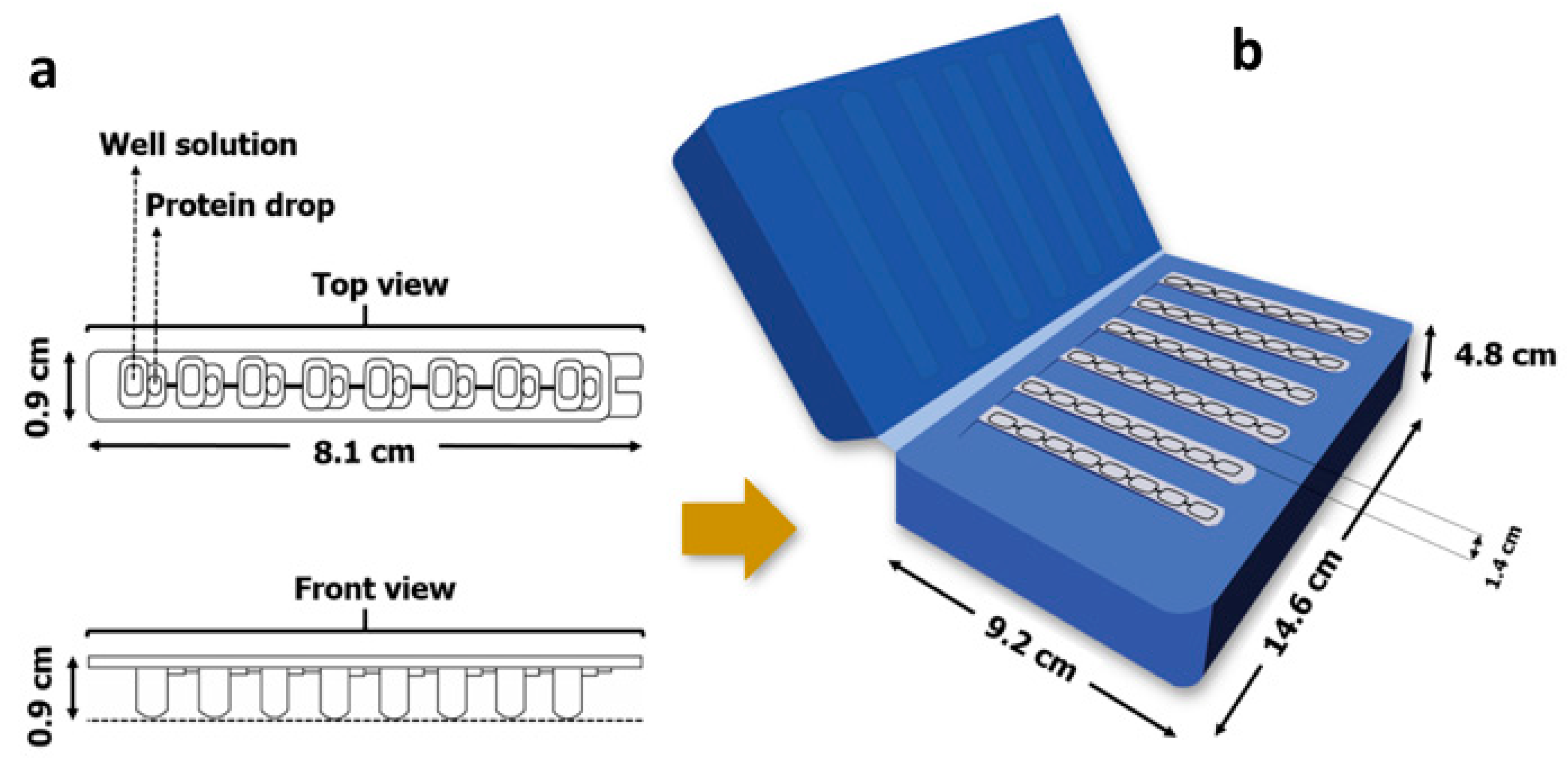
2.2. Crystal Growth Devices
2.3. X-ray Data Diffraction and Data Processing
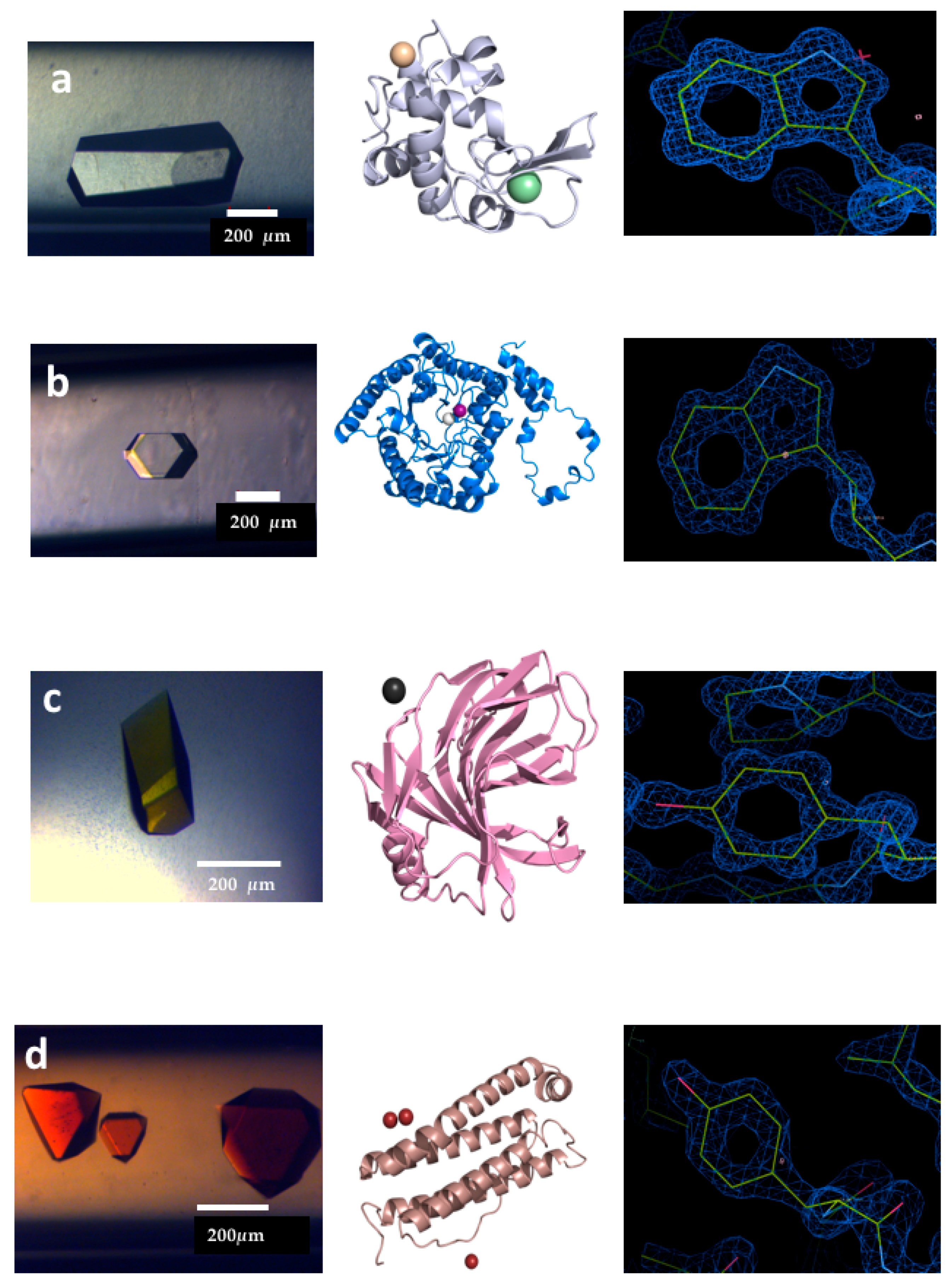
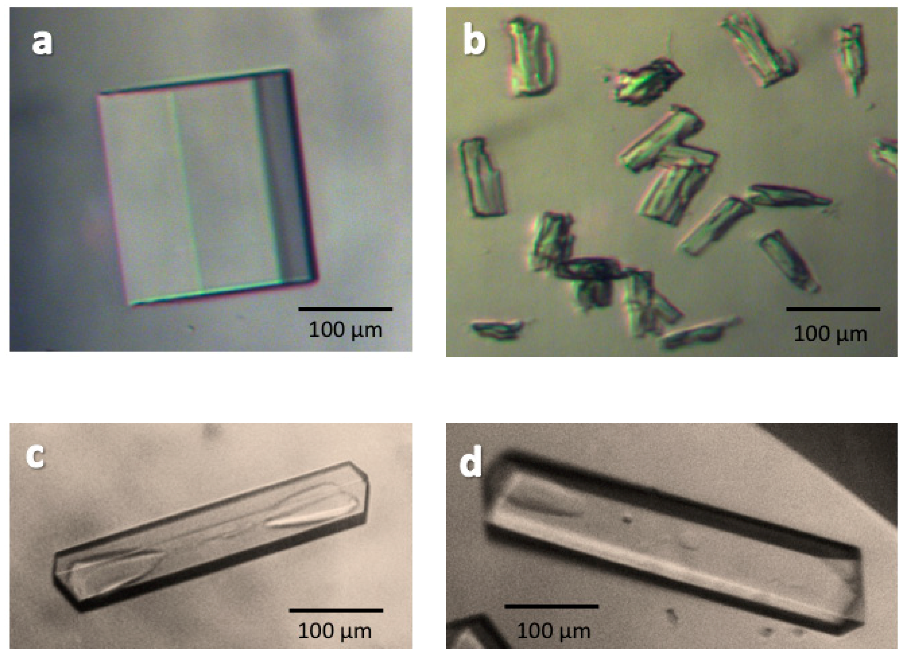
3. Results and Discussion
4. Conclusions
Author Contributions
Acknowledgments
Conflicts of Interest
References
- Lorber, B.; Sauter, C.; Theobald-Dietrich, A.; Moreno, A.; Schellenberger, P.; Robert, M.C.; Capelle, B.; Sanglier, S.; Potier, N.; Giege, R. Crystal growth of proteins, nucleic acids, and viruses in gels. Prog. Biophys. Mol. Biol. 2009, 101, 13–25. [Google Scholar] [CrossRef] [PubMed]
- Dauter, Z.; Wlodawer, A. Progress in Protein Crystallography. Protein Peptide Lett. 2016, 23, 201–210. [Google Scholar] [CrossRef]
- Fuertes, G.; Banterlea, N.; Ruff, K.M.; Chowdhury, A.; Mercadante, D.; Koehler, C.; Kachala, M.; Girona, G.E.; Milles, S.; Mishra, A.; et al. Decoupling of size and shape fluctuations in heteropolymeric sequences reconciles discrepancies in SAXS vs. FRET measurements. Proc. Natl. Acad. Sci. USA 2017, 114, E6342–E6351. [Google Scholar] [CrossRef] [PubMed]
- Sugiki, T.; Kobayashi, N.; Fujiwara, T. Modern Technologies of Solution Nuclear Magnetic Resonance Spectroscopy for Three-dimensional Structure Determination of Proteins Open Avenues for Life Scientists. Comput. Struct. Biotechnol. 2017, 15, 328–339. [Google Scholar] [CrossRef] [PubMed]
- Kay, L.E. Nuclear magnetic resonance methods for high molecular weight proteins: A study involving a complex of maltose binding protein and beta-cyclodextrin. Method Enzymol. 2001, 339, 174–203. [Google Scholar]
- Ovchinnikov, S.; Kim, D.E.; Wang, R.Y.R.; Liu, Y.; DiMaio, F.; Baker, D. Improved de novo structure prediction in CASP11 by incorporating coevolution information into Rosetta. Proteins 2016, 84, 67–75. [Google Scholar] [CrossRef] [PubMed]
- Rambo, R.P.; Tainer, J.A. Super-resolution in solution X-ray scattering and its applications to structural systems biology. Annu. Rev. Biophys. 2013, 42, 415–441. [Google Scholar] [CrossRef] [PubMed]
- Jolley, C.C.; Wells, S.A.; Fromme, P.; Thorpe, M.F. Fitting low-resolution Cryo-EM maps of proteins using constrained geometric simulations. Biophys. J. 2008, 94, 1613–1621. [Google Scholar] [CrossRef] [PubMed]
- Stagno, J.R.; Liu, Y.; Bhandari, Y.R.; Conrad, C.E.; Panja, S.; Swain, M.; Fan, L.; Nelson, G.; Li, C.; Wendel, D.R.; et al. Structures of riboswitch RNA reaction states by mix-and-inject XFEL serial crystallography. Nature 2017, 541, 242. [Google Scholar] [CrossRef] [PubMed]
- Liu, W.; Wacker, D.; Gati, C.; Won-Han, G.; James, D.; Wang, D.; Nelson, G.; Weierstall, U.; Katritch, V.; Barty, A.; et al. Serial Femtosecond Crystallography of G Protein-Coupled Receptors. Science 2013, 342, 1521–1524. [Google Scholar] [CrossRef] [PubMed]
- Zander, U.; Cianci, M.; Foos, N.; Silva, C.S.; Mazzei, L.; Zubieta, C.; de Maria, A.; Nanao, M.H. Mergin of synchrotron serial crystallographic data by a genetic algorithm. Acta Cryst. 2016, D72, 1026–1035. [Google Scholar]
- Giege, R.; Mikol, V. Crystallogenesis of Proteins. Trends Biotechnol. 1989, 7, 277–282. [Google Scholar] [CrossRef]
- Rosenberger, F. Protein crystallization. J. Cryst. Growth 1996, 166, 40–54. [Google Scholar] [CrossRef]
- Mcpherson, A. Crystallization of Proteins by Variation of Ph or Temperature. Methods Enzymol. 1985, 114, 125–127. [Google Scholar] [PubMed]
- Moller, J.; Schroer, M.A.; Erlkamp, M.; Grobelny, S.; Paulus, M.; Tiemeyer, S.; Wirkert, F.J.; Tolan, M.; Winter, R. The Effect of Ionic Strength, Temperature, and Pressure on the Interaction Potential of Dense Protein Solutions: From Nonlinear Pressure Response to Protein Crystallization. Biophys. J. 2012, 102, 2641–2648. [Google Scholar] [CrossRef] [PubMed]
- Juarez-Martinez, G.; Garza, C.; Castillo, R.; Moreno, A. A dynamic light scattering investigation of the nucleation and growth of thaumatin crystals. J. Cryst. Growth 2001, 232, 119–131. [Google Scholar] [CrossRef]
- Rosenberger, F.; Howard, S.B.; Sowers, J.W.; Nyce, T.A. Temperature-Dependence of Protein Solubility—Determination and Application to Crystallization in X-Ray Capillaries. J. Cryst. Growth 1993, 129, 1–12. [Google Scholar] [CrossRef]
- Martinez-Caballero, S.; Cuellar-Cruz, M.; Demitri, N.; Polentarutti, M.; Rodriguez-Romero, A.; Moreno, A. Glucose Isomerase Polymorphs Obtained Using an Ad Hoc Protein Crystallization Temperature Device and a Growth Cell Applying an Electric Field. Cryst. Growth Des. 2016, 16, 1679–1686. [Google Scholar] [CrossRef]
- Strelov, V.I.; Zakharov, B.G.; Bezbakh, I.Z.; Safronov, V.V.; Chernyshev, B.V.; Dutyshev, I.N. Implementation of Temperature-Controlled Method of Protein Crystallization in Microgravity. Crystallogr. Rep. 2018, 63, 149–153. [Google Scholar] [CrossRef]
- Chen, R.Q.; Lu, Q.Q.; Cheng, Q.D.; Ao, L.B.; Zhang, C.Y.; Hou, H.; Liu, Y.M.; Li, D.W.; Yin, D.C. An ignored variable: Solution preparation temperature in protein crystallization. Sci. Rep. 2015, 5, 7797. [Google Scholar] [CrossRef] [PubMed]
- Ferreira, C.; Crespo, R.; Martins, P.M.; Gales, L.; Rocha, F.; Damas, A.M. Small temperature oscillations promote protein crystallization. CrystEngComm 2011, 13, 3051–3056. [Google Scholar] [CrossRef]
- Budayova-Spano, M.; Dauvergne, F.; Audiffren, M.; Bactivelane, T.; Cusack, S. A methodology and an instrument for the temperature-controlled optimization of crystal growth. Acta Crystallogr. Sect. D Biol. Crystallogr. 2007, 63, 339–347. [Google Scholar] [CrossRef] [PubMed]
- Berg, M.; Urban, M.; Dillner, U.; Muhlig, P.; Mayer, G. Development and characterization of temperature-controlled microreactors for protein crystallization. Acta Crystallogr. Sect. D 2002, 58, 1643–1648. [Google Scholar] [CrossRef]
- Junius, N.; Oksanen, E.; Terrien, M.; Berzin, C.; Ferrer, J.L.; Budayova-Spano, M. A crystallization apparatus for temperature-controlled flow-cell dialysis with real-time visualization. J. Appl. Crystallogr. 2016, 49, 806–813. [Google Scholar] [CrossRef] [PubMed]
- Astier, J.P.; Veesler, S. Using Temperature To Crystalize Proteins: A. Mini-Review. Cryst. Growth Des. 2008, 8, 4215–4219. [Google Scholar] [CrossRef]
- Snyder, R.C.; Veesler, S.; Doherty, M.F. The evolution of crystal shape during dissolution: Predictions and experiments. Cryst. Growth Des. 2008, 8, 1100–1101. [Google Scholar] [CrossRef]
- Zhang, C.Y.; Yin, D.C.; Lu, Q.Q.; Guo, Y.Z.; Guo, W.H.; Wang, X.K.; Li, H.S.; Lu, H.M.; Ye, Y.J. Cycling Temperature Strategy: A Method to Improve the Efficiency of Crystallization Condition Screening of Proteins. Cryst. Growth Des. 2008, 8, 4227–4232. [Google Scholar] [CrossRef]
- Juarez-Martinez, G.; Steinmann, P.; Roszak, A.W.; Isaacs, N.W.; Cooper, J.M. High-throughput screens for postgenomics: Studies of protein crystallization using microsystems technology. Anal. Chem. 2002, 74, 3505–3510. [Google Scholar] [CrossRef] [PubMed]
- Kiefersauer, R.; Than, M.E.; Dobbek, H.; Gremer, L.; Melero, M.; Strobl, S.; Dias, J.M.; Soulimane, T.; Huber, R. A novel free-mounting system for protein crystals: Transformation and improvement of diffraction power by accurately controlled humidity changes. J. Appl. Crystallogr. 2000, 33, 1223–1230. [Google Scholar] [CrossRef]
- Pareja-Rivera, C.; Cuellar-Cruz, M.; Esturau-Escofet, N.; Demitri, N.; Polentarutti, M.; Stojanoff, V.; Moreno, A. Recent Advances in the Understanding of the Influence of Electric and Magnetic Fields on Protein Crystal Growth. Cryst. Growth Des. 2017, 17, 135–145. [Google Scholar] [CrossRef]
- De la Mora, E.; Flores-Hernandez, E.; Jakoncic, J.; Stojanoff, V.; Siliqi, D.; Sanchez-Puig, N.; Moreno, A. SdsA polymorph isolation and improvement of their crystal quality using nonconventional crystallization techniques. J. Appl. Crystallogr. 2015, 48, 1551–1559. [Google Scholar] [CrossRef]
- Wally, J.; Halbrooks, P.J.; Vonrhein, C.; Rould, M.A.; Everse, S.J.; Mason, A.B.; Buchanan, S.K. The crystal structure of iron-free human serum transferrin provides insight into inter-lobe communication and receptor binding. J. Biol. Chem. 2006, 281, 24934–24944. [Google Scholar] [CrossRef] [PubMed]
- Belaidi, A.A.; Bush, A.I. Iron neurochemistry in Alzheimer’s disease and Parkinson’s disease: Targets for therapeutics. J. Neurochem. 2016, 139, 179–197. [Google Scholar] [CrossRef] [PubMed]
- Booyjzsen, C.; Scarf, C.A.; Moreton, B.; Portman, I.; Scrivens, J.H.; Costantini, G.; Sadler, P.J. Fibrillation of transferrin. Bba-Gen. Subj. 2012, 1820, 427–436. [Google Scholar] [CrossRef] [PubMed]
- Apostolakis, S.; Kypraiou, A.M. Iron in neurodegenerative disorders: Being in the wrong place at the wrong time? Rev. Neurosci. 2017, 28, 893–911. [Google Scholar] [CrossRef] [PubMed]
- Torronen, A.; Harkki, A.; Rouvinen, J. Three-dimensional structure of the endo 1,4-beta-xylanase II from Trichodema reesei: Two conformational states in the active site. EMBO J. 1994, 13, 2493–2501. [Google Scholar] [PubMed]
- Lausi, A.; Polentarutti, M.; Onesti, S.; Plaisier, R.J.; Busetto, E.; Bais, G.; Barba, L.; Cassetta, A.; Campi, G.; Lamba, D.; et al. Status of the crystallography beamlines at Elettra. Eur. Phys. J. Plus 2015, 130, 1–8. [Google Scholar] [CrossRef]
- Minor, W.; Cymborowski, M.; Otwinowski, Z.; Chruszcz, M. HKL-3000: The integration of data reduction and structure solution—From diffraction images to an initial model in minutes. Acta Crystallogr. D 2006, 62, 859–866. [Google Scholar] [CrossRef] [PubMed]
- Kabsch, W. XDS. Acta Cryst. 2010, D66, 125–132. [Google Scholar] [CrossRef] [PubMed]
- Adams, P.D.; Afonine, P.V.; Bunkoczi, G.; Chen, V.B.; Davis, I.W.; Echols, N.; Headd, J.J.; Hung, L.W.; Kapral, G.J.; Grosse-Kunstleve, R.W.; et al. PHENIX: A comprehensive Python-based system for macromolecular structure solution. Acta Crystallogr. D 2010, 66, 213–221. [Google Scholar] [CrossRef] [PubMed]
- Emsley, P.; Cowtan, K. Coot: Model-building tools for molecular graphics. Acta Crystallogr. D 2004, 60, 2126–2132. [Google Scholar] [CrossRef] [PubMed]
- Afonine, P.V.; Grosse-Kunstleve, R.W.; Chen, V.B.; Headd, J.J.; Moriarty, N.W.; Richardson, J.S.; Richardson, D.C.; Urzhumtsev, A.; Zwart, P.H.; Adams, P.D. phenix.model_vs_data: A high-level tool for the calculation of crystallographic model and data statistics. J. Appl. Crystallogr. 2010, 43, 669–676. [Google Scholar] [CrossRef] [PubMed]
- De Lano, W.L. The PyMOL Molecular Graphics System; DeLano Scientific LLC: San Carlos, CA, USA, 2002. [Google Scholar]


| Data Collection Facility | Inhouse X-ray Diffraction Facility | Inhouse X-ray Diffraction Facility | Inhouse X-ray Diffraction Facility | Inhouse X-ray Diffraction Facility |
|---|---|---|---|---|
| Protein | Lysozyme | Glucose Isomerase | Xylanase | Ferritin |
| Wavelength (Å) | 1.54 | 1.54 | 1.54 | 1.54 |
| Space group | P43212 | I222 | P212121 | F432 |
| Unit cell dimensions [a, b, c (Å)] | 78.14, 78.14, 37.39 90,90,90 | 93.05, 98.28, 102.13 90,90,90 | 48.91, 58.39, 69.82 90, 90, 90 | 182.42, 182.42, 182.42 90,90,90 |
| Resolution (Å) | 39.20–1.45 | 35.41–1.70 | 40.06–1.00 | 50.00–2.00 |
| No. of unique reflections | 18,837 | 267,071 | 111,185 | 18,108 |
| Completeness (%) | 99.82 (100) | 99.8 (98.7) | 99.70 (95.1) | 99.7 (100) |
| Redundancy | 12.6(13.3) | 5.2(4.8) | 5.7 (2.9) | 11.4 (15.2) |
| Rmerge (%) | 0.038 (0.71) | 0.08 (0.37) | 0.064 (0.871) | 0.075 (0.57) |
| Mean ⟨I/σ(I)⟩ | 35.33(4.3) | 11.1(3.) | 33.4 (1.6) | 12.0(3.1) |
| Data Collection Beamline | XRD1 (Elettra) | XRD1 (Elettra) | XRD1 (Elettra) | XRD1 (Elettra) |
|---|---|---|---|---|
| Protein | Lysozyme | Glucose Isomerase | Xylanase | Ferritin |
| Wavelength (Å) | 0.9794 | 0.9794 | 0.9794 | 0.9794 |
| Space group | P 43212 | I 222 | P 212121 | F 432 |
| Unit cell parameters (Å) | a = 78.45, b = 78.45, c = 37.21 | a = 93.03, b = 98.56, c = 102.47 | a = 48.98, b = 58.62, c = 69.90 | a = 181.84, b = 181.84, c = 181.84 |
| Resolution range (Å) | 39.22–0.91 (0.94–0.91) | 40.08–1.102 (1.141–1.102) | 37.59–1.00 (1.036–1.00) | 40.66–1.75 (1.81–1.75) |
| No. of reflections | 835,727 (10,161) | 1,168,535 (105,218) | 667,833 (57,308) | 984,926 (100,812) |
| No. of unique reflections | 75,009 (3210) | 185,699 (17,833) | 108,893 (10,737) | 26,552 (2469) |
| Multiplicity | 11.1 (2.9) | 6.3 (5.8) | 6.1 (5.3) | 37.1 (38.7) |
| Completeness (%) | 99.59 (39.90) | 98.36 (95.58) | 99.73 (99.60) | 98.85 (93.97) |
| Mean I/σ (I) | 21.72 (0.20) | 9.64 (0.70) | 28.84 (4.58) | 32.21 (1.86) |
| Rmerge1 | 0.0414 (4.594) | 0.0759 (2.433) | 0.0370 (0.3304) | 0.1127 (2.305) |
| Rmeas2 | 0.0433 (5.403) | 0.0827 (2.668) | 0.0403 (0.3679) | 0.1144 (2.336) |
| CC 1/23 | 100 (88) | 99 (68) | 99 (95) | 88 (90) |
| Mosaicity (°) | 1.3 | 1.2 | 0.6 | 2.2 |
| Wilson B-factor (Å2) | 12.68 | 14.3 | 8.79 | 32.04 |
| Refinement | ||||
| Rwork/Rfree | 0.1582 (0.4441)/0.1876 (0.5569) | 0.2547 (0.5306)/0.2582 (0.5569) | 0.1394 (0.1509)/0.1684 (0.1859) | 0.2309 (0.6569)/0.2541 (0.6351) |
| Working reflections | 74,646 (3210) | 185,400 (17,825) | 108,816 (10,736) | 26,252 (2446) |
| Testing reflections | 3718 (170) | 9110 (856) | 5462 (551) | 1293 (112) |
| Non-H atoms | 1276 | 3072 | 2042 | 1557 |
| Protein | 129 | 390 | 190 | 171 |
| Ligands | 2 | 10 | 12 | 3 |
| Water molecules | 262 | 28 | 523 | 178 |
| Mean B factors (Å2) | ||||
| Protein | 16.39 | 17.69 | 10.14 | 31.15 |
| Ligands | 14.11 | 17.70 | 16.48 | 67.71 |
| Water molecules | 37.85 | 28 | 35.36 | 41.98 |
| rmsd bond lengths (Å) | 0.009 | 0.008 | 0.006 | 0.007 |
| rmsd angles (°) | 1.4 | 1.33 | 1.36 | 1.05 |
| Ramachandran plot statistics | ||||
| Favored | 98.43% | 97.14% | 98.4% | 97.63% |
| Outliers | 0% | 0.26% | 0% | 0.59% |
| Allowed | 1.57% | 2.60% | 1.6% | 1.78% |
| Inhouse X-ray Diffraction Facility | Synchrotron Facility | |
|---|---|---|
| Wavelength (Å) | 1.5418 | 0.9184 |
| Resolution range (Å) | 41.57–3.51 (3.63–3.51) | 29.60–2.91 (2.99–2.91) |
| Space group | P 21 21 21 | P 21 21 21 |
| Unit cell (Å) | a = 84.99, b = 101.41, c = 200.327 | a = 85.40, b = 101.60, c = 199.60 |
| Total reflections | 221,910 | 253,338 (13,314) |
| Unique reflections | 22,370 | 37,976 (2411) |
| Multiplicity | 9.9 | 6.7 (5.5) |
| Completeness (%) | 99.9 | 98.8 (86.6) |
| Mean I/sigma(I) | 1.9 | 12.20 (1.30) |
| R-merge | 0.163 | 0.100 (0.728) |
| CC1/2 | 69.6 | 99.8 (81.3) |
© 2018 by the authors. Licensee MDPI, Basel, Switzerland. This article is an open access article distributed under the terms and conditions of the Creative Commons Attribution (CC BY) license (http://creativecommons.org/licenses/by/4.0/).
Share and Cite
Flores-Ibarra, A.; Campos-Escamilla, C.; Guerra, Y.; Rudiño-Piñera, E.; Demitri, N.; Polentarutti, M.; Cuéllar-Cruz, M.; Moreno, A. Novel Devices for Transporting Protein Crystals to the Synchrotron Facilities and Thermal Protection of Protein Crystals. Crystals 2018, 8, 340. https://doi.org/10.3390/cryst8090340
Flores-Ibarra A, Campos-Escamilla C, Guerra Y, Rudiño-Piñera E, Demitri N, Polentarutti M, Cuéllar-Cruz M, Moreno A. Novel Devices for Transporting Protein Crystals to the Synchrotron Facilities and Thermal Protection of Protein Crystals. Crystals. 2018; 8(9):340. https://doi.org/10.3390/cryst8090340
Chicago/Turabian StyleFlores-Ibarra, Andrea, Camila Campos-Escamilla, Yasel Guerra, Enrique Rudiño-Piñera, Nicola Demitri, Maurizio Polentarutti, Mayra Cuéllar-Cruz, and Abel Moreno. 2018. "Novel Devices for Transporting Protein Crystals to the Synchrotron Facilities and Thermal Protection of Protein Crystals" Crystals 8, no. 9: 340. https://doi.org/10.3390/cryst8090340
APA StyleFlores-Ibarra, A., Campos-Escamilla, C., Guerra, Y., Rudiño-Piñera, E., Demitri, N., Polentarutti, M., Cuéllar-Cruz, M., & Moreno, A. (2018). Novel Devices for Transporting Protein Crystals to the Synchrotron Facilities and Thermal Protection of Protein Crystals. Crystals, 8(9), 340. https://doi.org/10.3390/cryst8090340