Article

Adsorption, Desorption, Surface Diffusion, Lattice Defect Formation, and Kink Incorporation Processes of Particles on Growth Interfaces of Colloidal Crystals with Attractive Interactions

1
Department of Applied Chemistry, Graduate School of Science and Technology, Tokushima University, 2-1 Minamijosanjima, Tokushima 770-8506, Japan
2
Department of Chemical Science and Technology, Faculty of Engineering, Tokushima University, 2-1 Minamijosanjima, Tokushima 770-8506, Japan
3
Institute for Materials Research, Tohoku University, 2-1-1 Katahira, Aoba-ku, Sendai 980-8577, Japan
4
Faculty of Pharmaceutical Sciences, Nagoya City University, 3-1 Tanabe, Mizuho, Nagoya, Aichi 467-8603, Japan
*
Author to whom correspondence should be addressed.
Crystals 2016, 6(7), 80; https://doi.org/10.3390/cryst6070080
Submission received: 8 June 2016 / Revised: 4 July 2016 / Accepted: 15 July 2016 / Published: 18 July 2016
(This article belongs to the Special Issue Colloidal Crystals)

Abstract

:
Good model systems are required in order to understand crystal growth processes because, in many cases, precise incorporation processes of atoms or molecules cannot be visualized easily at the atomic or molecular level. Using a transmission-type optical microscope, we have successfully observed in situ adsorption, desorption, surface diffusion, lattice defect formation, and kink incorporation of particles on growth interfaces of colloidal crystals of polystyrene particles in aqueous sodium polyacrylate solutions. Precise surface transportation and kink incorporation processes of the particles into the colloidal crystals with attractive interactions were observed in situ at the particle level. In particular, contrary to the conventional expectations, the diffusion of particles along steps around a two-dimensional island of the growth interface was not the main route for kink incorporation. This is probably due to the number of bonds between adsorbed particles and particles in a crystal; the number exceeds the limit at which a particle easily exchanges its position to the adjacent one along the step. We also found novel desorption processes of particles from steps to terraces, attributing them to the assistance of attractive forces from additionally adsorbing particles to the particles on the steps.

Graphical Abstract

1. Introduction

Growth kinetics of colloidal crystals have attracted a great deal of attention as model crystallization systems, since it has been expected that the incorporation processes of particles into crystals would be easily observed in situ using optical microscopes at the particle level [1,2]. After some pioneering experiments, extensive fundamental studies on colloidal crystals have been conducted so far [3,4,5,6,7,8,9,10,11,12,13,14,15,16,17,18,19,20,21]. On the other hand, the application of colloidal crystals as photonic bandgap (PBG) crystals has also received much attention. After the first proposal of the concept of PBG crystals [22], many studies on the application of colloidal crystals as PBG crystals have been examined [23,24,25,26,27,28,29,30,31,32]. Even most recently, both fundamental and application studies of colloidal crystals have been published actively and continuously [33,34,35,36,37,38,39,40,41,42,43,44,45,46,47,48,49,50,51,52,53,54,55].
However, almost all systems reported so far have mainly utilized repulsive interactions between particles for colloidal crystallization basically [1,2,3,4,5,6,7,8,9,10,11,12,13,14,15,16,17,18,20,21,25,26,27,28,29,30,31,32,33,34,35,36,37,38,39,40,42,43,44], whereas the growth of crystals of atoms or molecules usually proceeds via attractive interactions. In such systems, in situ observation of growth interfaces of colloidal crystals by optical microscopy is usually difficult because of high particle concentrations. Although a few exceptions have shown the dynamic character of growth interfaces of colloidal crystals at the particle level and claimed the existence of attractive interactions between particles [56,57,58], these studies involved only in situ observation of cross sections of the growth interfaces; there are no reports on the two-dimensional dynamics of particles on the growth interfaces. Furthermore, particles in the crystals in those studies separated from each other; repulsive interactions were still dominant.
If attractive interactions between particles can be utilized for colloidal crystallization, we can achieve in situ observation of particle incorporation into crystals on the growing surface from a dilute dispersion of particles at the particle level. However, van der Waals attraction between large particles often causes irreversible coagulation, owing to a deep primary minimum of its potential curve of the Derjaguin-Landau-Verwey-Overbeek (DLVO) theory [59]; van der Waals attraction is too strong to allow particles to reversibly attach to and detach from each other. Thus, we focus on depletion interaction between large particles that are suspended in a dilute polymer electrolyte solution as the second candidate [60]. The depletion interaction is also an attractive interaction, whereas a reversible formation of crystalline aggregate has been reported by Kose and Hachisu [61]. Although those authors showed clear microscopic images that indicate layer-by-layer stacking of two-dimensional regular arrays of polystyrene particles at the particle level, they did not focus on the surface dynamics of the particles. Although Toyotama et al. recently studied particle-level eutectic-formation processes from binary and ternery mixtures of polystyrene particles in depletion-induced attractive colloidal crystals [62], there are still a lot of unclarified elementary processes of crystal growth in this depletion-induced colloidal crystallization system.
In this study, we first use an optical microscope to examine adsorption, desorption, and diffusion of polystyrene particles on 2D regular arrays (crystals) of the particles in an aqueous sodium polyacrylate solution that induces depletion interactions between particles. We then examine lattice defect formation and incorporation processes of the particles into the crystals.

2. Results and Discussion

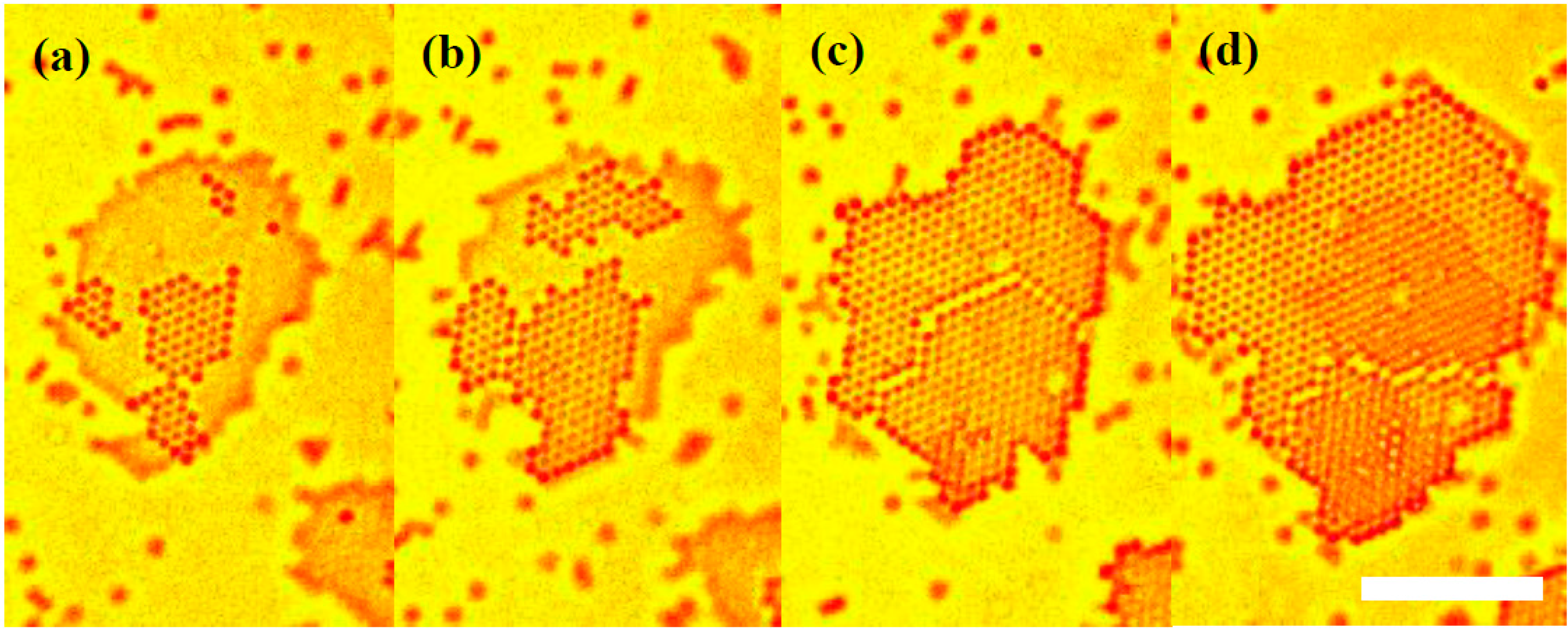
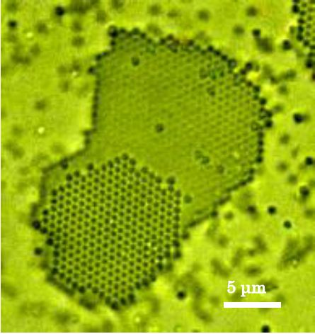
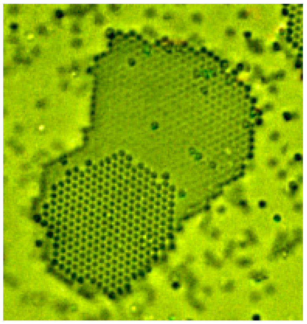
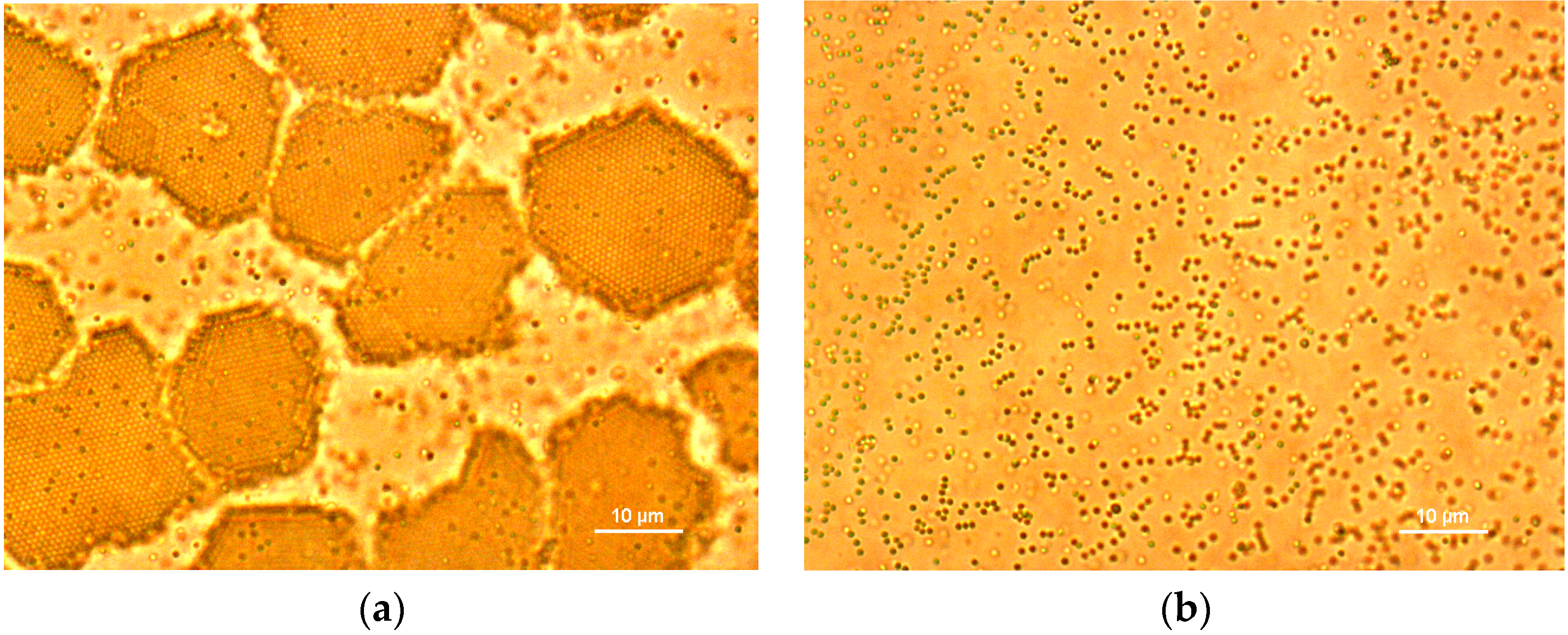
2.1. In Situ Observation of Growth Interface of Crystals

Growth surfaces of colloidal crystals were observed simply by using a 100× oil immersion objective (Figure 1). Not only the first layer of the crystals, but also the first three layers were easily resolved at the particle level. Particles on a layer moved around from one specific lattice position to another rather discretely, while those in bulk suspension moved continuously via normal bulk Brownian motion at the same time (Video S1). Such “hopping” behaviors of particles between adjacent lattice cites would provide more detailed information about activation processes of surface diffusion, if we conduct higher-frame-rate observation using high-speed cameras as the future works. Straight-shaped steps were also confirmed at the edges of a layer. From the viewpoint of periodic bond chain (PBC) theory [63], the straightness of the steps would reflect the strength of attractive interactions between particles along the steps. Furthermore, roughness of the steps is a direct measure of roughening transitions of steps. In the present setup, we can solve these problems by real-time and dynamical analyses of particle incorporation processes into the steps. We will also conduct these measurements in the near future.

2.2. Equilibrium Conditions

Equilibrium conditions (solubility, melting point, saturation pressure, and so on) are prerequisites for the quantitative analysis of crystallization kinetics. Thus, we first tried to find the equilibrium condition at which the growth and dissolution rates of crystals are equal. To determine the equilibrium conditions, we recorded the growth processes of crystals with time. At first, crystals nucleated, started to grow, and then grew layer-by-layer up to several thicknesses and sizes. Finally, the growth rates approached zero asymptotically; the system seemed to reach equilibrium. After that, however, the crystals started to dissolve, and in several days the crystals had disappeared completely and quite amazingly (Figure 2). Thus, we could not determine the equilibrium conditions in this system at the present stage. Although we considered Ostwald ripening and observed all parts of the growth cell repeatedly, we could not find any larger crystals, and all particles over the entire volume in the cell completely re-dispersed at the end of every experiment. In addition, no leaks of particles out of the growth cell were observed. Some other slow relaxation processes (interaction between water molecules and polyacrylic ions, for instance) would take several days. Although the mechanism of re-dispersion has not been clarified yet, crystallization processes in the early stages (at least on the first day under the conditions shown in Figure 2, for instance) seemed to be normal. Thus, in this study we focus on the dynamical behavior of particles on the growth interface of colloidal crystals with attractive interactions only in the early stages. Of course, we have to keep in mind that this “crystallization” process is examined under a dynamic and non-equilibrium environment at the present stage.

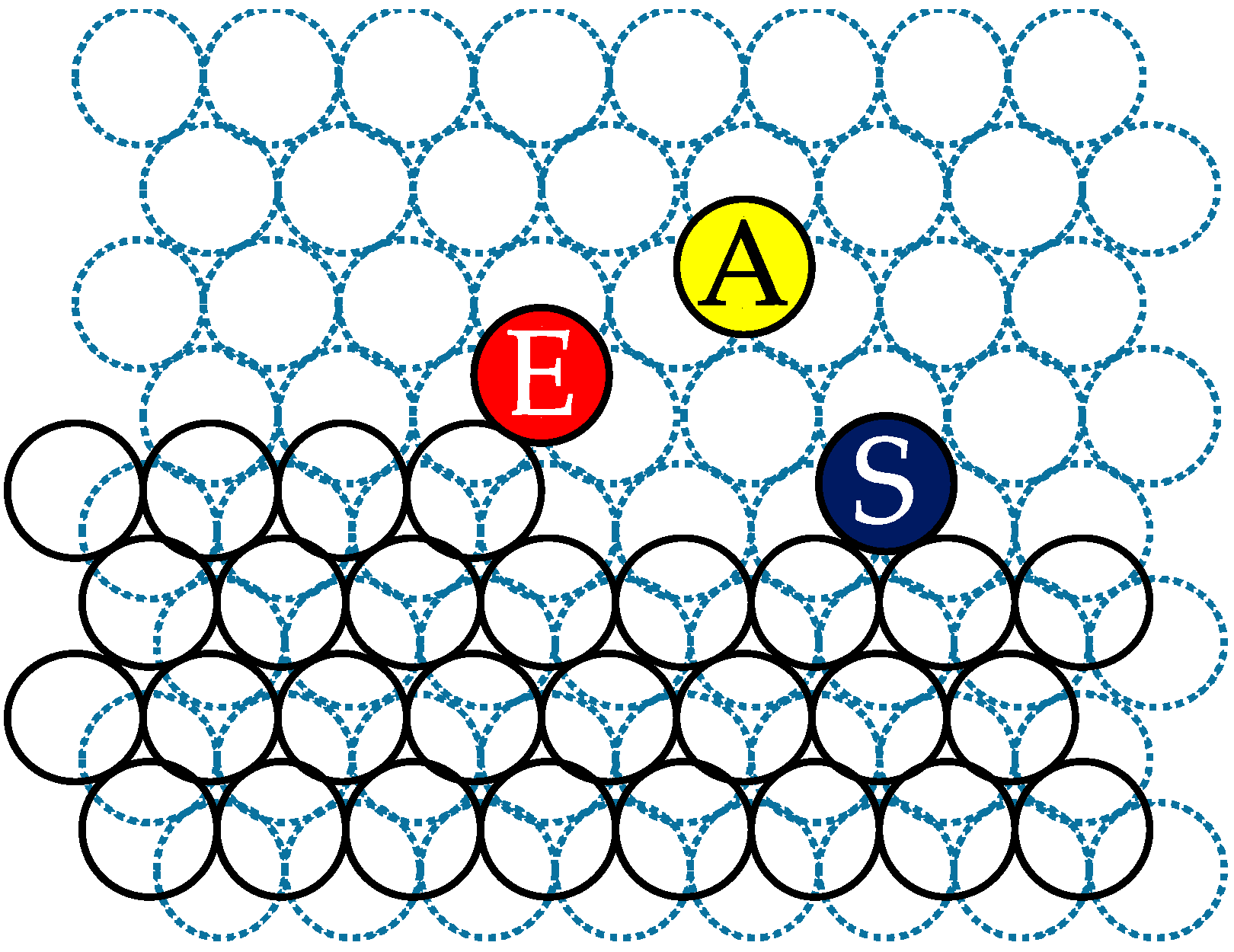
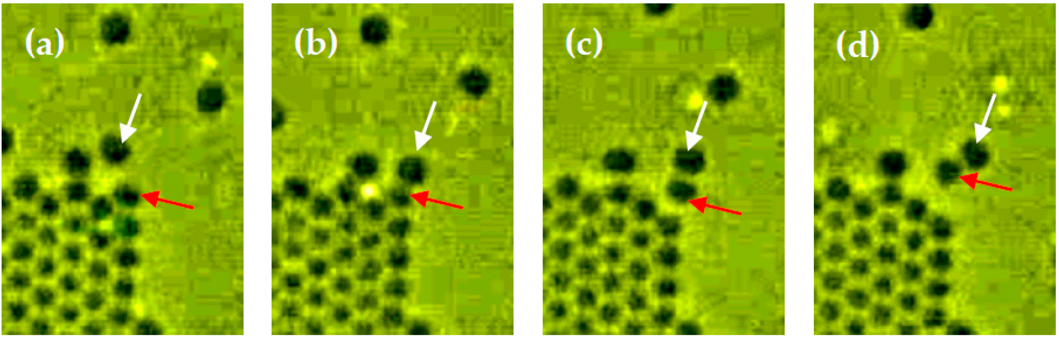
2.3. Surface Diffusion of Particles

The diffusion of particles on the growth interface of crystals was successfully observed as described in Section 2.1. The surface diffusion coefficient Ds of particles was estimated using an equation expressed as,
Ds = xs2/τ
where xs is the mean displacement of an adsorbed particle and τ is the mean lifetime of an adsorbed particle before being dissolved again into the surrounding solution [64]. Using 10 particles which adsorbed, diffused on the growth interfaces, and desorbed again into the surrounding solution, we directly measured xs and τ (Table 1). As a result, Ds of particles was calculated to be (2 ± 1) × 10−13 m2·s−1. This value is an order of magnitude smaller than the bulk diffusion coefficients of polystyrene particles in aqueous polyelectrolyte solutions [65]. This is probably due to the activation energy for surface diffusion; the activation barrier for surface diffusion is probably higher than that for bulk diffusion in the solution. In addition, Video S1 shows that the speed of particle movements on a growth interface is clearly lower than that in the bulk solution, for instance.

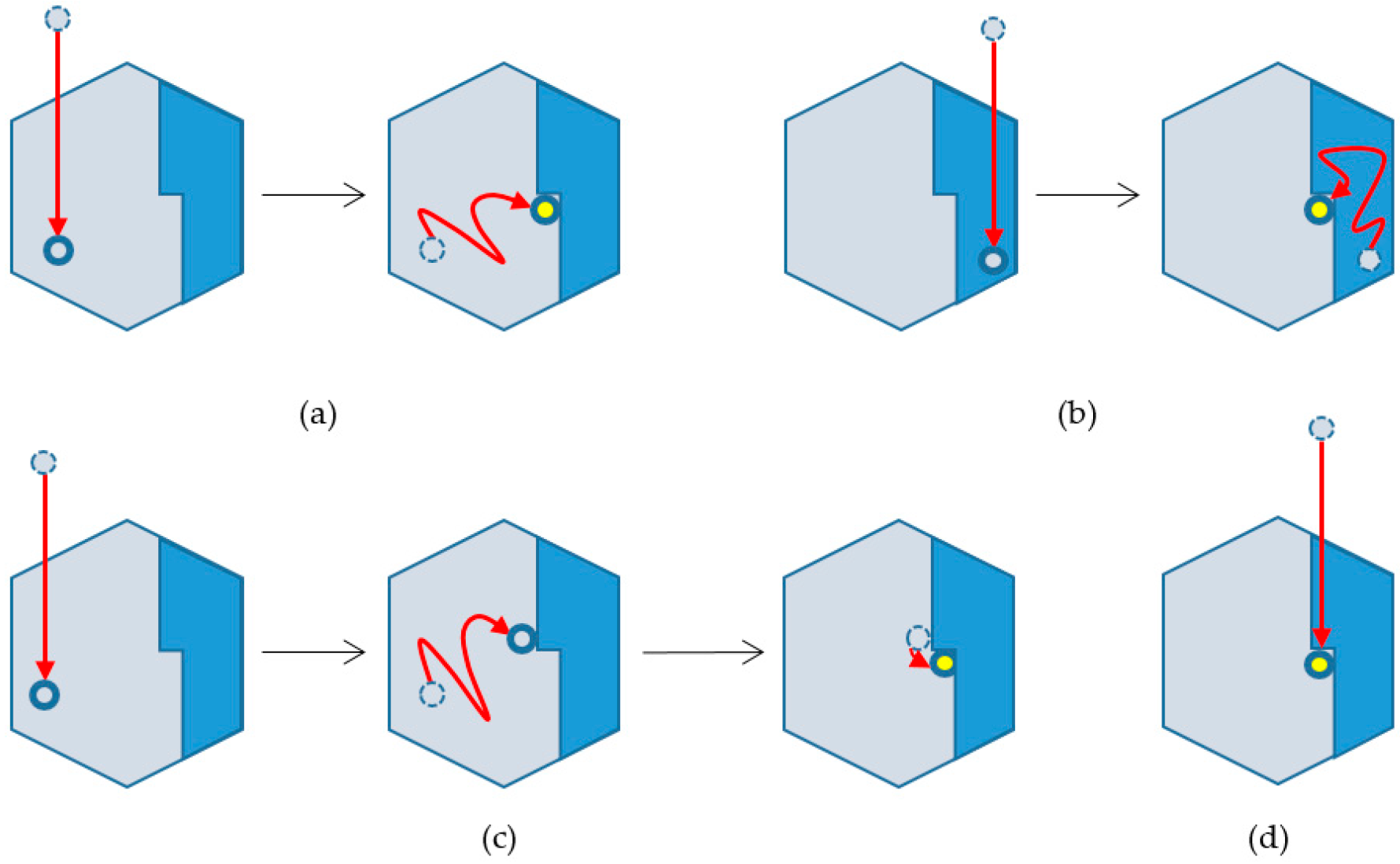
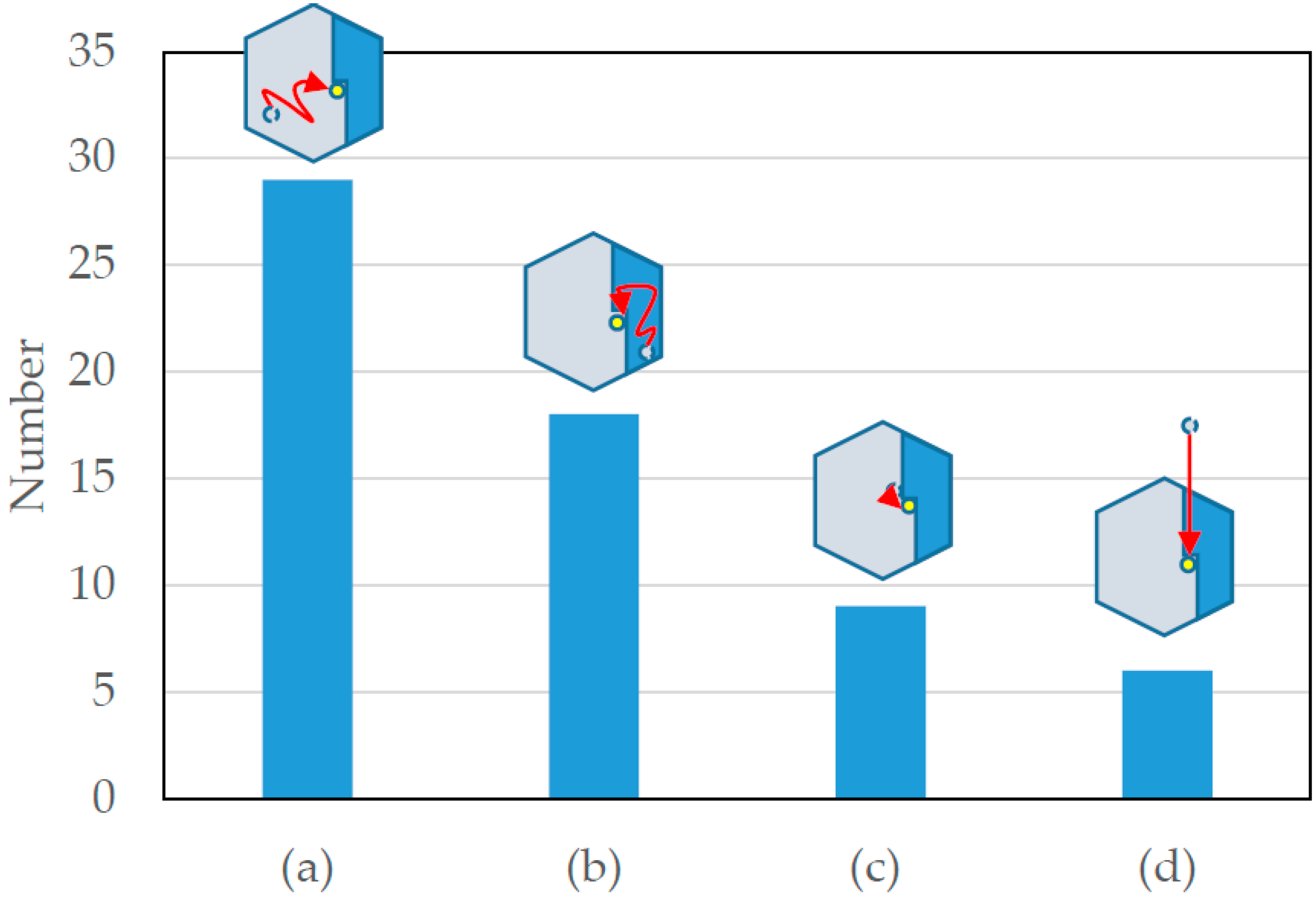
2.4. Classification of Particle Incorporation Processes into Crystals

Particle incorporation processes were successfully observed at the particle level. Although solute incorporation into a crystal is well known to be an important elementary process of crystallization, to our knowledge there has been no direct and precise analysis of the process. First, we classified the routes of particles to be finally incorporated into a crystal at a kink site, as schematically shown in Figure 3.
Four routes are classified as follows.
  • Kink incorporation processes via surface diffusion on lower layers
  • Those via surface diffusion on upper layers
  • Those via surface diffusion on lower layers and adsorption to the particle next to the kink site
  • Those via direct incorporation from solution
Although, in general, another classification—that via diffusion along the steps—would be added to this list, in the present study we did not observe kink incorporation via diffusion along the steps as far as we know; particles desorbed from the steps before arriving at the kink sites. Only in the “special” case of (c), particles incorporated into the kink site along the steps. Using 62 particles that were finally incorporated into kink sites, the number distribution of the incorporation routes was confirmed and are shown below as a bar graph (Figure 4).
These results indicate that surface diffusion processes (a) and (b) are major routes for kink incorporation processes, although we have not fully confirmed whether (a) is more frequent than (b) owing to Ehrlich-Schwoebel effects [66]. The difference would depend on the difference in the area at this stage, since the order of magnitude of the size of the layers corresponds to that of xs in this study. A second major process is via adsorption to the particle at the edge of the kink site (c). Although a direct incorporation process from bulk solutions (d) is a minor one compared to the other processes shown in Figure 4, the possibility of finally incorporating particles into a kink site using this process is much greater than that of the diffusion process of particles along steps. These results are probably attributable to the difference in the number of recovered bonds of adsorbed particles, as shown in Figure 5. Dotted and solid circles represent the lower and upper layers, respectively. A, E, and S particles in Figure 5 are those that are adsorbed and form respectively three, four, and five bonds with particles in a crystal. In the (a) and (b) processes, adsorbed particles are classified as A particles. Therefore, the surface diffusion of A particles needs the activation energy to cut three bonds of A particles and move to adjacent lattice points. In the (c) process, adsorbed particles are classified as E particles. To move to the adjacent kink site, E particles should cut four bonds with three particles of the lower layer and the edge particle at the kink site; this higher activation energy for kink incorporation results in a clearly reduced probability that the (c) process will achieve kink incorporation relative to the probability for the (a) or (b) processes.
Furthermore, S particles are adsorbed on steps and form five bonds; the probability that these particles will arrive at the kink site via the diffusion along the steps probably becomes much lower than the others.

2.5. Formation of Stacking Disorders

Formation processes of lattice defects were observed in situ at the particle level. Figure 6 shows the formation of stacking disorders on the triangle lattice via two–dimensional nucleation and growth on different lattice sites. Similar images of stacking disorders had been already observed in our previous experiments, and we assumed that the stacking disorder was formed via two–dimensional nucleation and growth of colloidal crystals on the wall of the growth container [35], whereas the image was an ex-situ scanning-electron-microscope micrograph. Furthermore, the displacement of the boundary between different stacking layers was observed. The movement of the boundary is mainly promoted by the hopping of particles beyond the boundary between the different stacking layers. More detailed observation on the formation of lattice defects are planned as a future work.

2.6. Particle Desorption from Steps by Attractive Forces from the Other Particles on the Terrace

Novel desorption processes of a particle on a step of growth surfaces were found. Desorption occurred by the adsorption of other particles to the desorbing particle, as if the adsorbed particles peeled the desorbing particle off the step (Figure 7). Although no textbook on crystal growth has dealt with such a phenomenon, this occurrence seems to be naturally acceptable if the attractive interaction between particles is considered. Actually, not only this “peeling off” but also other cooperative rearrangements of multiple particles around steps were also observed. They also seemed to be due to the attractive interactions between particles and will be reported elsewhere. More detailed observation of these cooperative desorption and rearrangement of particles would be useful to clarify the elementary processes on dissolution processes of normal crystals deeply.

3. Materials and Methods

3.1. Materials

Polystyrene particles (Thermo Fischer Scientific, Waltham, MA, USA, 5065A (diameter d = 0.65 mm)) was used without further purification or modification. Sodium polyacrylate (Kishida Chemical, Osaka, Japan, molecular weight Mw = 22,000~66,000) was used as the crystallizing agent and without further purification. 0.6% (volume fraction φ = 0.006) polystyrene particles and 2.5 × 10⁻⁴ g·mL−1 sodium polyacrylate were dissolved into Milli-Q water (Millipore, resistivity is larger than 18.2 MΩ·cm) just before the experiments were started.

3.2. Methods

An inverted microscope (Nikon, Tokyo, Japan, TE-2000U) was used with a 100× oil immersion objective (Nikon, CFI Plan Achromat 100× oil). Crystal growth in particle dispersion was observed using a handmade observation cell (Figure 7). The dispersion of particles is sandwiched between a glass slide (Matsunami Glass Industries, Osaka, Japan, micro-slide glass S1126 76 × 26 mm) and cover glass (Matsunami Glass Industries, Osaka, Japan, micro-cover glass 18 × 18 mm), spaced with polystyrene strips (Evergreen Scale Models, 14″ dimensional strips; Item 102 0.28 × 1.0 × 350 mm), and sealed with a silicone adhesive (Konishi, Osaka, Japan, #46842) as schematically shown in Figure 8. To suppress vaporization of water from the dispersion during experiments, liquid paraffin was injected around the strips and filled into possible pores or hollows in thin solidified adhesive layers between spacers and glasses.

4. Conclusions

As a good model system of general crystal growth processes, we have successfully observed in situ adsorption, desorption, surface diffusion, lattice defect formation, and kink incorporation of particles on growth interfaces of colloidal crystals of polystyrene particles in aqueous sodium polyacrylate solutions using a transmission-type optical microscope. Although no ground breaking results are observed, the results support current understanding of crystal growth essentially. Furthermore, some results show completely novel phenomena. The key findings obtained in this study are summarized as follows:
(1)
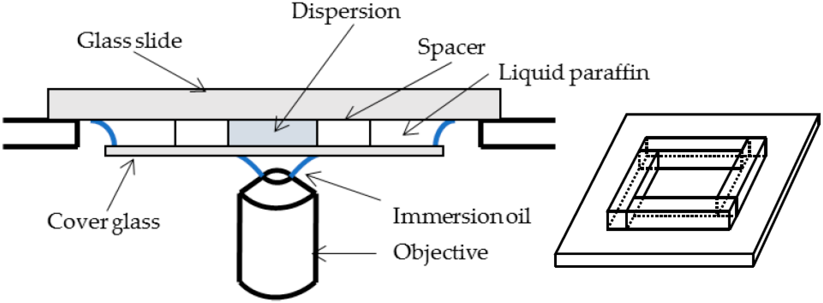
Using a simple optical microscope setup, we observed dynamical behaviors of two-dimensional diffusion of particles on the growth interface of colloidal crystals at the particle level.
(2)
First day of the experiment, crystals grew normally, and the growth rates approached zero asymptotically; the system seemed to reach equilibrium. After that, however, the crystals started to dissolve, and in several days the crystals had disappeared completely and quite amazingly. The mechanism of re-dispersion is unclear at the present moment. Thus, we only use the results at the early stages of the experiment in the first day.
(3)
Surface diffusion coefficients were directly evaluated using the mean displacement of an adsorbed particle x s and the mean lifetime of an adsorbed particle before being dissolved again into the surrounding solution 𝜏 . The average coefficient was an order of magnitude smaller than that of bulk diffusion. This is probably due to the high activation barrier for surface diffusion; the activation barrier for surface diffusion is probably higher than that for bulk diffusion in the solution.
(4)
Kink incorporation processes were precisely examined. Contrary to the expectation, the process via diffusion along steps were not observed essentially. This is probably attributable to the difference in the number of recovered bonds of adsorbed particles; particles adsorbed on steps recovered the largest number of bonds, and thus, the particles hardly cut the bonds to move along the steps.
(5)
Formation of stacking disorders proceeded via two-dimensional nucleation of particle layers at the different lattice sites and growth. The displacement of the boundary between different stacking layers was also observed. The displacement is mainly promoted by the hopping of particles beyond the boundary between the different stacking layers.
(6)
Desorption of particles from a step by the adsorption of other particles to the desorbing particle was observed for the first time, as if the adsorbed particles peeled the desorbing particle off the step.

Supplementary Materials

The following is available online at www.mdpi.com/2073-4352/6/7/80/s1, Video S1: Brownian motion of particles around growing crystals.

Acknowledgments

The authors wish to thank Qingfeng Yan of Tsinghua University for inviting them to submit this paper to the special issue “Colloidal Crystals”. Yoshihisa Suzuki was partially supported by JSPS KAKENHI Grant Numbers 24656016 and 26390054.

Author Contributions

Yoshihisa Suzuki conceived and designed the experiments; Yoshiaki Hattori performed the experiments; Yoshihisa Suzuki and Yoshiaki Hattori analyzed the data; Jun Nozawa, Satoshi Uda, Akiko Toyotama, and Junpei Yamanaka gave basic and important ideas as well as precise information; Jun Nozawa and Satoshi Uda showed precedence data and movies on this depletion colloidal crystallization system at the 45th National Conference on Crystal Growth (19aC01, 19 September 2015, Sapporo, Japan); Yoshihisa Suzuki wrote the paper.

Conflicts of Interest

The authors declare no conflict of interest.

References

  1. Kose, A.; Ozaki, M.; Takano, K.; Kobayashi, Y.; Hachisu, S. Direct observation of ordered latex suspension by metallurgical microscope. J. Colloid Int. Sci. 1973, 44, 330–338. [Google Scholar] [CrossRef]
  2. Hachisu, S.; Yoshimura, S. Optical demonstration of crystalline superstructures in binary mixtures of latex globules. Nature 1980, 283, 188–189. [Google Scholar] [CrossRef]
  3. Takano, K.; Hachisu, S. Pressure of Kirkwood—Alder transition in monodisperse latex. J. Chem. Phys. 1977, 67, 2604–2608. [Google Scholar] [CrossRef]
  4. Pusey, P.N.; van Megen, W. Phase behaviour of concentrated suspensions of nearly hard colloidal spheres. Nature 1980, 320, 340–342. [Google Scholar] [CrossRef]
  5. Davis, K.E.; Russel, W.B.; Glantschnig, W.J. Disorder-to-order transition in settling suspensions of colloidal silica: X-ray measurements. Science 1989, 245, 507–510. [Google Scholar] [CrossRef] [PubMed]
  6. Monovoukas, Y.; Gast, A.P. Microstructure identification during crystallization of charged colloidal suspensions. Phase Transit. 1990, 21, 183–195. [Google Scholar] [CrossRef]
  7. Gast, A.P.; Monovoukas, Y. A new growth instability in colloidal crystallization. Nature 1991, 351, 551–555. [Google Scholar] [CrossRef]
  8. Okubo, T. Giant colloidal single crystals of polystyrene and silica spheres in deionized suspension. Langmuir 1994, 10, 1695–1702. [Google Scholar] [CrossRef]
  9. Van Blaaderen, A.; Wiltzius, P. Real-space structure of colloidal hard-sphere glasses. Science 1995, 270, 1177–1179. [Google Scholar] [CrossRef]
  10. Weiss, J.A.; Oxtoby, D.W.; Grier, D.G.; Murray, C.A. Martensitic transition in a confined colloidal suspension. J. Chem. Phys. 1995, 103, 1180–1190. [Google Scholar] [CrossRef]
  11. Van Blaaderen, A.; Ruel, R.; Wiltzius, P. Template-directed colloidal crystallization. Nature 1997, 385, 321–324. [Google Scholar] [CrossRef]
  12. Míguez, H.; Meseguer, F.; López, C.; Mifsud, A.; Moya, J.S.; Vázquez, L. Evidence of FCC crystallization of SiO2 nanospheres. Langmuir 1997, 13, 6009–6011. [Google Scholar] [CrossRef]
  13. Sawada, T.; Suzuki, Y.; Toyotama, A.; Iyi, N. Quick fabrication of gigantic single-crystalline colloidal crystals for photonic crystal applications. Jpn. J. Appl. Phys. 2001, 40, L1226–L1228. [Google Scholar] [CrossRef]
  14. Gasser, U.; Weeks, E.R.; Schofield, A.; Pusey, P.N.; Weitz, D.A. Real-space imaging of nucleation and growth in colloidal crystallization. Science 2001, 292, 258–262. [Google Scholar] [CrossRef] [PubMed]
  15. Auer, S.; Frenkel, D. Prediction of absolute crystal-nucleation rate in hard-sphere colloids. Nature 2001, 409, 1020–1023. [Google Scholar] [CrossRef] [PubMed]
  16. Hoogenboom, J.P.; Derks, D.; Vergeer, P.; van Blaaderen, A. Stacking faults in colloidal crystals grown by sedimentation. J. Chem. Phys. 2002, 117, 11320–11328. [Google Scholar] [CrossRef]
  17. Yamanaka, J.; Murai, M.; Iwayama, Y.; Yonese, M.; Ito, K.; Sawada, T. One-directional crystal growth in charged colloidal silica dispersions driven by diffusion of base. J. Am. Chem. Soc. 2004, 126, 7156–7157. [Google Scholar] [CrossRef] [PubMed]
  18. Schall, P.; Cohen, I.; Weitz, D.A. Visualization of dislocation dynamics in colloidal crystals. Science 2004, 305, 1944–1948. [Google Scholar] [CrossRef] [PubMed]
  19. Leunissen, M.E.; Christova, C.G.; Hynninen, A.-P.; Patrick Royall, C.; Campbell, A.I.; Imhof, A.; Dijkstra, M.; van Roij, R.; van Blaaderen, A. Ionic colloidal crystals of oppositely charged particles. Nature 2005, 437, 235–240. [Google Scholar] [CrossRef] [PubMed]
  20. Mori, A.; Yanagiya, S.-I.; Suzuki, Y.; Sawada, T.; Ito, K. Monte Carlo simulation of crystal-fluid coexistence states in the hard-sphere system under gravity with stepwise control. J. Chem. Phys. 2006, 124. [Google Scholar] [CrossRef] [PubMed]
  21. Mori, A.; Suzuki, Y.; Yanagiya, S.-I.; Sawada, T.; Ito, K. Shrinking stacking fault through glide of the Shockley partial dislocation in hard-sphere crystal under gravity. Mol. Phys. 2007, 105, 1377–1383. [Google Scholar] [CrossRef]
  22. Ohtaka, K. Energy band of photons and low-energy photon diffraction. Phys. Rev. B 1979, 19, 5057–5067. [Google Scholar] [CrossRef]
  23. Yablonovitch, E. Inhibited spontaneous emission in solid-state physics and electronics. Phys. Rev. Lett. 1987, 58, 2059–2062. [Google Scholar] [CrossRef] [PubMed]
  24. John, S. Strong localization of photons in certain disordered dielectric superlattices. Phys. Rev. Lett. 1987, 58, 2486–2489. [Google Scholar] [CrossRef] [PubMed]
  25. Holtz, J.H.; Asher, S.A. Polymerized colloidal crystal hydrogel films as intelligent chemical sensing materials. Nature 1997, 389, 829–832. [Google Scholar] [CrossRef]
  26. Yang, S.M.; Ozin, G.A. Opal chips: Vectorial growth of colloidal crystal patterns inside silicon wafers. Chem. Commun. 2000, 2507–2508. [Google Scholar] [CrossRef]
  27. Xia, Y.; Gates, B.; Yin, Y.; Lu, Y. Monodispersed colloidal spheres: Old materials with new applications. Adv. Mater. 2000, 12, 693–713. [Google Scholar] [CrossRef]
  28. Blanco, A.; Chomski, E.; Grabtchak, S.; Ibisate, M.; John, S.; Leonard, S.W.; Lopez, C.; Meseguer, F.; Miguez, H.; Mondia, J.P.; et al. Large-scale synthesis of a silicon photonic crystal with a complete three-dimensional bandgap near 1.5 micrometres. Nature 2000, 405, 437–440. [Google Scholar] [PubMed]
  29. Vlasov, Y.A.; Bo, X.-Z.; Sturm, J.C.; Norris, D.J. On-chip natural assembly of silicon photonic bandgap crystals. Nature 2001, 414, 289–293. [Google Scholar] [CrossRef] [PubMed]
  30. Matsuo, S.; Fujine, T.; Fukuda, K.; Juodkazis, S.; Misawa, H. Formation of free-standing micropyramidal colloidal crystals grown on silicon substrate. Appl. Phys. Lett. 2003, 82, 4283–4285. [Google Scholar] [CrossRef]
  31. Yin, Y.; Li, Z.-Y.; Xia, Y. Template-directed growth of (100)-oriented colloidal crystals. Langmuir 2003, 19, 622–631. [Google Scholar] [CrossRef]
  32. Fudouzi, H.; Sawada, T. Photonic rubber sheets with tunable color by elastic deformation. Langmuir 2006, 22, 1365–1368. [Google Scholar] [CrossRef] [PubMed]
  33. Suzuki, Y.; Sawada, T.; Tamura, K. Colloidal crystallization by a centrifugation method. J. Cryst. Growth 2011, 318, 780–783. [Google Scholar] [CrossRef]
  34. Hilhorst, J.; Petukhov, A.V. Variable dislocation widths in colloidal crystals of soft thermosensitive spheres. Phys. Rev. Lett. 2011, 107, 095501-1-5. [Google Scholar] [CrossRef] [PubMed]
  35. Suzuki, Y.; Mori, A.; Fujiwara, T.; Tamura, K. Precise characterization of grain structures, stacking disorders and lattice disorders of a close-packed colloidal crystal. J. Cryst. Growth 2011, 322, 109–113. [Google Scholar] [CrossRef]
  36. Suzuki, Y.; Endoh, J.; Mori, A.; Yabutani, T.; Tamura, K. Gravitational annealing of colloidal crystals. Defect Diffus. Forum 2012, 323–325, 555–558. [Google Scholar] [CrossRef]
  37. Sato, M.; Katsuno, H.; Suzuki, Y. Formation of a crystal of Brownian particles under a uniform external force. Phys. Rev. E 2013, 87, 032403. [Google Scholar] [CrossRef]
  38. Suzuki, Y.; Sazaki, G.; Hashimoto, K.; Fujiwara, T.; Furukawa, Y. Colloidal crystallization utilizing interfaces of unidirectionally growing ice crystals. J. Cryst. Growth 2013, 383, 67–71. [Google Scholar] [CrossRef]
  39. Hashimoto, K.; Mori, A.; Tamura, K.; Suzuki, Y. Enlargement of grains of silica colloidal crystals by centrifugation in an inverted-triangle internal-shaped container. Jpn. J. Appl. Phys. 2013, 52, 030201-1-3. [Google Scholar] [CrossRef]
  40. Hilhorst, J.; de Winter, D.A.M.; Wolters, J.R.; Post, J.A.; Petukhov, A.V. Defect engineering in sedimentary colloidal photonic crystals. Langmuir 2013, 29, 10011–10018. [Google Scholar] [CrossRef] [PubMed]
  41. Zhang, C.; Macfarlane, R.J.; Young, K.L.; Choi, C.H.J.; Hao, L.; Auyeung, E.; Liu, G.; Zhou, X.; Mirkin, C.A. A general approach to DNA-programmable atom equivalents. Nat. Mater. 2013, 12, 741–746. [Google Scholar] [CrossRef] [PubMed]
  42. Mori, A.; Suzuki, Y.; Sato, M. Gravitational tempering in colloidal epitaxy to reduce defects further. Cryst. Growth Des. 2014, 14, 2083–2086. [Google Scholar] [CrossRef]
  43. Suzuki, Y.; Mori, A.; Sato, M.; Katsuno, H.; Sawada, T. Colloidal crystallization on tilted substrates under gravitational fields. J. Cryst. Growth 2014, 401, 905–909. [Google Scholar] [CrossRef]
  44. Mori, A.; Suzuki, Y. Identification of triangular-shaped defects often appeared in hard-sphere crystals grown on a square pattern under gravity by Monte Carlo simulations. Phys. B Condens. Matter 2014, 452, 58–65. [Google Scholar] [CrossRef]
  45. Mahynski, N.A.; Panagiotopoulos, A.Z.; Meng, D.; Kumar, S.K. Stabilizing colloidal crystals by leveraging void distributions. Nat. Commun. 2014, 5, 4472-1-8. [Google Scholar] [CrossRef] [PubMed]
  46. De Nijs, B.; Dussi, S.; Smallenburg, F.; Meeldijk, J.D.; Groenendijk, D.J.; Filion, L.; Imhof, A.; van Blaaderen, A.; Dijkstra, M. Entropy-driven formation of large icosahedral colloidal clusters by spherical confinement. Nat. Mater. 2015, 14, 56–60. [Google Scholar] [CrossRef] [PubMed]
  47. Peng, Y.; Wang, F.; Wang, Z.; Alsayed, A.M.; Zhang, Z.; Yodh, A.G.; Han, Y. Two-step nucleation mechanism in solid–solid phase transitions. Nat. Mater. 2015, 14, 101–108. [Google Scholar] [CrossRef] [PubMed]
  48. Pal, A.; Malik, V.; He, L.; Ern, B.H.; Yin, Y.; Kegel, W.K.; Petukhov, W.K. Tuning the colloidal crystal structure of magnetic particles by external field. Angew. Chem. Int. Ed. 2015, 54, 1803–1807. [Google Scholar] [CrossRef] [PubMed]
  49. Statt, A.; Virnau, P.; Binder, K. Finite-size effects on liquid-solid phase coexistence and the estimation of crystal nucleation barriers. Phys. Rev. Lett. 2015, 114, 026101-1-5. [Google Scholar] [CrossRef] [PubMed]
  50. Wang, Z.; Wang, F.; Peng, Y.; Han, Y. Direct observation of liquid nucleus growth in homogeneous melting of colloidal crystals. Nat. Commun. 2015, 6, 6942-1-9. [Google Scholar] [CrossRef] [PubMed]
  51. Lu, F.; Yager, K.G.; Zhang, Y.; Xin, H.; Gang, O. Superlattices assembled through shape-induced directional binding. Nat. Commun. 2015, 6, 6912-1-10. [Google Scholar] [CrossRef] [PubMed]
  52. Petukhov, A.V.; Meijer, J.-M.; Vroege, G.J. Particle shape effects in colloidal crystals and colloidal liquid crystals: Small-angle X-ray scattering studies with microradian resolution. Curr. Opin. Colloid Interface Sci. 2015, 20, 272–281. [Google Scholar] [CrossRef]
  53. Qi, W.; Peng, Y.; Han, Y.; Bowles, R.K.; Dijkstra, M. Nonclassical nucleation in a solid–solid transition of confined hard spheres. Phys. Rev. Lett. 2015, 115, 185701-1-5. [Google Scholar] [CrossRef] [PubMed]
  54. Cabane, B.; Li, J.; Artzner, F.; Botet, R.; Labbez, C.; Bareigts, G.; Sztucki, M.; Goehring, L. Hiding in plain view: Colloidal self-assembly from polydisperse populations. Phys. Rev. Lett. 2016, 116, 208001-1-5. [Google Scholar] [CrossRef] [PubMed]
  55. Zhao, Q.; Finlayson, C.E.; Snoswell, D.R.E.; Haines, A.; Schäfer, C.; Spahn, P.; Hellmann, G.P.; Petukhov, A.V.; Herrmann, L.; Burdet, P.; et al. Large-scale ordering of nanoparticles using viscoelastic shear processing. Nat. Commun. 2016, 7, 11661-1-10. [Google Scholar] [CrossRef] [PubMed] [Green Version]
  56. Ise, N.; Okubo, T.; Sugimura, M.; Ito, K.; Nolte, H.J. Ordered structure in dilute solutions of highly charged polymer lattices as studied by microscopy. I. Interparticle distance as a function of latex concentration. J. Chem. Phys. 1983, 78, 536–540. [Google Scholar] [CrossRef]
  57. Ito, K.; Nakamura, H.; Yoshida, H.; Ise, N. On the dynamic character of “ordered” structure in polymer latex suspensions. J. Am. Chem. Soc. 1988, 110, 6955–6963. [Google Scholar] [CrossRef]
  58. Larsen, A.E.; Grier, D.G. Like-charge attractions in metastable colloidal crystallites. Nature 1997, 385, 230–233. [Google Scholar] [CrossRef]
  59. Overbeek, J.T.G. Recent developments in the understanding of colloid stability. J. Colloid Int. Sci. 1977, 58, 408–422. [Google Scholar] [CrossRef]
  60. Asakura, S.; Oosawa, F. Interaction between particles suspended in solutions of macromolecules. J. Polym. Sci. 1958, 33, 183–192. [Google Scholar] [CrossRef]
  61. Kose, A.; Hachisu, S. Ordered structure in weakly flocculated monodisperse latex. J. Colloid Int. Sci. 1976, 55, 487–498. [Google Scholar] [CrossRef]
  62. Toyotama, A.; Okuzono, T.; Yamanaka, J. Spontaneous formation of eutectic crystal structures in binary and ternary charged colloids due to depletion attraction. Sci. Rep. 2016, 6, 23292. [Google Scholar] [CrossRef] [PubMed]
  63. Chernov, A.A. Modern Crystallography III Crystal Growth; Springer: Berlin, Germany, 1984; pp. 10–13. [Google Scholar]
  64. Burton, W.K.; Cabrera, N.; Frank, F.C. The growth of crystals and the equilibrium structure of their surfaces. Philos. Trans. R. Soc. Lond. A 1951, 243, 299–358. [Google Scholar] [CrossRef]
  65. Dunstan, D.E.; Stokes, J. Diffusing probe measurements of polystyrene latex particles in polyelectrolyte solutions: Deviations from Stokes–Einstein behavior. Macromolecules 2000, 33, 193–198. [Google Scholar] [CrossRef]
  66. Schwoebel, R.L.; Shipsey, E.J. Step motion on crystal surfaces. J. Appl. Phys. 1966, 37, 3682–3686. [Google Scholar] [CrossRef]
Figure 1. An obtained colloidal crystal containing three layers of regular arrays of polystyrene particles. Individual particles are clearly resolved at the particle level. The focal plane in this picture is adjusted at the third layer.
Crystals 06 00080 g001
Figure 2. Re-dispersion of particles from grown crystals. (a) 1 day after sample preparation; (b) 6 days after sample preparation. In a day, crystals grow up to several layers as shown in (a) at 25 °C. However, 3 days after sample preparation, the crystals start to dissolve, and 6 days after, the crystals have disappeared completely.
Crystals 06 00080 g002
Figure 3. Schematic of the classification of particle incorporation processes into a kink site. Hexagons indicate crystals. Gray and indigo parts show the first (lower) and second (upper) layers, respectively. The borders between these layers are mono-particulate steps. Kinks on the steps indicate kink sites at which particles are finally incorporated into the crystal. Yellow circles represent the particles that are just incorporated into the crystal at the kink sites on the steps. (a) kink incorporation processes via surface diffusion on lower layers; (b) those via surface diffusion on upper layers; (c) those via surface diffusion on lower layers and adsorption to the particle at the edge of the kink site; (d) direct incorporation from the solution. Dashed and solid circles at both ends of arrows indicate particles at the previous and present positions, respectively.
Crystals 06 00080 g003
Figure 4. Number distribution of incorporation process routes. (a) to (d) indicate the same classifications described in Figure 3. At the top of bars, schematics used in Figure 3 are placed.
Crystals 06 00080 g004
Figure 5. Schematic of adsorption conditions. A, E, and S particles are those that form respectively three, four, and five bonds with particles in a crystal. Dotted and solid circles represent the lower and upper layers, respectively.
Crystals 06 00080 g005
Figure 6. Formation processes of stacking disorders. (a) Two-dimensional nucleation on the first triangular layer (90 min. after the injection of a particle dispersion into a growth cell); (b) Growth of the nucleated layers (120 min); (c) Complete occupation of the second layer and formation of the boundary between two different stacks (200 min); (d) Displacement of the boundary accompanied by the growth of the third layers (280 min). Scale bar represents 10 µm.
Crystals 06 00080 g006
Figure 7. The time course of a “peeling off” process caused by the additional adsorption of a particle from bulk solution onto the other particle on a step. (a) A particle approaches a top layer; (b) The particle adsorbs to the edge particle of the layer; (c) The edge particle is desorbing from the layer with the adsorbed particle; (d) The particle pair is completely peeled off the top layer of particles.
Crystals 06 00080 g007
Figure 8. Schematic of an in situ observation cell. Settled particles on the cover glass formed crystals. Growth processes of the crystals were precisely observed using an oil immersion objective. A right inset figure shows the arrangement of spacers cut into four pieces (0.28 × 1.0 × 10 mm) on the glass slide. Thickness of the inner space is 0.28 mm.
Crystals 06 00080 g008
Table 1. xs and τ of 10 particles on growth interfaces.
Particle Numberxs/×10−6 mτ/s
11.06.83
21.66.12
30.54.62
41.06.09
50.95.48
61.712.00
71.310.51
80.89.16
91.710.35
101.28.37

