Article

Incidental Polymorphism, Non-Isomorphic and Isomorphic Substitution in Calcium-Valine Coordination Polymers

Institute of Inorganic Chemistry, RWTH Aachen University, Landoltweg 1, 52074 Aachen, Germany
*
Author to whom correspondence should be addressed.
Crystals 2015, 5(2), 261-272; https://doi.org/10.3390/cryst5020261
Submission received: 23 April 2015 / Revised: 20 May 2015 / Accepted: 22 May 2015 / Published: 29 May 2015

Abstract

:
Five coordination polymers with the stoichiometry CaX2(valine)2(H2O)2 (X = Cl, Br) were obtained from the corresponding calcium halides and either racemic and enantiopure valine. In all cases the zwitterionic amino acid is exclusively O coordinated and the halides act as counteranions for the resulting one-dimensional cationic chains. The enantiopure chloride shows dimorphism; both forms differ in connectivity from the bromide. In contrast to this structural variability for L-valine, the derivatives of the racemic amino acid are isomorphous.

Graphical Abstract

1. Introduction

While amino acids are often used in chemistry and certainly represent an essential class of compounds in biochemistry, their fundamental coordination chemistry has been investigated to a lesser extent than expected. Although numerous structures containing amino acids in some form are reported, only a few systematic investigations with focus on amino acid coordination chemistry have been pursued.
Tackling this gap, we recently reported on the coordination chemistry of the amino acids proline and alanine with the halide salts of the oxophilic cations manganese and calcium. Both amino acids have shown to be useful ligands, since their chirality and hydrogen bond interactions led to a versatile asset of coordination compounds. On the one hand, L-proline formed coordination polymers with manganese chloride, bromide, and iodide in equimolar stoichiometry. On the other hand, a series of molecular, isomorphous products in 1:2 stoichiometry was obtained with racemic proline [1]. Enantiopure alanine even allowed for isomorphous substitution of the heavy manganese and calcium halides to form a series of chiral coordination polymers in 1:3 stoichiometry, while racemic alanine yields different structures for the two metals [2]. Calcium, a cation without crystal field effects exerted from partially filled d-orbitals, exhibited its versatility in the coordination chemistry with proline: two polymorphs were found for reactions of CaCl2 with both enantiopure and racemic proline. One molecular complex resembles a structure from MnCl2, two unique coordination networks with Ca in octahedral environment are more characteristic for transition metals, and one coordination polymer features 7-fold Ca2+ coordination; all these solids show unusual Ca–Cl contacts [3]. No polymorphism is encountered for the CaBr2 derivatives. Here the network motif is established for enantiopure proline, whereas racemic proline once again yields a unique polymer with eight-fold coordination [4]. A comprehensive overview of crystal structures from amino acids has been published by Fleck and Petrosyan [5]. With respect to coordinative bonds, zwitterionic amino acids can be considered a special case of carboxylates, and carboxylates count among the most widely used ligands for the design of extended structures (e.g., [6,7,8,9]).
In this contribution we compile five structures, formed by reaction of CaCl2 or CaBr2 with the amino acid valine and discuss polymorphism, isomorphism and other structural relationships. Reaction of L-valine with CaCl2 in aqueous solution reproducibly yields coordination polymer 1a. In one instance an individual crystal of a second polymorph 1b was detected, and this alternative structure is included here for comparison. Despite variations in solvent, stoichiometry, concentration and crystallisation temperature, we have not been able to reproduce this second crystal form. Even the bulk material from which the single 1b specimen was selected consisted of the ubiquitous phase 1a, and the alternative polymorph 1b did not contribute detectable reflections to the powder pattern. A different polymer was obtained by reaction with the heavier CaBr2 (2). Two isomorphous structures are obtained by using racemic valine with CaCl2 (3) and CaBr2 (4). The chloride analogue has been reported before [10] but we provide a low temperature crystal structure for comparison. A scheme of the discussed structures is given as Figure 1.
Figure 1. Overview of the performed reactions and reaction products.
Crystals 05 00261 g001

2. Results and Discussion

All structures obtained from calcium chloride and bromide with valine in enantiopure and racemic form are coordination polymers with bridging carboxylato groups and uncoordinated halides. They all share the formal composition CaX2(valine)2(H2O)2 but differ significantly with respect to their detailed connectivity. Data collection and refinement details for all structures are given in Table 1.
Table 1. Data collection and refinement details for all structures.
Compound/CCDC1a/10613421b/10613432/10613443/10613454/1061346
Number [13]
Crystal Data
Chemical formulaC10H26CaN2O6Cl2C10H26CaN2O6Cl2C10H26CaN2O6Br2C10H26CaN2O6Cl2C10H26CaN2O6Br2
Mr381.31381.31470.23381.31470.23
Crystal systemTriclinicOrthorhombicMonoclinicOrthorhombicOrthorhombic
Space groupP1P21212C2PccaPcca
a (Å)4.9856 (18)17.420 (3)13.879 (8)24.180 (7)24.657 (7)
b (Å)9.542 (4)20.468 (4)20.293 (12)9.861 (3)10.010 (3)
c (Å)10.303 (4)5.0236 (9)7.841 (5)7.666 (2)7.710 (2)
α (°)77.743 (6)
β (°)76.975 (7) 117.282 (10)
γ (°)83.919 (7)
V3)465.8 (3)1791.2 (6)1963 (2)1827.9 (9)1903.0 (9)
Z14444
μ (mm−1)0.650.674.410.664.55
Crystal size (mm)0.21 × 0.08 × 0.030.25 × 0.05 × 0.050.25 × 0.05 × 0.040.34 × 0.12 × 0.110.19 × 0.05 × 0.03
Data collection
Tmin, Tmax0.600, 0.7460.678, 0.7450.550, 0.7450.630, 0.7450.524, 0.745
Rint0.0430.1130.080.0960.145
(sinθ/λ)max−1)0.7190.6260.6130.610.61
Measured, independent and observed [I > 2σ(I)] reflections7257, 5245, 455421760, 3677, 311311174, 3754, 323918905, 1737, 146017812, 1815, 1325
Refinement
R[F2 > 2σ (F2)], wR(F2)0.071, 0.1740.047, 0.1040.046, 0.0950.034, 0.0860.043, 0.095
S1.031.031.001.091.05
No. of reflections5245 3677375417371815
No. of parameters224234225113113
No. of restraints24212344
Δρmax, Δρmin (e Å−3)0.76, −0.580.44, −0.480.63, −0.430.33, −0.240.60, −0.51
Absolute structure parameter0.02 (8)−0.02 (4)−0.006 (18)
Number of quotients177411081316
Flack x determined using quotients method [(I+)-(I-)]/[(I+)+(I-)] [14].

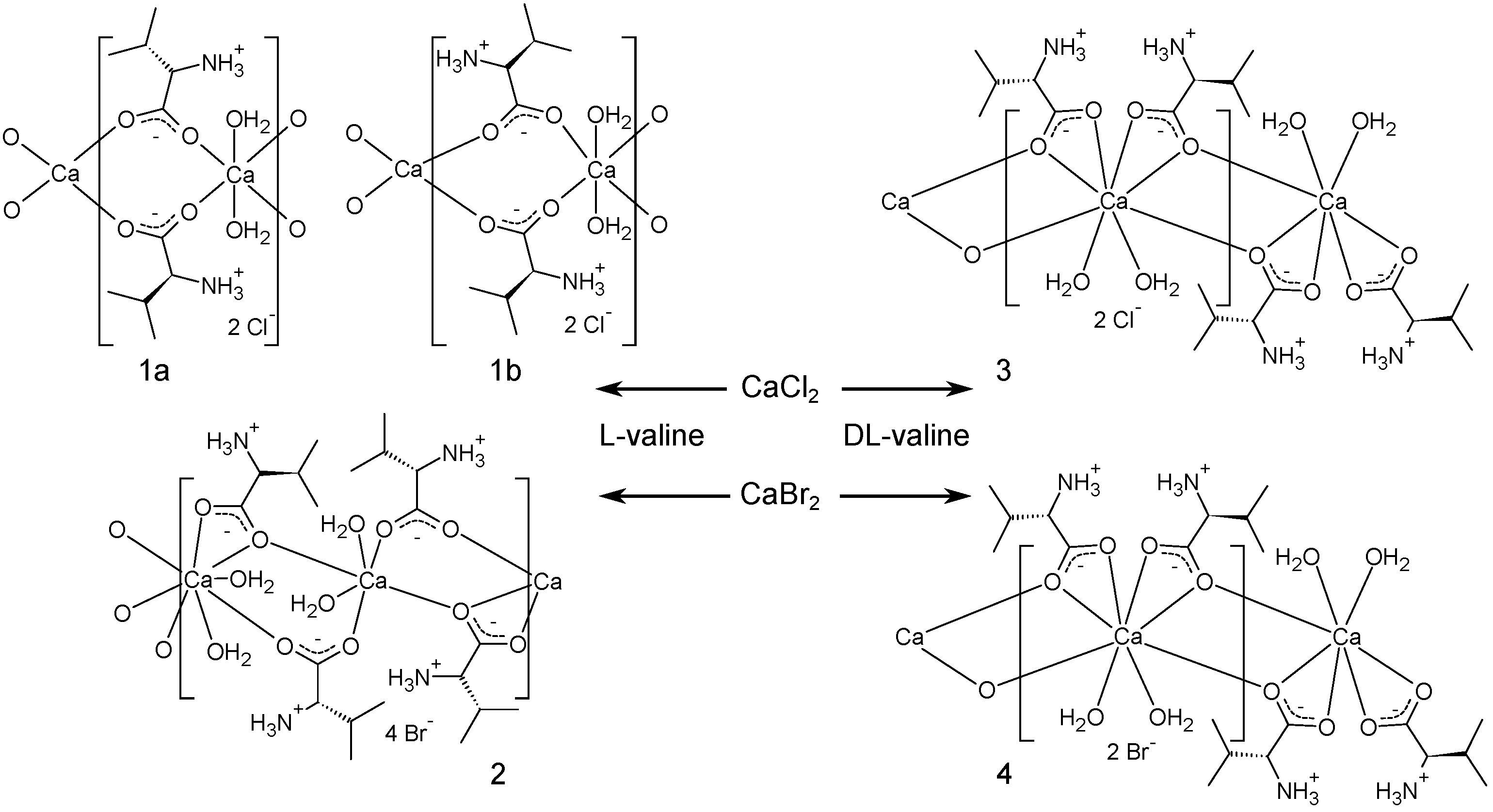
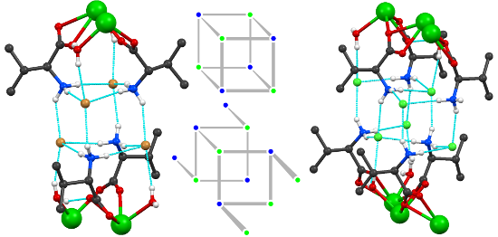
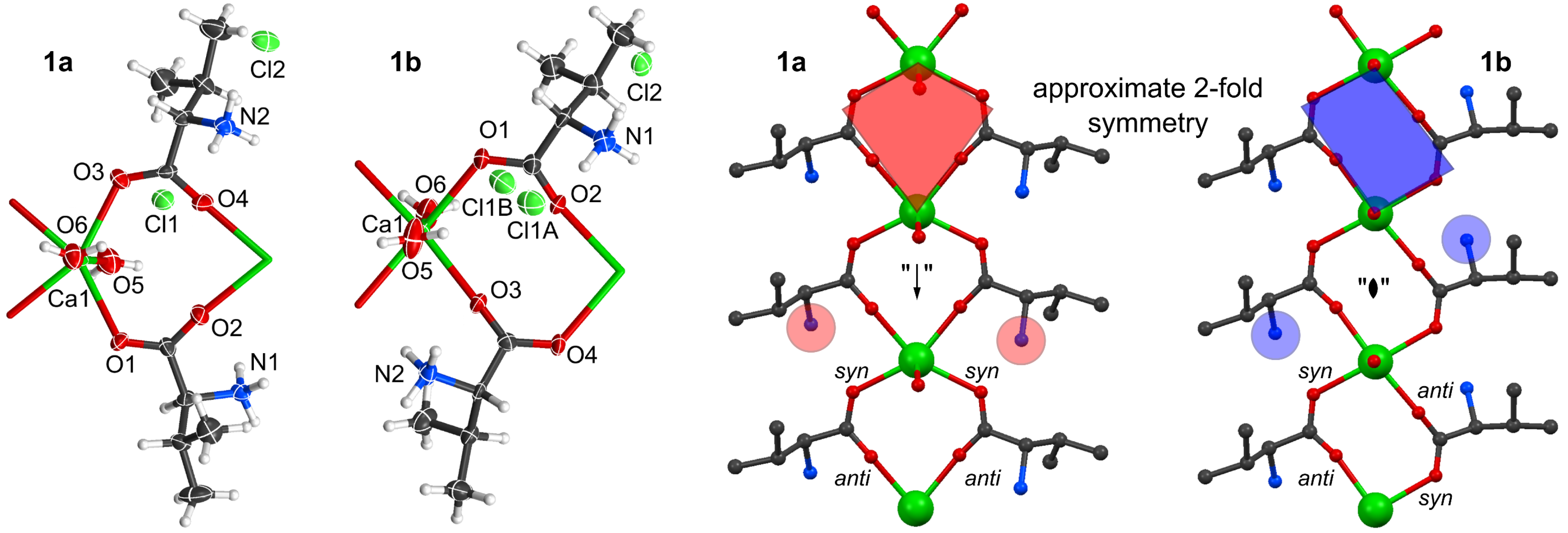
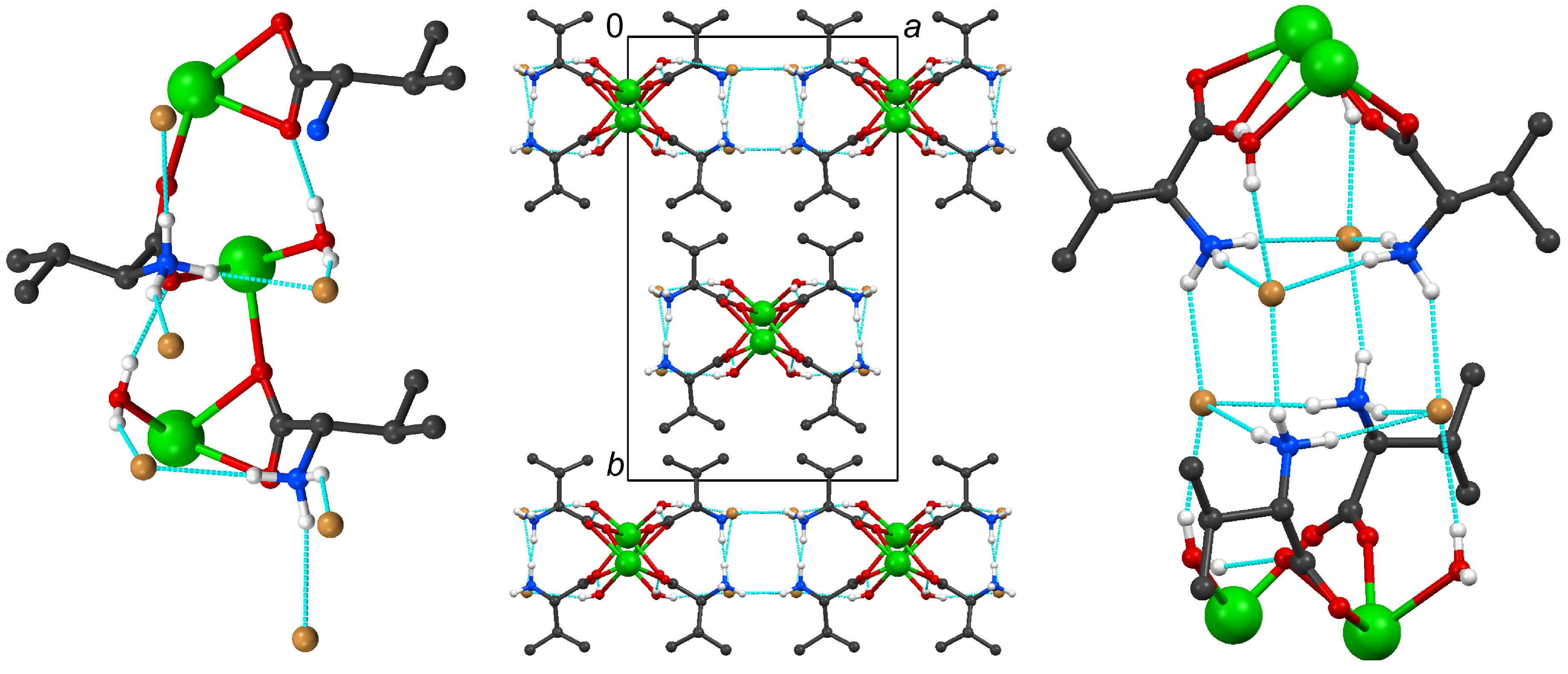
2.1. Structural Details of 1a and 1b

1a and 1b, the alternative polymorphs of CaCl2(L-valine)2(H2O)2, are associated with a different ratio of volume per formula unit (V/Z , see Table 1); the formula unit requires a larger volume in 1a (465.8 Å3) than in 1b (447.8 Å3). This relationship is of course also reflected in a higher linear absorption coefficient and density of the latter compound. Although 1b shows positional disorder for a chloride counteranion (see refinement section), it is associated with better space filling. We wish to communicate this fact but cannot provide a simple explanation; we are aware of examples for the opposite case, i.e., more efficient packing for the well-ordered phase [11,12].
The main polymorph 1a crystallises in the triclinic spacegroup P1 with one Ca(II) cation, two bridging L-valine molecules, two coordinating water ligands, and two uncoordinated chlorides in the asymmetric unit (see Figure 2). The same applies for 1b but in the orthorhombic spacegroup P21212. In both polymorphs, the calcium atom is enclosed in distorted octahedral geometry by two axial water ligands and four equatorially bridging µ2η:η carboxylate groups in syn-anti conformation. Ca–O distances range from 2.300 (5) to 2.390 (5) Å in 1a and from 2.272 (3) to 2.400 (3) Å in 1b (see Table 2).
Figure 2. Left: Asymmetric unit of 1a and 1b with labeling scheme as displacement ellipsoid plot (80% probability) and adjacent atoms as stick model. Right: Polymeric chains of 1a and 1b with kite (red) and parallelogram (blue) shaped bridges, approximate twofold symmetry with circles highlighting the stereo chemistry of the amino groups, and labeled syn and anti bridges.
Crystals 05 00261 g002
Table 2. Selected interatomic distances of all structures.
Compound1a1b 2 34
Ca1–O1 (Å)2.390(5)2.386(3)Ca1–O1 (Å)2.660(7)Ca1–O1 (Å)2.3484(16)2.357(4)
Ca1–O2 a (Å)2.319(6)2.272(3)Ca1–O3 b (Å)2.373(7)Ca1–O1 c (Å)2.6276(17)2.639(4)
Ca1–O3 (Å)2.386(6)2.275(3)Ca1–O5 (Å)2.521(7)Ca1–O2 c (Å)2.4403(17)2.440(4)
Ca1–O4 a (Å)2.300(5)2.400(3)Ca1–O6 (Å)2.385(8)Ca1–O3 (Å)2.3802(17)2.376(4)
Ca1–O5 (Å)2.360(5)2.366(4)Ca2–O1 (Å)2.279(6)
Ca1–O6 (Å)2.363(5)2.331(3)Ca2–O2 (Å)2.320(8)
Ca2–O4 (Å)2.372(4)
Symmetry operations: a −1 + x, y, z; b x, y, 1 + z; cx, −y, 1 − z.
While both resulting coordination polymers extend parallel to the shortest unit cell axis, their Ca· · · Ca separation differs significantly (compare a = 4.9856 (18) Å in 1a to c = 5.0236 (9) Å in 1b). This originates from the different sequence of the syn-anti coordinating carboxylates in the two polymorphs. While in 1a both coordinating L-valine molecules exhibit syn-anti conformation in chain direction, one valine molecule is syn-anti bridging in 1b whereas the other coordinates as anti-syn bridge. This results in a kite shaped form with approximate twofold symmetry along the chain in 1a contrary to a parallelogram shape with approximate 2-fold symmetry perpendicular to the chain in 1b (Figure 2). Consequently, the orientation of the amino groups is different and the resulting intermolecular interactions lead to different packing of the chains in the two polymorphs. While each amino group forms an N–H· · · O hydrogen bond towards the anti coordinating carboxylato-O of the next chain fragment, all other hydrogen bond donor groups find chlorides as suitable acceptors (see Table 3). The chains in 1a are crosslinked by those intermolecular hydrogen bonds in c-direction to form a two dimensional network. In 1b multiple hydrogen bonds link two neighbouring chains, forming a double strand that encloses the chlorides. The water molecule (O6) crosslinks these strands via O–H· · · Cl hydrogen bonds to form an overall three dimensional network (Figure 3).
Table 3. Hydrogen bond geometry in 1a and 1b (for the split positions of Cl1 only the shorter hydrogen bond is listed).
1aD· · · A (Å)D–H (Å)H· · · A (Å)DH· · · A (°)1bD· · · A (Å)D–H (Å)H· · · A (Å)DH· · · A (°)
N1–H1A· · · O1 a2.814(8)0.93(8)1.93(8)159(7)N1–H1C· · · O1 f2.826(5)0.93(4)1.92(4)166(3)
N1–H1C· · · Cl1 b3.137(5)0.93(7)2.25(6)161(7)N1–H1A· · · Cl1A3.023(5)0.95(3)2.10(4)164(4)
N1–H1B· · · Cl2 c3.167(6)0.93(4)2.26(5)164(6)N1–H1B· · · Cl23.184(4)0.93(3)2.27(3)167(3)
N2–H2A· · · O3 a2.813(7)0.92(5)1.90(6)172(7)N2–H2C· · · O4 c2.870(5)0.93(4)1.94(4)174(4)
N2–H2B· · · Cl13.156(6)0.92(8)2.26(6)158(7)N2–H2A· · · Cl1B g2.935(6)0.94(3)2.05(4)174(4)
N2–H2C· · · Cl23.145(6)0.92(8)2.27(8)158(7)N2–H2B· · · Cl2 h3.197(4)0.94(4)2.30(4)158(4)
O5–H5E· · · Cl2 c3.247(6)0.82(6)2.47(6)157(8)O5–H5D· · · Cl1A h3.182(5)0.82(5)2.40(5)160(5)
O5–H5D· · · Cl2 e3.124(7)0.83(9)2.32(9)164(8)O5–H5E· · · Cl1B3.037(5)0.82(4)2.28(5)152(5)
O6–H6B· · · Cl13.210(6)0.83(9)2.42(8)159(8)O6–H6A· · · Cl2 i3.170(4)0.82(4)2.37(4)167(4)
O6–H6A· · · Cl1 d3.136(5)0.83(6)2.32(6)166(7)O6–H6B· · · Cl2 j3.127(4)0.83(4)2.32(4)168(4)
Symmetry operations: a 1 + x, y, z; b 1 + x, y, –1 + z; c x, y, −1 + z; d −1 + x, y, z; e −1 + x, y, −1 + z; f x, y, 1 + z; g 1 − x, 1 − y, z; h 1 − x, 1 − y, −1 + z; i −0.5 + x, 0.5 − y, −z; j −0.5 + x, 0.5 − y, 1 − z.
Figure 3. Left: Hydrogen bonds (dashed light blue lines) extending from the asymmetric units of 1a and 1b. Right: Networks built by intermolecular hydrogen bonds in 1a and 1b.
Crystals 05 00261 g003

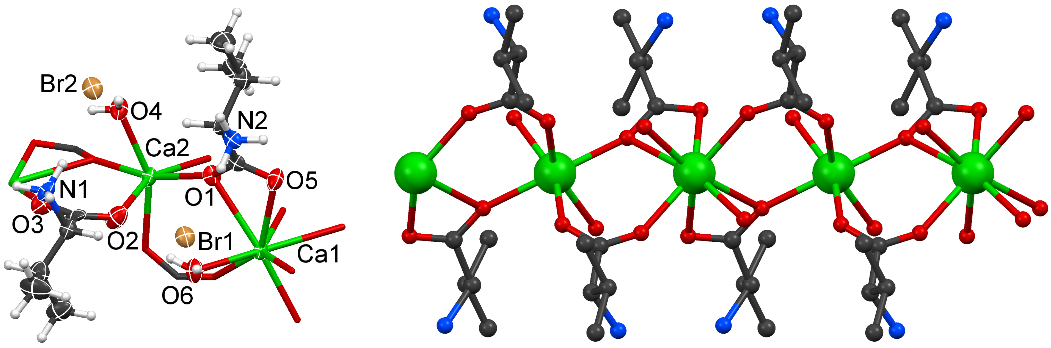
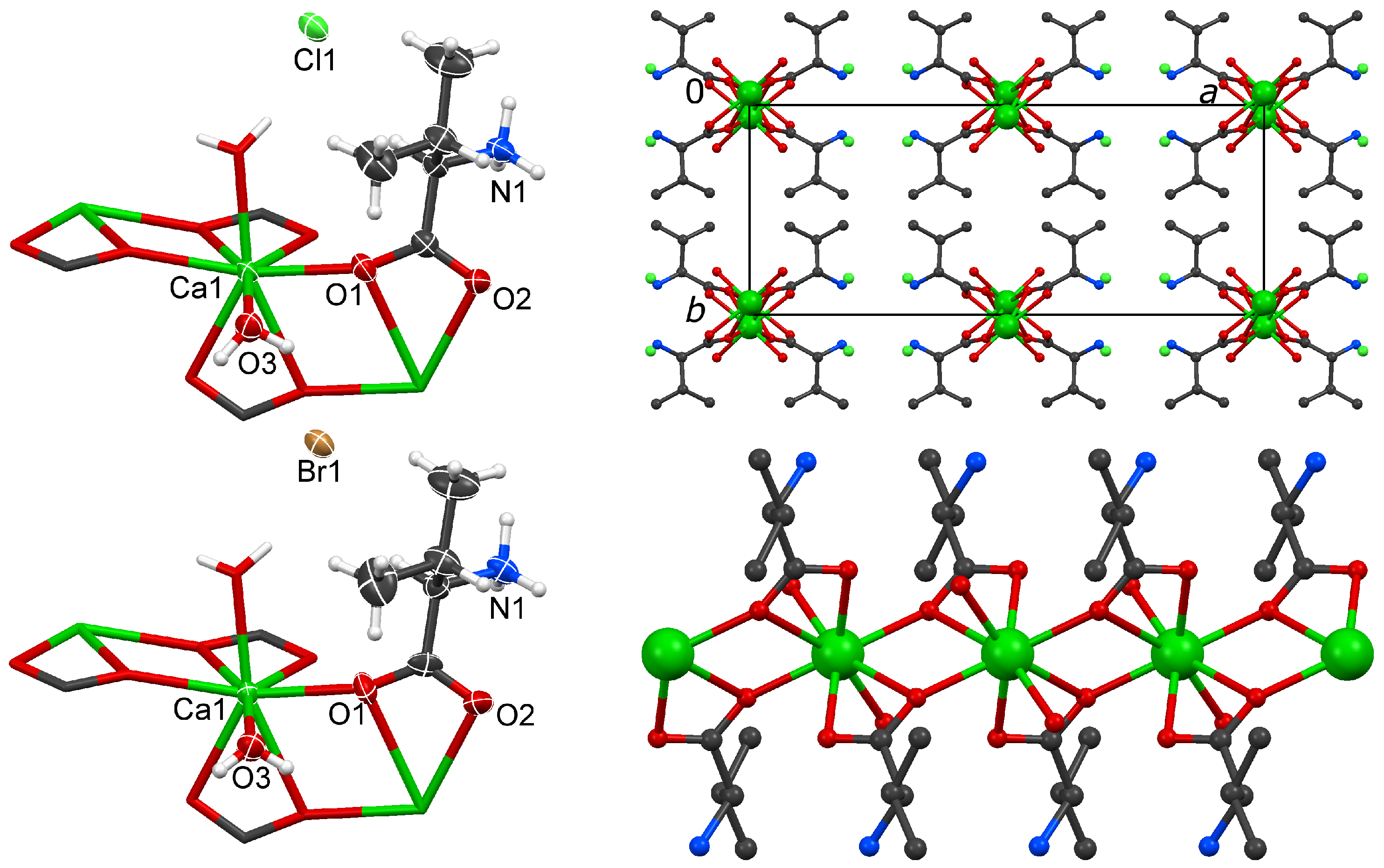
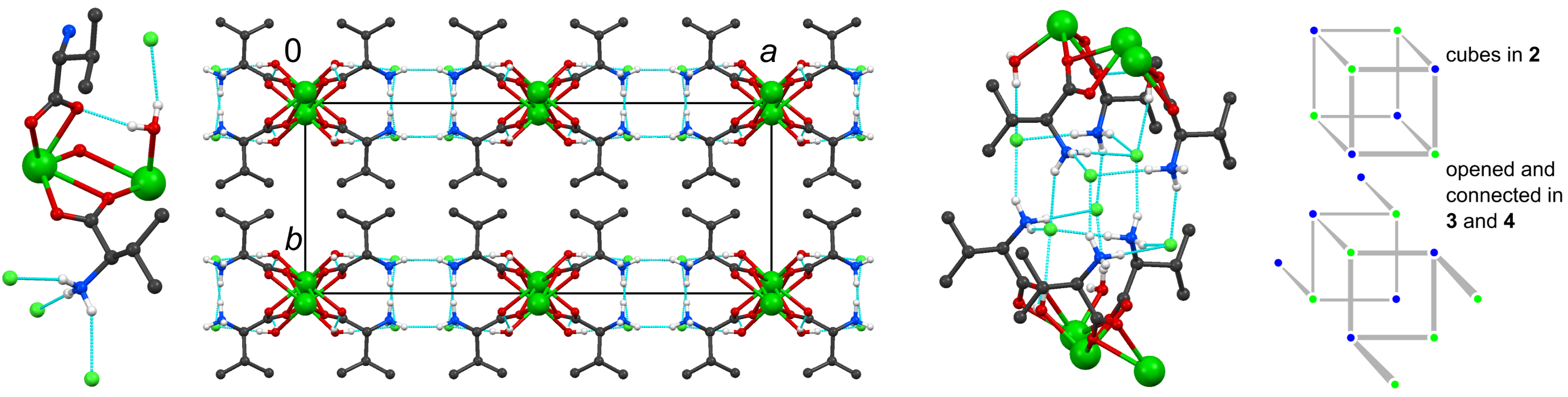
2.2. Structural Details of 2

Solid 2 is formed from L-valine and calcium bromide and crystallises in the monoclinic spacegroup C2. The asymmetric unit is formed by two independent Ca atoms on twofold axes (Wyckoff positions 2a and 2b), one simultaneously chelating and bridging L-valine lingand (µη2:η1), one syn-syn bridging L-valine ligand, two coordinating water molecules and two bromide counterions (Figure 4). The resulting cationic polymer chain (extending along the shortest unit cell axis c) is build by alternating eightfold coordinated Ca1 and six-coordinated Ca2. Ca–O distances range from 2.279 to 2.660 Å (see Table 2). The higher coordination number of Ca1 is due to the two chelating carboxylato groups, whereas Ca2 is only coordinated by one oxygen of each of the four connected amino acids. By action of the crystallographic axes perpendicular to the polymer, each chain segment consists of the same residues: one bridging and one simultaneously bridging and chelating carboxylate (Figure 4).
Figure 4. Left: Asymmetric unit of 2 drawn as ellipsoid pot at 80% probability and connected atoms as stick model. Right: Cationic chain of 2.
Crystals 05 00261 g004
All hydrogen bond donors find suitable acceptors. Contrary to many other amino acid coordination polymers, no N–H· · · O hydrogen bonds are formed along the chain. Instead, each amino group connects to three uncoordinated bromides. One intra chain hydrogen bond is provided by each water molecule towards a carboxylate-O, whereas the remaining donor groups coordinate bromide ions. Details about hydrogen bond geometry are compiled in Table 4. Overall, interstrand hydrogen bonds connect adjacent polymeric chains in a direction to form a two dimensional network in the ac plane. Herein, four amino groups and four bromide ions form a cube shaped motif (see Figure 5).
Table 4. Hydrogen bond geometry in 2.
2D· · · A (Å)D–H (Å)H· · · A (Å)DH· · · A (°)
N1–H2· · · Br23.356(8)0.95(5)2.42(4)168(7)
N1–H1· · · Br1 a3.361(9)0.93(6)2.46(7)163(7)
N1–H3· · · Br1 b3.331(7)0.94(6)2.48(6)150(7)
N2–H14· · · Br13.353(8)0.93(4)2.42(4)173(7)
N2–H16· · · Br2 c3.449(9)0.93(7)2.53(6)169(7)
N2–H15· · · Br2 b3.302(8)0.95(9)2.51(6)141(7)
O4–H13· · · O5 a2.755(10)0.83(8)1.96(9)161(10)
O4–H12· · · Br23.242(7)0.82(5)2.45(6)161(7)
O6–H26· · · O22.878(10)0.82(8)2.07(9)170(9)
O6–H25· · · Br13.334(6)0.83(5)2.56(5)157(8)
Symmetry operations: a x, y, −1 + z; b 1 − x, y, 1 − z; c x, y, 1 + z.
Figure 5. Left: Hydrogen bonds extending from the asymmetric unit of 2. Middle: Resulting two dimensional network built by intermolecular hydrogen bonds in 2. Right: Cube motif built by intermolecular hydrogen bonds in 2.
Crystals 05 00261 g005

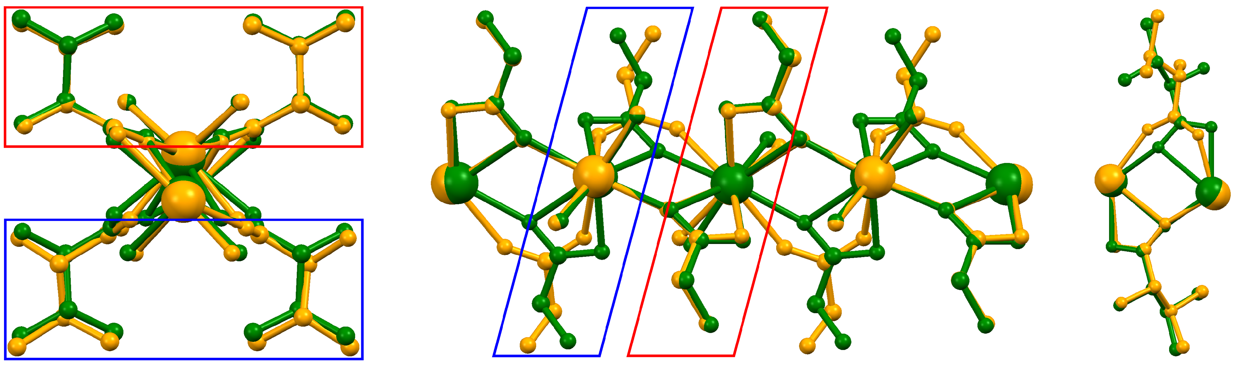
2.3. The Isomorphous Structures 3 and 4

The two compounds obtained from racemic valine with CaCl2 (3) or CaBr2 (4) are isomorphous and crystallise in the orthorhombic spacegroup Pcca. Their asymmetric unit comprises only half the size of that in the aforementioned structures: Ca2+ is situated on Wyckoff position 4c (twofold axis), and one simultaneously bridging and chelating valine molecule, one aqua ligand, and one uncoordinated chloride are in general position (Figure 6).
Figure 6. Left: Asymmetric units of 3 and 4 as displacement ellipsoid plots (80% probability) and adjacent atoms as stick model. Right: Packing of a cationic chain exemplary shown for 3 (hydrogen atoms omitted for clarity).
Crystals 05 00261 g006
Thus, the metal cation is in eightfold coordination geometry, enclosed by two chelating valine molecule of one enantiomer (four oxygen atoms), two water molecules, and two bridging valine molecules from the adjacent chain fragments (two oxygen atoms of the opposite enantiomer).
Ca–O distances are compiled in Table 2 and differ slightly between the two structures: While only one distance of the chelating carboxylate group is elongated in 4, the water coordination actually gets shorter whereas the chain-separating Ca–O bridge gets longer. This is reflected in the Ca· · · Ca distances within the chain direction c (3.9927(12) Å in 3 vs 4.0043(13) Å in 4).
Although 3 and 4 are racemic and only show µη2:η1 configured carboxylate bridging, the chain structure has high similarity to 2, which is reflected in similar c axes. An overlay of the chains (Figure 7) shows that the µη2:η1 coordinating valine in 2 coincides with the L-valine positions in 3 and 4, whereas the non-chelating µη:η carboxylate of 2 comprises almost the same space as the D-valine molecules in 3 and 4.
Figure 7. Overlay of 2 (yellow) and 4 (green). View in chain direction (left) and perpendicular (middle) with highlighted overlap of same (red) and opposite (blue) chirality. Right: Closeup of one chain fragment.
Crystals 05 00261 g007
Through the adoption of a different coordination mode both structure types allow for almost the same intermolecular interactions: The amino group engages in three hydrogen bonds towards neighbouring halide ions, whereas the water ligand forms one hydrogen bond towards a carboxylate group of the next chain fragment and a second towards a halide. Details about hydrogen bond geometry are provided in Table 5.
Table 5. Hydrogen bond geometry in 3 and 4.
3D· · · A (Å)D–H (Å)H· · · A (Å)DH· · · A (°)4D· · · A (Å)D–H (Å)H· · · A (Å)DH· · · A (°)
N1–H2· · · Cl1 a3.288(3)0.90(2)2.41(2)163(2)N1–H2· · · Br1 a3.405(5)0.97(3)2.51(3)154(3)
N1–H3· · · Cl1 b3.185(2)0.92(2)2.27(2)178(2)N1–H3· · · Br1 b3.312(4)0.96(3)2.36(3)173(3)
N1–H1· · · Cl1 c3.166(2)0.90(2)2.33(2)154(2)N1–H1· · · Br1 c3.296(4)0.96(4)2.42(4)151(4)
O3–H12· · · O2 d2.775(2)0.83(2)2.00(2)157(2)O3–H12· · · O2 d2.776(6)0.79(4)2.02(4)161(5)
O3–H13· · · Cl1 e3.130(2)0.84(2)2.32(2)162(2)O3–H13· · · Br1 e3.257(4)0.79(4)2.53(4)152(4)
Symmetry operations: a x, y, 1 + z; b x, −y, 0.5 + z; c 0.5 − x, y, 0.5 + z; dx, y, 1.5 − z; ex, y, 0.5 − z.
The intermolecular hydrogen bonds link neighbouring chains in a-direction to form a two dimensional layer in the ac-plane. In contrast to the staggered arrangement in the C centered cell of 2, successive layers in 3 and 4 are eclipsed. The cube-shaped motif of four amino groups and four chlorides between two chains remains. Though, altering the orientation of two amino groups because of the different chirality (and hence, different coordination) of every second valine ligand, switches two hydrogen bond vertices to open the cube-motif and connect it with the square base of the next cube (Figure 8).
Figure 8. Left to right: Hydrogen bonds emerging from the asymmetric unit in 3; the situation for 4 is analogous. View in c direction illustrating the resulting two dimensional network. Opened cube motif connecting two chains. Schematic drawing of cube and opened cube motifs.
Crystals 05 00261 g008

3. Conclusions

CaCl2(L-valine)2(H2O)2 is an instructive example for a dimorphic compound with an easily available main polymorph and a second crystal form discovered by serendipity. We have not been able to reproduce this second phase, despite considerable efforts including alteration of the crystallisation temperature, deliberate contamination with trace amounts of related compounds, and variation of the pH-value. The relationship between the alternative forms is surprising, with higher space filling for the disordered phase.
Both polymorphs and also the other three solids obtained from calcium chloride or bromide and from L- or DL-valine share common features: In all structures, the carboxylato groups act as bridges between neighbouring Ca2+. The cations are exclusively oxygen coordinated, and the halides act as counteranions for the resulting cationic scaffolds. In addition to electrostatic interactions, classical hydrogen bonds link the ionic residues. Each individual structure is unexceptional, but their relationship can teach a lesson with respect to isomorphous substitution: Retention of a structure type among these halides may be as unpredictable as their polymorphism.

4. Experimental Section

4.1. Instrumentation

Powder diffraction experiments were performed at room temperature on flat samples with a Stoe&Cie (Darmstadt, Germany) STADI P diffractometer equipped with an imageplate detector with constant ω-angle of 55° using germanium-monochromated Cu-Kα1 radiation (λ = 1.54051 Å). Single crystal intensity data were collected with a Bruker (Karlsruhe, Germany) D8 goniometer with APEX CCD area detector and an Incoatec microsource (Mo-Kα radiation, λ = 0.71073 Å, multilayer optics) at 100 K (Oxford Cryostream 700 instrument, Oxford, UK).

4.2. Syntheses

All products were obtained quantitatively as crystalline solids by dissolving the reactands in water in the stoichiometry of the target products. Subsequently, the solvent was evaporated under controlled conditions [2] at 50 °C. Variations in stoichiometry only altered the yield but not the composition of the product. 1a was obtained as main phase from 44 mg CaCl2 (0.4 mmol) and 92 mg L-valine (0.8 mmol) in 1 mL H2O in form of colorless elongated plates after two days. A needle shaped single crystal of 1b could be isolated from the same reaction. However, this polymorph could not be reproduced and has only been present as minority in the original sample. Seeding with 1b could therefore not be carried out as only one individual crystal of 1b could be isolated. However, several crystallisation experiments were done, including crystallisation at higher and lower temperature, intentional contamination with small amounts of other calcium salts/metal chlorides/amino acids, and variation of the pH-value. 2 was obtained from 87 mg CaBr2· xH2O (0.4 mmol) and 92 mg L-valine (0.8 mmol) under the same conditions as needle shaped colorless crystals. Using a racemic mixture of both valine enantiomers yielded the products 3 (with CaCl2) and 4 (with CaBr2). X-ray powder diffractograms are shown as Figure 9.
Figure 9. Simulated and experimental X-ray powder diffractograms of all products. Experimental data collected at room temperature. The asterisk indicates a reflection from unreacted valine.
Crystals 05 00261 g009

4.3. Data Collection and Refinement

Data were integrated with SAINT-Plus [15] and corrected for absorption with SADABS [15]. The structures were solved by the Patterson method and refined by full matrix least squares procedures on F2 as implemented in SHELXL-13 [16]. Non hydrogen atoms were refined with anisotropic displacement parameters. Hydrogen atoms connected to carbon were set to idealized positions and treated as riding with Uiso(H) = 1.2Ueq(C) for CHR3 and Uiso(H) = 1.5Ueq(C) for methyl groups. Hydrogen atoms attached to hetero atoms were found in difference Fourier maps. In each structure O-H and N-H distances were each restrained to similarity (SADI, σ = 0.02 Å). Uiso of the hydrogen atoms were constrained to multiple values of the Ueq of their parent atoms. Additionally, the H separation of the water molecules in 2 were also restrained to similarity (SADI, σ = 0.02 Å). In structure 1b two alternative positions were found for Cl1. Both could be assigned independent anisotropic displacement parameters; the sum of their refined occupancies (0.517(8) for Cl1A and 0.483(8) for Cl1B) was constrained to unity .

Acknowledgments

The authors thank Evonik Industries for providing amino acids and Stefanie Langenstück for her extensive crystallisation attempts for reproducing 1b.

Author Contributions

Experimental work, crystallographic studies and interpretation were carried out by Kevin Lamberts. Ulli Englert is the leading scientist of the group.

Conflicts of Interest

The authors declare no conflict of interest.

References and Notes

  1. Lamberts, K.; Englert, U. Structures from MnX2 and proline: Isomorphous racemic compounds and a series of chiral non-isomorphous chain polymers. Acta Crystallogr. 2012, 68, 610–618. [Google Scholar] [CrossRef] [PubMed]
  2. Lamberts, K.; Möller, A.; Englert, U. Enantiopure and racemic alanine as bridging ligands in Ca and Mn chain polymers. Acta Crystallogr. 2014, 70, 989–998. [Google Scholar] [CrossRef] [PubMed]
  3. Lamberts, K.; Porsche, S.; Hentschel, B.; Kuhlen, T.; Englert, U. An unusual linker and an unexpected node: CaCl2 dumbbells linked by proline to from square lattice networks. CrystEngComm 2014, 16, 3305–3311. [Google Scholar] [CrossRef]
  4. Lamberts, K.; Şerb, M.-D.; Englert, U. One- and two-dimensional polymers from proline and calcium bromide. Acta Crystallogr. 2015, C71, 311–317. [Google Scholar]
  5. Fleck, M.; Petrosyan, A.M. Salts of Amino Acids, Crystallization, Structure and Properties; Springer: Berlin, Germany, 2014. [Google Scholar]
  6. Ye, B.-H.; Tong, M.-L.; Chen, X.-M. Metal-organic molecular architectures with 2,2’-bipyridyl-like and carboxylate ligands. Coord. Chem. Rev. 2005, 249, 545–565. [Google Scholar] [CrossRef]
  7. Einspahr, H.; Bugg, C. E. The Geometry of Calcium-Carboxylate Interactions in Crystalline Complexes. Acta Crystallogr. 1981, 37, 1044–1052. [Google Scholar] [CrossRef]
  8. Kaufman Katz, A.; Glusker, J.P.; Beebe, S.A.; Bock, C.W. Calcium Ion Coordination: A Comparison with That of Beryllium, Magnesium, and Zinc. J. Am. Chem. Soc. 1996, 118, 5752–5763. [Google Scholar] [CrossRef]
  9. Burgess, K.M.N.; Xu, Y.; Leclerc, M.C.; Bryce, D.L. Alkaline-Earth Metal Carboxylates Characterized by 43Ca and 87Sr Solid-State NMR: Impact of Metal-Amine Bonding. Inorg. Chem. 2014, 53, 552–561. [Google Scholar] [CrossRef] [PubMed]
  10. Glowiak, T.; Ciunik, Z. The crystal structure of bis(DL-valine) calcium chloride dihydrate and bis(DL-2-aminobutyric acid) calcium chloride dihydrate. Bull. Acad. Pol. Sci. Ser. Sci. Chim. 1978, 26, 43–51. [Google Scholar]
  11. Batsanov, A.S.; Howard, J.A.K.; Wu, N.; Yang, Z.; Marder, T.B. An irreversible phase transition in 1-n-butylindeno[2,1-c]pyran-3,9-dione. Acta Crystallogr. 2012, 68, o413–o416. [Google Scholar] [CrossRef] [PubMed]
  12. Fábry, J.; Fridrichová, M.; Dušek, M.; Fejfarová, K.; Krupková, R. Two polymorphs of bis(2-carbamoylguanidinium) fluorophosphonate dihydrate. Acta Crystallogr. 2012, 68, o71–o75. [Google Scholar] [CrossRef] [PubMed]
  13. CCDC 1061342–1061346 Contain the Supplementary Crystallographic Data for This Paper. These Data can be Obtained Free of Charge via http://www.ccdc.cam.ac.uk/conts/retrieving.html (or from the CCDC, 12 Union Road, Cambridge CB2 1EZ, UK; Fax: +44-1223-336-033; E-Mail: [email protected]).
  14. Parsons, S.; Flack, H. Precise absolute-structure determination in light-atom crystals. Acta Crystallogr. 2004, 60. [Google Scholar] [CrossRef]
  15. SAINT-Plus and SADABS; Bruker AXS Inc.: Madison, WI, USA, 2008.
  16. Sheldrick, G.M. Crystal structure refinement with SHELXL. Acta Crystallogr. 2015, 71, 3–8. [Google Scholar]

