Article

Structural Insights into the N-Terminus and a Flexible P-x-P Loop near the Active Site of a Class III Aminotransferase


1
Research Unit for Low-Temperature Convergence Technology, Korea Polar Research Institute, Incheon 21990, Republic of Korea
2
Department of Polar Sciences, University of Science and Technology, Incheon 21990, Republic of Korea
*
Authors to whom correspondence should be addressed.
Crystals 2026, 16(9), 576; https://doi.org/10.3390/cryst16090576
Submission received: 10 August 2026 / Revised: 28 August 2026 / Accepted: 1 September 2026 / Published: 3 September 2026
(This article belongs to the Section Biomolecular Crystals)

Abstract

Class III aminotransferases represent a structurally and functionally unique subgroup. However, the contribution of specific loop elements to their active-site architecture and cofactor-dependent structural transitions remain underexplored. We elucidated the structural framework underlying the catalytic function of class III fold Pyridoxal 5′-phosphate-dependent aminotransferase from the Antarctic bacterium Hymenobacter sp. PAMC 26554 (HyAT), and report its crystal structure at 2.31 Å resolution. The structure revealed a canonical class III fold organized as a functional homotetramer. Structural analysis identified a proline-containing motif (P-x-P) within the α10-α11 loop, which induces intrinsic disorder at the active-site entrance in the apo-form and revealed that a cooperative disorder-to-order transition is requisite for active-site assembly upon cofactor binding. We propose that this flexible loop region may be involved in modulating substrate access. Notably, this proline motif was conserved in homologs from Hyperthermophiles, despite the cold-adapted nature of HyAT. This convergence implies a common evolutionary strategy where the geometric constraints of proline are exploited to decouple local active-site dynamics from global scaffold stability, thereby addressing the stability–activity trade-off across diverse thermal environments. Our findings provide new molecular insights into the structural dynamics of class III aminotransferases and highlight evolutionary strategies for tuning enzyme flexibility in extreme environments.

1. Introduction

Pyridoxal 5′-phosphate (PLP)-dependent aminotransferases constitute a ubiquitous and functionally diverse enzyme family that catalyzes reversible amino group transfer reactions between amino acids and keto acids. These enzymes play central roles in cellular nitrogen metabolism, amino acid biosynthesis, and degradation pathways across all domains of life. Beyond their physiological importance, PLP-dependent aminotransferases have attracted considerable interest as versatile biocatalysts for the asymmetric synthesis of chiral amines, pharmaceutical intermediates, and fine chemicals, owing to their high stereoselectivity and broad substrate tolerance [1,2,3].
Although they have not yet been assigned a formal EC number due to insufficient experimental data, aminotransferases (EC.2.6.1) are classified into several distinct classes (I–V) based on sequence similarity [4], and enzymes in each class possess a distinct structural fold and catalytic features. Among them, class III aminotransferases represent a structurally and functionally unique subgroup [5,6], possibly due to the fact that class III aminotransferases independently diverged from one of the PLP-dependent enzymes (PLP-fold type IV) in their evolution, whereas the other classes I, II, and IV evolved from PLP-fold type I enzymes [7,8]. They are characterized by a compact fold composed of a central β-sheet flanked by α-helices and typically adopt a dimeric or tetrameric organization as a functional unit, with the active sites formed at the subunit interface [9]. Members of this class include enzymes involved in specialized metabolic pathways, such as omega-transaminases [10] and taurine–pyruvate aminotransferases [11], yet they remain less extensively characterized than the well-studied classes I, II, and IV aminotransferases. In particular, the structural determinants governing substrate recognition, cofactor-induced conformational changes, and active-site organization in class III aminotransferases can significantly contribute to a well-defined classification system to elucidate the evolution of the aminotransferase family.
PLP-dependent catalysis generally relies on dynamic conformational rearrangements that accompany cofactor binding and progression through catalytic intermediates such as pyridoxamine 5′-phosphate (PMP). Previous structural studies have demonstrated that flexible loop regions proximal to the active site play critical roles in regulating cofactor accommodation, substrate access, and catalytic efficiency, acting as “gating” elements [12]. However, for class III aminotransferases, the contribution of specific loop elements to active-site architecture and cofactor-dependent structural transitions has remained largely unexplored. Specifically, the conformational dynamics of loop regions connecting late α-helical segments (e.g., helices α10 and α11) and their potential interactions with other structural motifs have not been systematically analyzed.
In this study, we focused on a previously uncharacterized class III fold PLP-dependent aminotransferase from the Antarctic bacterium Hymenobacter sp. PAMC 26554 (HyAT). By determining and analyzing its crystal structure, we aim to elucidate the structural framework underlying its catalytic function. Special attention has been given to the α10-α11 loop region located adjacent to the active-site cavity. Unlike the rigid loops observed in homologs, the HyAT loop exhibits significant conformational plasticity, potentially linked to a unique sequence motif. Through comparative structural analysis with homologs, we identified a unique P-x-P-containing motif (P-x-P) within the α10–α11 loop that distinguishes HyAT from mesophilic counterparts. The intrinsic disorder of this loop in the apo-state may provide conformational flexibility that could influence ligand access to the active site. Furthermore, we revealed that the ordering of this loop is structurally associated with the stabilization of the N-terminal region, suggesting a coordinated disorder-to-order transition mechanism for active-site assembly.

2. Materials and Methods

2.1. Protein Expression and Purification

A hypothetical protein sequence (NCBI Accession No. AMR27191.1) was searched based on sequence similarity using class-III aminotransferase as a query. It was discovered from the complete genome sequence of Hymenobacter psoromatis PAMC 26554, which was isolated from Antarctic lichens [13] and the domain sequence was annotated as a class-III aminotransferase using InterProScan [14]. Selected coding gene from Hymenobacter sp. PAMC 26554, hereafter HyAT, was used for cloning, expression, and purification. The coding sequence was codon-optimized, synthesized, and cloned into the pET28a expression vector using NdeI and XhoI restriction sites (Bioneer, Daejeon, South Korea). The recombinant plasmid was transformed into Escherichia coli BL21 (DE3) cells for protein overexpression. Transformed cells were cultured in 4 L of Luria–Bertani (LB) medium supplemented with 50 μg/mL kanamycin at 37 °C with shaking at 120 rpm. When the optical density at 600 nm (OD600) of the cell culture reached 0.6, 0.5 mM isopropyl β-D-1-thiogalactopyranoside (IPTG) was added for inducing protein overexpression. Then, the culture was further incubated at 25 °C with shaking at 120 rpm overnight. The cells were harvested by centrifugation at 5000 rpm for 20 min at 4 °C. The pellet was resuspended in cell lysis buffer (20 mM Tris-HCl (pH 8.0), 200 mM NaCl, and 5 mM imidazole) and disrupted by ultrasonication (Vibra-Cell, Sonics & Materials, Inc., Newtown, CT, USA) at 4 °C. The soluble supernatant fraction was collected via centrifugation at 15,000 rpm for 40 min at 4 °C. The supernatant containing the HyAT was applied onto a nickel–nitrilotriacetic acid (Ni-NTA) agarose-packed column which has been equilibrated with cell lysis buffer. After washing with buffer containing 30 mM imidazole (20 mM Tris-HCl [pH 8.0], 200 mM NaCl, and 30 mM imidazole) to remove non-specifically bound proteins, our target protein was eluted using buffer containing 300 mM imidazole (20 mM Tris-HCl [pH 8.0], 200 mM NaCl, and 300 mM imidazole). To remove the N-terminal poly-histidine tag, the eluted fraction was incubated with thrombin from bovine plasma overnight at 4 °C. After successful removal of the His-tag, further purification was conducted by size-exclusion chromatography (SEC) using FPLC system (ÄKTA avant; Cytiva, Marlborough, MA, USA) with a HiLoad Superdex 200 pg column (Cytiva). The column was equilibrated with buffer consisting of 20 mM Tris-HCl (pH 8.0) and 200 mM NaCl before running. The purified protein was verified with SDS-PAGE and concentrated to 50 mg/mL using an Amicon Ultra Centrifugal Filter (10 kDa MWCO; Merck, Darmstadt, Germany). The purified protein was freshly employed for crystallization and stored at −80 °C until next use.

2.2. Crystallization and Data Collection

As an initial crystallization screening, the purified HyAT protein was crystallized in 96-well sitting-drop plates at 23 °C. In detail, 400 nL of purified protein (15 and 30 mg/mL, respectively) was mixed with 400 nL of crystallization screening solution using a Mosquito crystallization robot (SPT Labtech, Melbourn, UK). For initial screening, commercial crystallization screening kits (MCSG I-IV; Anatrace, Maumee, OH, USA, PGA Screen; Molecular Dimensions, Rotherham, UK) and a customized suite (SGC; Molecular Dimensions) were used. The optimal crystals were observed after 1 day in a condition of 0.1 M Tris-HCl (pH 8.5), 0.2 M magnesium chloride, and 15% of PEG 8000.
The crystal was mounted at the macromolecular crystallography beamline (BL-5C) of the Pohang Light Source II (PLS-II) (PAL, Pohang, Republic of Korea) for the X-ray diffraction experiment. Before mounting on the goniometer, a single crystal was briefly soaked to cryoprotectant, reservoir solution supplemented with 20% (v/v) final glycerol concentration. Diffraction images were collected at every 1° oscillation per frame at an energy of 12.658 keV (0.97949 Å) using an Eiger 9M detector (Dectris, Baden, Switzerland). The diffraction data were indexed, integrated, and scaled using the HKL2000 software package [15]. The crystals belonged to the space group P21 with unit cell parameter a = 100.81 Å, b = 152.26 Å, c = 117.37 Å, and α, γ = 90°, β = 104.9°. The processed file was converted to MTZ format using Scalepack2mtz in the CCP4i suite [16].

2.3. Structure Determination and Refinement

The initial model was built using the structure of taurine-2-oxo-glutarate aminotransferase (PDB code, 6JIX) as a template in the molecular replacement method [17]. While running MOLREP in CCP4i, eight molecules were built in crystallographic data based on Matthews Coefficient value (VM, 2.42) and solvent content (Vs, 49.24%) in the asymmetric unit [18]. Manual model building and refinement were conducted in Coot [19], and iterative auto-refinement was performed using phenix.refinement in the Phenix suite [20]. The final structure was validated using MolProbity [21] and atomic coordinates and structural factors have been deposited in Protein Data Bank (PDB) under the accession code of 22PO. All structural figures in this article were generated using PyMoL v.2.3 [22]. X-ray diffraction data collection and refinement statistics are summarized in Table 1.

2.4. Analytical Ultracentrifugation

The oligomerization state of HyAT was analyzed by ultracentrifuge (Proteome Lab XL-A, Beckman Coulter, Inc., Brea, CA, USA) using an AN-60 Ti rotor (Beckman Coulter, Brea, CA, USA). HyAT was prepared in 20 mM Tris-HCl (pH 8.0) and 200 mM NaCl buffers. The partial specific volume of the sample and density and viscosity of buffer were evaluated by SEDNTEPR v.3.0.4, respectively (0.7356 mL/g, 1.00709 g/mL, and 0.01026 Poise at 20 °C) [23]. The sample was accelerated and initialized to 42,000 rpm for 10 min at 20 °C. The sedimentation profile of HyAT was subsequently monitored at 280 nm at every 5 min and the total 40 absorbance scan data were analyzed using the SEDFIT v.18.1 [24]. Data were visualized using GraphPad Prism v.5.02 (GraphPad Software, San Diego, CA, USA). The protein–protein interfaces within the tetrameric assembly observed in the crystal structure of HyAT were analyzed using PDBePISA [25].

3. Results

3.1. Overall Structure

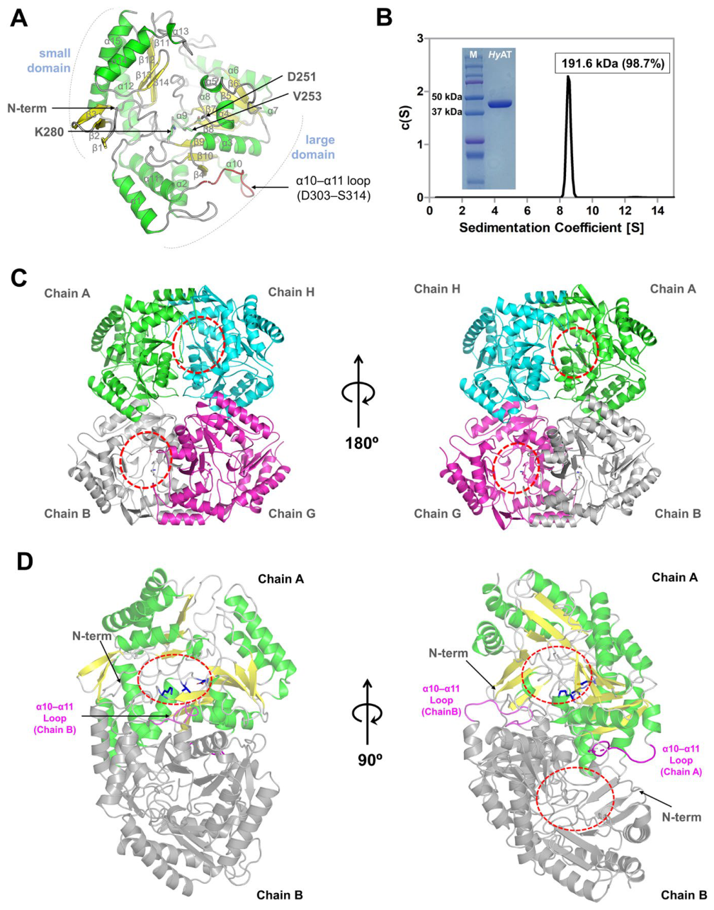
The crystal structure of the PLP-dependent aminotransferase from Hymenobacter sp. PAMC 26554 was determined at a high resolution of 2.31 Å. HyAT crystallizes in the P21 space group, and the asymmetric unit contains eight monomers, corresponding to two tetramers. Each monomer adopts the canonical class III aminotransferase fold, characterized by a central seven-stranded β-sheet flanked by α-helices (Figure 1A).
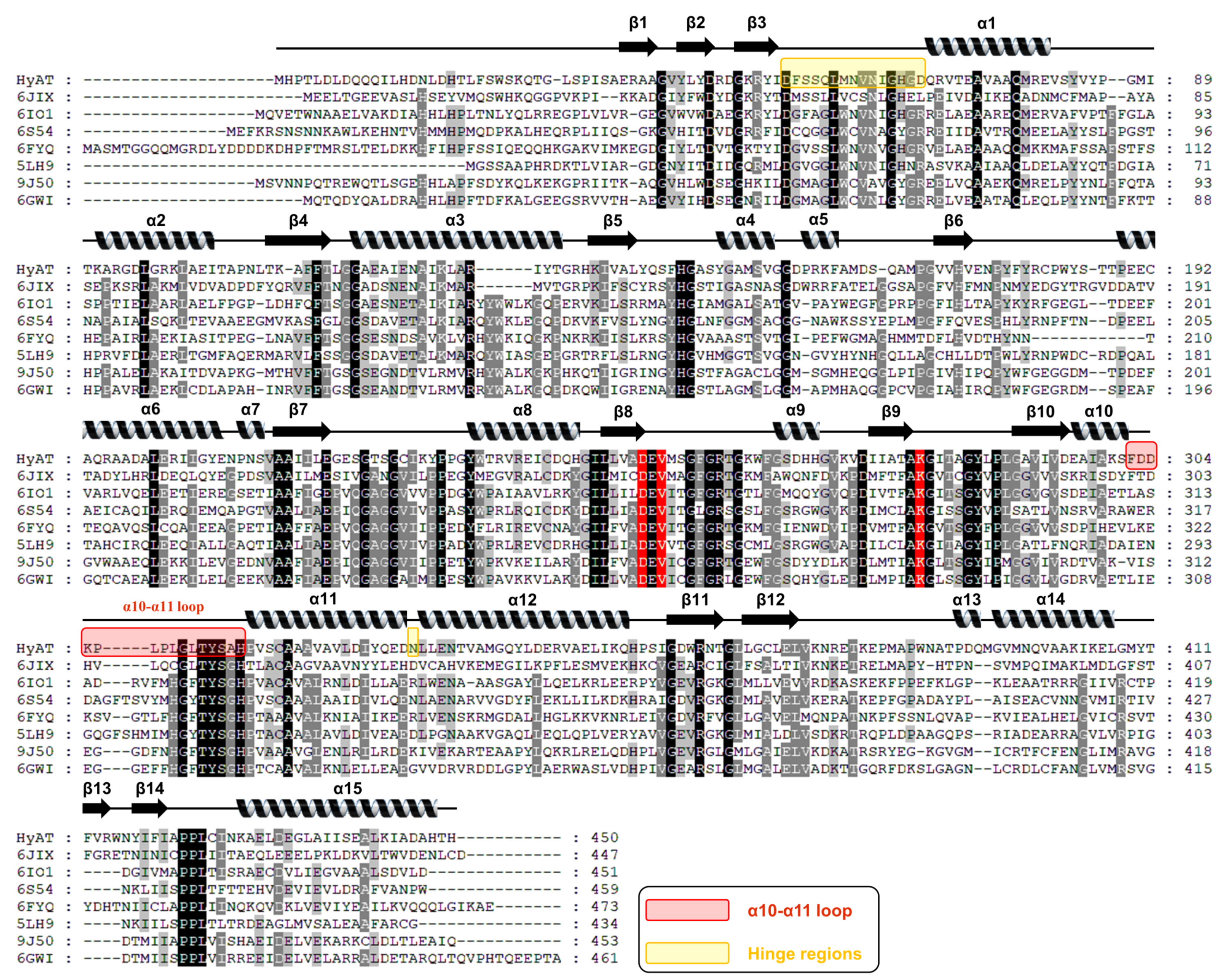
Specifically, the monomeric structure is divided into two distinct domains: a large and a small domain. The large domain forms the core of the enzyme and is composed of a central β-sheet formed by strands β4-β10, which is surrounded by solvent-accessible α-helices. The small domain is formed by discontinuous segments from both the N- and C-termini; it comprises three β-strands from the N-terminus and four β-strands from the C-terminus, along with helices α12, α14, and α15. The N- and C-terminal domains are connected by two structural hinge regions. The first hinge region is located between β3 and α1 (D53-D67) in the N-terminal region. The second hinge is observed between α11 and α12, where an asparagine residue induces a local helical kink.
Consistent with the conserved architecture of class III aminotransferases, the catalytic cavity of HyAT is formed by the central β-sheet of one monomer and the α-helices flanking the large domain (α1, α2, and α11) of the neighboring subunit (Table 2). Within this inter-subunit cleft, key catalytic residues, including D251 and the conserved active-site residue, K280, are spatially arranged to form the cofactor-binding pocket (Figure 1A,C). This conserved inter-subunit architecture supports the obligate dimeric nature of HyAT for enzymatic catalysis, as formation of the functional active site requires the precise alignment of structural elements from both monomers to support catalysis.

3.2. Oligomeric State of HyAT

Members of the class III aminotransferase family typically function as homodimers or homotetramers (Table 2). Although the crystallographic asymmetric unit of HyAT contains eight protomers, implying a complex assembly, it is essential to distinguish the biologically relevant oligomer from crystal packing artifacts. To determine the functional assembly of HyAT in solution, we performed sedimentation velocity analytical ultracentrifugation (AUC). The AUC analysis confirmed that HyAT exists as a stable tetramer in solution (Figure 1B). Interface analysis using PDBePISA revealed that the primary assembly is a homodimer (Chain A–B, C–D, E–F, G–H) as an enzymatic functional unit, and the average of interface area was estimated as 2709.9 Å 2 (p = 0.004). The dimerization interface was stabilized by extensive hydrophobic and electrostatic interactions between the helices and β-strands near the catalytic core, placing the active-site cavities at the subunit interface (Figure 1C,D). Subsequently, two functional homodimers (A–B and G–H) associate through a smaller secondary interface, with an average buried surface area of 1133.2 Å 2, to form a tetrameric assembly in a back-to-back configuration (Figure 1C). In this arrangement, the active-site cavities of both primary dimers remain exposed to the solvent-accessible outer surface of the tetramer. Structural superposition of the HyAT monomer with the corresponding monomer of the homologous enzyme (PDB: 5LH9) yielded an r.m.s.d. of 1.317 Å, indicating a highly similar monomeric fold. Consistent with this similarity, the tetrameric assemblies of HyAT and 5LH9 also exhibit closely comparable quaternary arrangements, in which two functional homodimers associate in a back-to-back configuration.

3.3. Structural Plasticity of the α10-α11 Loop and N-Terminus in Apo-HyAT

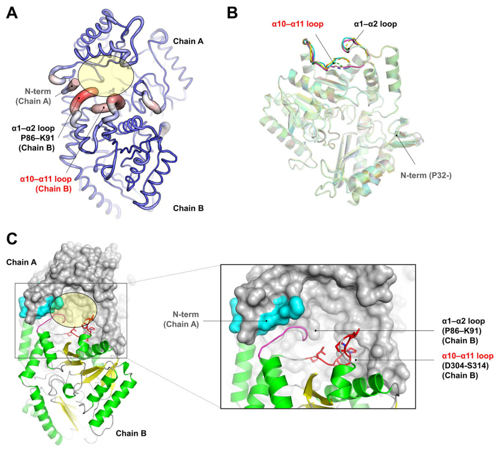
The high-resolution electron density maps enabled a model of a majority of the polypeptide chain to be built. However, despite the overall high quality of the data (Table 1), two specific regions lacked interpretable electron density. First, the loop regions connecting helices α1 and α2 (P86–K91), and α10 and α11 (D303–S314) exhibited consistently weak or discontinuous density, preventing the modeling of a rigid conformation (Figure 2A). Second, the apo-HyAT structure was characterized by a lack of interpretable electron density for the N-terminal region. While the core domain was well-defined, the N-terminal segment (approximately residues 1–31) was completely invisible in the electron density map. The absence of density for the preceding N-terminal residues suggests that this segment is highly disordered and does not adopt a stable structure in the apo-form.
To investigate the extent of this flexibility, we superimposed the eight independent monomers contained within the asymmetric unit (Figure 2B). While the overall core structure was highly conserved, aligning within a root mean square deviation (RMSD) of 0.30 Å, the backbone trajectories of the α10–α11 loop displayed a high degree of structural divergence. Consistent with this flexibility, the N-terminal region exhibited uniform disorder across the asymmetric unit. In a majority of the eight chains, the polypeptide chain could only be modeled starting from residue P32, leaving residues 1–31 structurally unresolved. This structural heterogeneity reveals that the flexibility indicated by the electron density maps is not an isolated artifact restricted to a single chain, but is a consistent feature observed across multiple subunits. Consequently, this region exists as a structural ensemble rather than a single unified conformation, reflecting the intrinsic dynamics of the enzyme in its apo-state.
Topological analysis of the active site revealed that this flexible loop is positioned adjacent to the catalytic cleft. The α10–α11 loop, including the α1–α2 loop, constitutes one of the sidewalls defining the entrance channel to the active site (Figure 2C). The intrinsic flexibility of this channel wall in the apo-state likely plays a critical role in substrate recognition.
To validate the role of this flexibility in the cofactor binding mechanism, we performed a structural comparison between apo-HyAT and the class III aminotransferase from Vibrio fluvialis (VfAT) (Figure S1). VfAT serves as an ideal structural surrogate because it is one of the few homologs for which high-resolution structures of both the apo (PDB: 5ZTX) and PMP-bound (PDB: 4E3Q) states have been determined, thereby allowing direct visualization of cofactor-induced conformational changes. We calculated and compared the normalized B-factors of apo-HyAT with those of the VfAT structures using BANΔIT (2020) software [30,31,32]. Although the overall structures of HyAT and VfAT shared high similarity (RMSD of 1.18Å), the B-factor analysis provided critical insights into the dynamics of the α10–α11 loop region. As expected, the apo-VfAT structure displayed elevated B-factors in this region, similar to apo-HyAT, indicating that the loop possesses inherent flexibility in the absence of a ligand. Contrarily, in the PMP-bound VfAT, the B-factors in the corresponding loop were reduced. This comparison demonstrates that while the apo-state is flexible, cofactor binding acts as a localized “switch” that rigidifies the loop. This observation suggests our hypothesis that the α10–α11 loop of HyAT may adopt a more ordered conformation depending on ligand binding state.

3.4. Sequence-Encoded Flexibility and Associated N-Terminal Dynamics

To elucidate the molecular basis of the loop’s intrinsic flexibility observed in the α10–α11 loop, we analyzed its sequence composition. Structural homology search was performed using the Dali server [33] and comparative analysis with homologous enzymes revealed that HyAT contains a unique P-x-P-containing motif (303-DDKPLPLGLTYS-314) embedded within the α10–α11 loop (Figure 3). The presence of two proline residues (P306, P308), known as potent helix breakers, likely disrupts the formation of a rigid secondary structure [34]. This unique sequence feature may underlie the elevated flexibility and partial disorder of the α1–α2 and α10–α11 loops observed in the apo-HyAT structure. Consistent with this interpretation, normalized B-factor analysis showed that these loop regions exhibit substantially higher flexibility in HyAT than in the corresponding regions of structurally homologous enzymes (Figure S2), in which the equivalent loops generally display lower normalized B-factors and more ordered conformations.
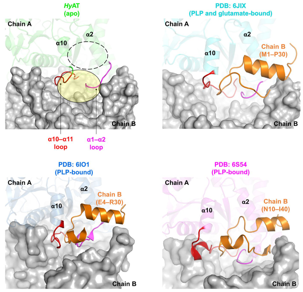
Notably, this loop flexibility appears to be structurally associated with the N-terminal region. To determine whether this feature is unique to HyAT, we compared our structure with its closest structural homologs identified by a DALI search against the PDB (Table 2). Among the top-ranked hits, we selected three representative structures—PDB 6JIX, 6IO1, and 6S54—that superimpose onto the HyAT monomer with Cα RMSD values of 1.000, 1.042, and 1.218 Å, respectively, and retain a fully ordered N-terminal segment, thereby permitting a direct comparison of this region. In our HyAT model, the N-terminal segment preceding residue S31 is invisible due to disorder, whereas all three homologs display a stable helix–turn–helix motif in this region that packs across the dimer interface and shields the α10–α11 and α1–α2 loops of the partner subunit (Figure 4). Superposition with these homologs revealed that the ordered N-terminus typically docks between the α1–α2 loop and the α10–α11 loop of the neighboring subunit. Superposition with these homologs revealed that the ordered N-terminus typically docks within an inter-subunit groove formed by the α1–α2 and α10–α11 loops of the neighboring subunit. In 6JIX, 6IO1, and 6S54, the N-terminal segment forms multiple polar and nonpolar contacts with residues in these loop regions, supporting stable inter-subunit docking (Figure S3). In contrast, the corresponding N-terminal region is not resolved in apo HyAT, in which the α1–α2 and α10–α11 loops exhibit comparatively high flexibility compared to other homologous structures. Thus, the well-defined N-terminal docking arrangement observed in these homologs is not apparent in HyAT. Without the rigid loop acting as a docking scaffold, the N-terminus fails to stabilize. This suggests an associated dynamic mechanism where the stabilization of the α10–α11 loop (potentially by substrate or cofactor binding) is a prerequisite for the ordering of the N-terminus, thereby completing the active site assembly.

4. Discussion

Our findings suggest that the local conformational variability of the active-site loop is associated with disorder in the N-terminus. By solving the high-resolution structure of HyAT, our comparative analysis revealed structural features that may contribute to this plasticity, indicating that the active-site loop and the adjacent N-terminal region adopt distinct conformations between apo (HyAT) and cofactor-bound (homologs) structures. These observations are consistent with conformational rearrangements associated with ligand binding. Furthermore, sequence comparison and structural modeling of HyAT-related proteins suggest that the local P-x-P motif occupies a corresponding position within this group. This motif may contribute to the local conformational flexibility of the loop. Notably, this observation is restricted to HyAT-related proteins, as the corresponding loop region is variable among the broader class III aminotransferase family.
The structural interaction observed between the disordered α10–α11 loop and the N-terminal region suggests a coordinated loop-mediated gating mechanism. We propose that in the apo-state, the P-x-P-containing loop adopts a flexible and open conformation near the active-site entrance. Such flexibility may be associated with accessibility to the active site for the PLP cofactor or substrates. Upon ligand binding, the loop likely undergoes a disorder-to-order transition to a rigid, closed state. This conformational freezing would, in turn, stabilize the N-terminal helix of the neighboring subunit, thereby locking the active site for catalysis. This mechanism ensures that the catalytic machinery is fully assembled only when the correct ligand is present, preventing non-productive side reactions. However, the further ligand-bound structural and biochemical studies will be required to define the functional role of this loop.
Although active-site gating is a common feature in enzyme catalysis, HyAT employs a different structural mechanism. In contrast to homologs that rely on rigid body movements of helices, HyAT utilizes intrinsic disorder mediated by the P-x-P motif to regulate substrate access. This indicates that while the catalytic core of class III aminotransferases is conserved, the surrounding loops, particularly the α10–α11 region, show structural variation. Consequently, the structural plasticity of this loop in HyAT exemplifies the contribution of intrinsically disordered regions to enzyme function. These findings suggest that local sequence variations, such as the insertion of proline residues, modulate conformational dynamics, providing a structural basis for the diversity observed in the Class III aminotransferase family.
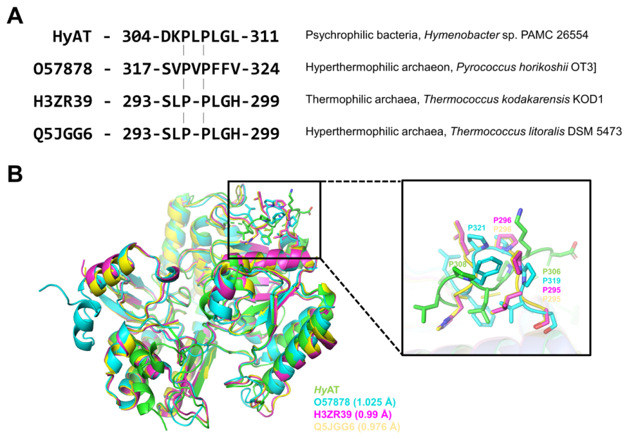
Comparative sequence analysis revealed that the distinct P-x-P-containing motif observed in HyAT was also conserved in homologous aminotransferases from hyperthermophiles, such as Pyrococcus horikoshii OT3 [35,36], Thermococcus kodakarensis KOD1 [37], and Thermococcus litoralis DSM 5473 [38] (Figure 5A). In addition, structural superposition of HyAT with homologous structures showed that the P-x-P-containing motif is conserved in the corresponding loop regions (Figure 5B). This observation is noteworthy because HyAT originates from Hymenobacter sp. PAMC 26554, a bacterium isolated from the Antarctic environment; whereas the homologs belong to organisms thriving in high-temperature niches. This observation suggests a unified evolutionary strategy to maintain function despite the opposing environmental constraints.
Generally, enzymes face a fundamental trade-off between stability and activity [39]. High thermal stability typically requires a rigid protein structure, but this rigidity often restricts the conformational flexibility required for catalysis. We propose that the conserved proline motif serves as a strategic solution to decouple local active site dynamics from the global stability of the protein scaffold. This strategy enables adaptation to extreme temperatures without compromising enzyme function.
In the case of hyperthermophiles, the protein core must be extremely rigid to prevent denaturation at high temperatures. However, such global rigidity can lead to a kinetically trapped active site that hinders the structural rearrangements necessary for substrate turnover. Here, the P-x-P-containing motif likely introduces essential local mobility into the otherwise rigid scaffold. By acting as a mechanical hinge or a structural disruptor that prevents the formation of an overly rigid helix, the motif ensures that the active site loop retains the specific flexibility required for catalysis. This overcomes the conflicting demands of structural rigidity and catalytic activity.
Conversely, for psychrotolerant enzymes such as HyAT, the challenge is to maintain activity at low temperatures where molecular motions are naturally dampened. Consistent with the observation that structural stability weaknesses are often enriched in cold-adapted enzymes to support catalysis [39], HyAT utilizes the same proline motif to locally enhance entropy at the active site. By acting as a helix breaker, the motif maintains the loop in an intrinsically disordered state. This local flexibility lowers the activation energy barrier for loop movement and facilitates substrate access even under energy-deprived cold conditions.
Collectively, these findings suggest a convergent evolutionary strategy. Rather than altering the global flexibility of the entire enzyme, both psychrophiles and hyperthermophiles exploit the unique geometric properties of proline residues to fine-tune the local dynamics of the active site. This local adaptation allows enzymes to preserve their catalytic competence across diverse environmental extremes. It ensures that the active site remains functionally dynamic regardless of whether the global scaffold has evolved for rigidity or stability.
In summary, the crystal structure of HyAT reveals a distinct mechanism of active-site organization in class III aminotransferases, governed by the intrinsic plasticity of the α10–α11 loop. Our structural analysis, supported by data from eight independent monomers, demonstrates that a unique P-x-P-containing motif is associated with local disorder in the apo-state. Notably, this loop flexibility is structurally associated with the N-terminal region, suggesting that a cooperative disorder-to-order transition is required to assemble the functional active site. Furthermore, the identification of this motif in both Antarctic and hyperthermophilic homologs provides an example of evolutionary convergence. It suggests that the unique geometric constraints of proline residues are exploited to fine-tune loop dynamics, thereby overcoming the stability–activity trade-off in extreme thermal environments. Collectively, these findings provide a structural blueprint for understanding how local sequence variations orchestrate the optimal balance between rigidity and flexibility essential for enzyme catalysis.

Supplementary Materials

The following supporting information can be downloaded at: https://www.mdpi.com/article/10.3390/cryst16090576/s1; Figure S1: Comparative analysis of normalized B-factor between apo and ligand-binding structures; Figure S2: Comparative analysis of normalized B-factor of α1–α2 and α10–α11 loop region of HyAT with its structural homologs using BANΔIT web-server; Figure S3: Comparative analysis of active-site formation loops and N-terminal region of partner subunit.

Author Contributions

Conceptualization, Supervision and Project administration: H.D. and J.H.L.; Investigation and Methodology: J.H. and H.D.; Data curation: J.H.; Formal analysis: J.H. and H.D.; Validation: J.H. and H.D.; Visualization: J.H. and H.D.; Writing—original draft: J.H. and H.D.; Writing—review and editing; H.D. and J.H.L. All authors have read and agreed to the published version of the manuscript.

Funding

This work was supported by a Korea Polar Research Institute grant funded by the Ministry of Oceans and Fisheries (KOPRI Grant numbers PE26150 and PE26220).

Data Availability Statement

The structural data set generated in this study is available from the RCSB PDB Protein Data Bank repository. The PDB accession number is 22PO for HyAT.

Acknowledgments

We would like to thank the staff at the X-ray core beamline (BL-5C) of the Pohang Light Source (PLS-II, Pohang, Republic of Korea) for their kind help with data collection. During the preparation of this manuscript, the authors used ChatGPT (GPT-5.6, OpenAI) to improve the clarity, grammar, and readability of the English text. The authors have reviewed and edited the output and take full responsibility for the content of this publication.

Conflicts of Interest

The authors declare no conflicts of interest.

Abbreviations

The following abbreviations are used in this manuscript:
PLPPyridoxal 5′-phosphate
HyATHymenobacter sp. PAMC 26554
P-x-PProline-containing motif
LBLuria–Bertani
OD600Optical density at 600 nm
Ni-NTANickel–nitrilotriacetic acid
SECSize-exclusion chromatography
PLS-IIPohang Light Source II
PDBProtein Data Bank
AUCAnalytical ultracentrifugation
RMSDRoot mean square deviation
VfATVibrio fluvialis

References

  1. Kelly, S.A.; Pohle, S.; Wharry, S.; Mix, S.; Allen, C.C.R.; Moody, T.S.; Gilmore, B.F. Application of ω-transaminases in the pharmaceutical industry. Chem. Rev. 2018, 118, 349–367. [Google Scholar] [CrossRef] [Scilit] [PubMed]
  2. Jia, D.X.; Peng, C.; Li, J.L.; Wang, F.; Liu, Z.Q.; Zheng, Y.G. Redesign of (R)-omega-transaminase and its application for synthesizing amino acids with bulky side chain. Appl. Biochem. Biotechnol. 2021, 193, 3624–3640. [Google Scholar] [CrossRef] [Scilit] [PubMed]
  3. Pandya, S.; Gupte, A. Transaminases for green chemistry: Recent progress and future prospects. Microbiol. Biotechnol. Lett. 2023, 51, 333–352. [Google Scholar] [CrossRef] [Scilit]
  4. Sigrist, C.J.A.; Cuche, B.A.; de Castro, E.; Coudert, E.; Redaschi, N.; Bridge, A. The PROSITE database for protein families, domains, and sites. Nucleic Acids Res. 2026, 54, D451–D458. [Google Scholar] [CrossRef] [Scilit] [PubMed]
  5. Mehta, P.K.; Hale, T.I.; Christen, P. Aminotransferases: Demonstration of homology and division into evolutionary subgroups. Eur. J. Biochem. 1993, 214, 549–561. [Google Scholar] [CrossRef] [Scilit] [PubMed]
  6. Schiroli, D.; Peracchi, A. A subfamily of PLP-dependent enzymes specialized in handling terminal amines. Biochim. Biophys. Acta 2015, 1854, 1200–1211. [Google Scholar] [CrossRef] [Scilit] [PubMed]
  7. Koper, K.; Han, S.W.; Kothadia, R.; Salamon, H.; Yoshikuni, Y.; Maeda, H.A. Multisubstrate specificity shaped the complex evolution of the aminotransferase family across the tree of life. Proc. Natl. Acad. Sci. USA 2024, 121, e2405524121. [Google Scholar] [CrossRef] [Scilit] [PubMed]
  8. Koper, K.; Han, S.W.; Pastor, D.C.; Yoshikuni, Y.; Maeda, H.A. Evolutionary origin and functional diversification of aminotransferases. J. Biol. Chem. 2022, 298, 102122. [Google Scholar] [CrossRef] [Scilit] [PubMed]
  9. Roura Padrosa, D.; Alaux, R.; Smith, P.; Dreveny, I.; López-Gallego, F.; Paradisi, F. Enhancing PLP-binding capacity of Class-III ω-transaminase by single residue substitution. Front. Bioeng. Biotechnol. 2019, 7, 282. [Google Scholar] [CrossRef] [Scilit] [PubMed]
  10. Koszelewski, D.; Tauber, K.; Faber, K.; Kroutil, W. ω-Transaminases for the synthesis of non-racemic α-chiral primary amines. Trends Biotechnol. 2010, 28, 324–332. [Google Scholar] [CrossRef] [Scilit] [PubMed]
  11. Li, M.; Wei, Y.; Yin, J.; Lin, L.; Zhou, Y.; Hua, G.; Cao, P.; Ang, E.L.; Zhao, H.; Yuchi, Z.; et al. Biochemical and structural investigation of taurine:2-oxoglutarate aminotransferase from Bifidobacterium kashiwanohense. Biochem. J. 2019, 476, 1605–1619. [Google Scholar] [CrossRef] [Scilit] [PubMed]
  12. Börner, T.; Rämisch, S.; Reddem, E.R.; Bartsch, S.; Vogel, A.; Thunnissen, A.M.W.H.; Adlercreutz, P.; Grey, C. Explaining operational instability of amine transaminases: Substrate-induced inactivation mechanism and influence of quaternary structure on enzyme–cofactor intermediate stability. ACS Catal. 2017, 7, 1259–1269. [Google Scholar] [CrossRef] [Scilit]
  13. Oh, T.J.; Han, S.R.; Ahn, D.H.; Park, H.; Kim, A.Y. Complete genome sequence of Hymenobacter sp. strain PAMC26554, an ionizing radiation-resistant bacterium isolated from an Antarctic lichen. J. Biotechnol. 2016, 227, 19–20. [Google Scholar] [CrossRef] [Scilit] [PubMed]
  14. Blum, M.; Andreeva, A.; Florentino, L.C.; Chuguransky, S.R.; Grego, T.; Hobbs, E.; Pinto, B.L.; Orr, A.; Paysan-Lafosse, T.; Ponamareva, I.; et al. InterPro: The protein sequence classification resource in 2025. Nucleic Acids Res. 2025, 53, D444–D456. [Google Scholar] [CrossRef] [Scilit] [PubMed]
  15. Otwinowski, Z.; Minor, W. Processing of X-ray diffraction data collected in oscillation mode. Methods Enzymol. 1997, 276, 307–326. [Google Scholar] [CrossRef] [Scilit] [PubMed]
  16. Winn, M.D.; Ballard, C.C.; Cowtan, K.D.; Dodson, E.J.; Emsley, P.; Evans, P.R.; Keegan, R.M.; Krissinel, E.B.; Leslie, A.G.W.; McCoy, A.; et al. Overview of the CCP4 suite and current developments. Acta Crystallogr. D Struct. Biol. 2011, 67, 235–242. [Google Scholar] [CrossRef] [Scilit] [PubMed]
  17. Evans, P.; McCoy, A. An introduction to molecular replacement. Acta Crystallogr. D Struct. Biol. 2008, 64, 1–10. [Google Scholar] [CrossRef] [Scilit] [PubMed]
  18. Kantardjieff, K.A.; Rupp, B. Matthews coefficient probabilities: Improved estimates for unit cell contents of proteins, DNA, and protein–nucleic acid complex crystals. Protein Sci. 2003, 12, 1865–1871. [Google Scholar] [CrossRef] [Scilit] [PubMed]
  19. Emsley, P.; Cowtan, K. Coot: Model-building tools for molecular graphics. Acta Crystallogr. D Struct. Biol. 2004, 60, 2126–2132. [Google Scholar] [CrossRef] [Scilit] [PubMed]
  20. Adams, P.D.; Afonine, P.V.; Bunkóczi, G.; Chen, V.B.; Davis, I.W.; Echols, N.; Headd, J.J.; Hung, L.W.; Kapral, G.J.; Grosse-Kunstleve, R.W.; et al. Phenix: A comprehensive Python-based system for macromolecular structure solution. Acta Crystallogr. D Struct. Biol. 2010, 66, 213–221. [Google Scholar] [CrossRef] [Scilit] [PubMed]
  21. Williams, C.J.; Headd, J.J.; Moriarty, N.W.; Prisant, M.G.; Videau, L.L.; Deis, L.N.; Verma, V.; Keedy, D.A.; Hintze, B.J.; Chen, V.B.; et al. MolProbity: More and better reference data for improved all-atom structure validation. Protein Sci. 2018, 27, 293–315. [Google Scholar] [CrossRef] [Scilit] [PubMed]
  22. Schrödinger LLC (New York, NY, USA). The PyMOL Molecular Graphics System, version 2.0; 2020. Available online: http://www.pymol.org/pymol (accessed on 21 January 2026).
  23. Philo, J.S. SEDNTERP: A calculation and database utility to aid interpretation of analytical ultracentrifugation and light scattering data. Eur. Biophys. J. 2023, 52, 233–266. [Google Scholar] [CrossRef] [Scilit] [PubMed]
  24. Schuck, P.; Perugini, M.A.; Gonzales, N.R.; Howlett, G.J.; Schubert, D. Size-distribution analysis of proteins by analytical ultracentrifugation: Strategies and application to model systems. Biophys. J. 2002, 82, 1096–1111. [Google Scholar] [CrossRef] [Scilit] [PubMed]
  25. Krissinel, E.; Henrick, K. Inference of macromolecular assemblies from crystalline state. J. Mol. Biol. 2007, 372, 774–797. [Google Scholar] [CrossRef] [Scilit] [PubMed]
  26. Kwon, S.; Lee, J.H.; Kim, C.M.; Jang, H.; Yun, H.; Jeon, J.-H.; So, I.; Park, H.H. Structural basis of substrate recognition by a novel thermostable (S)-enantioselective ω-transaminase from Thermomicrobium roseum. Sci. Rep. 2019, 9, 6958. [Google Scholar] [CrossRef] [Scilit] [PubMed]
  27. Guidi, B.; Planchestainer, M.; Contente, M.L.; Laurenzi, T.; Eberini, I.; Gourlay, L.J.; Romano, D.; Paradisi, F.; Molinari, F. Strategic single point mutation yields a solvent- and salt-stable transaminase from Virgibacillus sp. in soluble form. Sci. Rep. 2018, 8, 16441. [Google Scholar] [CrossRef] [Scilit] [PubMed]
  28. Das, P.; Noronha, S.; Bhaumik, P. Structural insights and rational design of Pseudomonas putida KT2440 omega transaminases for enhanced biotransformation of (R)-PAC to (1R, 2S)-norephedrine. J. Biol. Chem. 2025, 301, 110289. [Google Scholar] [CrossRef] [Scilit] [PubMed]
  29. Planchestainer, M.; Hegarty, E.; Heckmann, C.M.; Gourlay, L.J.; Paradisi, F. Widely applicable background depletion step enables transaminase evolution through solid-phase screening. Chem. Sci. 2019, 10, 5952–5958. [Google Scholar] [CrossRef] [Scilit] [PubMed]
  30. Barthels, F.; Schirmeister, T.; Kersten, C. BANΔIT: B’-factor analysis for drug design and structural biology. Mol. Inform. 2021, 40, e2000144. [Google Scholar] [CrossRef] [Scilit] [PubMed]
  31. Midelfort, K.S.; Kumar, R.; Han, S.; Karmilowicz, M.J.; McConnell, K.; Gehlhaar, D.K.; Mistry, A.; Chang, J.S.; Anderson, M.; Villalobos, A.; et al. Redesigning and characterizing the substrate specificity and activity of Vibrio fluvialis aminotransferase for the synthesis of imagabalin. Protein Eng. Des. Sel. 2013, 26, 25–33. [Google Scholar] [CrossRef] [Scilit] [PubMed]
  32. Shin, Y.C.; Yun, H.; Park, H.H. Structural dynamics of the transaminase active site revealed by the crystal structure of a co-factor free omega-transaminase from Vibrio fluvialis JS17. Sci. Rep. 2018, 8, 11454. [Google Scholar] [CrossRef] [Scilit] [PubMed]
  33. Holm, L. Dali server: Structural unification of protein families. Nucleic Acids Res. 2022, 50, W210–W215. [Google Scholar] [CrossRef] [Scilit] [PubMed]
  34. Macarthur, M.W.; Thornton, J.M. Influence of proline residues on protein conformation. J. Mol. Biol. 1991, 218, 397–412. [Google Scholar] [CrossRef] [Scilit] [PubMed]
  35. Kawakami, R.; Ohmori, T.; Sakuraba, H.; Ohshima, T. Identification of a novel amino acid racemase from a hyperthermophilic archaeon Pyrococcus horikoshii OT-3 induced by d-amino acids. Amino Acids 2015, 47, 1579–1587. [Google Scholar] [CrossRef] [Scilit] [PubMed]
  36. Kawakami, R.; Sakuraba, H.; Ohmori, T.; Ohshima, T. First characterization of an archaeal amino acid racemase with broad substrate specificity from the hyperthermophile Pyrococcus horikoshii OT-3. J. Biosci. Bioeng. 2017, 124, 23–27. [Google Scholar] [CrossRef] [Scilit] [PubMed]
  37. Kawakami, R.; Kinoshita, C.; Kawase, T.; Sato, M.; Hayashi, J.; Sakuraba, H.; Ohshima, T. Characterization of a novel moderate-substrate specificity amino acid racemase from the hyperthermophilic archaeon Thermococcus litoralis. Biosci. Biotechnol. Biochem. 2021, 85, 1650–1657. [Google Scholar] [CrossRef] [Scilit] [PubMed]
  38. Zheng, R.C.; Lu, X.F.; Tomita, H.; Hachisuka, S.I.; Zheng, Y.G.; Atomi, H. TK1211 encodes an amino acid racemase towards leucine and methionine in the hyperthermophilic archaeon Thermococcus kodakarensis. J. Bacteriol. 2021, 203, e00617-20. [Google Scholar] [CrossRef] [Scilit] [PubMed]
  39. Hou, Q.; Rooman, M.; Pucci, F. Enzyme stability-activity trade-off: New insights from protein stability weaknesses and evolutionary conservation. J. Chem. Theory Comput. 2023, 19, 3664–3671. [Google Scholar] [CrossRef] [Scilit] [PubMed]
Figure 1. Overall structure and oligomeric assembly of HyAT. (A) Ribbon representation of the HyAT monomer. The structure consists of a small domain and a large domain, with α-helices colored in green and β-strands in yellow. The flexible α10–α11 loop (residues D303–S314) is highlighted in red. Key active site residues (D251, V253, and K280) are shown as stick models. (B) Oligomeric state determination by sedimentation velocity analytical ultracentrifugation (SV-AUC). The c(S) distribution reveals a molecular mass of 191.6 kDa (98.7%), indicating that HyAT exists as a stable tetramer in solution. The inset shows the SDS-PAGE analysis of the purified protein. (C) Tetrameric organization of HyAT shown in two views related by a 180° rotation. The tetramer adopts a back-to-back arrangement of two dimers. Chains A, B, G, and H are shown in green, magenta, gray, and cyan, respectively. The active-site regions are indicated by red dashed circles. (D) Structure of the functional homodimer. Chain A is colored by secondary structure, and Chain B is shown in gray. Each homodimer contains two symmetric active sites at the inter-subunit interface, highlighted by red dashed circle. The view on the right is rotated by 90 °C relative to the left view.
Crystals 16 00576 g001
Figure 2. Structural plasticity and active-site topology ofHyAT. (A) B-factor putty representation of the HyAT monomer. The tube thickness and color gradient (blue to red) correspond to the crystallographic B-factors, where blue/thin indicates low B-factors (rigid) and red/thick indicates high B-factors (flexible). The regions exhibiting the highest flexibility, specifically the α1–α2 loop and the α10–α11 loop, are labeled. The approximate location of the active site is marked with a yellow oval. (B) Structural superposition of the eight independent monomers within the asymmetric unit. The alignment reveals significant structural divergence in the flexible regions, highlighting the conformational heterogeneity of the α1–α2 and the α10–α11 loops across the subunits. (C) Surface and cartoon representation of the HyAT dimer (left) and a close-up view of the active-site entrance (right). The flexible α1–α2 and the α10–α11 loops constitute the sidewalls of the active-site channel. In the close-up view, the cyan region in Chain A indicates the start of the visible N-terminal structure. The N-terminal segment preceding this point (residues 1–31) lacks electron density due to disorder and is therefore absent from the model.
Crystals 16 00576 g002
Figure 3. Multiple sequence alignment of HyAT with representative structural homologs class III aminotransferases. Amino acid sequences of homologous class III aminotransferases are retrieved from blastP search against the PDB database. Conserved residues are highlighted in black, and partially conserved residues are shown in gray and especially, conserved catalytic residues are colored in red. Secondary structure elements of HyAT are indicated above the alignment, with α-helices and β-strands labeled accordingly. The α10-α11 loop region is marked as red colored box, demonstrating sequence variability among homologs that may underlie observed different loop conformation and its role in catalysis. The hinge regions between large and small domain are marked as yellow colored box.
Crystals 16 00576 g003
Figure 4. Structural interaction between loop flexibility and N-terminal disorder, and evolutionary convergence. Comparative view of the active-site environment (indicated by a yellow oval) of apo-HyAT and structural homologs, such as Bifidobacterium kashiwanohense (PDB: 6JIX), Thermomicrobium roseum (PDB: 6IO1), and Silicibacter pomeroyi (PDB: 6S54). In the HyAT and homologous structures, flexible loops connecting helices α1–α2 loop and the α10–α11 loop are colored as magenta and red, respectively. The region corresponding to the disordered and invisible N-terminus in apo-HyAT is highlighted with a dotted oval. In contrast, the ordered N-terminal helical arm, which is typically conserved in the homologous enzymes, is shown in orange, illustrating the structural divergence in this region.
Crystals 16 00576 g004
Figure 5. Multiple sequence alignment and structural superposition of the α10–α11 loop region. (A) Partial sequence alignment of HyAT with homologous Class III aminotransferases from Hyperthermophilic archaea: Pyrococcus horikoshii OT3 (UniProt: O57878), Thermococcus kodakarensis KOD1 (UniProt: H3ZR39), and Thermococcus litoralis DSM 5473 (UniProt: Q5JGG6). (B) Structural superposition of HyAT and its homologs, highlighting the active-site region. The inset shows the conserved P-x-P-containing loop and corresponding proline residues. HyAT, O57878, H3ZR39, and Q5JGG6 are shown in green, cyan, magenta, and yellow, respectively. RMSD values (Å) relative to HyAT are indicated.
Crystals 16 00576 g005
Table 1. X-ray diffraction data collection and refinement statistics.
Data SetHyAT
X-ray sourcePAL-5C, PAL
Space groupP21
Unit-cell parameters (Å, °)A = 100.81 b = 152.26 c = 117.37, α, γ = 90 β = 104.9
Wavelength (Å)0.97949
Resolution (Å)50−2.31 (2.35–2.31)
Total reflections960,661 (92,794)
Unique reflections147,794 (14,267)
Average I/σ (I) 17.32 (1.8)
Rmerge a0.084 (0.724)
Redundancy6.5 (6.1)
Completeness (%)99.3 (98.2)
Refinement
Resolution range (Å)33.56–2.31 (2.40–2.31)
No. of reflections of working set147,794 (14,268)
No. of reflections of test set7440 (706)
No. of amino acid residues3334
No. of water molecules802
Rcryst b0.196 (0.241)
Rfree c0.25 (0.315)
R.m.s. bond length (Å)0.009
R.m.s. bond angle (°)1.09
Average B value (Å 2) (protein)40.77
Average B value (Å 2) (solvent)38.97
a Rmerge = ∑|<I>–I|/∑<I>. b Rcryst = ∑||Fo|–|Fc||/∑|Fo|. c Rfree calculated with 5% of all reflections excluded from refinement stages using high-resolution data. Values in parentheses refer to the highest resolution shells.
Table 2. Structural homolog search results for HyAT using the DaliLite server.
Protein NamePDB CodeDali
Z-Score
UniprotKB
/NCBI
Accession Code
Sequence Identity (%) with HyATOligomer
State
(Method)
Reference
HyAT22PO-AMR27191.1100Tetramer
(GPC a)
This study
Taurine-2-oxoglutarate aminotransferase6JIX56.3A0A0A7I43538Dimer
(GPC)
[11]
ω-transaminase6IO153.6B9L0K938Dimer
(SEC-MALS)
[26]
Class-III -ω-Transaminase6S5453.0WP_081786405.140Tetramer
(XRD b)
[9]
Transaminase6FYQ52.9A0A4P1LYG136Dimer
(GPC)
[27]
Amine transaminase5LH952.9A0A1W2VMW535Tetramer
(DLS c, GPC)
[12]
ω-transaminase9J5052.2Q9I6J233Dimer
(XRD)
[28]
Transaminase6GWI52.2E1V91332Dimer
(XRD)
[29]
a GPC: Gel Permeation Chromatography; b XRD: X-ray Diffraction; c DLS: Dynamic Light Scattering.
Disclaimer/Publisher’s Note: The statements, opinions and data contained in all publications are solely those of the individual author(s) and contributor(s) and not of MDPI and/or the editor(s). MDPI and/or the editor(s) disclaim responsibility for any injury to people or property resulting from any ideas, methods, instructions or products referred to in the content.