Article

2D Layered Uranyl Coordination Framework: Tetracycline Photodegradation and Selective Fe3+ Sensing


1
School of Pharmacy, Xi’an Medical University, Xi’an 710021, China
2
College of Chemistry and Materials, Northwest University, Xi’an 710069, China
*
Author to whom correspondence should be addressed.
Crystals 2026, 16(7), 443; https://doi.org/10.3390/cryst16070443
Submission received: 30 May 2026 / Revised: 3 July 2026 / Accepted: 7 July 2026 / Published: 9 July 2026
(This article belongs to the Section Hybrid and Composite Crystalline Materials)

Abstract

As a typical representative of antibiotic contaminants, tetracycline (TC) remains persistent in surface water and wastewater. Coordination polymers have been confirmed to represent a highly efficient strategy for pollutant removal. In this study, a novel U(VI)-containing polymer, [UO2(Htci)]·7.5H2O, was obtained hydrothermally using uranyl nitrate hexahydrate and tris(2-carboxyethyl) isocyanurate (H3tci). Structural characterization by single-crystal X-ray diffraction indicated a 2D layered crystalline architecture. The compound is interconnected by 3-connected Htci2− anions to afford a characteristic (6, 3) honeycomb topological network. The ligand displayed a special cis-cis-trans conformation, and all carboxylic acid groups were bis-chelating. In addition, the compound was characterized by elemental analysis, FT-IR spectroscopy, powder X-ray diffraction (PXRD), thermal analysis, and photoluminescence spectroscopy. The photodegradation efficiency of TC reached 93.2% after 120 min under irradiation with UV light. At the same time, metal ion sensing of the compound revealed selectivity in recognition of Fe3+, with a detection limit of 0.77 mg·L−1 being achieved.

1. Introduction

Coordination polymers (CPs) have attracted wide attention and found widespread applications in numerous industries due to their diverse structural and functional properties [1,2,3,4]. An increasing number of coordination polymers (CPs) have been synthesized, with their applications undergoing continuous exploration and development. Although there are now huge crystal databases based on transition metals and lanthanides, research on 5f actinide polymers remains relatively uncommon. Actinides exhibit multiple oxidation states, as their 5f, 6d, 7s, and 7p orbitals readily participate in bonding interactions, thereby giving rise to their exceptionally diverse coordination chemistry. Within the actinide series, uranium stands out as the most intensively studied element, garnering the greatest research interest. Uranium-based coordination polymers not only display unique topological structures and various geometric frameworks [5,6], but also demonstrate significant potential in applications such as photocatalysis [7,8], separation [9], luminescence [10], recognition [11], etc. Uranyl complexes, which are among the most competitive candidates for photocatalytic systems, display intense visible light absorption, outstanding photocatalytic activity, and promising application potential [8,12].
Uranyl cations (UO22+), which serve as the predominant pattern of uranium-based polymers, typically exhibit tetrahedral, pentagonal, and hexagonal bipyramidal coordination geometries. Moreover, the nature of the ligand is critical in dictating the nuclearity and spatial arrangement of the resulting complexes. Uranyl ions show a strong affinity for carboxylic ligands, affording an extensive scope for exploring diverse carboxylic acid derivatives in the construction of uranyl coordination polymers. Polycarboxylates, widely utilized as ligands for uranyl ions, exhibit diverse structures and notable properties, and have demonstrated proven efficacy in constructing uranyl coordination polymers. In particular, among the various polycarboxylic ligands, H3tci features a flexible tripodal configuration with an isocyanurate ring. The three carboxylate arms (-CH2-CH2-COOH) of H3tci may adopt two possible conformations (cis–cis–cis or cis–cis–trans), regulated by the geometric constraints of metal ions during the self-assembly process. As a highly flexible tripod ligand, H3tci has gained significant traction in coordination chemistry in recent years [13,14,15,16,17]. In our previous work, H3tci was employed to construct some uranyl polymers [18,19,20]; these display diverse structural characteristics and exhibit promising application potentials.
In order to further explore the uranium-based coordination chemistry of H3tci and develop novel multifunctional polymers, we selected the flexible H3tci as the organic ligand to construct new uranyl-based coordination polymers and investigate their potential applications. In this study, a two-dimensional uranyl polymer expressed as [UO2(Htci)]·7.5H2O was presented. X-ray crystallography was adopted for crystal structure determination, and the compound was further investigated using IR, PXRD, and thermal analysis, along with an investigation of its luminescence properties. The results demonstrated that the complex featured a honeycomb-like uranyl framework. Furthermore, the photocatalytic performance of the uranyl polymer in degrading TC antibiotics was investigated. The metal ion sensing performance of the uranyl compound was also studied in detail.

2. Materials and Methods

2.1. Synthesis of the Complex

H3tci (0.0518 g, 0.15 mmol) was added into an 8 mL deionized water solution containing UO2(NO3)2·6H2O (0.0753 g, 0.15 mmol); this was followed by efficient stirring. Afterwards, 200 μL of 10% diluted pyridine was slowly added dropwise to the resulting mixture until the pH was adjusted to 4. The above solution was sealed in a Teflon-lined stainless steel autoclave; this was maintained at 160 °C for 96 h, and subsequently cooled to 30 °C at 10 °C·h−1. Upon completion of cooling, yellow rod-shaped crystals were successfully isolated. Yield: 59% (basis of uranium). Anal. calc for C12H28N3O18.5U (%): C, 19.26; N, 5.61; H, 3.77. Found: C, 19.43; N, 5.77; H, 3.43.

2.2. Photocatalytic Activity Measurements

TC was selected as a model antibiotic pollutant to evaluate the photodegradation activity of the title uranyl polymer toward aqueous solution under UV light (365 nm, 220 V, 30 W) and Xenon lamp (300 W, simulated sunlight). The compound was dispersed in 100 mL of 48 mg·L−1 TC aqueous solution within a quartz reactor. Dark stirring of the suspension was performed for 30 min to establish adsorption–desorption equilibrium, followed by continuous stirring under UV/Xenon lamp illumination. Periodically, a sample was withdrawn from the reactor and analyzed using a UV-vis spectrophotometer. The corresponding concentrations were calculated based on the standard curve of TC. Then, the graph of Ct/C0 changing with time was plotted. The degradation rate, denoted as d, was determined using the formula below.
d = ( 1 A t A 0 ) × 100 % = ( 1 C t C 0 ) × 100 %
d (%): degradation rate; At/A0: absorbance of the solution at the initial stage or at the t reaction time of the photocatalytic reaction; Ct/C0: concentration (g·L−1) of TC at the initial stage or at the t reaction time. All spectral measurements were repeated three times, and the data employed for graphing represented the average.

2.3. Sensing of Metal Ions

To investigate the metal ion selectivity of the complex, the sample (3 mg) was uniformly dispersed in aqueous metal chloride solution (M(Cl)x·n(H2O)(M = Na+, K+, Mg2+, Ca2+, Mn2+, Co2+, Ni2+, Zn2+, Cu2+, Cd2+, Fe3+, Cr3+, Cs+, Sr2+), 3 mL, 200 mg·L−1). The suspensions were sonicated for 30 min and then allowed to stand, forming a stable and homogeneous solution. The above solutions were characterized by the fluorescence emission spectra at an excitation of 325 nm. All data for plotting were the average values of three parallel tests.
The Fe3+ sensitivity measurements were performed in accordance with the above procedures, excluding variations in ion concentration.

3. Results and Discussion

3.1. Structural Analysis

X-ray single-crystal diffraction analysis showed that the compound crystallizes in the orthorhombic system with the Pbca space group. The structure was solved and refined with Olex2 [21], SHELXL-2019 [22] and SHELXT [23], details of the single crystals are listed in Tables S1 and S2 (CCDC number: 2466734).
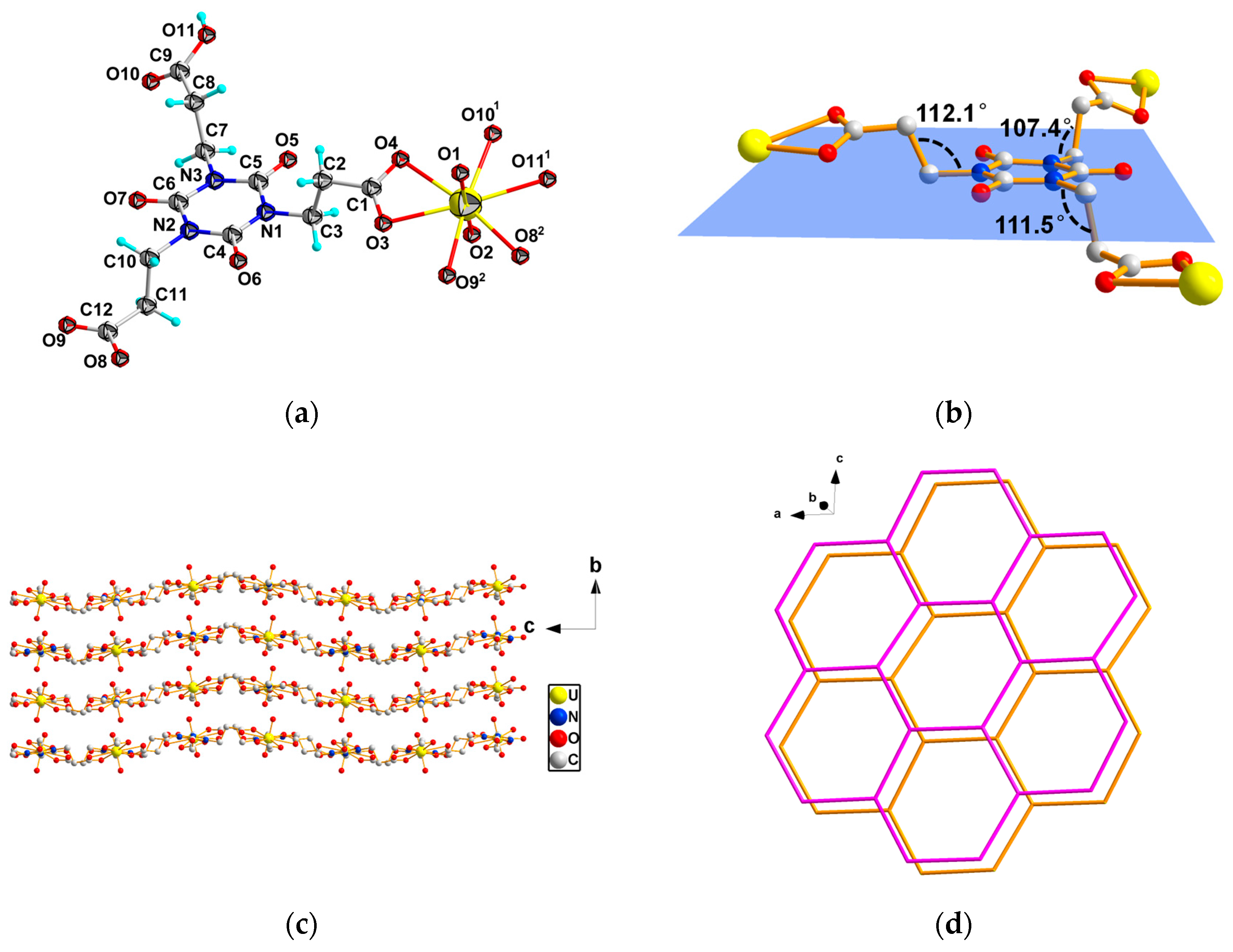
The asymmetric unit is mainly composed of one Htci2− ligand and a uranyl center. Each uranyl center is coordinated by eight oxygen atoms, displaying an eight-coordinated hexagonal bipyramidal geometric configuration (Figure 1a). With the uranyl [O1=U=O2]2+ as the axial direction, the bond lengths are 1.786(10) and 1.8291(11) Å, and the bond angle is 178.8(5)°. The six equatorial oxygen atoms (O3, O4, O82, O92, O101, O111), all originating from carboxyl groups, exhibit U-O bond lengths ranging from 2.403(10) to 2.498(11) Å, with an average value of 2.464(10) Å. The valence of U, calculated through the bond lengths around the uranium atom, is 5.79, which accords well with the hexavalent oxidation state of the uranium center.
The ligand Htci2− exhibits a chair-shaped coordination configuration in space (Figure 1b). The carboxyl groups of Htci2−, featuring highly flexible propionyloxy moieties, adopt a cis-cis-trans coordination configuration. The angles between –CH2COO and the triazine ring plane are 109.0, 110.2, and 112.2°. However, the reported uranyl complexes based on H3tci represent cis-cis-cis [18,19] and cis-cis-trans [19,20] configurations. It has been demonstrated that the carboxylate groups in H3tci are distorted, and this makes H3tci a feasible ligand for forming uranyl coordination polymers with diverse spatial structures and topologies. Three carboxyl groups of H3tci are fully deprotonated. Three propionic acid groups of H3tci keep turning and extending in space, forming an ABAB-packing two-dimensional spatial wavy structure along the bc plane (Figure 1c). Each tripodal Htci2− ligand binds to three UO22+ metal centers, and each of the metal centers is bonded to three Htci2− ligands. These are interconnected and expanded along the ac plane, forming a honeycomb-typed structure. Two layers along the ac plane do not completely overlap with each other, but form a two-dimensional spatial layered structure with staggered overlap.
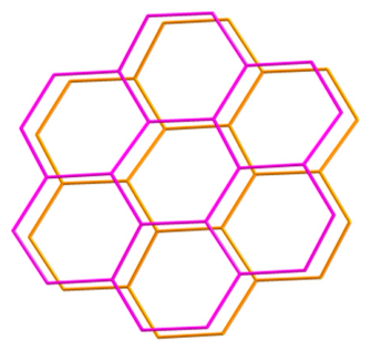
To further analyze the topology of the polymer, the Htci2− ligand is simplified as a 3-connected Y-type node to bridge three UO22+ metal centers. Similarly, each UO22+ serves as a three-connected node linking three corresponding ligands. The skeleton of the complex comprises a (6, 3) honeycomb-like network and spreads throughout the ac plane (Figure 1d). The honeycomb-like polymers of uranyl are not unique because of the non-extensible UO22+. Some tripodal ligands have a good tendency to construct honeycomb-like structures [18,24,25,26,27]. Table 1 lists the structural formulas for all reported examples in uranyl complexes with Htci2−, as well as illustrations of the spatial configurations. The complexes in Table 1 differ in terms of their counterions and the pH conditions during the synthesis process, both of which obviously play structure-directing roles. The formation of such structural topological variations may not be regarded as surprising, given the flexible ligand and the capping effect of UO22+.

3.2. Structural Characterization

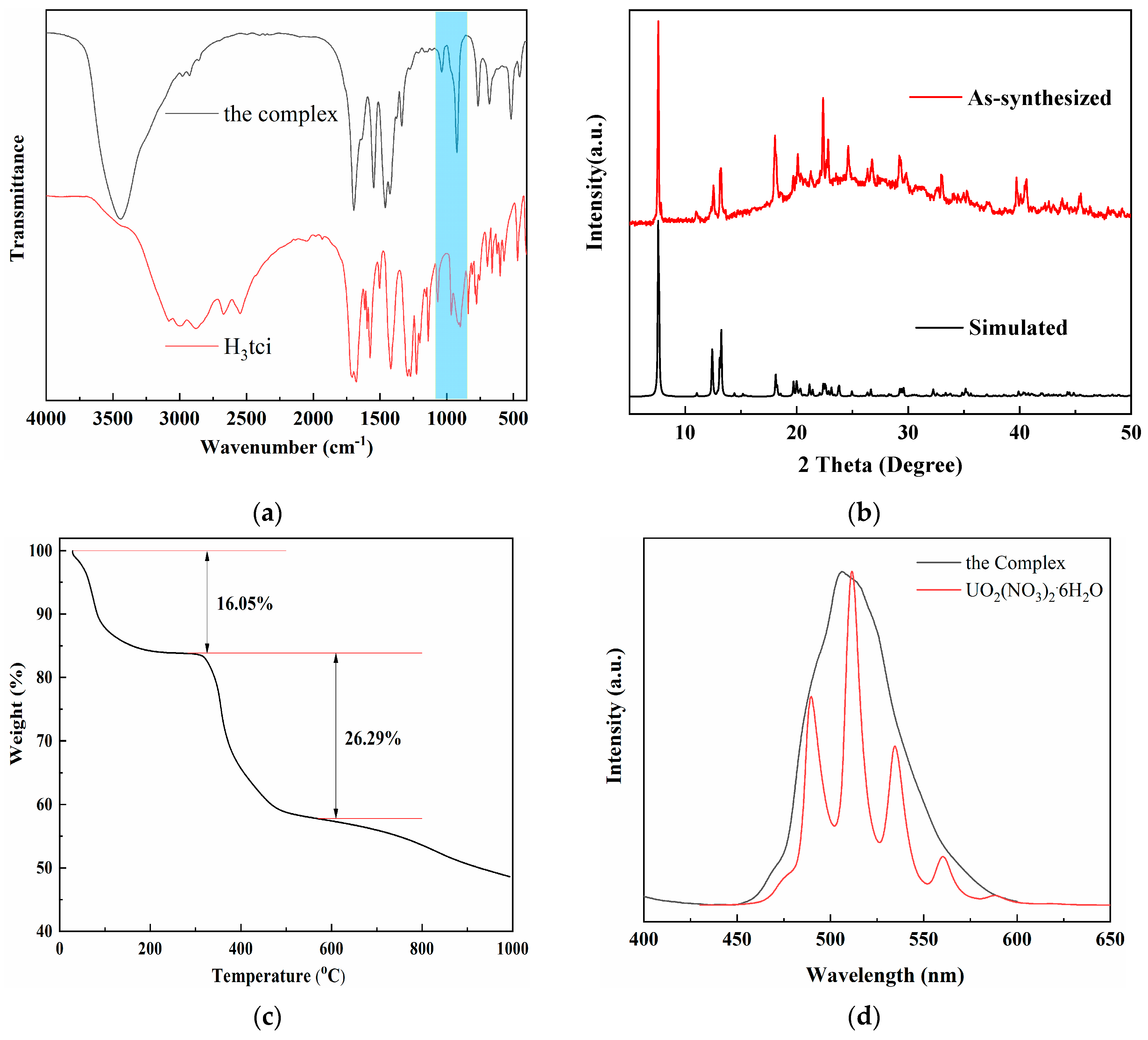
FT-IR spectra of the compound and H3tci are displayed in Figure 2a. Compared with the uncoordinated ligand, the uranyl complex exhibits characteristic absorption maxima at approximately 1039 and 926 cm−1, which correspond to the vsym (U=O) and vasym (U=O) vibrations, respectively. The disappearance of a band at 1695 cm−1, typical of the free carboxylic acid moiety in H3tci, verifies the full deprotonation of the carboxylate groups and their coordination to the uranyl centers. The strong absorption bands at 1549, 1462, and 1427 cm−1 in the complex are assigned to the vasym and vsym of the coordinated carboxylate groups. Additionally, the bands at 768, 683, and 518 cm−1 are ascribed to the skeletal vibrations of the nitrogen-containing triazine rings. These spectral characteristics are consistent with the coordination modes determined by X-ray diffraction.
The PXRD pattern of the polymer was collected under room temperature (Figure 2b). The synthesized compound shows a close match with the simulated crystal diffraction pattern. The minor discrepancies in peak intensities are likely due to variations in crystallinity introduced during sample preparation. Thus, the sample is suitable for the following investigations.
To assess the thermal stability of the uranyl polymer, thermo-gravimetric measurement was performed under a N2 atmosphere from 25 °C to 1000 °C at 10 K·min−1 (Figure 2c). The TG curve indicates that the uranyl compound experiences two distinct weight-loss stages. The first step in the range of ambient temperature to 305 °C is ascribed to the loss of free water molecules (obsd: 16.05%; calcd: 18.05%). The second rapid weight-loss step, corresponding to the collapse of the network, takes place between 320 and 582 °C. The second stage is poorly resolved, owing to the partial decomposition of the polymeric structure. The remaining 48.50% might be U3O8 (calcd: 1/3U3O8, 37.51%).
The solid-state fluorescence emission measurement of the polymer was carried out at ambient temperature under 330 nm excitation (Figure 2d). The emission spectrum measured in the range of 400–600 nm was compared with that of uranyl nitrate hexahydrate. In contrast to the characteristic emission peaks of uranyl nitrate (476, 489, 511, 534, 560, 588 nm), the polymer exhibits less resolved emission features. The spectrum of the polymer converges as a wide and unsmooth band spanning 460~580 nm, with a central wavelength at 506 nm indicating a slight blue shift of approximately ~5 nm relative to uranyl nitrate hexahydrate. It is not uncommon for uranyl polymers to have such broad fluorescence emission bands because of the diversity of uranyl structures [28,29,30].

3.3. Photocatalytic Performance

The presence of UO22+ in the polymer led to the attractive photocatalytic activities. To evaluate the catalytic degradation efficiency of the target polymer, TC was chosen as a representative contaminant under both UV-light and xenon-lamp irradiation.
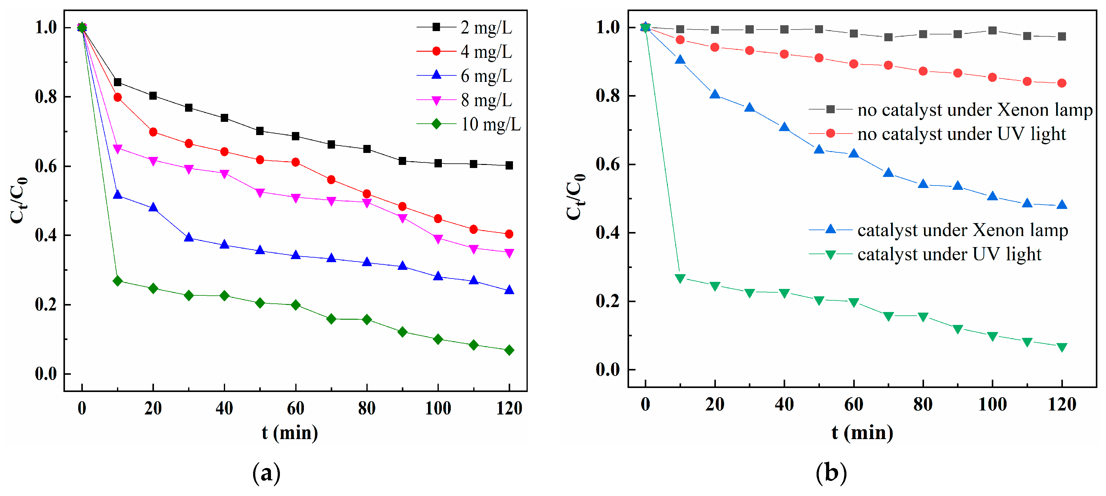
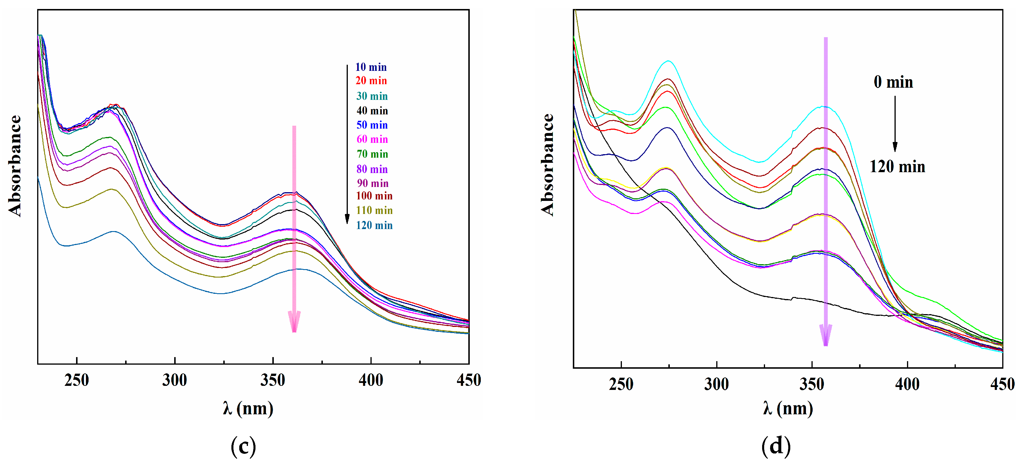
The degradation process of TC in aqueous solution was monitored in the presence of the target polymer. After being irradiated by UV lamp for 120 min, the photocatalytic degradation rates of TC containing the different concentrations of uranyl polymer (2, 4, 6, 8, and 10mg·L−1) were 39.8, 60.7, 64.9, 76.8, and 93.2%, respectively (Figure 3a). Taking into account both the photocatalytic degradation efficiency and the catalyst concentration, the optimal polymer dosage was determined to be 10 mg·L−1 for the purposes of further experiments. Under xenon-lamp irradiation, the photocatalytic degradation rate of the polymer for TC was 52.1% at 120 min (2.9% no catalyst under xenon lamp). Meanwhile, under irradiation with UV light, the degradation rate was 93.2% at 120 min (11.7% no catalyst under UV light) (Figure 3b). The decreasing UV-Vis absorption spectra of the TC under Xenon lamp and UV light are displayed in Figure 3c,d. These results clearly demonstrate that the uranyl-based polymers exert a considerable effect on the photodegradation of TC [25,31].

3.4. Sensing Property for Fe3+

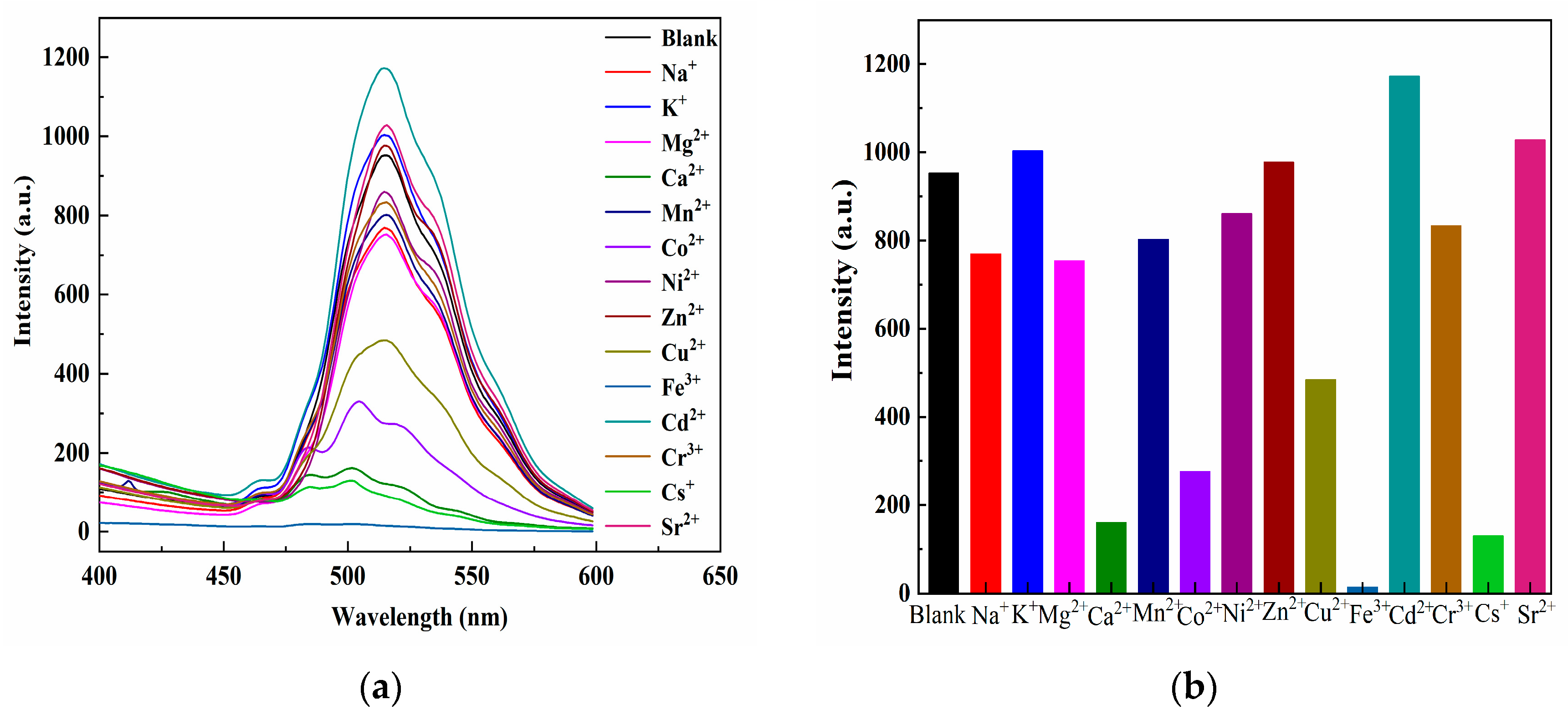
Fluorescence spectra of the complex in the presence of diverse metal ions exhibited similar profiles to the blank sample. No obvious shifts in peak positions or variations in peak shapes were observed; however distinct changes in fluorescence intensity did occur. Fluorescence intensity increased markedly in Cd2+, remained nearly unchanged in K+, Zn2+ and Sr2+, decreased slightly in Na+, Mg2+, Mn2+, Ni2+ and Cr3+, dropped obviously in Ca2+, Co2+, Cu2+and Cs+, and was almost fully quenched in Fe3+ (Figure 4a,b). The results show that the complex exhibits a sharp quenching of fluorescence emission intensity upon the addition of Fe3+, indicating high selectivity for Fe3+ among the various metal cations.
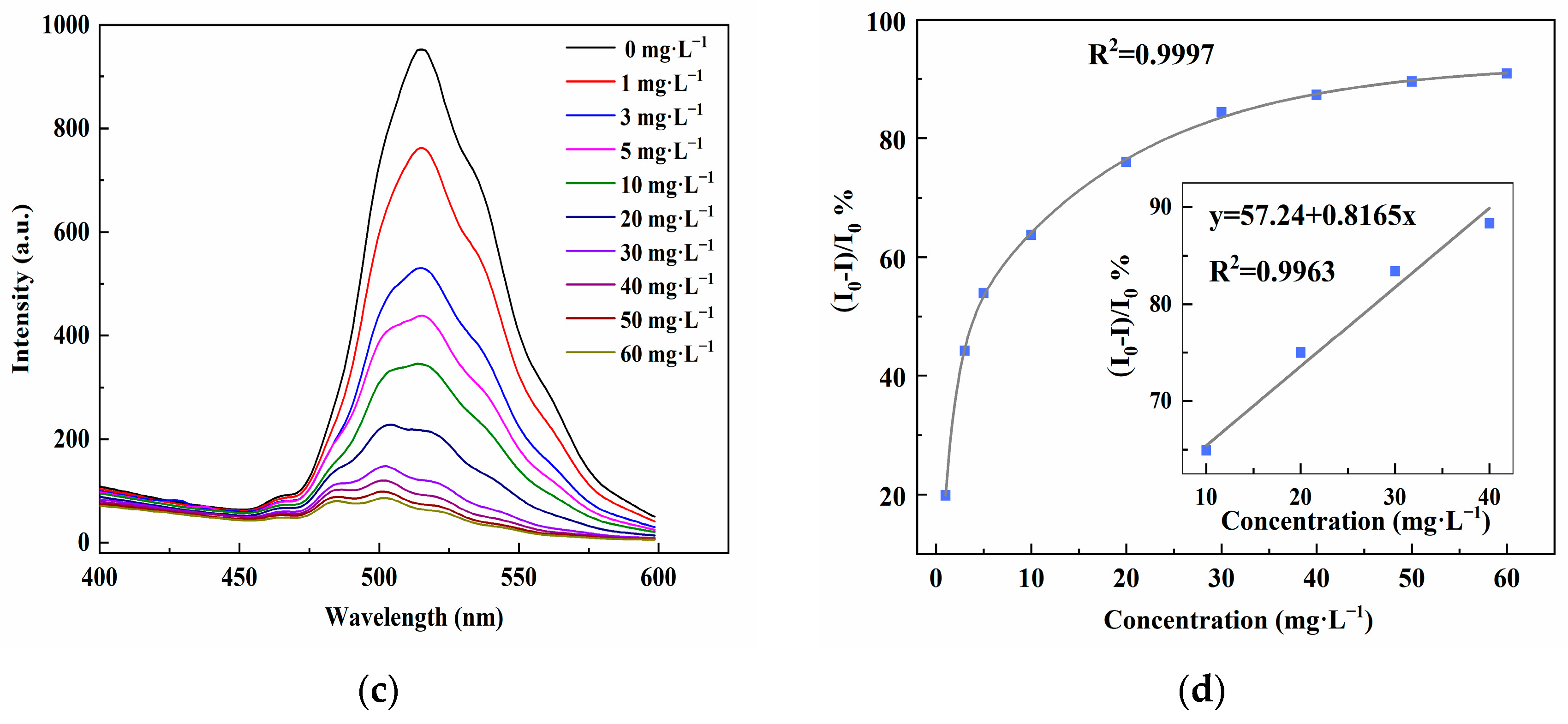
Based on the selectivity of the complex for sensing Fe3+, further studies of its sensitivity under different Fe3+ concentrations were conducted. The quenching constant (Ksv) was obtained using the Stern–Volmer equation, I0/I − 1 = Ksv[C]. The calculated Ksv value for Fe3+ reached 3.35 × 10⁵, further demonstrating the high sensitivity and selectivity of the compound toward Fe3+. It is interesting to note that the increase in Fe3+ concentration from 0 to 60 mg·L−1 leads to a rapid decline in luminescence intensity (Figure 4c). A correlation analysis between the luminescence quenching ratio [(I0I)/I0%] and the Fe3+ content was performed. The Langmuir fitting (R2 = 0.9997) confirmed the effective response of the sensor. Notably, a well-fitted linear relationship (y = 57.24 + 0.8165x, R2 = 0.9963) was observed in the concentration range of 10–40 mg·L−1 (Figure 4d). This sensing method exhibits high sensitivity toward Fe3+ over a detection range of 0–60 mg·L−1, with the limit detection calculated to be 0.77 mg·L−1, according to the 3σ/k criterion.
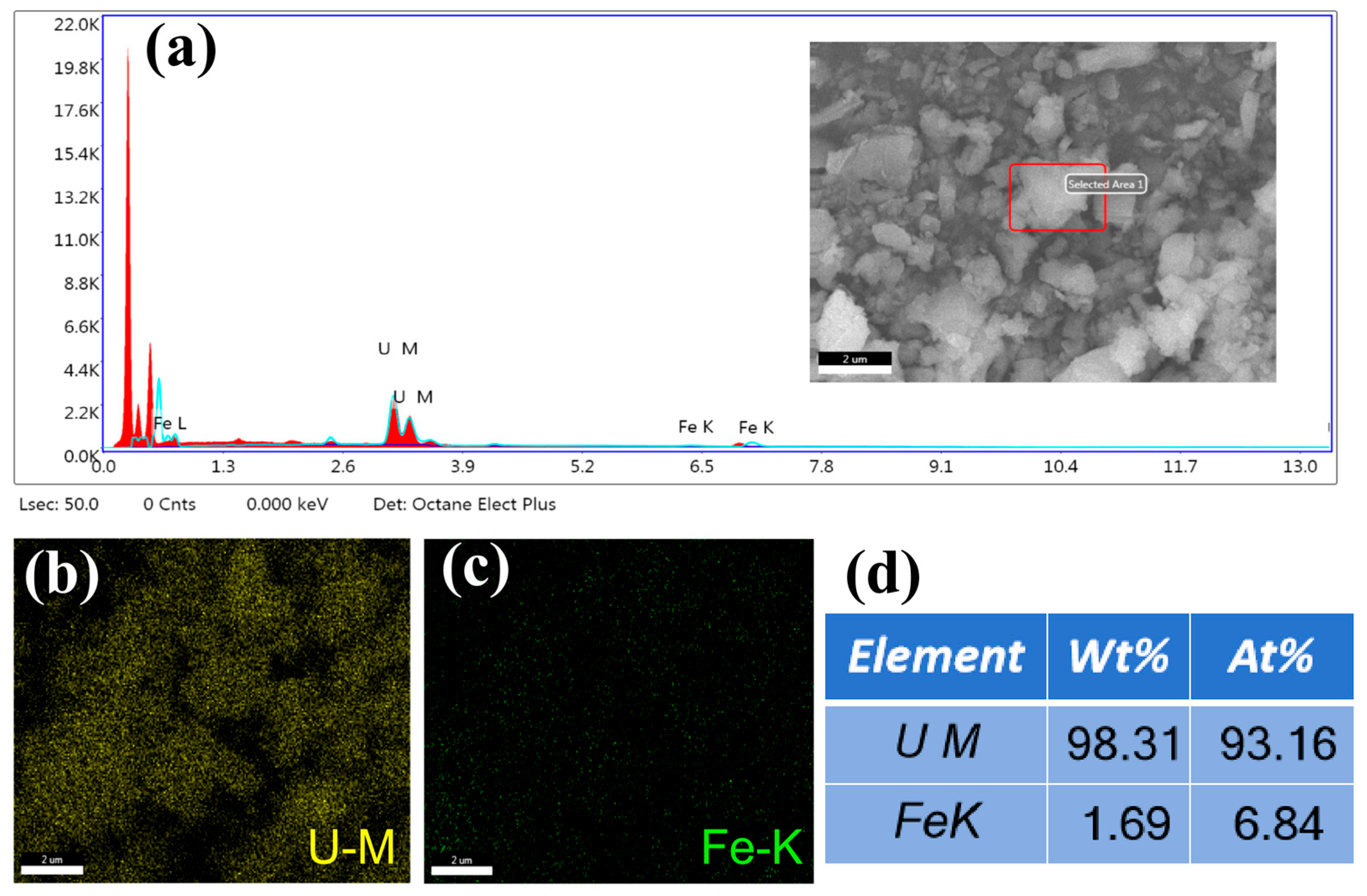
Fluorescence quenching of complex materials by metal ions occurs mainly via three mechanisms: structure disruption, central cation exchange, and competitive light absorption between adsorbed metal ions and the complex. SEM and EDS results confirm that iron species are loaded on the compound (Figure 5a). EDS mapping shows the elemental distribution on the surface of the complex (Figure 5b,c), with the atomic% of Fe being 6.84% (Figure 5d). The characteristic absorption peaks in the FTIR spectra demonstrate that the basic structure of the complex remains unchanged after catalysis (Figure S2). Consistently, the PXRD results further confirm that no obvious structural variation occurs in the complex after the catalytic reaction (Figure S3). Accordingly, the fluorescence quenching is attributed to competitive light absorption and energy transfer between Fe3+ and uranyl complex [20]. The excitation spectrum of the uranyl complex partially overlaps with the absorption spectrum of Fe3+. The dominant portion of excitation energy is taken up by Fe3+ loaded on the material surface. This effect reduces the absorption intensity of the complex, resulting in reduced luminescence or even total quenching.

4. Conclusions

A high-purity uranyl polymer constructed from H3tci was successfully prepared via a hydrothermal route. Structural characterization indicates that the polymer features a honeycomb-like 2D layered structure, with the ligand adopting a cis-cis-trans conformation. The luminescent property suggests that the polymer can be conducted as a potentially fluorescent material. Notably, the polymer exhibits remarkable photocatalytic activity toward TC degradation under UV irradiation. The photodegradation efficiency of TC solution reached about 93.2% in 120 min, which provides the theoretical and experimental basis for the application of uranyl polymers. Meanwhile, the metal ion sensing of the compound revealed a selective recognition of Fe3+, with a detection limit of 0.77 mg·L−1. These results establish the practical foundation for utilizing the uranyl polymer as an efficient photocatalyst in treatment of antibiotic-contaminated wastewater.

Supplementary Materials

The following supporting information can be downloaded at: https://www.mdpi.com/article/10.3390/cryst16070443/s1, Table S1: Crystal data and structure parameters of the complex; Table S2: Selected bond lengths (Å) and angles (deg) of the complex; Figure S1: The solid-state UV-vis spectra of the uranyl complex; Figure S2: FT-IR spectra comparison of the complex before and after photocatalysis; Figure S3: PXRD patterns comparison of the complex before and after photocatalysis.

Author Contributions

Conceptualization, L.-L.L.; methodology, L.-L.L.; formal analysis, Z.-Y.L., T.-T.L. and Y.-Z.Z.; investigation, Z.-Y.L., T.-T.L. and Y.-Z.Z.; writing—original draft preparation, L.-L.L.; writing—review and editing, L.-L.L.; project administration, L.-L.L. and J.-S.Z. All authors have read and agreed to the published version of the manuscript.

Funding

This research was funded by National Natural Science Foundation (No. 21671157), Natural Science Basic Research Program of Shaanxi (Nos. 2025JC-YBMS-178 and 2025JC-YBQN-136), Xi’an Medical University (Nos. 2022NLTS056, 2025HXZR08, 2025XSKY46 and 2025XSKY49).

Data Availability Statement

Crystallographic data for the uranyl complex (CCDC number: 2466734) can be obtained free of charge via http://www.ccdc.cam.ac.uk//data_request/cif (accessed on 3 May 2026).

Conflicts of Interest

The authors declare no conflicts of interest.

References

  1. Alawadhi, A.H.; Chheda, S.; Stroscio, G.D.; Rong, Z.C.; Kurandina, D.; Nguyen, H.L.; Rampal, N.; Zheng, Z.L.; Gagliardi, L.; Yaghi, O.M. Harvesting water from air with high-capacity, Stable furan-based metal-organic frameworks. J. Am. Chem. Soc. 2024, 146, 2160–2166. [Google Scholar] [CrossRef] [PubMed]
  2. Kim, M.; Lee, Y.L.; Moon, H.R. Carboxylate-based metal-organic framework and coordination polymer glasses: Progress and perspectives. Acc. Chem. Res. 2024, 57, 2347–2357. [Google Scholar] [CrossRef] [PubMed]
  3. Achenbach, B.; Yurdusen, A.; Stock, N.; Maurin, G.; Serre, C. Synthetic aspects and characterization needs in MOF chemistry-from discovery to applications. Adv. Mater. 2025, 37, 2411359. [Google Scholar] [CrossRef] [PubMed]
  4. Lin, Z.X.; Richardson, J.J.; Zhou, J.J.; Caruso, F. Direct synthesis of amorphous coordination polymers and metal-organic frameworks. Nat. Rev. Chem. 2023, 7, 273–286. [Google Scholar] [CrossRef] [PubMed]
  5. Thuéry, P.; Harrowfield, J. Bifunctional ligands in uranyl chemistry: Metalloligands and zwitterions. Coord. Chem. Rev. 2024, 510, 215821. [Google Scholar] [CrossRef]
  6. Gurzhiy, V.V.; Nazarchuk, E.V.; Tagirova, Y.G.; Krivovichev, S.V. Results of 25-Year studies of uranium compounds at the Department of Crystallography of St. Petersburg State University. Crystallogr. Rep. 2025, 70, 235–270. [Google Scholar] [CrossRef]
  7. Cowie, B.E.; Purkis, J.M.; Austin, J.; Love, J.B.; Arnold, P.L. Thermal and photochemical reduction and functionalization chemistry of the uranyl dication, [UVIO2]2+. Chem. Rev. 2019, 119, 10595–10637. [Google Scholar] [CrossRef] [PubMed]
  8. Gao, P.; Shen, Z.W.; Chen, Y.N.; Jiang, T.; Ji, Z.Y.; Zhao, G.X.; Yue, G.R.; Hu, Y.Z.; Wang, X.K.; Huang, X.B.; et al. A review on uranyl-based photocatalysts in photocatalytic organic transformation. J. Catal. 2025, 442, 115900. [Google Scholar] [CrossRef]
  9. Luan, X.F.; Wang, C.Z.; Wu, Q.Y.; Lan, J.H.; Chai, Z.F.; Xia, L.S.; Shi, W.Q. Theoretical insights on improving amidoxime selectivity for potential uranium extraction from seawater. J. Phys. Chem. A 2022, 126, 406–415. [Google Scholar] [CrossRef] [PubMed]
  10. Zhao, H.; Qi, C.; Yan, X.; Ji, J.; Chai, Z.; Wang, S.; Zheng, T. A multifunctional porous uranyl phosphonate framework for cyclic utilization: Salvages, uranyl leaking prevention, and fluorescent sensing. ACS Appl. Mater. Interfaces 2022, 14, 14380–14387. [Google Scholar] [CrossRef] [PubMed]
  11. Debsharma, K.; Dutta, B.; Dey, S.; Sinha, C. Metal-organic coordination polymers: A review of electrochemical sensing of environmental pollutants. Cryst. Growth Des. 2024, 24, 6503–6530. [Google Scholar] [CrossRef]
  12. Li, Y.; Yan, B.J.; Khan, M.A.; Zhang, J.C.; Cao, X.W.; Zhang, J.; Wang, N.; Yuan, Y.H. Uranyl-incorporated two-dimensional hydrogen-bonded organic framework for efficient triple-pathway H2O2 production. Appl. Catal. B Environ. Energy 2026, 386, 126428. [Google Scholar] [CrossRef]
  13. Sun, X.; Wang, Y.; Tang, Y.; Zhang, B.; Wei, W.; Li, X.; Fei, X.; Liu, X. Synthesis of isocyanurate-based imidazole carboxylate as thermal latent curing accelerator for thermosetting epoxy resins. J. Appl. Polym. Sci. 2020, 137, e49221. [Google Scholar] [CrossRef]
  14. Li, H.; Xu, Z.; Zhao, B.; Jia, Y.; Ding, R.; Hou, H.; Fan, Y. Nuclearity control of manganese polymers dependent on structural differences in the coligands and magnetic properties studies. CrystEngComm 2014, 16, 2470–2479. [Google Scholar] [CrossRef]
  15. Huang, H.Y.; Zhong, S.Q.; Chen, Y.W.; Gong, W.Q.; Ye, C.S.; Qiu, T.; Chen, J. Engineering sulfonated polymers for the removal of ultra-trace complexed Cr(III) in tris(2-carboxyethyl) isocyanurate photoresist resin monomers. Ind. Chem. Mater. 2025, 3, 618–630. [Google Scholar] [CrossRef]
  16. Ramathulasamma, M.; Bommakanti, S.; Das, S.K. Diverse coordination architectures based on a flexible multidentate carboxylate ligand and N-donor linkers: Synthesis, structure, supramolecular chemistry and related properties. Polyhedron 2023, 230, 116216. [Google Scholar] [CrossRef]
  17. Ghosh, S.K.; Bureekaew, S.; Kitagawa, S. A dynamic, isocyanurate-functionalized porous coordination polymer. Angew. Chem. Int. Ed. 2008, 47, 3403–3406. [Google Scholar] [CrossRef]
  18. Liang, L.L.; Cai, Y.G.; Weng, N.S.; Zhang, R.L.; Zhao, J.S.; Wang, J.F.; Wu, H.L. A novel uranyl complex UO2(tci)(C3H5N2)·H2O: Synthesis, crystal structure and characterization. Inorg. Chem. Commun. 2009, 12, 86–88. [Google Scholar] [CrossRef]
  19. Liang, L.L.; Zhang, R.L.; Zhao, J.S.; Liu, C.Y.; Weng, N.S. Two actinide-organic frameworks constructed by a tripodal flexible ligand: Occurrence of infinite {(UO2)O2(OH)3}4n and hexanuclear {Th6O4(OH)4} motifs. J. Solid State Chem. 2016, 243, 50–56. [Google Scholar] [CrossRef]
  20. Liang, L.L.; Zhang, R.L.; Zhao, J.S. Counterion-controlled formation of layered honeycomb and polythreading uranyl networks and the highly sensitive and selective detection of Fe3+ in aqueous media. Inorg. Chem. 2020, 59, 7980–7990. [Google Scholar] [CrossRef] [PubMed]
  21. Dolomanov, O.V.; Bourhis, L.J.; Gildea, R.J.; Howard, J.A.K.; Puschmann, H. OLEX2: A complete structure solution, refinement and analysis program. J. Appl. Cryst. 2009, 42, 339–341. [Google Scholar] [CrossRef]
  22. Sheldrick, G.M. ShelXT-integrated space-group and crystal-structure determination. Acta Crystallogr. 2015, A71, 3–8. [Google Scholar] [CrossRef]
  23. Sheldrick, G.M. Crystal structure refinement with Shelxl. Acta Crystallogr. 2015, C71, 3–8. [Google Scholar] [CrossRef]
  24. Wang, Y.; Liu, Z.; Li, Y.; Bai, Z.; Liu, W.; Xu, X.; Xiao, C.; Sheng, D.; Diwu, J.; Su, J.; et al. Umbellate distortions of the uranyl coordination environment result in a stable and porous polycatenated framework that can effectively remove cesium from aqueous solutions. J. Am. Chem. Soc. 2015, 137, 6144–6147. [Google Scholar] [CrossRef] [PubMed]
  25. Plaman, A.S.; Knapp, J.G.; Xie, H.M.; Kirlikovali, K.O.; Farha, O.K. Exploring energy-density relationships in uranyl nun-topology MOFs and their topological isomers. ACS Mater. Lett. 2024, 6, 4889–4896. [Google Scholar] [CrossRef]
  26. Kusumoto, S.; Atoini, Y.; Masuda, S.; Koide, Y.; Kim, J.Y.; Hayami, S.; Kim, Y.; Harrowfield, J.; Thuery, P. Flexible aliphatic diammonioacetates as zwitterionic ligands in UO22+ complexes: Diverse topologies and interpenetrated structures. Inorg. Chem. 2023, 62, 3929–3946. [Google Scholar] [CrossRef] [PubMed]
  27. Thuéry, P.; Harrowfield, J. Contrasting networks and entanglements in uranyl ion complexes with adipic and trans, trans-muconic acids. Inorg. Chem. 2022, 61, 2790–2803. [Google Scholar] [CrossRef] [PubMed]
  28. Zhang, Y.; Chen, L.; Guan, J.; Wang, X.; Wang, S.; Diwu, J. A unique uranyl framework containing uranyl pentamers as secondary building units: Synthesis, structure, and spectroscopic properties. Dalton Trans. 2020, 49, 3676–3679. [Google Scholar] [CrossRef] [PubMed]
  29. Harrowfield, J.; Atoini, Y.; Thuéry, P. Plumbing the uncertainties of solvothermal synthesis involving uranyl ion carboxylate complexes. CrystEngComm 2022, 24, 1475–1484. [Google Scholar] [CrossRef]
  30. Thuéry, P.; Atoini, Y.; Harrowfield, J. Functionalized aromatic dicarboxylate ligands in uranyl–organic assemblies: The cases of carboxycinnamate and 1,2-/1,3-Phenylenedioxydiacetate. Inorg. Chem. 2020, 59, 2923–2936. [Google Scholar] [CrossRef] [PubMed]
  31. Wang, J.C.; Wang, X.; Zhang, Q.Y.; Guo, L.L.; Zhang, Q.S.; Liu, Y.L.; Zeng, Q.Y. A self-driven solar coupling system with activated carbon felt cathode for resourcefully purifying uranium and organic contaminated water. Sep. Purif. Technol. 2025, 354, 129233. [Google Scholar] [CrossRef]
Figure 1. (a) Asymmetric unit with symmetry codes: 1 1 − x, −1/2 + y, +3/2 − z; 2 1/2 − x, −1/2 + y, z; (b) Arms of the ligand twisted in the space showed different bond angles; (c) 2D layered structure and adopted ABAB packing mode; (d) Layered honeycomb-like networks of the complex.
Crystals 16 00443 g001
Figure 2. (a) FT-IR spectra of the complex and H3tci. (b) Simulated PXRD patterns from single-crystal data and experimental patterns of the as-synthesized compound. (c) TG curves of the complex. (d) Emission spectra of the complex and UO2(NO3)2·6H2O.
Crystals 16 00443 g002
Figure 3. (a) Photocatalytic degradation of TC with varying concentrations of the uranyl polymer. (b) Photocatalytic degradation of TC under xenon-lamp and UV-light irradiation. (c,d) Time-dependent UV-Vis absorption spectra of TC during degradation under a xenon lamp (c) and UV light (d).
Crystals 16 00443 g003aCrystals 16 00443 g003b
Figure 4. (a,b) Emission spectra and intensities of the complex dispersed into different metal salt solutions. (c) Emission spectra of the complex at various Fe3+ solutions (0–60 mg·L−1). (d) Simulated correlation between (I0I)/I0% and Fe3+ concentration; the inset displays the linear fitting range from 10 to 40 mg·L−1.
Crystals 16 00443 g004aCrystals 16 00443 g004b
Figure 5. (a) EDS analysis of the Fe loaded on the compound; inset is the SEM photograph of the compound. (b,c) EDS mapping of U and Fe elements showed the elemental distribution on the surface. (d) Results of EDS analysis of the compound for U and Fe.
Crystals 16 00443 g005
Table 1. Overview of spatial configurations in uranyl complexes with H3tci.
CompoundConfigurationsRef.
UO2(tci)(C3H5N2)·H2OCrystals 16 00443 i001
2D irregular layered structure
[18]
(UO2)4(tci)2(OH)4[(UO2)2(H2O)4]1/2Crystals 16 00443 i002
3D extended network
[19]
[(UO2)2(tci)2]1/2·[Mn2O(H2O)8]1/2·2H2OCrystals 16 00443 i003
2D crossed honeycomb-like network
[20]
[UO2(tci)]·(C10H9N2)+·H2OCrystals 16 00443 i004
2D wave-like layered structure
[24]
[UO2(Htci)]·7.5H2OCrystals 16 00443 i005
2D overlapping honeycomb-like network
This work
Disclaimer/Publisher’s Note: The statements, opinions and data contained in all publications are solely those of the individual author(s) and contributor(s) and not of MDPI and/or the editor(s). MDPI and/or the editor(s) disclaim responsibility for any injury to people or property resulting from any ideas, methods, instructions or products referred to in the content.