Article

Structure of Agmatinase from Klebsiella pneumoniae and the Active Site Comparison with Its Structural Homologues

College of Pharmacy, Chung-Ang University, Seoul 06974, Republic of Korea
*
Author to whom correspondence should be addressed.
Crystals 2026, 16(5), 285; https://doi.org/10.3390/cryst16050285
Submission received: 26 March 2026 / Revised: 21 April 2026 / Accepted: 22 April 2026 / Published: 25 April 2026
(This article belongs to the Section Biomolecular Crystals)

Abstract

Agmatinase (SpeB) catalyzes the hydrolysis of agmatine to produce putrescine, a key step in bacterial polyamine biosynthesis. Here, we report the crystal structure of SpeB from Klebsiella pneumoniae (kpSpeB) and characterize its oligomeric and active-site architecture. SEC–MALS analysis demonstrates that kpSpeB forms a canonical hexamer in solution. Structural comparison reveals high similarity to Escherichia coli SpeB and other members of the arginase superfamily, including proclavaminic acid amidino hydrolase (PAH) and guanidine hydrolase (GdmH). Despite strong conservation of residues coordinating the binuclear Mn2+ center, subtle differences in metal positioning and cavity geometry were observed. Surface analysis indicates variations in active-site cavity volume among homologues, with partial occlusion in GdmH due to a bulky tryptophan residue. These findings suggest that minor adjustments in metal coordination and cavity architecture may fine-tune substrate selectivity while preserving the conserved catalytic framework of the arginase superfamily.

1. Introduction

Polyamines are ubiquitous polycationic molecules that play essential roles in cellular growth, nucleic acid stabilization, and stress responses across all domains of life [1,2,3]. In bacteria, one of the major polyamines, putrescine, is synthesized from agmatine through a hydrolytic reaction catalyzed by agmatinase (SpeB; EC 3.5.3.11) [4,5]. This enzymatic step constitutes a key branch of the polyamine biosynthetic pathway and directly links nitrogen metabolism to cellular growth regulation [6].
Agmatinase belongs to the evolutionarily conserved arginase superfamily, a group of binuclear metal-dependent hydrolases that catalyze the hydrolysis of guanidinium-containing substrates to generate urea and corresponding amine products [7,8]. Members of this superfamily share a characteristic α/β/α structural fold and typically assemble into oligomeric complexes—most commonly hexamers (trimers of dimers)—with each monomer harboring an independent binuclear metal-dependent active site [7,9,10]. The catalytic mechanism involves two closely spaced metal ions—most frequently Mn2+—that activate a bridging hydroxide for nucleophilic attack on the guanidinium carbon [9,10]. Agmatinase has recently emerged as a potential metabolic target, as inhibition of bacterial agmatinase can directly affect polyamine homeostasis and cellular physiology [11]. Structural studies of canonical arginases and related enzymes, including proclavaminic acid amidinohydrolase (PAH), have demonstrated that substrate specificity is largely determined by subtle variations in the architecture of the substrate-binding cavity while maintaining a highly conserved metal coordination geometry [9,12,13]. PAH, identified in Streptomyces clavuligerus, is involved in clavulanic acid biosynthesis and represents a well-characterized member of the arginase superfamily. Recent structural and biochemical investigations have further expanded the functional diversity within this superfamily. Notably, the discovery of a Ni2+-dependent guanidine hydrolase (GdmH), which has been identified in Pseudomona aeruginosa and catalyzes the hydrolysis of guanidine and exhibits distinct substrate specificity through modification of its active-site architecture, revealed that the modification of metal preference and constriction of the substrate-binding pocket can dramatically alter substrate specificity while preserving the conserved catalytic core [14].
Despite the availability of a couple of agmatinase structures from different bacterial species, detailed structural comparisons remain limited due to the lack of structural information on this enzyme family. In this study, we determined the crystal structure of SpeB from Klebsiella pneumoniae (kpSpeB) and characterized its oligomeric state and active-site organization. SEC–MALS analysis revealed that kpSpeB forms a hexamer in solution, consistent with the canonical assembly observed in arginase-family enzymes. Structural comparison showed high similarity to Escherichia coli SpeB (ecSpeB) as well as related arginase superfamily members, including PAH [13] and GdmH [14]. Despite strong conservation of active-site residues, subtle differences in metal positioning and cavity architecture were observed among homologues, suggesting potential structural determinants of substrate specificity.

2. Materials and Methods

2.1. Protein Expression and Purification

The kpSpeB gene (Accession: WP_369382317.1) was synthesized by Bionics (Dae-Jeon, Republic of Korea) and cloned into the pET28a expression vector using the NdeI and XhoI restriction sites. The resulting plasmid was transformed into E. coli BL21(DE3) cells. A single colony was selected and cultured at 37 °C for 16 h in 10 mL of lysogeny broth (LB) containing 50 μg/mL of kanamycin. The culture was subsequently inoculated into 1 L of LB media supplemented with the same antibiotics and grown at 37 °C with shaking at 220 rpm until the optical density at 600 nm reached approximately 0.7. Protein expression was induced by adding 0.25 mM isopropyl β-D-1-thiogalactopyranoside (IPTG), followed by incubation at 20 °C for 16 h. Cells were harvested by centrifugation and resuspended in 20 mL lysis buffer (20 mM Tris-HCl, pH 8.0; 500 mM NaCl; and 25 mM imidazole). The resuspended cells were lysed by sonication on ice in the presence of 1 mM phenylmethanesulfonyl fluoride (PMSF; Sigma-Aldrich, St. Louis, MO, USA). The lysate was centrifuged at 10,000× g for 30 min at 4 °C.
The clarified supernatant was incubated with nickel nitrilotriacetic acid (Ni-NTA) resin (QIAGEN, Hilden, Germany) for 2 h at 4 °C with gentle mixing. The resin was loaded onto a gravity-flow column and washed with 25 mL washing buffer (20 mM Tris-HCl, pH 8.0; 500 mM NaCl; and 60 mM imidazole). The bound protein was eluted with elution buffer (Tris-HCl, pH 8.0; 500 mM NaCl; and 250 mM imidazole). The eluted protein was further purified by size-exclusion chromatography (SEC) using an ÄKTA Explorer system (GE Healthcare, Chicago, IL, USA) equipped with a Superdex 200 Increase 10/300 GL column (24 mL; GE Healthcare), pre-equilibrated with SEC buffer (20 mM Tris-HCl, pH 8.0, and 150 mM NaCl). Peak fractions were analyzed by sodium-dodecyl sulfate–polyacrylamide gel electrophoresis (SDS-PAGE), and fractions containing the target protein were pooled and concentrated to 9.6 mg/mL.

2.2. Crystallization and Data Collection

Crystallization of kpSpeB was performed using the hanging-drop vapor diffusion method at 20 °C. Initial crystallization screening was performed using commercially available sparse-matrix screening kits (e.g., Wizard screening I, II, and III; Crystal Screen I and II; and PEG/Ion screens). Protein solution and reservoir solution were mixed at a 1:1 (v/v) ratio to form 2 μL hanging drops and equilibrated against 300 μL reservoir solution. Crystals were first obtained from Wizard Screening I (Rigaku, Tokyo, Japan), condition No. 1 (0.1 M CHES, pH 9.5, and 15% ethanol). Conditions were optimized by varying precipitant concentration, pH, and salt composition. The optimized crystals appeared after 9 days in a condition containing 1 M potassium sodium tartrate; 0.1 M CHES/sodium hydroxide, pH 9.5; and 0.2 M lithium sulfate. Crystals were flash-frozen in liquid nitrogen at −178 °C prior to data collection. X-ray diffraction data were collected at beamline 5C of the Pohang Accelerator Laboratory (PAL) (Pohang, Republic of Korea). Diffraction data were indexed, integrated, and scaled using HKL-2000 [15].

2.3. Structure Determination and Analysis

The structure of kpSpeB was determined by molecular replacement using Phaser [16] implemented in the PHENIX package (Version 2.0) [17]. An AlphaFold3-predicted model was used as the search model for molecular replacement. Model building and refinement were carried out using Coot [18] and phenix.refine [17]. The six protomers in the asymmetric unit were refined independently without applying non-crystallographic symmetry restraints in PHENIX. Model quality was validated using MolProbity [19], and structural figures were prepared using PyMOL version 3.1 (Schrödinger, LLC, New York, NY, USA).

2.4. SEC-Multi Angle Light Scattering (MALS) Analysis

The absolute molar mass of kpSpeB was determined by SEC-MALS. Protein samples were loaded onto a Superdex 200 10/300 gel filtration column (GE Healthcare) equilibrated with purification buffer (20 mM Tris-HCl, pH 8.0, and 150 mM NaCl) at room temperature. The chromatography system (ÄKTA Explorer) (GE Healthcare) was connected to a DAWN-TREOS MALS detector (Wyatt Technology, Santa Barbara, CA, USA). The molecular mass was calculated using ASTRA software Version 6 (Wyatt Technology).

3. Results

3.1. The Overall Structure and Oligomeric State of kpSpeB

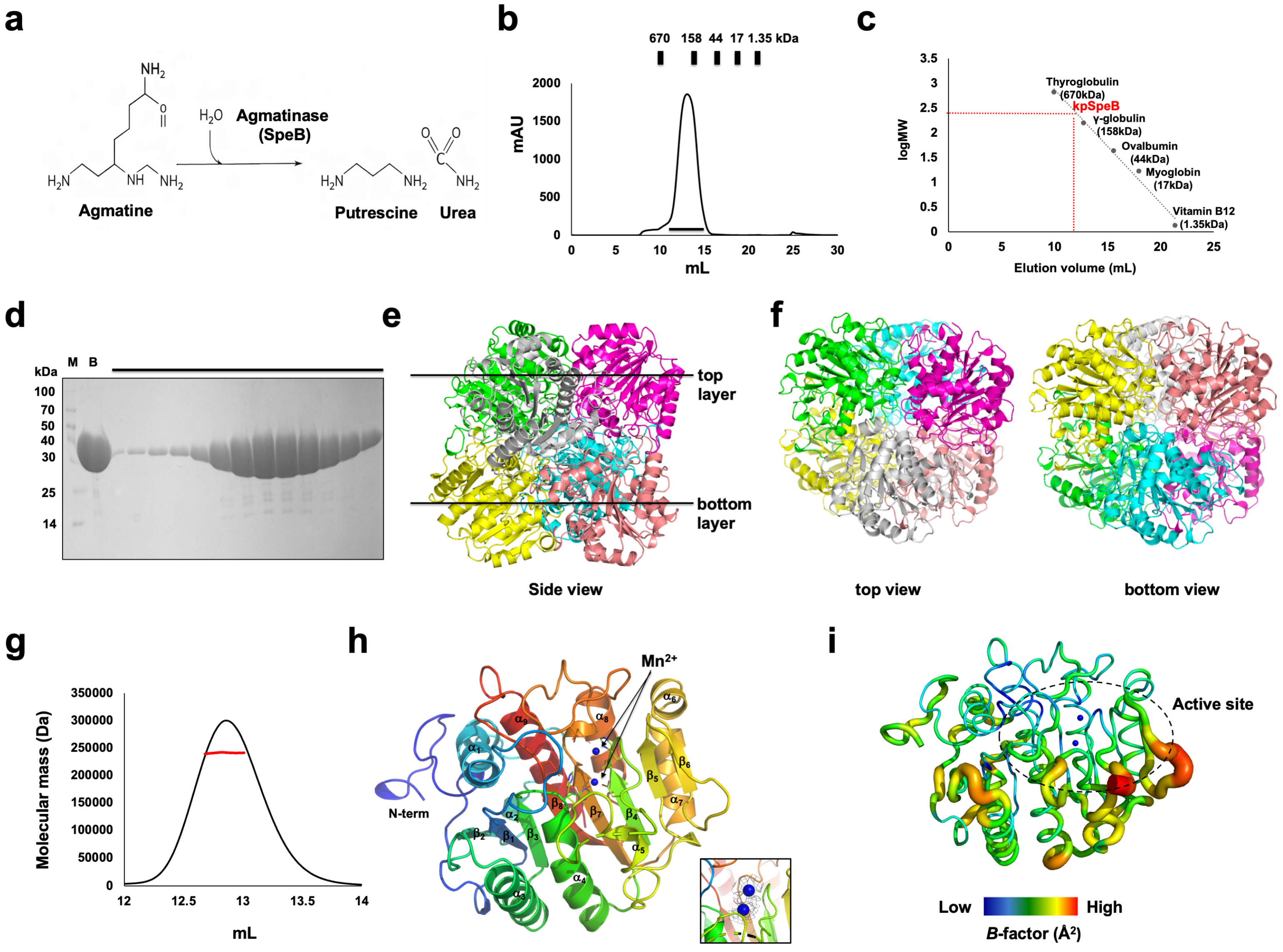
SpeB is an agmatinase that catalyzes the hydrolysis of agmatine into putrescine and urea, constituting a key step in the polyamine biosynthetic pathway (Figure 1a). A full-length SpeB (residues 1-306, molecular weight 38.6 kDa) from K. pneumoniae (hereafter called kpSpeB) was purified through a quick two-step chromatography (affinity chromatography followed by SEC) to study the structure. In the SEC profile, kpSpeB was eluted at approximately 13 mL over γ-globulin (158 kDa), indicating that kpSpeB may exist in a hexameric form within the solution (Figure 1b,c). During SEC, a single symmetric peak was observed, and SDS–PAGE analysis of the corresponding fractions confirmed that the peak contained highly purified kpSpeB (Figure 1d). The purified target protein was successfully crystallized, and the 2.77 Å crystal structure was determined through the MR phasing method, using the predicted structure from AlphaFold3 as a search model. The final structural model was refined to Rwork = 26.40% and Rfree = 29.22%. Table 1 summarizes the crystallographic and refinement statistics.
The crystallographic asymmetric unit (ASU) contains six identical kpSpeB molecules, forming a hexameric assembly composed of two stacked trimers arranged in a two-layer architecture (Figure 1e,f). Although SpeB is generally known to function as a hexamer, we performed SEC coupled with multi-angle light scattering (SEC–MALS) to independently confirm its stoichiometry in solution. The SEC–MALS analysis revealed a molecular mass of approximately 241.8 kDa (Figure 1g). Given that the calculated molecular weight of a kpSpeB monomer is 38.6 kDa, the theoretical mass of a hexamer is 231.6 kDa. The experimentally determined molecular weight is in close agreement with the predicted hexameric mass, indicating that kpSpeB exists as a hexamer in solution. These results further demonstrate that the biologically relevant assembly corresponds to the hexamer formed by the six protomers observed in the ASU.
The overall monomeric structure of kpSpeB conveyed the canonical structural fold of agmatinase, containing a central α/β domain with 9 α-helices around an inner 8 β-sheets (Figure 1h). The RMSD between the crystal structure and the AlphaFold3 model was calculated based on Cα atoms and was found to be 0.5 Å, indicating a high level of structural similarity. SpeB is a metal-dependent enzyme that requires two Mn2+ ions at its active site for catalytic activity [9,12]. In our kpSpeB structure, we observed clear electron density corresponding to two metal ions in the active-site region. Based on the coordination geometry and comparison with previously characterized SpeB structures, these densities were assigned as Mn2+ ions (Figure 1h). No Mn2+ was intentionally added during purification or crystallization; therefore, the observed metal ions are likely derived from trace amounts present in the expression system. B-factor analysis showed that kpSpeB overall exhibits relatively low B-factor values (average 44 Å2), especially near the active site, indicating a rigid structural framework. However, several loops surrounding the catalytic center display higher B-factor values, suggesting that these regions possess greater conformational flexibility (Figure 1i).

3.2. Structural Comparison of kpSpeB with Other Structural Isoforms

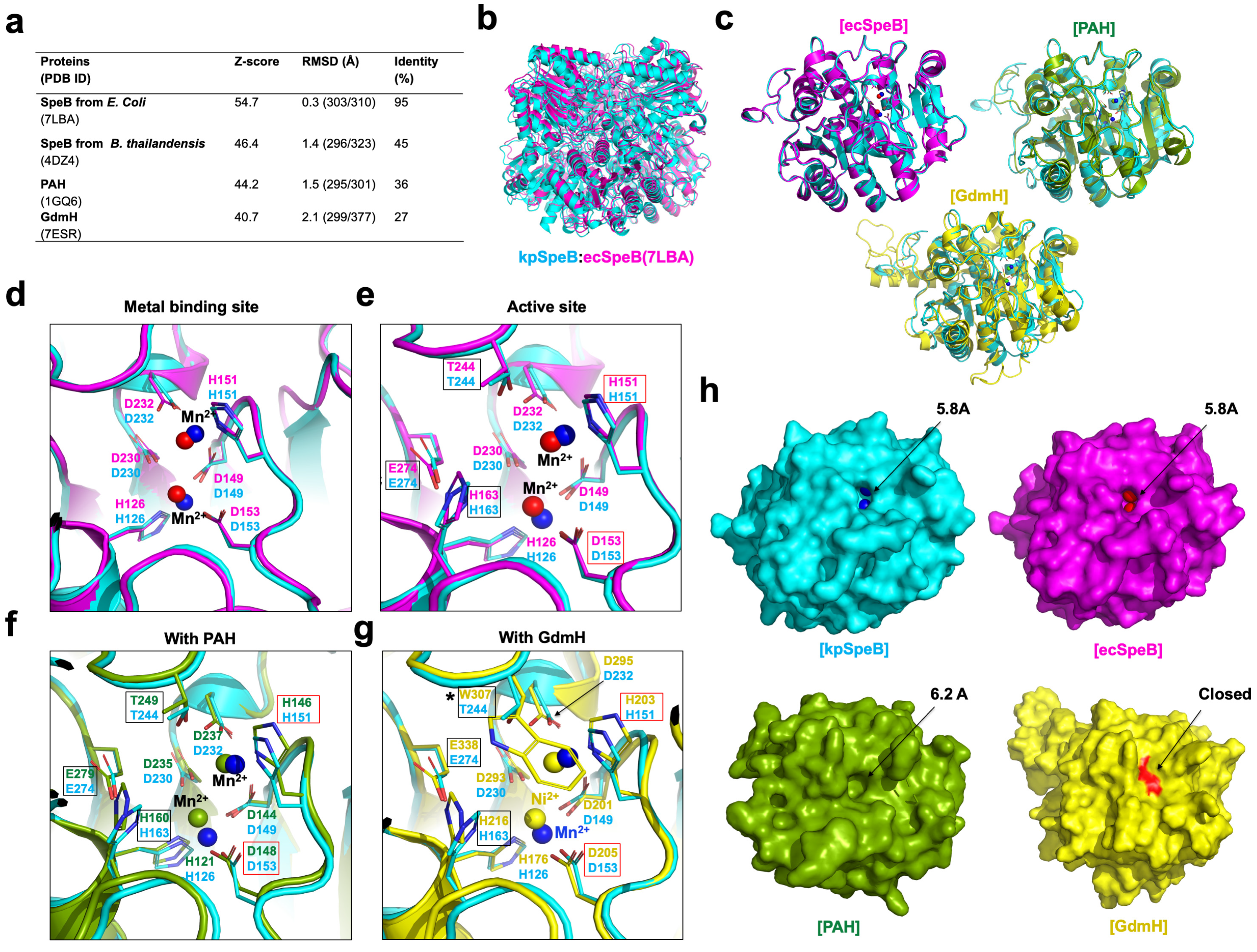
Structural homology analysis using the DALI server revealed that SpeB structures from Escherichia coli (ecSpeB, PDB ID: 7LBA) [9] and Burkholderia thailandensis (btSpeB, PDB ID: 4DZ4) [10] are the closest structural homologues of kpSpeB, with Z-scores of 54.7 and 46.4, respectively (Figure 2a). These correspond to the two SpeB structures reported to date, and their high structural similarity to kpSpeB is consistent with their classification within the same agmatinase family. In addition, members of the arginase superfamily, including PAH (PDB ID: 1GQ6) [13] and guanidine hydrolase (PDB ID: 7ESR) [14], were also identified as structurally related proteins (Figure 2a). Both ecSpeB and btSpeB were also reported to form hexameric assemblies [9,10,12], similar to kpSpeB, and their hexameric architectures are highly similar to that observed in kpSpeB (Figure 2b). The amino acid sequence identity between kpSpeB and ecSpeB is very high (~95%), whereas btSpeB shares a more moderate sequence identity of approximately 45% (Figure 2a). Consistent with this sequence similarity, pairwise structural superposition showed that kpSpeB is almost identical to ecSpeB, with an RMSD of approximately 0.3 Å (Figure 2a,c). RMSD values were calculated using Cα atoms only. In contrast, superposition with btSpeB resulted in an RMSD of 1.4 Å, indicating slightly greater structural divergence. For PAH and guanidine hydrolase, the sequence identities with kpSpeB are approximately 36% and 27%, respectively. Structural comparisons revealed RMSD values of 1.5 Å for PAH and 2.1 Å for guanidine hydrolase (Figure 2a,c). Despite the moderate level of sequence conservation, these results highlight the strong structural conservation among members of the arginase family.
It is well established that SpeB, as well as other members of the arginase family, requires two Mn2+ ions at the active site for catalytic activity [9,10,12,13]. The residues responsible for coordinating these metal ions have been well characterized. In the case of ecSpeB, residues H126, D149, D151, D153, D230, and D232 have been reported to coordinate the two Mn2+ ions [9]. Analysis of the metal-binding site in kpSpeB revealed that these six metal-coordinating residues are completely conserved and adopt similar positions to coordinate the Mn2+ ions (Figure 2d). In addition, previous structural studies of ecSpeB identified five residues—H151, D153, H163, T244, and E274—as critical components of the catalytic center [9]. These residues form the active-site architecture and are essential for enzymatic activity. Notably, all of these residues are also fully conserved in kpSpeB (Figure 2e). Taken together, these observations strongly suggest that kpSpeB shares the same metal-coordination scheme and catalytic mechanism as ecSpeB. Although the environments of the ion-binding site and the active site are highly similar, the positions of the two metal ions are not completely identical. In kpSpeB, the distance between the two Mn2+ ions is slightly larger than that observed in ecSpeB (Figure 2d,e). Structural comparison of the kpSpeB active site with that of PAH showed that all metal-coordinating residues and catalytic residues are fully conserved. Nevertheless, the exact positions of the metal ions are not identical (Figure 2f). Notably, among the conserved metal-binding residues, the side chains of H146 (equivalent to H151 in kpSpeB) and H160 (equivalent to H163 in kpSpeB) are positioned farther from the two metal ions than in kpSpeB (Figure 2f). Finally, comparison of the kpSpeB active site with that of GdmH revealed that most of the metal-coordinating residues and catalytic residues are conserved (Figure 2g). However, the most notable difference is that T244 in kpSpeB is replaced by W307 in GdmH. In addition, the side chains of H203 and H216 are positioned farther from the metal ions compared with those in kpSpeB, suggesting a weaker involvement in metal coordination.
Inspection of the surface representations of SpeB and other members of the arginase family revealed the presence of a hole-like opening surrounding the active site (Figure 2h). In both SpeB structures, the diameter of this opening is approximately 5.8 Å, whereas in PAH, the opening is slightly larger, measuring about 6.2 Å. In contrast, the corresponding region in GdmH appears to be blocked. This obstruction is caused by the bulky side chain of W307, which replaces T244 in kpSpeB. In the case of PAH, the larger hole size compared with SpeB can be explained by the positions of H203 and H216, whose side chains are located farther from the metal ions, thereby creating a wider opening around the active-site region.

4. Discussion

In this study, we report the crystal structure of agmatinase (SpeB) from Klebsiella pneumoniae (kpSpeB) and characterize its oligomeric state and active-site architecture. SEC–MALS analysis demonstrates that kpSpeB exists as a hexamer in solution, consistent with the canonical oligomeric organization of arginase-family enzymes. Structural comparison using the DALI server revealed high similarity to E. coli SpeB (95% sequence identity) as well as more distantly related homologues, including PAH and guanidine hydrolase. Remarkably, although the active-site residues coordinating the binuclear metal center are fully conserved, subtle differences in Mn2+ positioning were observed between kpSpeB and ecSpeB. Comparison with PAH (36% sequence identity) further showed complete conservation of nine residues involved in active-site formation and metal coordination, despite overall sequence divergence. In contrast, guanidine hydrolase contains a bulky tryptophan residue at the position corresponding to T244 in kpSpeB, a substitution known to restrict substrate access and confer specificity toward free guanidine.
Structural surface analysis revealed a distinct cavity at the active site of kpSpeB. This cavity is larger in PAH, whereas in GdmH it is partially occluded by the tryptophan side chain. These observations suggest that subtle differences in cavity volume—potentially influenced by minor variations in metal positioning and surrounding residues—may fine-tune substrate selectivity within the arginase superfamily. The observation that kpSpeB exhibits a cavity size intermediate between PAH and GdmH further supports the idea that small structural adjustments around the catalytic pocket modulate substrate accessibility without altering the conserved catalytic machinery.
Interestingly, the distance between the two Mn2+ ions in kpSpeB is slightly larger than that observed in ecSpeB. Because the binuclear metal center plays a critical role in activating the nucleophilic hydroxide during catalysis, even small variations in metal positioning may influence catalytic efficiency or substrate binding geometry. Such micro-variations have been reported in other arginase-family enzymes and are thought to reflect subtle adaptations to different physiological substrates while maintaining the same catalytic framework.
From an evolutionary perspective, the strong structural conservation observed across SpeB, PAH, and GdmH highlights the robustness of the arginase-family fold. Despite sequence divergence, the α/β/α scaffold and the binuclear metal center remain highly preserved. Functional diversification within this family therefore appears to arise primarily from local modifications surrounding the substrate-binding pocket rather than from large-scale rearrangements of the catalytic core. In particular, the substitution of T244 by W307 in GdmH represents a striking example of how a single bulky residue can reshape the substrate-access channel and alter enzyme specificity.
The biological significance of agmatinase is particularly relevant in pathogenic bacteria such as K. pneumoniae. Polyamine biosynthesis plays important roles in bacterial growth, stress adaptation, and virulence regulation. The structural information obtained here therefore provides a framework for understanding how agmatinase contributes to metabolic adaptation in this clinically important pathogen. Furthermore, because enzymes of the polyamine biosynthetic pathway are increasingly considered potential antimicrobial targets, the kpSpeB structure reported in this study may serve as a valuable template for future inhibitor design. Thus, structural insights into kpSpeB may contribute to a better understanding of metabolic adaptation mechanisms in K. pneumoniae and support the development of novel antimicrobial strategies targeting polyamine metabolism.
Together, our findings provide new structural insight into agmatinase architecture and highlight how minimal but strategically positioned variations in metal coordination and cavity geometry may drive functional diversification within the arginase superfamily. These results emphasize that substrate selectivity in this enzyme family is governed not by major alterations of the catalytic machinery but rather by fine structural tuning of the active-site environment. Although functional validation, such as enzymatic activity or thermostability analysis, was not performed in this study, such experiments will be important to further elucidate the biochemical properties of kpSpeB, particularly in the context of the environmental adaptability of K. pneumoniae.

Author Contributions

Conceptualization, H.H.P.; formal analysis, S.Y.L. and H.B.J.; data curation, S.Y.L. and H.B.J.; writing—original draft preparation, S.Y.L. and H.H.P.; writing—review and editing, S.Y.L. and H.H.P.; supervision, H.H.P.; project administration. All authors have read and agreed to the published version of the manuscript.

Funding

This study was supported by the Basic Science Research Program through the National Research Foundation of Korea (NRF) of the Ministry of Education, Science, and Technology (RS-2025-02316334) and supported by the Chung-Ang University Research Scholarship grants in 2024.

Institutional Review Board Statement

Not applicable.

Informed Consent Statement

Not applicable.

Data Availability Statement

Coordinating model and structural factors were deposited in the PDB under PDB ID: 23ZU. Further inquiries can be directed to the corresponding author.

Acknowledgments

We thank the 5C beamline staff at the Pohang Accelerator Laboratory (Pohang, Korea) for their data collection assistance.

Conflicts of Interest

The authors declare no conflicts of interest.

References

  1. Miller-Fleming, L.; Olin-Sandoval, V.; Campbell, K.; Ralser, M. Remaining Mysteries of Molecular Biology: The Role of Polyamines in the Cell. J. Mol. Biol. 2015, 427, 3389–3406. [Google Scholar] [CrossRef] [Scilit] [PubMed]
  2. Pegg, A.E. Functions of Polyamines in Mammals. J. Biol. Chem. 2016, 291, 14904–14912. [Google Scholar] [CrossRef] [Scilit] [PubMed]
  3. Zhang, C.; Zhen, Y.; Weng, Y.; Lin, J.; Xu, X.; Ma, J.; Zhong, Y.; Wang, M. Research progress on the microbial metabolism and transport of polyamines and their roles in animal gut homeostasis. J. Anim. Sci. Biotechnol. 2025, 16, 57. [Google Scholar] [CrossRef] [Scilit] [PubMed]
  4. Kobayashi, T.; Sakamoto, A.; Kashiwagi, K.; Igarashi, K.; Takao, K.; Uemura, T.; Moriya, T.; Oshima, T.; Terui, Y. Putrescine Biosynthesis from Agmatine by Arginase (TtARG) in Thermus thermophilus. J. Biochem. 2023, 174, 81–88. [Google Scholar] [CrossRef] [Scilit] [PubMed]
  5. Benitez, J.; Garcia, D.; Romero, N.; Gonzalez, A.; Martinez-Oyanedel, J.; Figueroa, M.; Salas, M.; Lopez, V.; Garcia-Robles, M.; Dodd, P.R.; et al. Metabolic strategies for the degradation of the neuromodulator agmatine in mammals. Metabolism 2018, 81, 35–44. [Google Scholar] [CrossRef] [Scilit] [PubMed]
  6. Michael, A.J. Polyamines in Eukaryotes, Bacteria, and Archaea. J. Biol. Chem. 2016, 291, 14896–14903. [Google Scholar] [CrossRef] [Scilit] [PubMed]
  7. Dowling, D.P.; Di Costanzo, L.; Gennadios, H.A.; Christianson, D.W. Evolution of the arginase fold and functional diversity. Cell. Mol. Life Sci. 2008, 65, 2039–2055. [Google Scholar] [CrossRef] [Scilit] [PubMed]
  8. Hernandez, V.M.; Arteaga, A.; Dunn, M.F. Diversity, properties and functions of bacterial arginases. FEMS Microbiol. Rev. 2021, 45, fuab034. [Google Scholar] [CrossRef] [Scilit] [PubMed]
  9. Chitrakar, I.; Ahmed, S.F.; Torelli, A.T.; French, J.B. Structure of the E. coli agmatinase, SPEB. PLoS ONE 2021, 16, e0248991. [Google Scholar] [CrossRef] [Scilit] [PubMed]
  10. Baugh, L.; Gallagher, L.A.; Patrapuvich, R.; Clifton, M.C.; Gardberg, A.S.; Edwards, T.E.; Armour, B.; Begley, D.W.; Dieterich, S.H.; Dranow, D.M.; et al. Combining functional and structural genomics to sample the essential Burkholderia structome. PLoS ONE 2013, 8, e53851. [Google Scholar] [CrossRef] [Scilit] [PubMed]
  11. Tassoulas, L.J.; Wackett, L.P. Insights into the action of the pharmaceutical metformin: Targeted inhibition of the gut microbial enzyme agmatinase. iScience 2024, 27, 108900. [Google Scholar] [CrossRef] [Scilit] [PubMed]
  12. Lee, S.J.; Kim, D.J.; Kim, H.S.; Lee, B.I.; Yoon, H.J.; Yoon, J.Y.; Kim, K.H.; Jang, J.Y.; Im, H.N.; An, D.R.; et al. Crystal structures of Pseudomonas aeruginosa guanidinobutyrase and guanidinopropionase, members of the ureohydrolase superfamily. J. Struct. Biol. 2011, 175, 329–338. [Google Scholar] [CrossRef] [Scilit] [PubMed]
  13. Elkins, J.M.; Clifton, I.J.; Hernandez, H.; Doan, L.X.; Robinson, C.V.; Schofield, C.J.; Hewitson, K.S. Oligomeric structure of proclavaminic acid amidino hydrolase: Evolution of a hydrolytic enzyme in clavulanic acid biosynthesis. Biochem. J. 2002, 366, 423–434. [Google Scholar] [CrossRef] [Scilit] [PubMed]
  14. Funck, D.; Sinn, M.; Fleming, J.R.; Stanoppi, M.; Dietrich, J.; Lopez-Igual, R.; Mayans, O.; Hartig, J.S. Discovery of a Ni2+-dependent guanidine hydrolase in bacteria. Nature 2022, 603, 515–521. [Google Scholar] [CrossRef] [Scilit] [PubMed]
  15. Otwinowski, Z.; Minor, W. Processing of X-ray diffraction data collected in oscillation mode. Methods Enzymol. 1997, 276, 307–326. [Google Scholar] [PubMed]
  16. McCoy, A.J. Solving structures of protein complexes by molecular replacement with Phaser. Acta Crystallogr. D Biol. Crystallogr. 2007, 63, 32–41. [Google Scholar] [CrossRef] [Scilit] [PubMed]
  17. Adams, P.D.; Afonine, P.V.; Bunkoczi, G.; Chen, V.B.; Davis, I.W.; Echols, N.; Headd, J.J.; Hung, L.W.; Kapral, G.J.; Grosse-Kunstleve, R.W.; et al. PHENIX: A comprehensive Python-based system for macromolecular structure solution. Acta Crystallogr. D Biol. Crystallogr. 2010, 66, 213–221. [Google Scholar] [CrossRef] [Scilit] [PubMed]
  18. Emsley, P.; Cowtan, K. Coot: Model-building tools for molecular graphics. Acta Crystallogr. D Biol. Crystallogr. 2004, 60, 2126–2132. [Google Scholar] [CrossRef] [Scilit] [PubMed]
  19. Chen, V.B.; Arendall, W.B., 3rd; Headd, J.J.; Keedy, D.A.; Immormino, R.M.; Kapral, G.J.; Murray, L.W.; Richardson, J.S.; Richardson, D.C. MolProbity: All-atom structure validation for macromolecular crystallography. Acta Crystallogr. D Biol. Crystallogr. 2010, 66, 12–21. [Google Scholar] [CrossRef] [Scilit] [PubMed]
Figure 1. Biochemical characterization and overall structure of kpSpeB: (a) Schematic representation of the catalytic reaction of agmatinase (SpeB), which hydrolyzes agmatine to produce putrescine and urea. (b) Size-exclusion chromatography (SEC) profile of purified kpSpeB, showing a single symmetric peak corresponding to a homogeneous protein population. (c) The elution volume line fitting in SEC versus the size marker and the kpSpeB molecular weight logarithm. The red point on the fitting line indicates the elution volume. (d) SDS–PAGE analysis of the peak fractions obtained from SEC, confirming the high purity of kpSpeB. M denotes the molecular weight marker, and B indicates the sample before injection. The black bar above the gel corresponds to the peak fractions indicated by the black bar in the SEC profile (b) and represents the samples loaded for SDS–PAGE. (e) Cartoon representation of the kpSpeB hexamer in the crystallographic asymmetric unit. Six protomers form a hexamer composed of two stacked trimers arranged in a two-layer architecture (side view). (f) Overall structure of the kpSpeB hexamer shown in different orientations (top and bottom views), with each protomer colored individually. (g) Elution profile used for SEC–MALS analysis, indicating the region used for molecular mass calculation (red line). (h) Cartoon representation of a kpSpeB monomer highlighting the overall fold and the location of the binuclear Mn2+ ions in the active site. The rainbow color scheme represents the protein from the N-terminus (blue) to the C-terminus (red). The box on the right shows the 2fo-fc electron density map around the ion, contoured at a 1σ cutoff. (i) B-factor distribution mapped onto the kpSpeB structure, colored from blue (low) to red (high), showing that the active-site region is relatively rigid while several surrounding loops exhibit higher flexibility. The active site is highlighted by a blue dotted circle.
Crystals 16 00285 g001
Figure 2. Structural comparison of kpSpeB with homologous proteins from the arginase family: (a) Structural homology analysis of kpSpeB obtained from the DALI server, showing the closest structural homologues and their corresponding Z-scores and RMSD values. (b) Overall structural superposition of hexameric kpSpeB (cyan) with hexameric ecSpeB (magenta; PDB ID: 7LBA). (c) Structural comparison of kpSpeB (cyan) with representative structural homologues, including ecSpeB, PAH, and guanidine hydrolase (GdmH), highlighting the conserved overall fold among arginase-family proteins. (d,e) Close-up views of the active-site region showing the coordination of the binuclear Mn2+ ions in kpSpeB (cyan) and ecSpeB (magenta). Active-site-forming and ion-coordinating residues are labeled. Residues forming the active site are indicated by boxes, and those involved in both active-site formation and metal-ion coordination are highlighted with red boxes. (f) Structural comparison of the kpSpeB active site with PAH (green), showing conserved catalytic residues and Mn2+-coordinating residues. (g) Structural comparison between kpSpeB (cyan) and GdmH (yellow). The bulky side chain of W307 in GdmH, which replaces T244 in kpSpeB, is indicated by *. (h) Surface representations of kpSpeB, ecSpeB, PAH, and GdmH illustrating the presence of an opening leading to the catalytic pocket (indicated by arrows).
Crystals 16 00285 g002
Table 1. Data collection and refinement statistics.
Data Collection
Space groupP 1
Unit cell parameter a, b, c (Å)
a, b, c (Å)a = 77.63, b = 78.63, c = 79.07
α, β, γ (°)α = 105.349, β = 110.048, γ = 98.04
Resolution range (Å) 129.28–2.77 (2.85–2.77)
Total reflections146,593 (14,896)
Unique reflections40,538 (4055)
Multiplicity3.6 (3.7)
Completeness (%) 197.11 (96.87)
Mean I/σ(I) 17.15 (1.88)
Rmerge (%) 1,215.93 (0.6566)
Wilson B-factor (Å2)44.16
Refinement
Resolution range (Å)29.28–2.77
Reflections40,498
Rwork (%)26.40
Rfree (%)29.22
No. of molecules in the asymmetric unit6
No. of non-hydrogen atoms13,518
    Macromolecules13,439
   Ligands12
    Solvent67
Average B-factor values (Å2)45.99
    Macromolecules46.01
   Ligands42.93
    Solvent42.67
Ramachandran plot:
    favored/allowed/outliers (%)98.72/1.28/0.00
Rotamer outliers (%)1.05
Clash score15.69
RMSD bonds (Å)/angles (°)0.005/1.02
1 Outermost resolution shell values are in parentheses. 2 Rmerge = Σh Σi |I(h)i − <I(h)>|/Σh Σi I(h)i, where I(h) is the observed reflection h intensity, and <I(h)> is the average intensity from multiple measurements.
Disclaimer/Publisher’s Note: The statements, opinions and data contained in all publications are solely those of the individual author(s) and contributor(s) and not of MDPI and/or the editor(s). MDPI and/or the editor(s) disclaim responsibility for any injury to people or property resulting from any ideas, methods, instructions or products referred to in the content.