Article

X-Ray Inside Clarifications Concerning the Acylation Reaction of 8-Hydroxyquinoline

by
Roxana Angela Tucaliuc
1,
Sergiu Shova
2,
Violeta Mangalagiu
3,4 and
Ionel I. Mangalagiu
5,*
1
Faculty of Horticulture, Ion Ionescu de la Brad Iasi University of Live Science (IULS), Aleea Mihail Sadoveanu 3, 700490 Iasi, Romania
2
Petru Poni Institute of Macromolecular Chemistry, Romanian Academy, Grigore Ghica Voda Alley 41 A, 700487 Iasi, Romania
3
Institute of Interdisciplinary Research—CERNESIM Centre, Alexandru Ioan Cuza University of Iasi, 11 Carol 1, 700506 Iasi, Romania
4
Faculty of Food Engineering, Stefan Cel Mare University of Suceava, 13 Universitatii Str., 720229 Suceava, Romania
5
Faculty of Chemistry, Alexandru Ioan Cuza University of Iasi, 11 Carol 1, 700506 Iasi, Romania
*
Author to whom correspondence should be addressed.
Crystals 2026, 16(4), 259; https://doi.org/10.3390/cryst16040259
Submission received: 20 March 2026 / Revised: 2 April 2026 / Accepted: 10 April 2026 / Published: 12 April 2026
(This article belongs to the Section Organic Crystalline Materials)

Abstract

We report here a thorough study concerning the acylation reaction products of 8-hydroxyquinoline with 2-chloroacyl chloride, with new insights and clarifications in respect to the obtained products brought by NMR and X-ray studies. According to the reaction conditions we employed, three compounds could be obtained: 1-(2-chloro-2-oxoethyl)pyridin-1-ium chloride 10, 8-hydroxyquinoline hydrochloride 11, and the acylated product 8-(2-chloroacetoxy)quinolin-1-ium chloride 12. A certain influence of the catalyst and the used solvent was observed, and feasible explanations for product formations were furnished. The structure of the compounds was proved by using 1H- and 13C-NMR spectra as well as single-crystal X-ray diffraction studies for compounds 12 and 11. According to X-ray crystallography, compounds 11 and 12 have a planar structure and exhibit an ionic crystal structure crystallized as a hydrochloride salt of the corresponding organic base. The crystal structures of both compounds are stabilized by intermolecular hydrogen bonds and π-π stacking interactions. In the crystals of compounds 11 and 12, the structural components are interconnected by a system of intermolecular hydrogen bonding, and a similar one-dimensional array is formed via hydrogen bonding and π-π stacking. The further assembling of the structure for 12 and 11 occurs with the formation of a three-dimensional supramolecular network.

1. Introduction

Over the past few decades, 8-hydroxyquinoline (8-HQ) has attracted many researchers because of its unique biological, physical, and chemical properties. Structurally, 8-HQ is a bicyclic fused pyridine with a phenolic hydroxyl attached at the eighth position. The pyridine ring in 8-HQ maintains its characteristics as an electron-deficient six-membered ring with a slightly basic nitrogen, enabling acid–base reactions, alkylations, oxidations, and more. The phenolic hydroxyl group at the eighth position of the quinoline ring is highly reactive and versatile, enabling a variety of chemical reactions, such as substitutions, complexation with various metals, diazonium coupling, and others [1,2,3,4,5].
On the other hand, 8-hydroxyquinoline and its derivatives are excellent scaffolds in medicinal chemistry, with a wide variety of biological and pharmacological applications, including anticancer, antibacterial, antifungal, antileishmanial, antitubercular, anti-HIV, antioxidant, anti-inflammatory, and anti-Alzheimer’s [4,5,6,7,8,9,10,11,12,13,14,15,16,17,18,19,20,21,22,23]. In fact, 8-HQ is a core scaffold in many drugs, such as Cloxiquine 1, Clioquinol 2, Chloroxine 3, Iodoquinol 4, Intestopan 5, Chlorquinaldol 6, and Nitroxoline 7 [24,25,26], as shown in Scheme 1.
Taking into account the above considerations, our expertise in the field of biologically active quinoline [27,28,29,30], and aiming to obtain new chemical entities derived from 8-HQ of potential interest in medicinal chemistry, we present herein a detailed study related to the acylation reaction product of 8-hydroxyquinoline with 2-chloroacyl chloride, with new insights and clarifications related to the structure of compounds brought by the NMR and the X-ray studies.

2. Materials and Methods

2.1. Materials

Reagents and solvents were acquired from Merck and Sigma-Aldrich and used as received. The melting points were measured with an A. Krüss Optronic Melting Point Meter KSPI. The thin-layer chromatography plates (60 F254, Merck Darmstadt, Darmstadt, Germany) were used to monitor the reactions under UV light (max = 254 or 365 nm). The NMR spectra were recorded with a Bruker Avance III 500 MHz spectrometer (Bruker, Vienna, Austria), operating at 125 MHz for 13C and 500 MHz for 1H. Chemical shifts (δ units) are reported in parts per million (ppm), with coupling constants (J) in Hz. Single-crystal X-ray diffraction data were collected on a Rigaku XtaLAB Synergy Dualflex HyPix diffractometer (Wilmington, MA, USA) using Cu Kα radiation. Unit cell calculation and data integration were performed with the CrysAlisPro package from Oxford Diffraction [31]. Structure solutions were acquired using the SHELXT program with intrinsic phasing, followed by refinement with SHELXL through least-squares minimization [32]. Olex2 served as the graphical interface for the SHELX programs [33]. Non-hydrogen atoms were refined anisotropically. Hydrogen atoms attached to carbon were added in idealized positions and refined using a riding model. The H atoms of OH and NH groups were located from Fourier maps, and their positions were verified by the geometric parameters of the corresponding hydrogen bonds. Selected crystallographic data and structure refinement details are provided in Table 1 and the relevant CIF files [see also Supplementary Materials, CCDC no. 2536830 (for compound 12) and 2536831 (for compound 11)]. The supplementary crystallographic data are available free of charge from the joint Cambridge Crystallographic Data Centre and Fachinformationszentrum Karlsruhe via https://www.ccdc.cam.ac.uk/services/structures (accessed on 28 March 2026) or deposit@ccdc.cam.ac.uk.

2.2. Methods

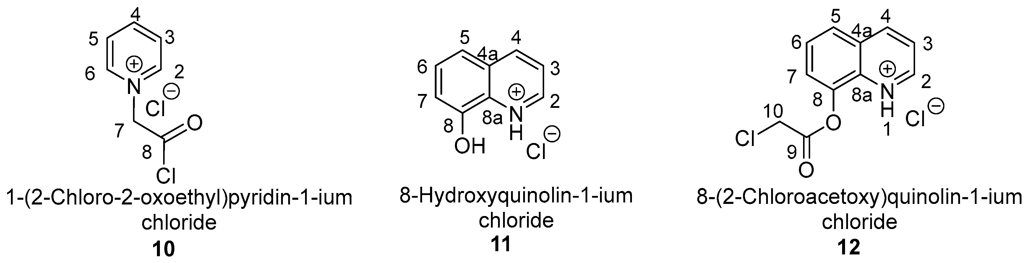
In Scheme 2, the chemical structure with atom numbering for the obtained compounds is shown.
1-(2-Chloro-2-oxoethyl)pyridin-1-ium chloride 10.
The experimental reaction setup was repeated according to the literature procedure described in former papers [34,35]: in a two-necked round-bottomed flask (250 mL), 0.04 mol of 8-hydroxyquinoline was dissolved in 100 mL of dry pyridine. Then 0.022 mol of freshly distilled 2-chloroacyl chloride was added dropwise and then stirred for 2 h at room temperature. The pyridinium salt 10 was obtained as beige crystals, ɳ = 92%, m.p. = 152–154 °C. 1H NMR (500 MHz, CDCl3, δ, ppm): 5.39 (2H, s, methylene protons from 7-th position), 8.16 (2H: H3 and H5, dd, J = 6.0 Hz, J = 8.0 Hz), 8.62 (1H: H4, t, J = 8.0 Hz, J = Hz), 9.04 (2H: H2 and H6, d, J = 6.0 Hz); 13C NMR (125 MHz, CDCl3, δ, ppm): 62.05 (carbon from methylene group from 7-th position), 127.42 (C3 and C5), 145.59 (C4), 145.93 (C2 and C6), 166.93 (C from ester C=O from 8th position).
General procedure to obtain salts 11 and 12.
In total, 1 mmol (0.145 g) of 8-hydroxyquinoline was dissolved in 15 mL of acetone in a 25 mL round-bottom flask. Then, 2 mmol (0.27 mL) of triethylamine was added. To the stirred solution, 5 mmol (0.4 mL) of 2-chloroacyl chloride was added dropwise. When salts 11 are to be obtained, the reaction mixture is stirred at room temperature overnight. If salts 12 are to be obtained, the reaction mixture was refluxed for 24 h. In either case, the mixture formed a white precipitate, which was separated by filtration and washed twice with diethyl ether to remove unreacted chloride.
8-Hydroxyquinolin-1-ium chloride 11.
The quinolinium chloride 11 was obtained as yellow crystals, ɳ = 98%, m.p.= 217–218 °C. 1H NMR (500 MHz, CDCl3, δ, ppm): 7.60 (1H: H7, dd, J = 3.0 Hz, J = 6.0 Hz), 7.74 (2H: H3, H6, m), 8.02 (1H: H5, q, J = 3.0 Hz, J = 8.5.0 Hz), 9.09 (2H: H2, H4, m), 12.18 [1H, broad singlet, proton from OH1-st position]; 13C NMR (125 MHz, CDCl3, δ, ppm): 115.75 (C7), 118.32 (C5), 122.36 (C6), 129.63 (C4a), 129.74 (C8), 130.25 (C3), 144.62 (C4), 145.55 (C8a), 149.00 (C2).
8-(2-Chloroacetoxy)quinolin-1-ium chloride 12.
The quinolinium salt 12 was obtained as white crystals, ɳ = 94%, m.p. = 107–110 °C. 1H NMR (500 MHz, CDCl3, δ, ppm): 4.94 (2H, s, methylene protons from 10-th position), 5.91 [1H, broad singlet, proton from NH1-st position], 7.72 (2H: H6 and H7), 7.75 (1H: H3, d, J = 3.5 Hz), 8.04 (1H: H5, d, J = 8.0 Hz), 8.65 (1H: H4, d, J = 8.0 Hz), 9.01 (1H: H2, d, J = 3.5 Hz); 13C NMR (125 MHz, CDCl3, δ, ppm): 41.34 (carbon from methylene group from 10-th position), 122.41 (C7), 122.45 (C5), 126.75 (C6), 127.05 (C3), 129.25 (C4), 130.90 (C4a), 138.27 (C8), 145.35 (C8a), 150.01 (C2), 166.31 (C from ester C=O from 9-th position).

3. Results and Discussions

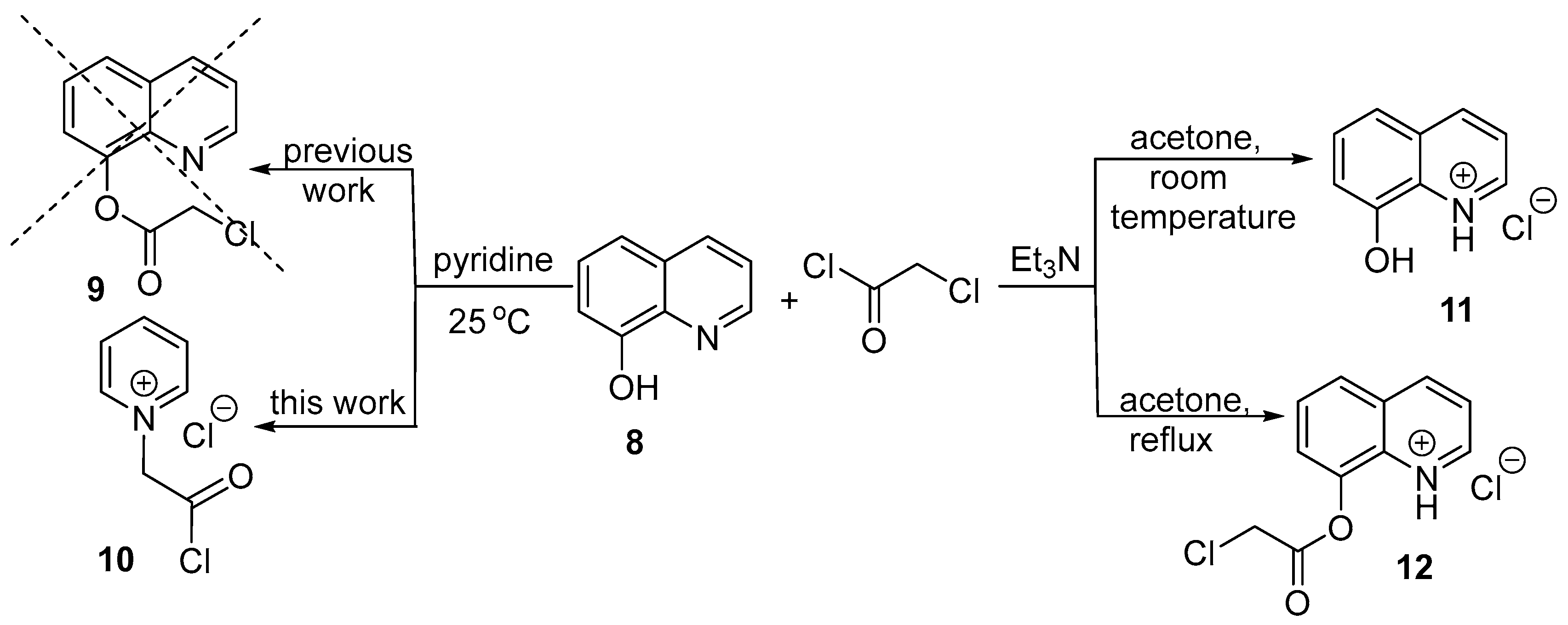
Our initial goal was to obtain the quinoline compound quinolin-8-yl 2-chloroacetate 9, a key intermediate for further synthesis of hybrid biologically active quinoline derivatives. In this respect, we accurately repeated an experimental literature procedure [34,35], where the authors claim that in the reaction of 8-hydroxyquinoline 8 with 2-chloroacyl chloride and using pyridine as a catalyst, they obtained compound 9; Scheme 3.
The above authors claim that they obtained the acylated product, quinolin-8-yl 2-chloroacetate 9, and attempted to confirm its structure using melting point and elemental analysis. Because melting point and elemental analysis are not enough to prove the structure of a compound, we performed the NMR (1H- and 13C-) spectra of the product obtained by using the literature procedure. The 1H- and 13C-NMR spectra of this compound reveal that the real product of synthesis is the salt 1-(2-chloro-2-oxoethyl)pyridin-1-ium chloride 10, and not the acylated product quinolin-8-yl 2-chloroacetate 9, as the literature authors claim. Table 2 lists the signals from the 1H- and 13C-NMR spectra of salt 10 (see also the Supplementary Materials for copies of 1H- and 13C-NMR spectra).
The protons from the α-position of the pyridine ring (H2 and H6) appear at 9.04 ppm as two equivalent protons, having a coupling constant of 6.0 Hz with protons H3/5 (d, J = 6.0 Hz). The protons from the β-position of the pyridine ring (H3 and H5) appear at 8.16 ppm, also as two equivalent protons, coupled with H2/6 and H4 (dd, J = 6.0 Hz, J = 8.0 Hz). The H4 proton from the γ-position of the pyridine ring appears at 8.62 ppm as a triplet (t, J = 8.0 Hz, J = Hz). These protons from the pyridine ring appear at such a high chemical shift because of the powerful unshielded effect exerted by the positive nitrogen from the pyridine ring: the most unshielded protons are H2 and H6 (α-position to the positive nitrogen), followed by H4 protons (γ-position to the positive nitrogen), and H3 and H5 (β-position to the positive nitrogen). The two H7 methylene protons appear at 5.39 ppm (s). This very unusual high chemical shift for these aliphatic methylene protons could be explained by the powerful unshielded effect exerted by the positive nitrogen (from the pyridine ring) and carbonyl ketone group from the vicinity. The carbons from the α-position of the pyridine ring (C2 and C6) appear at 145.93 ppm, those from the β-position of the pyridine ring (C3 and C5) appear at 127.42 ppm, and the C4 carbons from the γ-position appear at 145.59 ppm. The carbon from the methylene group appears at 62.05 ppm, while the carbon from the carbonyl group moiety appears at 166.93 ppm.
The formation of salt 1-(2-chloro-2-oxoethyl)pyridin-1-ium chloride 10 could be explained by the difference in basicity between the two heterocycles, pyridine and 8-hydroxyquinoline. Pyridine (pka = 5.2) is a stronger base compared with 8-hydroxyquinoline (pka = 9.9), and in the reaction conditions employed (room temperature), 2-chloroacyl chloride reagent realizes an alkylation reaction to the more basic nitrogen from pyridine, leading to salt 10.
Having in view that the above literature setup procedure does not lead to the desired compound, we were searching for another experimental procedure to obtain the quinolin-8-yl 2-chloroacetate 9. By using different reaction setups (different catalysts, solvents, molar ratios of reagents, reaction time, and temperature), we found that triethylamine as a catalyst and acetone as a solvent are suitable for this reaction (Scheme 2). Thus, if the reaction took place at room temperature, quinoline hydrochloride 11 is obtained, while using acetone on reflux leads to the 8-(2-chloroacetoxy)quinolin-1-ium chloride 12; Scheme 2. The structure of the obtained compounds was proven by elemental (C, H, N) and spectral analysis: 1H- and 13C-NMR spectra and single-crystal X-ray diffraction spectra (see also Supplementary Materials for copies of 1H- and 13C- NMR spectra and X-ray diffraction spectra of compounds 11 and 12).
The formation of quinoline hydrochloride 11 and salt 8-(2-chloroacetoxy)quinolin-1-ium chloride 12 could be explained by taking into consideration the basicity of heterocycles, the reactivity of 2-chloroacyl chloride, and the reaction conditions. If the reaction took place at room temperature, the system does not have enough energy to accomplish the acylation reaction of the hydroxyl functionality from 8-HQ. Instead, the existing hydrochloric acid from the 2-chloroacyl chloride reagent will achieve a protonation reaction of the nitrogen atom of the pyridine ring of 8-HQ, with the formation of quinoline hydrochloride 11. If the reaction took place at reflux, the system has enough energy to accomplish the acylation reaction of the hydroxyl functionality from 8-HQ, and simultaneously, the hydrochloric acid existing in the 2-chloroacyl chloride reagent will achieve a protonation reaction of the nitrogen atom of the pyridine ring of 8-HQ, with the formation of salt 12.
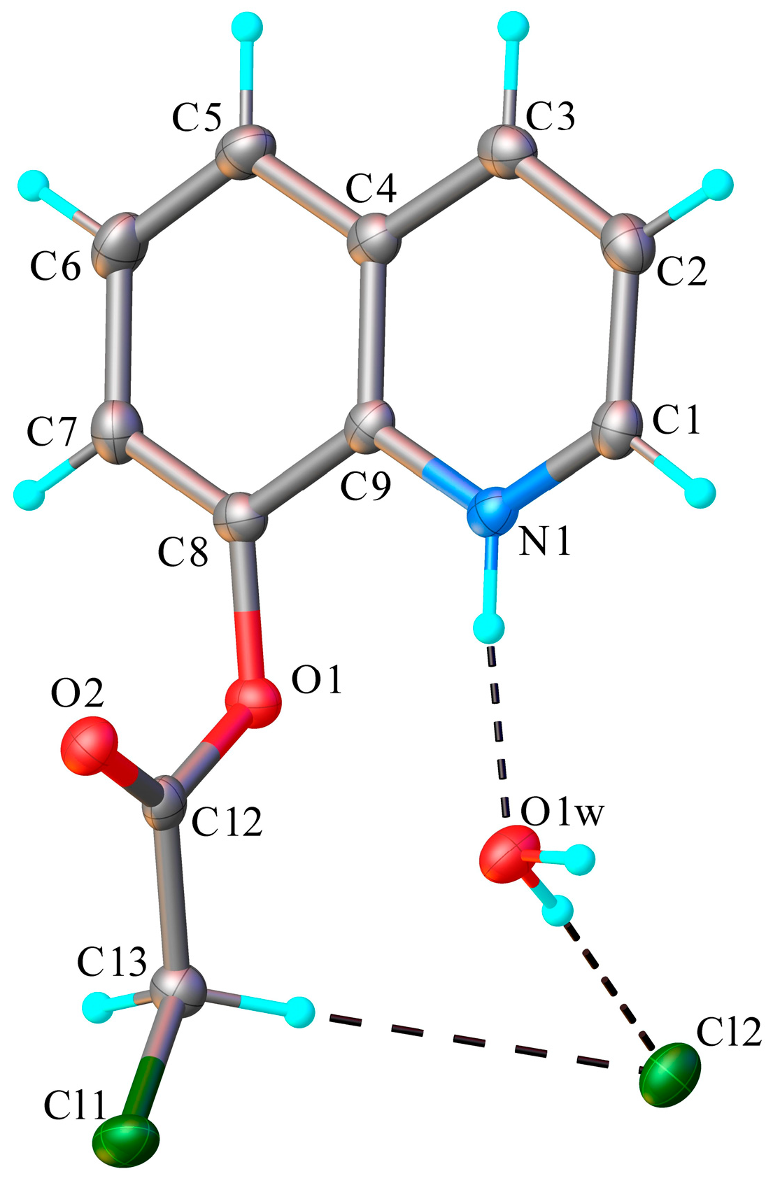
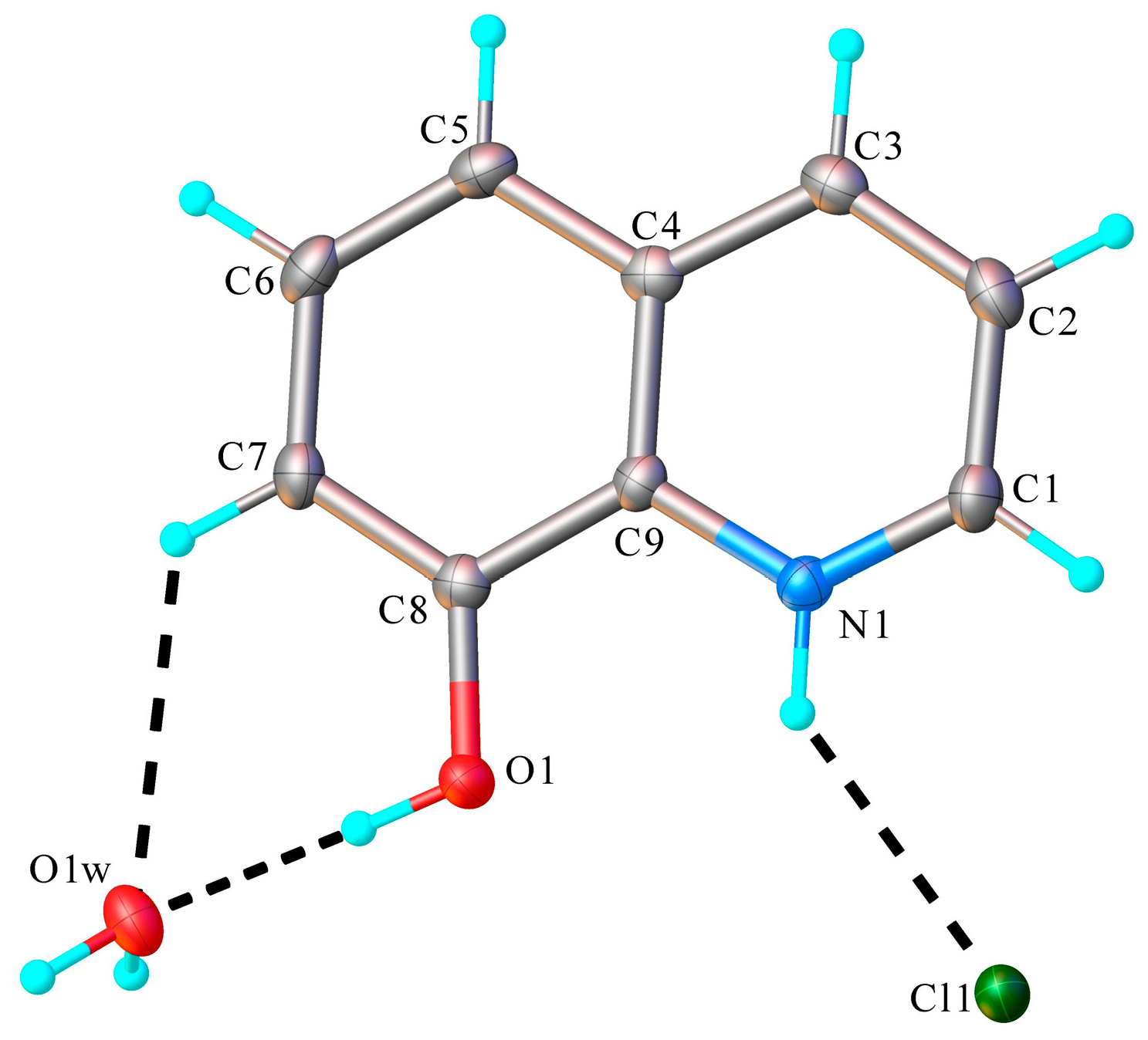
The X-ray diffraction analysis on monocrystal proved the structure of compounds 12 and 11 unambiguously. The obtained results of the X-ray diffraction studies are illustrated in Figure 1 and Figure 2. The bond distances and angles are summarized in Table S1 (see also Supplementary Materials).
As it turned out, both compounds have a planar structure, and in the case of salt 12, the chloroacetyl group from the eighth position of the quinoline ring is slightly out of plane. In the solid state, each of the two compounds 11 and 12 exhibits an ionic crystal structure crystallized as a hydrochloride salt of the corresponding organic base. There is also an interstitial water molecule in the asymmetric part of both crystals, and all the components of the structure are interconnected through a system of intermolecular hydrogen bonding (Figure 1 and Figure 2). In the case of salt 11, the water molecule is connected via hydrogen bonds with the hydrogen atom from hydroxyl group 8-HQ and the hydrogen atom from the seventh position of the 8-HQ moiety. In the case of acylated salt 12, the water molecule is connected via hydrogen bonds with the basic nitrogen atom N1 from the quinoline ring and the chorine atom. The H-bond characteristics are displayed in Table 3.
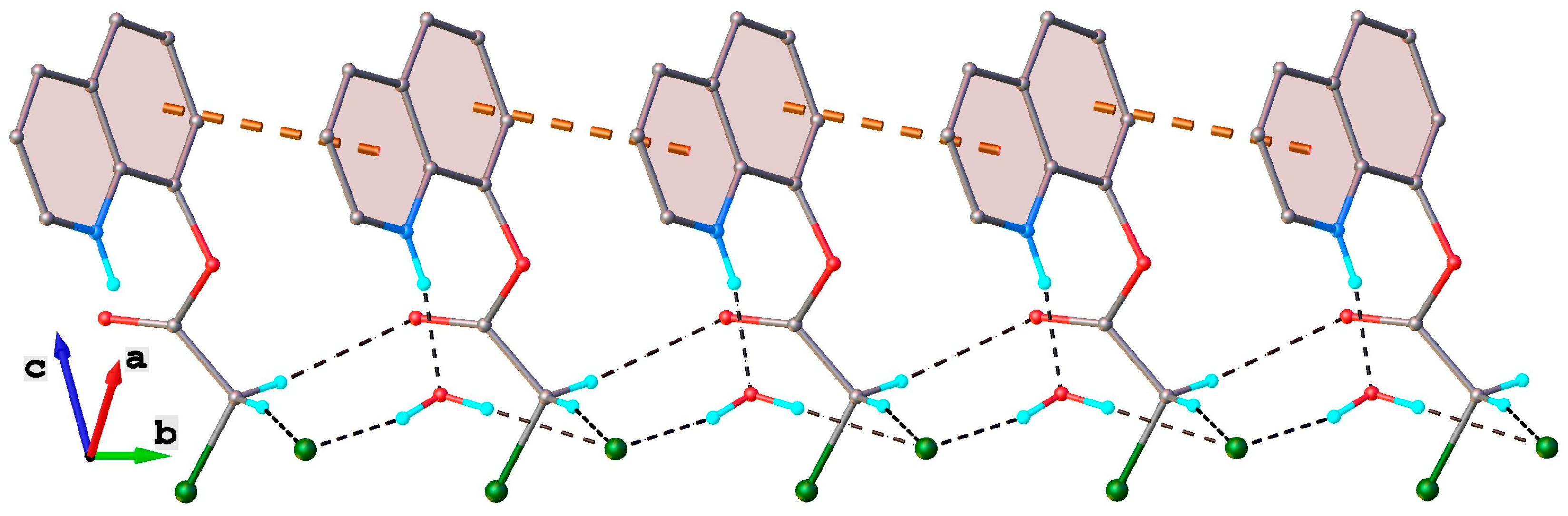
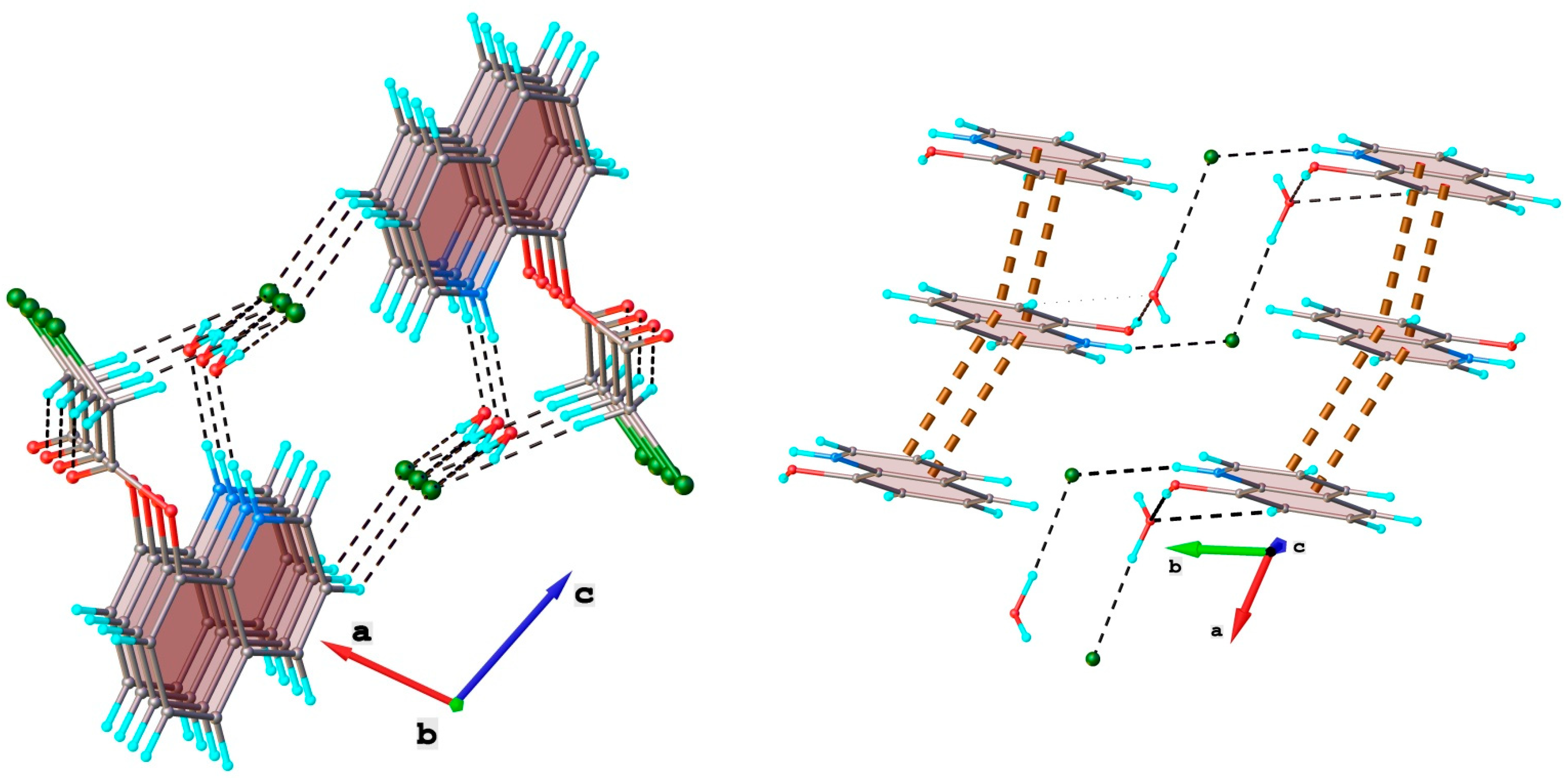
The crystal structures of both compounds 12 and 11 are stabilized by intermolecular hydrogen bonds and π-π stacking interactions. Analyzing the structure of compounds 12 and 11 from the point of view of short intermolecular contacts, it should be noticed that in both cases, besides the classical hydrogen bonding (C-H···O and C-H···Cl), the π-π stacking interactions involving aromatic rings have been identified to influence the crystal packing. Thus, in both crystal structures of 12 and 11, a similar one-dimensional supramolecular array formed via π-π stacking interactions has been observed, as shown in Figure 3 and Figure 4, respectively. Also, numerous hydrogen bonds are present in crystal structures (Table 3, Figure 3 and Figure 4), consolidating the supramolecular structures.
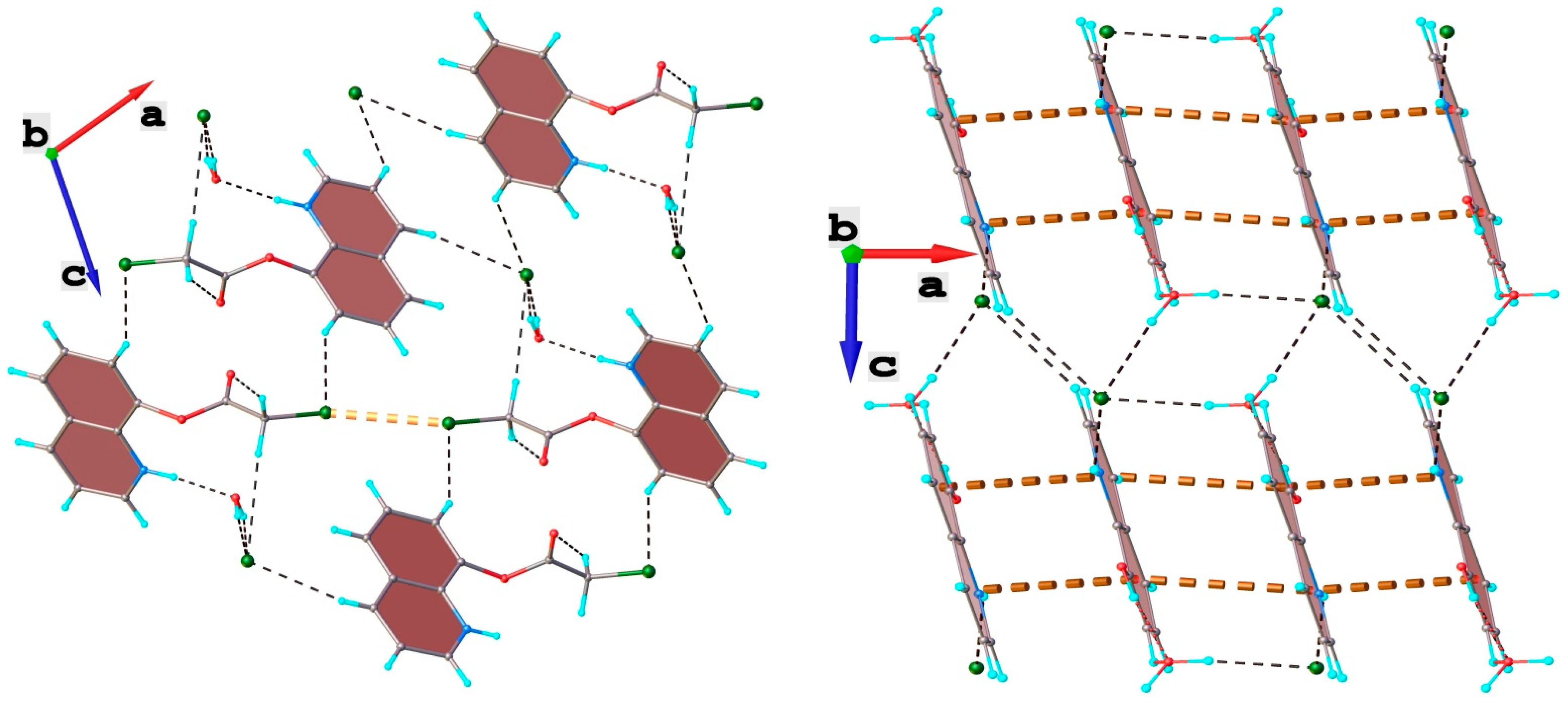
In turn, the intermolecular C-H···Cl hydrogen bonds (Table 3) in the case of crystal 12, and O-H···O, O-H···Cl, N-H···Cl in the case of 11, assemble the 1D chains into two-dimensional supramolecular layers, illustrated in Figure 5.
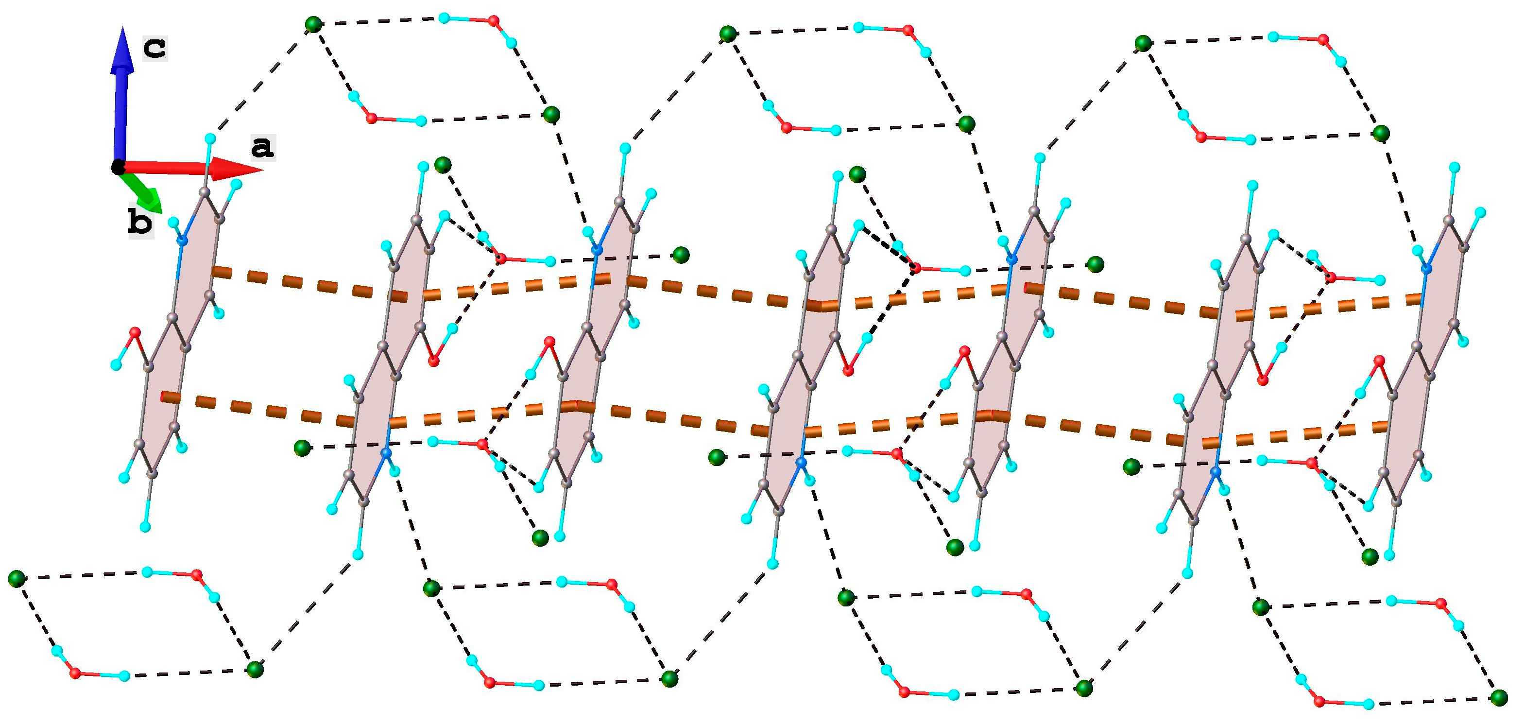
The further analysis has revealed that both structures represent a three-dimensional supramolecular module. In the crystal of 12, the assembly of 2D layers occurs through C3-H···Cl2 hydrogen bond (Table 3) and short interlayer Cl1···Cl1’ contacts at 3.316 Å (symmetry code: −x. 2—y, 1—z), as shown in Figure 6 (left side). In the crystal of compound 11, the 3D network is formed due to O1w-H···Cl1 interlayer H-bonds. A fragment of the 3D supramolecular structure in the crystal of compound 11 is shown in Figure 6 (right side).
The water-soluble salt 8-(2-chloroacetoxy)quinolin-1-ium chloride 12 will be used in further synthesis in an aqueous solution, which is an important asset from a medicinal and green chemistry point of view.

4. Conclusions

This paper presents a detailed study of the acylation reaction products of 8-hydroxyquinoline with 2-chloroacyl chloride, providing new insights and clarifications based on NMR and X-ray analyses. The products formed mainly depend on the catalyst used. When pyridine is employed as the catalyst, the reaction yields salt 1-(2-chloro-2-oxoethyl)pyridin-1-ium chloride 10. The formation of salt 10 could be explained by an alkylation reaction of the more basic nitrogen from pyridine (compared with 8-HQ). Using triethylamine as the catalyst results either in 8-hydroxyquinoline hydrochloride 11 at room temperature or in 8-(2-chloroacetoxy)quinolin-1-ium chloride 12 under reflux conditions. The formation of compounds 11 and 12 could be explained by taking into consideration, in addition to the catalysts, the basicity of heterocycles, the reactivity of 2-chloroacyl chloride, and the reaction conditions. The structures of these compounds were confirmed through 1H- and 13C-NMR spectra, as well as single-crystal X-ray diffraction studies of compounds 12 and 11. The X-ray analyses show that compounds 11 and 12 have a planar structure, and in the case of salt 12, the chloroacetyl group from the eighth position of the quinoline ring is slightly out of plane. Also, both compounds form ionic crystal structures as hydrochloride salts of the respective organic bases. Compound 12 crystallizes in the monoclinic P2_1/n space group, while compound 11 crystallizes in the P-1 space group, each with an interstitial water molecule in the asymmetric unit. The crystal structures are stabilized by intermolecular hydrogen bonds. In their crystal packing, compounds 12 and 11 form similar one-dimensional supramolecular chains through hydrogen bonds (C-H···O and C-H···Cl) and π-stacking interactions (involving aromatic rings), which reinforce the supramolecular architecture. The 1D chains assemble into two-dimensional layers via intermolecular C-H···Cl hydrogen bonds in compound 12, and O-H···O, O-H···Cl, and N-H···Cl hydrogen bonds in compound 11. The 3D supramolecular structure of compound 12 forms through C3-H···Cl2 hydrogen bonds and short interlayer Cl1···Cl1′ contacts, while that of compound 11 is stabilized by O1w-H···Cl1 interlayer hydrogen bonds.

Supplementary Materials

The following supporting information can be downloaded at https://www.mdpi.com/article/10.3390/cryst16040259/s1.

Author Contributions

Conceptualization and formal analysis, I.I.M. and V.M.; Investigation, R.A.T. and S.S.; Writing—original draft, review and editing, R.A.T., S.S., V.M. and I.I.M.; Supervision, I.I.M.; Project administration, R.A.T. and I.I.M. All authors have read and agreed to the published version of the manuscript.

Funding

This research received no external funding.

Data Availability Statement

CCDC 2536830 and 2536831 contain the full crystallographic data for this article. These are available free of charge from the joint Cambridge Crystallographic Data Centre and Fachinformationszentrum Karlsruhe via https://www.ccdc.cam.ac.uk/services/structures (accessed on 28 March 2026) or deposit@ccdc.ca.ac.uk.

Acknowledgments

The authors are grateful to Alexandru Ioan Cuza University of Iasi for financial support and to the Institute of Interdisciplinary Research, CERNESIM, and RECENRAIR centers for the use of their infrastructure.

Conflicts of Interest

The authors declare no conflicts of interest.

References

  1. Vollhardt, P.C.; Schore, N.E. Organic Chemistry: Structure and Function, 8th ed.; W. H. Freeman and Company: New York, NY, USA, 2018. [Google Scholar]
  2. Albrecht, M.; Fiege, M.; Osetska, O. 8-Hydroxyquinolines in metallo supramolecular chemistry. Coord. Chem. Rev. 2008, 252, 812–824. [Google Scholar] [CrossRef]
  3. Kepeňová, M.; Samolova, E.; Smolková, R.; Potočňák, I. Copper(II) complexes of 8-hydroxyquinoline derivatives: Alternative methods for obtaining crystals suitable for X-ray structural analysis. Transit. Met. Chem. 2025, 50, 905–918. [Google Scholar] [CrossRef]
  4. Oliveri, V.; Vecchio, G. 8-Hydroxyquinolines in medicinal chemistry: A structural perspective. Eur. J. Med. Chem. 2016, 120, 252–274. [Google Scholar] [CrossRef]
  5. Zhou, X. Insights of metal 8-hydroxylquinolinol complexes as the potential anticancer drugs. J. Inorg. Biochem. 2023, 238, 112051. [Google Scholar] [CrossRef]
  6. Sharma, R.; Sharma, A.; Borah, A.; Chandra, R.; Singh, S. Synthesis of 8-Hydroxy Quinoline Derivatives: A Revolutionary Target in Medicinal Chemistry (2020–2025). ChemistrySelect 2025, 10, e03182. [Google Scholar] [CrossRef]
  7. Gupta, R.; Luxami, V.; Paul, K. Insights of 8-hydroxyquinolines: A novel target in medicinal chemistry. Bioorg. Chem. 2021, 108, 104633. [Google Scholar] [CrossRef]
  8. Saadeh, H.; Sweidan, K.; Mubarak, M. Recent Advances in the Synthesis and Biological Activity of 8-Hydroxyquinolines. Molecules 2020, 25, 4321. [Google Scholar] [CrossRef] [PubMed]
  9. Song, Y.; Xu, H.; Chen, W.; Zhan, P.; Liu, X. 8-Hydroxyquinoline: A privileged structure with a broad-ranging pharmacological potential. Med. Chem. Commun. 2015, 6, 61–74. [Google Scholar] [CrossRef]
  10. Hegedus, D.; Szemerédi, N.; Petrinca, K.; Berkecz, R.; Spengler, G.; Szatmári, I. Synthesis of Tumor Selective Indole and 8-Hydroxyquinoline Skeleton Containing Di-, or Triarylmethanes with Improved Cytotoxic Activity. Molecules 2024, 29, 4176. [Google Scholar] [CrossRef]
  11. Gasparin, C.B.; Pilger, D.A. 8-Hydroxyquinoline, Derivatives and Metal-Complexes: A Review of Antileukemia Activities. ChemistrySelect 2023, 8, e202204219. [Google Scholar] [CrossRef]
  12. Pape, V.F.S.; Palkó, R.; Tóth, S.; Szabó, M.J.; Sessler, J.; Dormán, G.; Enyedy, É.A.; Soós, T.; Szatmári, I.; Szakács, G. Structure–activity relationships of 8-hydroxyquinoline-derived Mannich bases with tertiary amines targeting multi-drug-resistant cancer. J. Med. Chem. 2022, 65, 7729–7745. [Google Scholar] [CrossRef] [PubMed]
  13. Krawczyk, M.; Pastuch-Gawolek, G.; Mrozek-Wilczkiewicz, A.; Kuczak, M.; Skonieczna, M.; Musiol, R. Synthesis of 8-hydroxyquinoline glycoconjugates and preliminary assay of their B1,4-GalT inhibitory and anti-cancer properties. Bioorg. Chem. 2019, 84, 326–338. [Google Scholar] [CrossRef]
  14. Kozmenko, Y.V.; Khisamov, M.M.; Revtovich, S.V.; Korolev, S.P.; Sherman, D.K.; Spiridonov, V.V.; Kalnina, L.B.; Valuev-Elliston, V.T.; Gottikh, M.B.; Kochetkov, S.N.; et al. Anti-HIV and Antimicrobial Activity of 7-Hydrazino-8-hydroxyquinoline-Based Aromatic Hydrazones. Int. J. Mol. Sci. 2025, 26, 8402. [Google Scholar] [CrossRef]
  15. Peddapaka, J.; Nasreen, A.; Sanam, T.; Shaik, M.G.; Swain, B.; Sanwer, S.; Alvala, R.; Arifuddin, M.; Nerella, S.G. Facile synthesis, antimicrobial activity, and molecular docking analysis of 8-hydroxyquinoline-4-thiazolidinone hybrids. Future Med. Chem. 2025, 17, 435–447. [Google Scholar] [CrossRef]
  16. Krummenauer, M.E.; Camargo, M.d.S.; Gentz, C.d.B.; Silva Lopes, M.; Feliciani da Luz, L.; Silva Trentin, D.d.; Avila Rodrigues, B.; Rigon Zimmer, K.; Fernandes de Andrade, S.; Henning Vainstein, M. Antifungal Activity of 8-Hydroxyquinoline Derivatives Against Candida auris, Candida haemulonii, Cryptococcus neoformans, and Cryptococcus gattii Complex. Pathogens 2025, 14, 999. [Google Scholar] [CrossRef] [PubMed]
  17. Joaquim, A.R.; Boff, R.T.; Adam, F.C.; Lima-Morales, D.; Cesare, M.A.; Kaminski, T.F.; Teixeira, M.L.; Fuentefria, A.M.; Andrade, S.F.; Martins, A.F. Antibacterial and Synergistic Activity of a New 8-Hydroxyquinoline Derivative against Methicillin-Resistant Staphylococcus aureus. Future Microbiol. 2022, 17, 425–436. [Google Scholar] [CrossRef]
  18. Joaquim, A.R.; Gionbelli, M.P.; Gosmann, G.; Fuentefria, A.M.; Lopes, M.S.; Andrade, S.F. Novel antimicrobial 8-hydroxyquinoline-based agents: Current development, structure–activity relationships, and perspectives. J. Med. Chem. 2021, 64, 16349–16379. [Google Scholar] [CrossRef]
  19. Bhat, S.Y.; Jagruthi, P.; Srinivas, A.; Arifuddin, M.; Qureshi, I.A. Synthesis and characterization of quinoline-carbaldehyde derivatives as novel inhibitors for leishmanial methionine aminopeptidase. Eur. J. Med. Chem. 2020, 186, 111860. [Google Scholar] [CrossRef] [PubMed]
  20. Cherdtrakulkiat, R.; Boonpangrak, S.; Sinthupoom, N.; Prachayasittikul, S.; Ruchirawat, S.; Prachayasittikul, V. Derivatives (halogen, nitro and amino) of 8-hydroxyquinoline with highly potent antimicrobial and antioxidant activities. Biochem. Biophys. Rep. 2016, 6, 135–141. [Google Scholar] [CrossRef]
  21. Darby, C.M.; Nathan, C.F. Killing of non-replicating Mycobacterium tuberculosis by 8-hydroxyquinoline. J. Antimicrob. Chemother. 2010, 65, 1424–1427. [Google Scholar] [CrossRef] [PubMed]
  22. Bartolomeu, P.F.; Fortes, I.S.; Zimmer, A.R.; Lopes, M.S.; Andrade, S.F. 8-Hydroxyquinoline Derivatives as Drug Candidates for the Treatment of Alzheimer’s Disease. Curr. Med. Chem. 2026, 33, e09298673409645. [Google Scholar] [CrossRef] [PubMed]
  23. Guerguer, F.Z.; Rossafi, B.; Abchir, O.; Raouf, Y.S.; Albalushi, D.B.; Samadi, A.; Chtita, S. Potential Azo-8-hydroxyquinoline derivatives as multi-target lead candidates for Alzheimer’s disease: An in-depth in silico study of monoamine oxidase and cholinesterase inhibitors. PLoS ONE 2025, 20, e0317261. [Google Scholar] [CrossRef] [PubMed]
  24. Van Hau, T.; Ruankham, W.; Suwanjang, W.; Songtawee, N.; Wongchitrat, P.; Pingaew, R.; Prachayasittikul, V.; Prachayasittikul, S.; Phopin, K. Repurposing of nitroxoline drug for the prevention of neurodegeneration. Chem. Res. Toxicol. 2019, 32, 2182–2191. [Google Scholar] [CrossRef]
  25. Hongmanee, P.; Rukseree, K.; Buabut, B.; Somsri, B.; Palittapongarnpim, P. In Vitro Activities of Cloxyquin (5-Chloroquinolin-8-ol) against Mycobacterium tuberculosis. Antimicrob. Agents Chemother. 2007, 51, 1105–1106. [Google Scholar] [CrossRef]
  26. Jiang, H.; Taggart, J.E.; Zhang, X.; Benbrook, D.M.; Lind, S.E.; Ding, W.Q. Nitroxoline (8-hydroxy-5-nitroquinoline) is more a potent anti-cancer agent than clioquinol (5-chloro-7-iodo-8-quinoline). Cancer Lett. 2011, 312, 11–17. [Google Scholar] [CrossRef]
  27. Zbancioc, G.; Mangalagiu, I.I.; Moldoveanu, C. The Effective Synthesis of New Benzoquinoline Derivatives as Small Molecules with Anticancer Activity. Pharmaceuticals 2024, 17, 52. [Google Scholar] [CrossRef] [PubMed]
  28. Oniciuc, L.; Amăriucăi-Mantu, D.; Diaconu, D.; Mangalagiu, V.; Danac, R.; Antoci, V.; Mangalagiu, I.I. Benzoquinoline Derivatives: An Attractive Approach to Newly Small Molecules with Anticancer Activity. Int. J. Mol. Sci. 2023, 24, 8124. [Google Scholar] [CrossRef] [PubMed]
  29. Al-Matarneh, M.C.; Amarandi, R.-M.; Mangalagiu, I.I.; Danac, R. Synthesis and Biological Screening of New Cyano-Substituted Pyrrole Fused (Iso)Quinoline Derivatives. Molecules 2021, 26, 2066. [Google Scholar] [CrossRef] [PubMed]
  30. Diaconu, D.; Mangalagiu, V.; Amariucai-Mantu, D.; Antoci, V.; Giuroiu, C.L.; Mangalagiu, I.I. Hybrid Quinoline-Sulfonamide Complexes (M2+) Derivatives with Antimicrobial Activity. Molecules 2020, 25, 2946. [Google Scholar] [CrossRef]
  31. Rigaku Oxford Diffraction. CrysAlis Pro Software System; Rigaku Corporation: Oxford, UK, 2015.
  32. Sheldrick, G.M. SHELXT—Integrated Space-Group and Crystal-Structure Determination. Acta Crystallogr. Sect. A Found. Crystallogr. 2015, 71, 3–8. [Google Scholar] [CrossRef]
  33. Dolomanov, O.V.; Bourhis, L.J.; Gildea, R.J.; Howard, J.A.K.; Puschmann, H. OLEX2: A Complete Structure Solution, Refinement and Analysis Program. J. Appl. Crystallogr. 2009, 42, 339–341. [Google Scholar] [CrossRef]
  34. Abd-Alla, M.A.; Ahmed, A.H.N.; El-Zohry, M.F.; Omar, F.A. Synthesis and antibacterial activity of certain quinoline derivatives. Collect. Czechoslov. Chem. Commun. 1992, 57, 1547–1552. [Google Scholar] [CrossRef]
  35. Behera, B.; Rath, P.C.; Routh, M.K.J. New therapeutic agents of the quinoline series: Part II. Quinoline compounds with basic side chains. J. Indian Chem. Soc. 1967, 44, 119–122. [Google Scholar]
Scheme 1. 8-hydroxyquinoline scaffold in diverse drugs.
Crystals 16 00259 sch001
Scheme 2. Atom numbering for obtained compounds.
Crystals 16 00259 sch002
Scheme 3. Reaction pathway between 8-hydroxyquinoline and 2-chlorocetyl chloride.
Crystals 16 00259 sch003
Figure 1. View of the asymmetric part in crystal structure 12 with atom labeling and thermal ellipsoids at a 50% probability level. H-bonds are shown as black-dashed lines.
Crystals 16 00259 g001
Figure 2. View of the asymmetric part in crystal structure 11 with atom labeling and thermal ellipsoids at a 50% probability level. H-bonds are shown as black-dashed lines.
Crystals 16 00259 g002
Figure 3. A view of the one-dimensional supramolecular module showing the role of π-π stacking and hydrogen bonding in the crystal structure of 12. Hydrogen bonds and centroid-to-centroid distances (3.884 Å) are shown as black- and brown-dashed lines, respectively.
Crystals 16 00259 g003
Figure 4. A view of the one-dimensional supramolecular module showing the role of π-π stacking and hydrogen bonding in the crystal structure of 11. Hydrogen bonds and centroid-to-centroid distances (3.812 and 3.554 Å) are shown as black- and brown-dashed lines, respectively.
Crystals 16 00259 g004
Figure 5. Partial view of the crystal packing showing the formation of a two-dimensional supramolecular layer in the crystal of 12 (left); partial view of the crystal packing showing the formation of a two-dimensional supramolecular layer in the crystal of 11. Hydrogen bonds and centroid-to-centroid distances are shown as black- and brown-dashed lines, respectively (right).
Crystals 16 00259 g005
Figure 6. Partial view of the 3D network in the crystal of 12. Cl···Cl contacts are shown in dashed-orange lines (left side); partial view of the 3D network in the crystal of 11 (right side).
Crystals 16 00259 g006
Table 1. Crystal data and details of structure refinement of the X-ray analysis for compounds 12 and 11.
Compound1211
Emp. formulaC11H11Cl2NO3C9H10ClNO2
Fw276.11199.63
T [K]100100
Space groupP21/nP-1
a [Å]14.2286(4)7.2622(3)
b [Å]5.02309(14)8.2725(3)
c [Å]17.7425(5)8.4646(3)
α [°]9078.166(3)
β [°]106.435(3)86.011(3)
γ [°]9066.573(3)
V3]1216.27(6)456.65(3)
Z42
ρcalcd [g cm−3]1.5081.452
μ [mm−1]4.7893.432
Crystal size [mm]0.10 × 0.07 × 0.020.30 × 0.20 × 0.15
2Θ range7.064 to 153.86410.68 to 133.188
Refls. collected66754042
Indep. Refls., Rint2312, 0.02551603, 0.0140
Data/rests./params.2312/0/1601603/0/122
GOF1.0561.124
R1, wR2(all data)0.0272, 0.07360.0256, 0.0709
Table 2. Crystal 1H- NMR (up) and 13C-NMR spectra (down) of salt 1-(2-chloro-2-oxoethyl)pyridin-1-ium chloride 10.
H2 and H6H3 and H5H4H7 (from CH2)-
C2 and C6C3 and C5C4C7 (from CH2)C8, of C=O
109.048.168.625.39-
145.93127.42145.5962.05166.93
Table 3. Hydrogen bond parameters.
Compound 12
D-H···AdD-HdH-AdD-A∠D-H-A/°
N1-H···O1w0.891.762.643(2)170
C2-H···Cl20.952.783.549(2)139
C3-H···Cl20.952.753.642(2)156
C7-H···Cl10.952.793.654(2)152
C13-H···Cl20.992.893.858(2)168
C13-H···O20.992.503.434(2)157
O1w-H···Cl20.892.173.044(1)168(2)
O1w-H···Cl20.822.233.042(1)176(2)
Compound 11
D-H···AdD-HdH-AdD-A∠D-H-A/°
O1-H···O1w0.841.802.6382(13)174.3
N1-H···Cl10.882.373.0965(11)139.7
C1-H···Cl10.952.703.4729(14)139.1
C7-H···O1w0.952.633.2674(17)125.2
O1w-H···Cl10.872.263.1243(11)172.9
Disclaimer/Publisher’s Note: The statements, opinions and data contained in all publications are solely those of the individual author(s) and contributor(s) and not of MDPI and/or the editor(s). MDPI and/or the editor(s) disclaim responsibility for any injury to people or property resulting from any ideas, methods, instructions or products referred to in the content.