Article

Preparation of Polymerizable Mechanochromic Gelator

Department of Applied Chemistry, Graduate School of Engineering, University of Hyogo, 2167 Shosha, Himeji 671-2280, Japan
*
Author to whom correspondence should be addressed.
Crystals 2026, 16(3), 212; https://doi.org/10.3390/cryst16030212
Submission received: 25 December 2025 / Revised: 14 March 2026 / Accepted: 15 March 2026 / Published: 20 March 2026
(This article belongs to the Section Liquid Crystals)

Abstract

Mechanochromism is a phenomenon in which mechanical stimuli change the optical properties of a material, such as its color and emission properties. Various materials exhibiting this behavior have been intensively studied. Mechanochromic materials that exploit liquid crystals have been previously reported. Using liquid crystals, properties different from those of conventional materials, such as anisotropic response and multicolored luminescence due to intermediate aggregation phase stabilization, can be expected. Recently, we reported the preparation and evaluation of the optical properties of liquid-crystalline mechanochromic dyes with cholesterol terminals. The dyes formed gels in some solvents, changed their emission color, and exhibited a friable response without reaching a crystalline state. In addition, film-forming properties, processability, and responsiveness were improved in thin films mixed with polymers. However, the mechanical and thermal stabilities of the gels were low. In this study, a compound similar to the polymerizable unit was synthesized to produce tougher gels. In addition, triblock polymers with a mechanoresponsive dye in the hard segment were synthesized. The xerogel film prepared from the monomer showed an irreversible blue shift in photoluminescent color by mechanical grinding and also exhibited linearly polarized photoluminescence by uniaxial grinding due to force-induced alignment. On the other hand, the xerogel film prepared from the triblock copolymer showed a blue shift in photoluminescent color that can approximately revert to the initial state by thermal annealing, though it showed no anisotropy by uniaxial grinding, indicating that polymerization partially preserves mechanical responsiveness.

1. Introduction

In recent years, the development of stimuli-responsive dyes that reversibly change their color and luminescence in response to external stimuli has attracted considerable attention. Among these, mechanochromic (MC) materials, which exhibit color or luminescence changes in the solid state upon mechanical stimulation, have emerged as promising candidates for passive sensing applications because they operate without external energy inputs, such as heat or light, and can directly respond to physical contact or deformation. In particular, mechanochromic luminescent (MCL) materials are regarded as attractive for practical use because of their ability to visualize stress and strain through changes in light emission [1,2]. MCL materials are applicable to mechanical stress detection across diverse systems, including glass, polymers, and biological environments. For example, Sagara and Kato reported micelles that changed color from yellow to green under a mechanical force and demonstrated MCL responses in living cells [3]. Kwak et al. developed a mechanoresponsive polymer that can visualize fingerprints, in which trace lipids from fingertips diffuse into the polymer film and induce fluorescence [4]. These molecular mechanoresponsive mechanisms can be extended to macroscopic systems, such as structural components [5] and wearable devices [6]. The incorporation of MC or MCL dyes into polymer [4], microcapsule [7], and cellulose nanofiber [8] matrices has enabled the spatial mapping of force distribution and controllable mechanical responses, offering the potential for damage visualization, load monitoring, and enhanced maintenance safety. Because of their simple design in comparison to various electric sensing devices, MC and MCL dyes are key materials for realizing sustainable and adaptive sensing systems. Although previous studies have improved coloration efficiency and spectral tunability through the molecular design and optimization of conjugated frameworks, critical challenges remain in achieving stable dispersion, durable response, and the precise control of MC behavior for functional implementation. To address these limitations, the incorporation of liquid-crystalline (LC) anisotropy and self-assembly characteristics has proven effective. LC-based MCL systems exhibit multicolor luminescence through transitions between multiple solid-state packing structures [9,10], phase-dependent color control [11,12], and circularly polarized luminescence [13], and they can even detect the grinding direction via force-induced molecular alignment combined with polarized emission [14,15]. Furthermore, hydrogen-bonded supramolecular LC phases have demonstrated tunable MC responses, underscoring the role of the LC order in enhancing both sensitivity and structural complexity [16,17]. In addition to LC systems, MCL-active gels have gained attention as alternative supramolecular approaches. These dyes form unique solid architectures during gelation, enabling luminescence changes without forming crystalline aggregates [18]. Low-molecular-weight gels offer the additional advantage of easy processing into films owing to their solid-like mechanical properties. Among gelating units, cholesterol moieties are frequently employed because of their strong packing ability [19] and long-established role as mesogenic components. In our previous study, we developed MC dyes bearing cholesterol groups at their terminal positions and demonstrated polarization-dependent luminescence arising from their LC character. These dyes self-assembled into uniform xerogel films in dioxane and showed enhanced processability and mechanoresponsiveness when blended with polymers [20]. Moreover, by employing ionic liquids as dispersion media, we achieved electrochromic behavior by adding electrical responsiveness to the MC system [21]. However, conventional gel-based materials exhibit insufficient mechanical and thermal stabilities and a limited range of response modes, particularly for frictional stimuli. In this study, we aim to address these challenges by synthesizing MC dyes bearing polymerizable functional groups and constructing polymer gels based on these dyes, thereby developing MC gel materials with enhanced structural robustness and versatile deformation responses. Furthermore, we designed and synthesized triblock copolymers incorporating friction-responsive dyes into the hard segments to achieve both diverse MC behavior and improved material processability.

2. Results and Discussion

2.1. The General Properties of the Monomer

Figure 1 shows the molecular structure of the polymerizable dye used in this study. To achieve both mechanical responsiveness and processability, we designed a molecule in which the cholesteryl unit responsible for gel formation and LC behavior was covalently linked to a cyanostilbene derivative known for its MCL. This structure has a similar structure to a gelling agent called ALS (Aromatic group A, Linker L, Steroid group S), which forms one-dimensional fibrous aggregates that serve as the basic structure for forming physical gels by combining different intermolecular interactions acting on the steroid and aromatic moieties [22]. Cyanostilbene derivatives possess large dipole moments and adopt a twisted geometry around the central ethylene bond. They are also known to form various aggregated structures through multiple C–H···N and C–H···O hydrogen bonds, which can reversibly change upon external stimuli. Because of these properties, cyanostilbenes have frequently been utilized as functional cores in MCL materials [23]. In addition, their rod-like molecular shapes facilitate the formation of LC phases [24]. To enhance structural stability and material processability through polymerization, we extended the terminal alkyl chain and introduced a methacrylate group at the chain end. Methacrylates offer the advantage of facile polymerization via radical mechanisms, allowing for the fine-tuning of material properties through copolymerization or block formation. In general, incorporating dye molecules into a polymer backbone often alters aggregation behavior and crystallinity, which can affect the sensitivity of MCL dyes.
Accordingly, MCL dyes that operate through mechanisms without changes in solid-state aggregate structure (such as alterations in conformation [25] or chemical bonds [26]) have been considered suitable for polymerization. The compound derivative employed in this study lacks well-defined crystalline aggregates detectable by X-ray diffraction (XRD), and its luminescence changes are governed by subtle variations in local aggregation. Therefore, we expected that polymerization would minimally disrupt the aggregation behavior, enabling the polymerizable dye to retain its mechanoresponsive properties. The details of the synthetic procedures are provided in the Experimental Section.
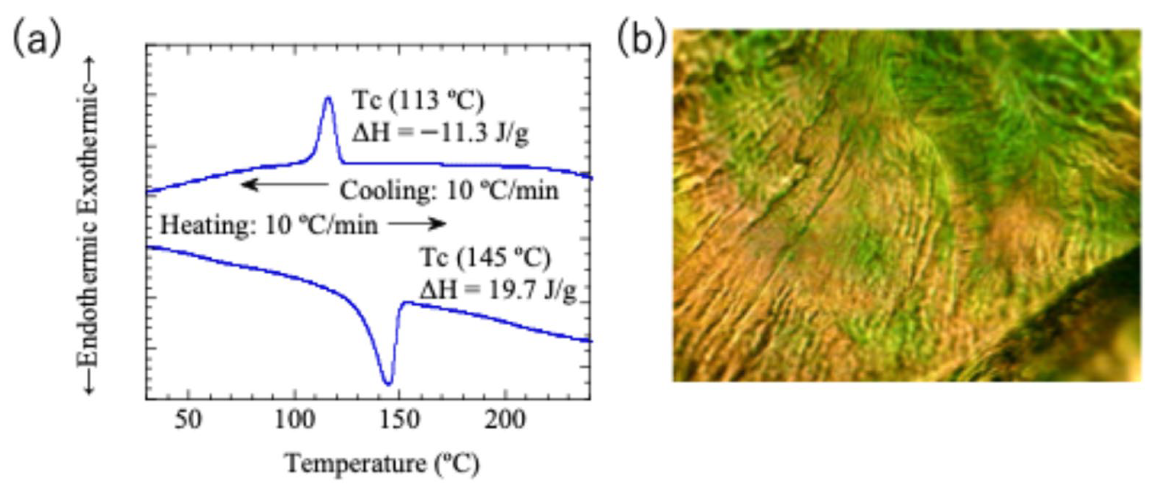
Differential scanning calorimetry (DSC) analysis revealed that the compound exhibits a single LC phase above 113 °C. As shown in Figure 2b, polarized optical microscopy (POM) observation at 140 °C displayed a characteristic oily streak texture, confirming that the material forms a cholesteric LC phase. Cholesteric liquid crystals exhibit MC behavior through both selective reflection and luminescence [27]. Although a similar response was expected for this compound, no selective reflection color was observed. This absence is likely due to the insufficient helical twisting power of the molecule, resulting in a short helical pitch that corresponds to the ultraviolet region rather than the visible range.
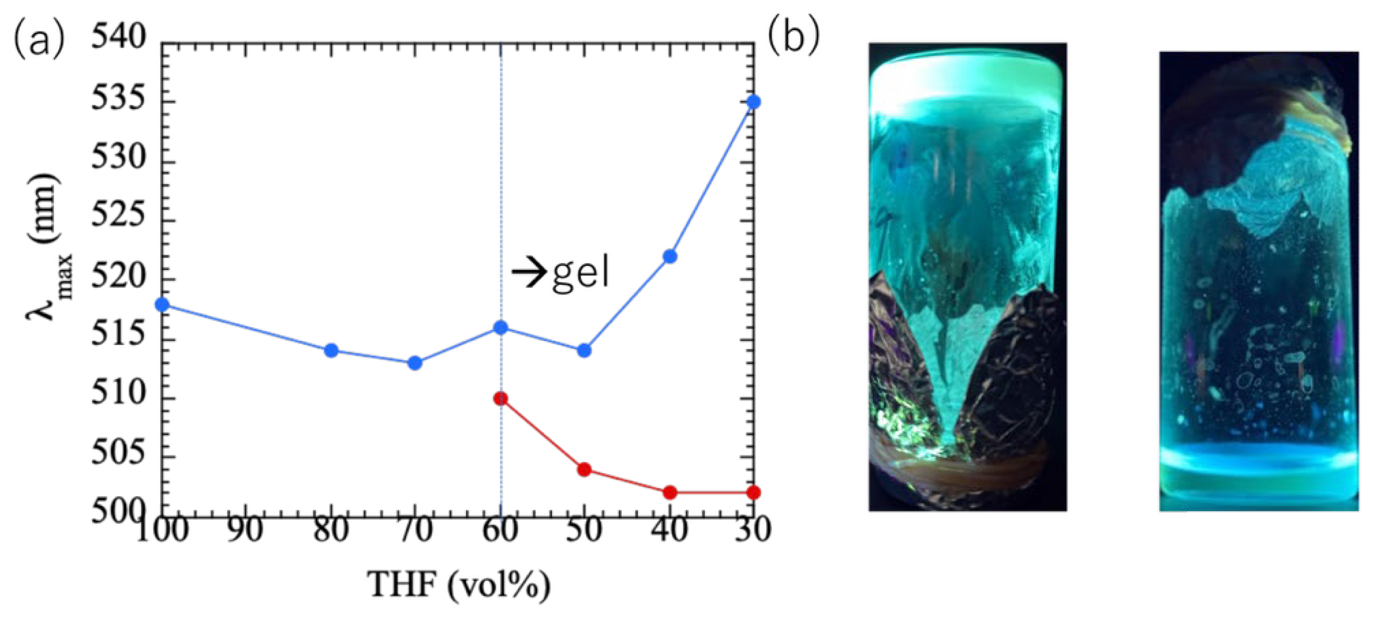
Figure 3a shows the changes in the maximum emission wavelength of the monomer in tetrahydrofuran (THF)/hexane mixtures with various volume ratios. The compound was highly soluble in THF and exhibited blue fluorescence with an emission maximum at 518 nm. As the hexane fraction increased, the emission maximum blue-shifted, reflecting the solvatochromic effect associated with a decrease in solvent polarity. At higher hexane concentrations (40 vol%), the solution formed a gel at room temperature, while it dissolved by heating, accompanied by a green emission centered at 516 nm. The blue shift is attributed to solvatochromism arising from changes in the interaction between the molecular charge distribution and solvent environment. In addition, the monomer formed physical gels when dissolved in 1,4-dioxane above 20 mg/mL (Figure 3b).
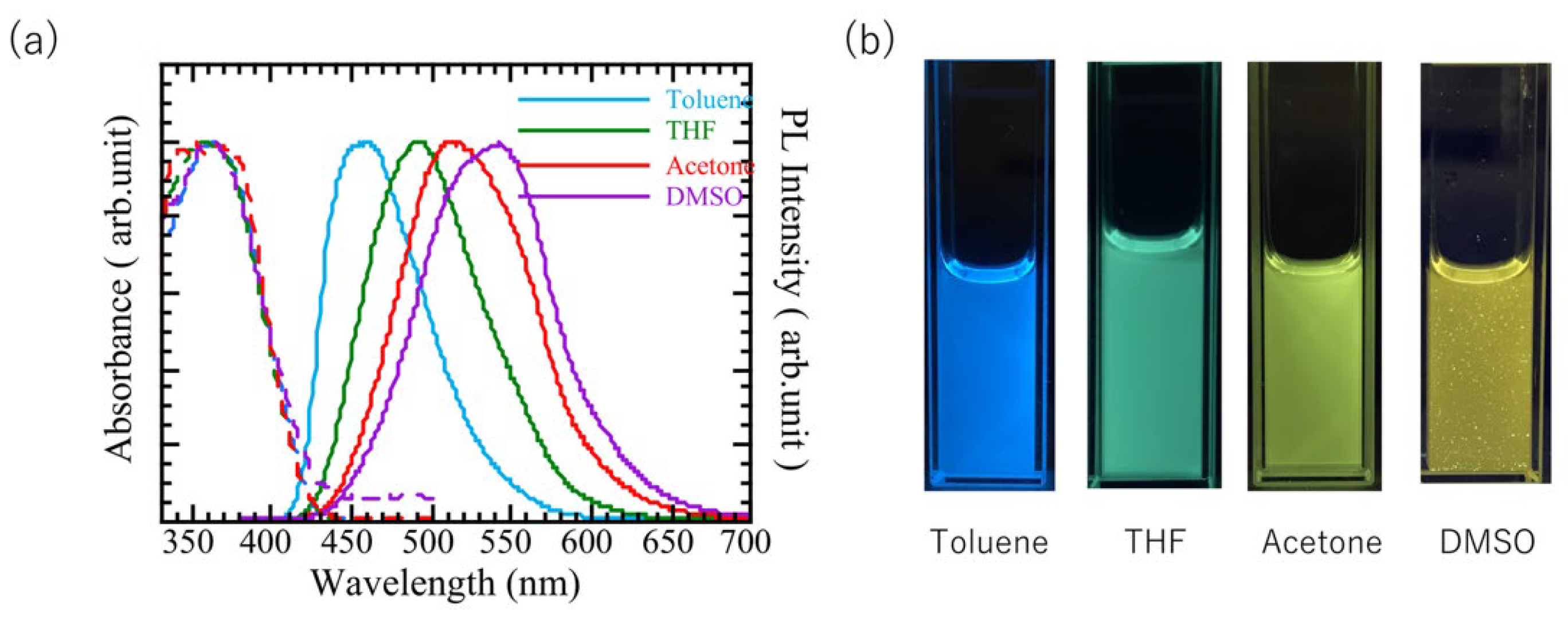
Although the compound was insoluble in pure hexane and its emission spectrum could not be obtained, measurements in solvents of different polarities supported this interpretation. A blue shift was observed in toluene, which has a lower polarity than THF, whereas red shifts occurred in more polar solvents, such as acetone and DMSO (Figure 4).
The red shift associated with gelation was ascribed to the stabilization of the emissive state caused by restricted intramolecular motion and enhanced intermolecular interactions in the aggregated network.
These results demonstrate that the present compound exhibits reversible luminescence changes depending on both the solvent environment and molecular assembly state, highlighting its potential as a stimuli-responsive luminescent material.

2.2. The Mechanoresponsive Behavior of the Monomer

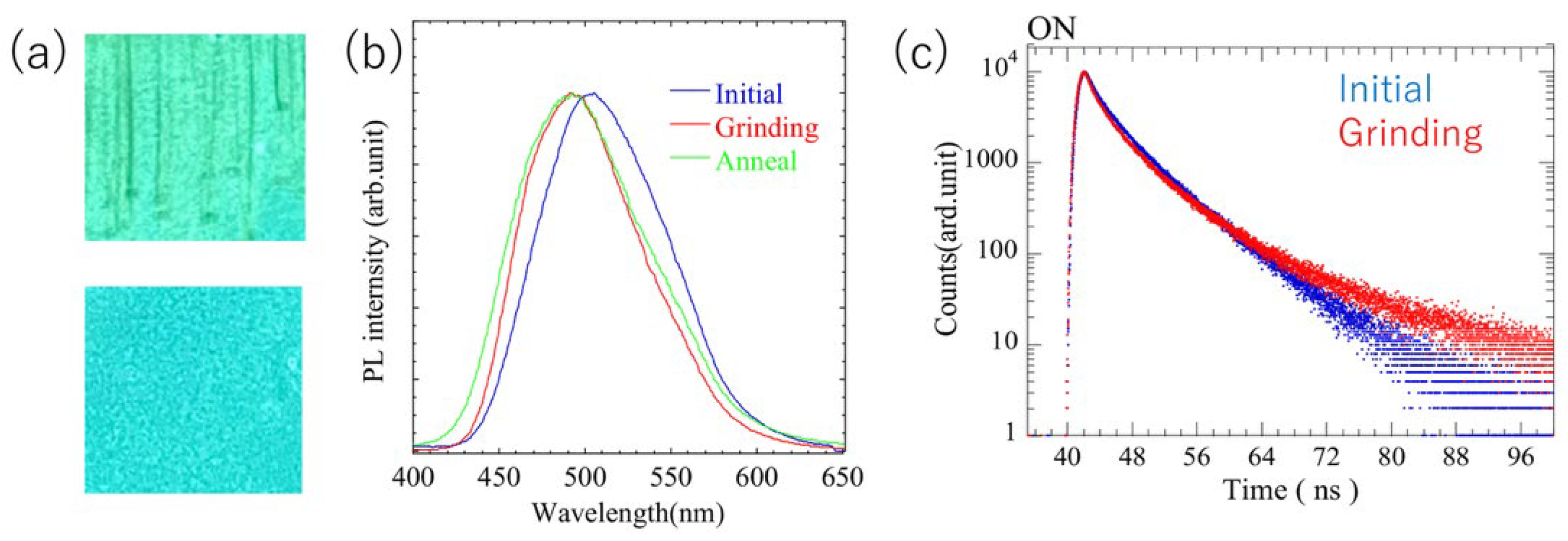
Figure 5a shows the photoluminescence (PL) spectra and fluorescence photographs of the xerogel thin films before and after grinding. Before grinding, the xerogel films exhibited a blue emission with a maximum wavelength of 505 nm. After grinding, the emission components at shorter wavelengths increased, and the maximum emission wavelength shifted to 492 nm, resulting in a visually bluer emission. The quantum yields before and after grinding were 1.5% and 1.3%, respectively, showing no significant difference. Fluorescence lifetime measurements revealed a slight increase in lifetime after grinding (Figure 5b). The emission color changed from blue (λ_max = 505 nm) to a shift towards shorter wavelengths (λ_max = 492 nm) after grinding. In previous studies, related compounds lacking polymerizable groups and shorter alkyl chains exhibited longer wavelengths and longer-lived emission components before grinding. This comparison suggests that the aggregate structure formed upon gelation depends strongly on the molecular design and the gelation mechanism does not necessarily coincide with the mechanoresponsive behavior induced by grinding. No distinct crystalline peaks were observed in the XRD patterns, suggesting amorphous aggregation. The SEM images revealed fibrous structures before grinding that disappeared after grinding, indicating the collapse of the fibrous network and reorganization of the fine aggregates.
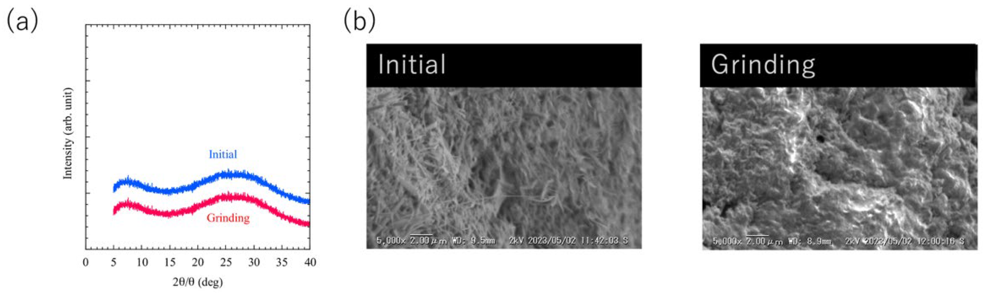
The aggregated structures of the xerogel thin films of the monomer before and after grinding were further analyzed by powder XRD and scanning electron microscopy (SEM), as shown in Figure 6.
The XRD patterns (Figure 6a) showed no distinct crystalline peaks in either state, indicating that both films possess amorphous or poorly ordered aggregate structures. SEM observations (Figure 6b) revealed fibrous structures in the film before grinding, which were disrupted and disappeared after grinding. These results suggest that the change in luminescent color upon grinding originates from microscopic rearrangements within the fibrous aggregates.
Cyanostilbene derivatives have been reported to exhibit mechanochromism through various intermolecular and intramolecular interactions. In the materials studied here, the absence of prominent peaks in XRD indicates that this phenomenon is not attributable to strong intermolecular periodic structures like crystalline structures. Furthermore, the minimal change in fluorescence lifetime, which consistently remains short, rules out excimer formation as the cause. Previous reports indicate that strong twisting is readily induced in cyanostilbene, and this twisting induces changes in the conjugated length [22]. Adopting this hypothesis, we presume that the conjugated structure stabilized by gelation becomes twisted during grinding, leading to short-wavelength shifts.
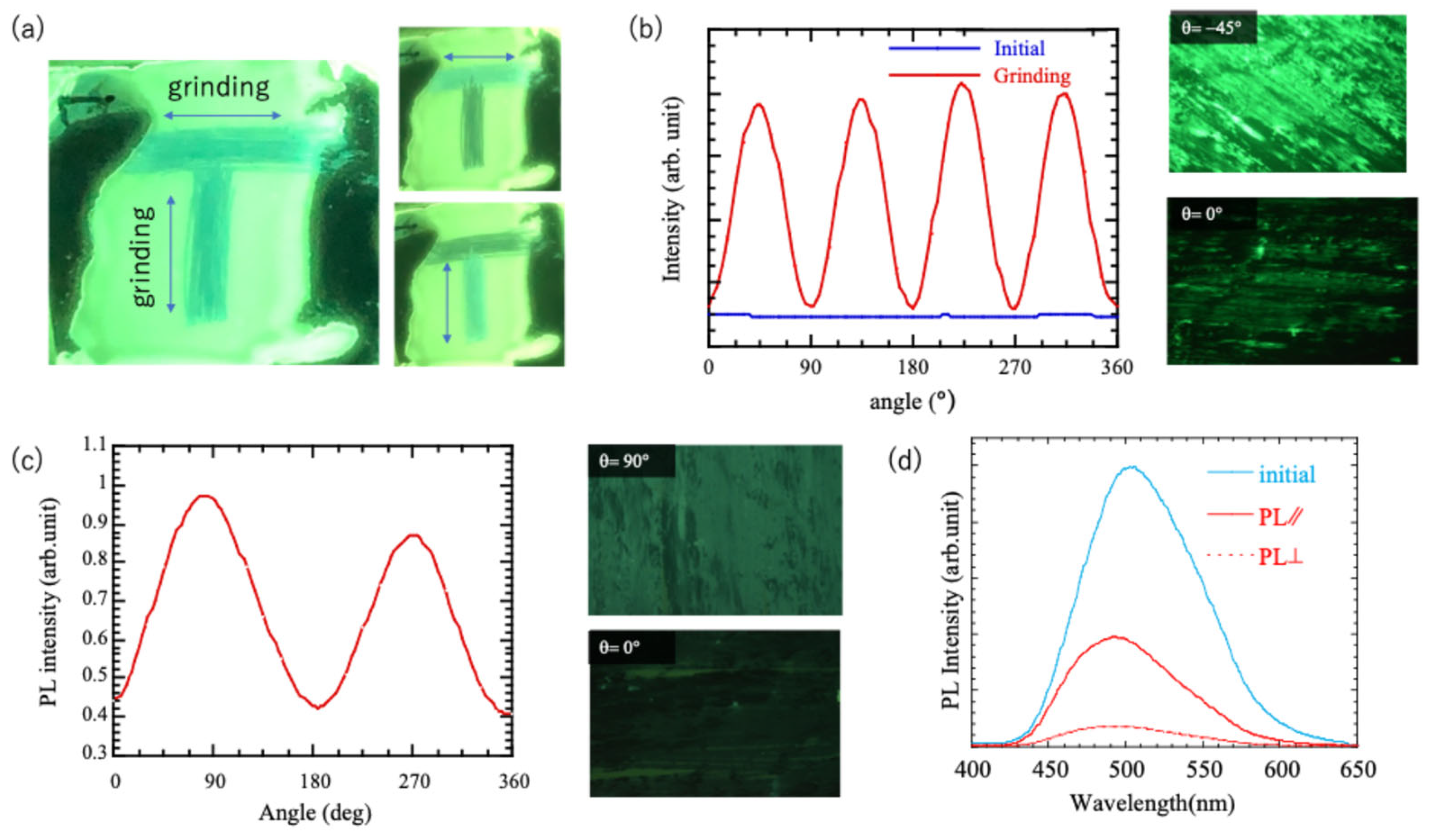
The directional grinding of the monomer film produced a characteristic optical anisotropy, as shown in Figure 7a. When the film surface was rubbed in a T-shaped pattern using a pen tip, blue emission corresponding to the written trace appeared. This indicates that applying mechanical pressure in two orthogonal directions induces anisotropic emissions. When the same film was viewed through a linear polarizer, the bright and dark regions alternated as the orientation of the polarizer was rotated, confirming that the emission intensity depended on the grinding direction (right panel in Figure 7a). Polarizing optical microscopy was used to examine the alignment behavior in the ground areas. As shown in Figure 7b, the transmitted light intensity exhibits periodic bright and dark changes at 45° intervals during rotation, revealing the birefringence induced by the force-directed molecular alignment (inset of Figure 7b). When nonpolarized UV light was used instead of transmitted light, blue emission was observed when the polarizer was parallel to the grinding direction. The emission intensity varied periodically every 180° (Figure 7c), indicating that polarized luminescence occurred along the direction of the force-induced alignment. Finally, polarized PL spectroscopy was performed to quantify anisotropy (Figure 7d). The emission peak in the parallel direction was slightly blue-shifted relative to that in the perpendicular direction. The intensity in the parallel direction was approximately 5.4 times greater than that in the perpendicular direction at the wavelength where the maximum emission intensity was observed. The luminescent polarization ratio p = ( I I ) / ( I + I )   , where I and I are PL intensities parallel and perpendicular to the rubbing direction, respectively, calculated from the respective luminescence intensities, was 0.68, confirming the occurrence of polarized luminescence induced by the grinding process.

2.3. The Mechanoresponsive Behavior of the Polymeric Composite and Polymers

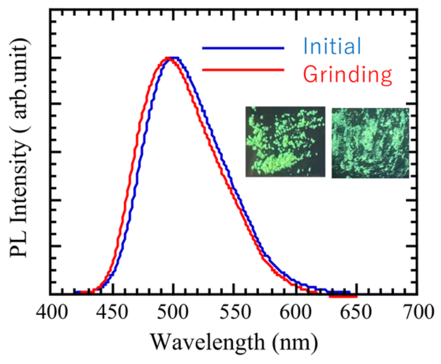
Bulk polymerization was used to prepare the homopolymer. The resulting product was insoluble in THF, confirming polymer formation. DSC analysis revealed an inflection point near 172 °C, and no significant change in texture was observed in the polarized optical microscopy images before and after this transition. These results suggest that the homopolymer retained a certain degree of LC order in the solid state. When the homopolymer was ground in the solid state, the maximum emission wavelength shifted from 501 to 495 nm (Figure 8). Although spectral changes were hardly observed, it was presumed that the grinding responsiveness could be maintained even when this chromophore was polymerized. However, the polymer exhibited extremely low solubility and poor film-forming ability, which prevented the evaluation of its degree of polymerization.
To ensure solubility while evaluating the aggregation structure and luminescence responsiveness of the dye, we adopted a strategy in which relatively low-molecular-weight dye oligomers were attached to a high-molecular-weight non-photoresponsive polymer. A block copolymer architecture was selected to stabilize the aggregated structure, and a commercial macroinitiator (VPE-0201) was used. This initiator contains a PEG chain (Mn ≈ 2000) linked by an AIBN unit, allowing dye homopolymer chains to be introduced at both PEG chain ends during polymerization. Thus, triblock copolymers could be synthesized without specialized polymerization techniques.
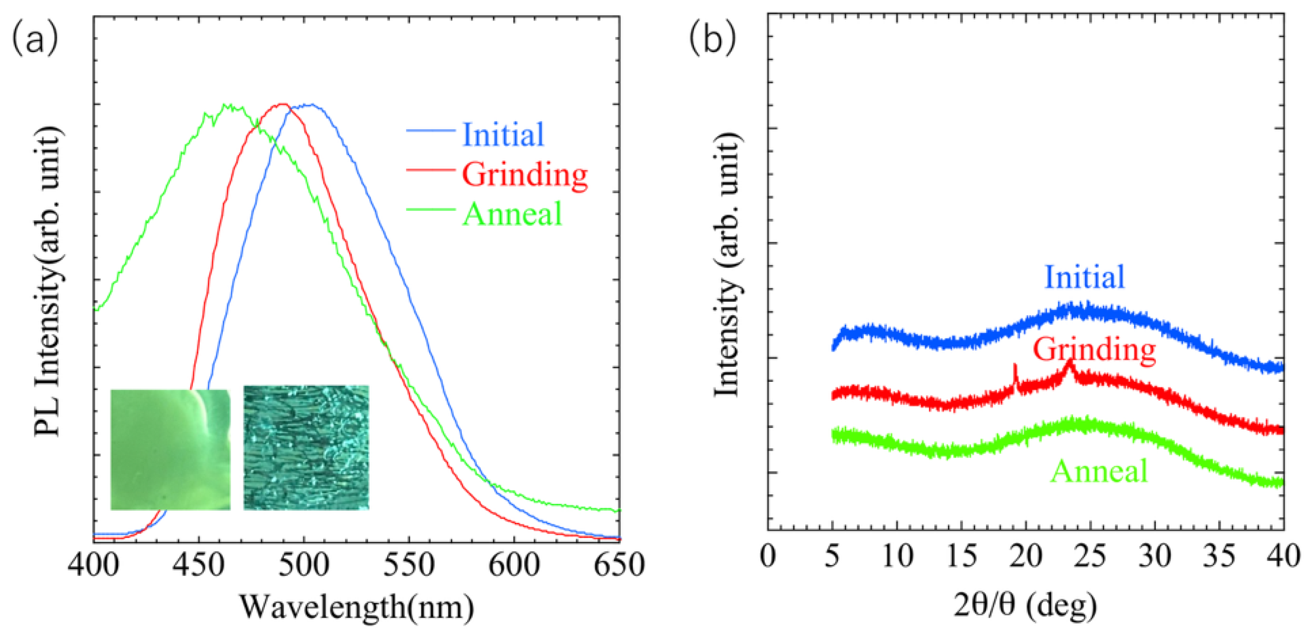
To examine whether the grinding-induced luminescence was preserved in the PEG matrix, a film containing 10 wt% of the monomer dispersed in PEG was prepared. The dispersed film exhibited a similar blue shift in emission upon grinding (Figure 9a), confirming that mechanoresponsive luminescence was retained. In contrast, heating the film at 138 °C caused a marked blue shift in the emission spectrum. PXRD measurements showed no change in the diffraction patterns upon grinding or heating (Figure 9b), suggesting that the dye did not crystallize within the PEG but instead formed a gel-like aggregated structure. The thermally induced blue shift is attributed to the partial disruption of this aggregation.
The block copolymer synthesized using the macroinitiator was analyzed using GPC, and its molecular weight and distribution were confirmed. Considering a PEG molecular weight of 2000, approximately five dye units were estimated to be introduced at each chain end. This copolymer readily dissolved in THF and formed physical gels when dissolved in dioxane, a THF/hexane mixture or chloroform above 30 mg/mL, suggesting that polymerization enhanced the gel-forming capability. However, the resulting films were mechanically fragile and lacked sufficient self-standing ability to apply mechanical stimuli, such as stretching. This limitation is likely due to insufficient molecular weight and may be improved through the optimization of the polymerization conditions.
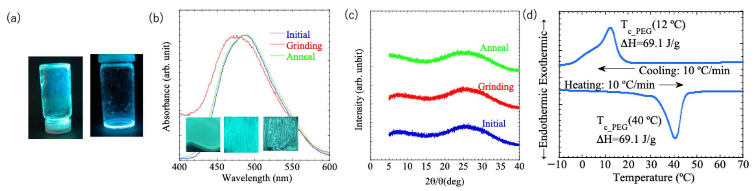
The xerogel films prepared from dioxane exhibited a blue emission at 489 nm in the initial state, which shifted to 476 nm after grinding (Figure 10a). Similarly to the homopolymer and monomer systems, no distinct PXRD peaks were observed (Figure 10b). In contrast, birefringence and polarized emission arising from stress-induced alignment were not detected. Remarkably, when the grinding film was heated to 70 °C, the emission wavelength shifted toward longer wavelengths, restoring its mechanoresponsiveness. DSC revealed an endothermic peak around 40 °C (Figure 10c). Because neither the monomer nor the homopolymer exhibits LC melting at this temperature, and considering that PEG melts near 40 °C, the recovery of the MC response is attributed to PEG block melting, which results in the incorporation of the dye oligomers into the PEG matrix and recovery of the characteristic aggregated structure. A comparison of the monomer and block copolymer is presented in Table 1.
Collectively, these results demonstrate that PEG-based block copolymerization enables the material to retain mechanoresponsive luminescence while improving its solubility and gel-forming ability.

3. Materials and Methods

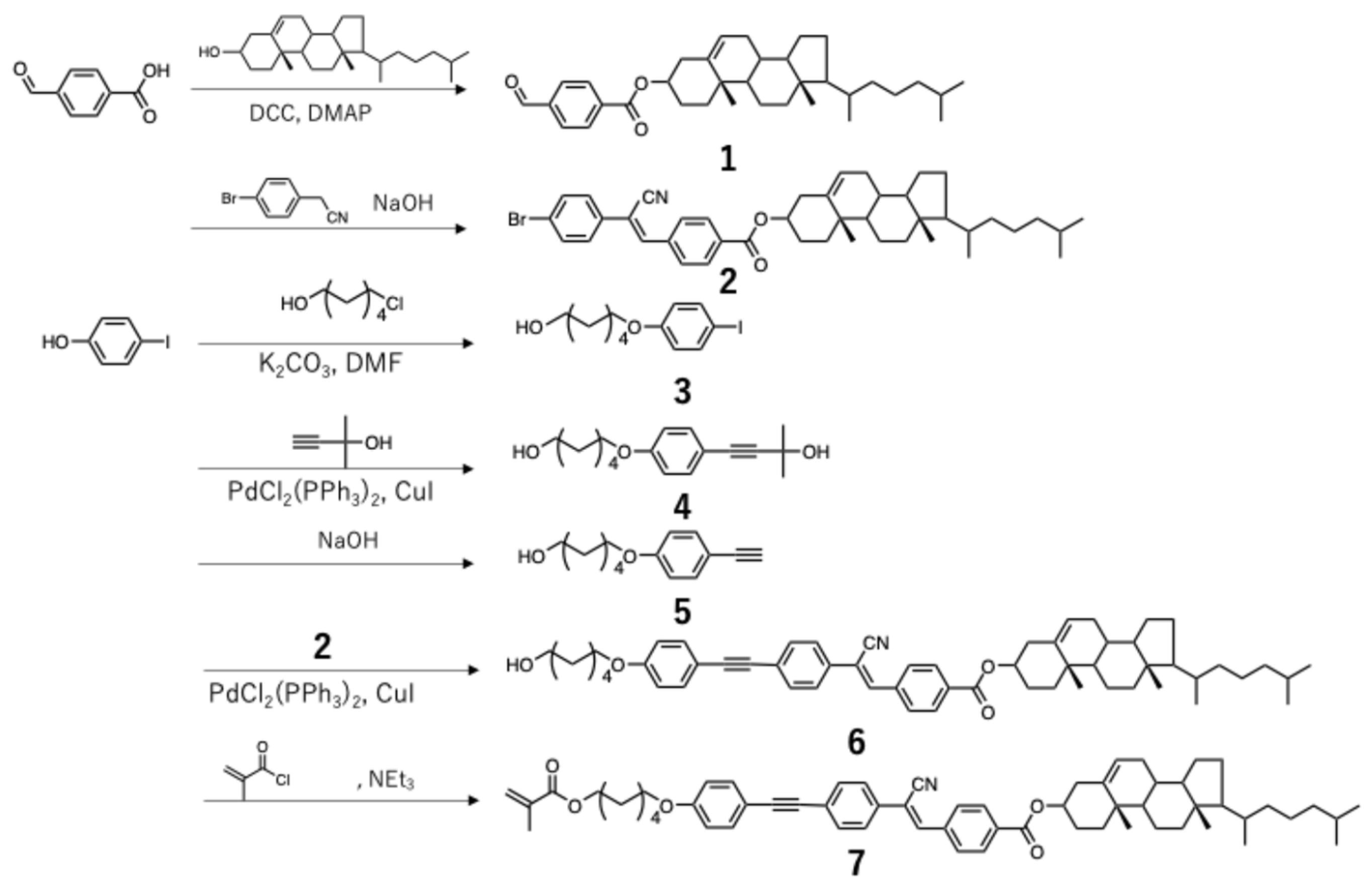
The synthesis of the luminophore used in this study is shown in Scheme 1.

3.1. Synthesis

  • Synthesis of compound 1
  • Terephthalaldehydic acid (2.7 g, 18.3 mmol), cholesterol (7.1 g, 18.3 mmol), and a catalytic amount of DMAP (8 g) were placed in a 200 mL three-necked flask and dissolved in dry THF (50 mL). DCC (5.0 g, 24.0 mmol) was added, and the reaction mixture was stirred at room temperature for 3 h. After completion, the reaction mixture was filtered to remove the precipitated salt, and the filtrate was concentrated under reduced pressure to obtain a white solid.
  • Yield: 5.9 g (11.4 mmol, 62%s) mp: 151–162 °C
  • 1H NMR (400 MHz, CDCl3) δ 10.13–10.11 (s, 1H), 8.28–8.24 (d, J = 8.2 Hz, 2H), 8.01–7.96 (d, J = 8.2 Hz, 2H), 5.41–5.36 (d, J = 4.6 Hz, 1H), 2.38–0.82 (m, 42H), 0.71–0.63 (s, 3H)
  • FT-IR (KBr, cm−1): 3296, 2938, 2860, 1717, 1544, 1468, 1382, 1347, 1316, 1271, 1253, 1236, 1200, 1013.
  • Synthesis of compound 2
  • A mixture of 4-Bromophenylacetonitrile (2.23 g, 11.4 mmol) and compound 1 (5.9 g, 11.5 mmol) was dissolved in THF (200 mL) in a three-necked flask. A solution of potassium tert-butoxide (1.2 g, 11 mmol) in THF (10 mL) was added dropwise, and the reaction was stirred at room temperature for 2 h. After completion, the solvent was evaporated under reduced pressure, and the residue was washed with hexane to afford a white solid.
  • Yield: 4.14 g (5.94 mmol, 52%) mp: 220–300 °C
  • 1H NMR (400 MHz, CDCl3) δ 8.13–8.11 (d, J = 8.2 Hz, 2H), 7.93–7.91 (d, J = 8.2 Hz, 2H), 7.60–7.54 (m, 5H), 5.42 (d, J = 4.1 Hz, 1H), 2.02–0.84 (m, 50H), 0.68 (s, 3H)
  • FT-IR (KBr, cm−1): 3458, 2937, 2866, 2216, 1717, 1635, 1276, 1111.
  • Synthesis of compound 3
  • A solution of 4-Iodophenol (25 g, 0.10 mol) in ethanol (60 mL) was prepared in a 500 mL three-necked flask, and KOH (7.0 g, 0.13 mol) was added and dissolved at 60 °C. The solution was refluxed at 78 °C for 1 h, followed by the dropwise addition of 6-chloro-1-hexanol (17 g, 0.12 mol). Potassium iodide (0.03 g, 0.18 mmol) was added, and the mixture was refluxed at 78 °C for 48 h. After completion (as confirmed by TLC), the reaction mixture was neutralized, dehydrated with sodium sulfate, and filtered. The filtrate was concentrated under reduced pressure to obtain a yellow liquid.
  • Yield: 31 g (0.10 mol, 84%) mp: 33–49 °C
  • 1H NMR (400 MHz, CDCl3) δ 7.44–7.38 (d, J = 8.7 Hz, 2H), 6.84–6.79 (d, J = 8.7 Hz, 2H), 3.90 (q, J = 6.3 Hz, 2H), 3.63 (m, 3H), 1.80–1.73 (m, 6H), 1.62–1.54 (m, 2H)
  • FT-IR (KBr, cm−1): 3301, 2935, 2857, 1589, 1484, 1288, 1245, 1172, 1014.
  • Synthesis of compound 4
  • Bis(triphenylphosphine)palladium(II)dichloride (3.3 g, 9.3 mmol), CuI (3.1 g, 16 mmol), triphenylphosphine (6.1 g, 23 mmol), and compound 3 (31 g, 0.10 mol) were added to a 500 mL three-necked flask under nitrogen. A mixture of 2-Methyl-3-butyn-2-ol (8.7 g, 0.10 mol), triethylamine (30 mL), and THF (60 mL) was prepared, and the reaction mixture was stirred at 65 °C for 24 h. The filtrate was washed with water and extracted with chloroform. The organic layer was dried over anhydrous sodium sulfate and concentrated under reduced pressure to yield a brown liquid.
  • Yield: 24 g (88 mmol, 90%) mp: 48–87 °C
  • 1H NMR (400 MHz, CDCl3) δ 7.34–7.30 (d, 2H), 6.80 (d, J = 2.5 Hz, 2H), 3.94 (t, J = 6.4 Hz, 2H), 3.75–3.72 (m, 5H), 3.68–3.63 (m, 6H), 1.84 (m, 5H), 1.77 (q, J = 7.2 Hz, 2H).
  • FT-IR (KBr, cm−1): 3382, 3245, 2933, 2869, 2218, 1571, 1509, 1474, 1252.
  • Synthesis of compound 5
  • A solution of compound 4 (24 g, 88 mmol) in THF (70 mL) was prepared in a 500 mL three-necked flask. A solution of NaOH (4.0 g, 0.10 mol) in methanol (40 mL) was added, followed by toluene (200 mL). The mixture was refluxed at 120 °C (solution temperature: 103 °C) for 4 h. After the completion of the reaction (as confirmed by TLC), the reaction mixture was filtered, dried over anhydrous sodium sulfate, and concentrated under reduced pressure. The crude product was purified using silica gel column chromatography (eluent: ethyl acetate/hexane = 1:1) to afford a brown liquid.
  • Yield: 9.0 g (12.9 mmol, 47%) mp: 28–48 °C
  • 1H NMR (400 MHz, CDCl3) δ 7.44–7.38 (d, 2H), 6.84–6.79 (d, 2H), 3.94 (t, J = 6.6 Hz, 2H), 3.71–3.51 (m, 4H), 2.98 (s, 1H), 1.85–1.72 (m, 3H), 1.66–1.35 (m, 10H).
  • FT-IR (KBr, cm−1): 3285, 2982, 2938, 2961, 1605, 1508, 1245.
  • Synthesis of compound 6
  • CuI (0.22 g, 1.1 mmol), PPh3 (0.30 g, 1.1 mmol), 5 (1.3 g, 5.9 mmol), triethylamine (15 mL), and THF (20 mL) were combined in a 300 mL three-necked flask. A solution of 2 (4.14 g, 6.0 mmol) in THF (120 mL) was added by heating, and the mixture was bubbled with nitrogen at 55 °C for 40 min. Subsequently, PdCl2(PPh3)2 (1.0 g, 5.2 mmol) dispersed in THF (20 mL) was added dropwise, and the reaction mixture was stirred for 22 h. After filtration, the residue was washed with methanol and diluted with HCl (10:1 v/v) to obtain a green solid.
  • Yield: 1.0 g (1.2 mmol, 20%) Phase transition: C 138 N 296 I
  • 1H NMR (400 MHz, CDCl3) δ 8.14–8.11 (d, J = 8.2 Hz, 2H), 7.94–7.92 (d, J = 8.2 Hz, 2H), 7.68–7.66 (d, J = 8.7 Hz, 2H), 7.59–7.57 (d, J = 8.2 Hz, 3H), 7.48–7.45 (d, J = 8.7 Hz, 2H), 6.88–6.86 (d, J = 8.7 Hz, 2H), 5.42 (d, J = 5.0 Hz, 1H), 2.02–1.71 (m, 7H), 1.34–0.85 (m, 26H), 0.68 (s, 3H)
  • FT-IR (KBr, cm−1): 3335, 3060, 3040, 2933, 2864, 2363, 2211, 1708, 1599, 1514, 1464, 1366, 1278, 1244, 1167, 1103, 1008.
  • Synthesis of compound 7
  • A mixture of 6 (1.0 g, 1.2 mmol), triethylamine (0.18 g, 1.2 mmol), hydroquinone (0.14 g, 1.2 mmol), and dry THF (20 mL) was placed in a 300 mL three-necked flask and stirred in an ice bath (~2 °C). A solution of methacryloyl chloride (0.25 g, 2.5 mmol) in dry THF (5 mL) was added dropwise, and the reaction mixture was stirred for 1 h. After completion, the mixture was filtered, and the filtrate was washed with water and extracted with chloroform. The organic layer was dried over sodium sulfate and concentrated under reduced pressure. The crude product was purified using silica gel chromatography (eluent: chloroform) to yield a yellow solid.
  • Yield: 0.6 g (0.66 mmol, 55%)
  • Phase transition: C 113 N* > 300 (heating), C 145 N* > 300 (cooling)
  • 1H NMR (400 MHz, CDCl3) δ 8.13–8.11 (d, J = 8.2 Hz, 2H), 7.94–0.92 (d, J = 8.7 Hz, 2H), 7.68–7.66 (d, J = 8.7 Hz, 2H), 7.59–7.64 (d, J = 8.2 Hz, 3H), 7.48–7.75 (d, J = 8.7 Hz, 2H), 6.88–6.86 (d, J = 9.2 Hz, 2H), 6.09 (s, 1H), 5.54 (d, J = 1.8 Hz, 1H), 5.42 (d, J = 4.1 Hz, 1H), 2.02–1.79 (m, 7H), 1.34–0.84 (m, 26H), 0.67 (s, J = 5.0 Hz, 3H).
  • FT-IR (KBr, cm−1): 3433, 3056, 3040, 2933, 2862, 2350, 2215 (C≡N), 1710 (C=O, Ph), 1637 (methacrylate), 1601 (C≡C), 1515, 1461, 1372, 1279 (PhCOO), 1245, 1172, 1105.
  • Anal: Calcd for C61H75NO4: C, 81.2; H, 8.38; N, 1.55. Found: C, 81.3; H, 8.20; N, 1.55. MS (LCMS, intensity) m/z Calcd: 901.56. Found: 902.57 (M+).
  • Polymerization of homopolymer
  • A mixture of compound 7 (6.2 mg, 0.041 mmol) and 0.04 mg (0.013 μmol) of 1,1′-Azobis(cyclohexane-1-carbonitrile) was placed in a sample tube and dissolved in THF. The solution was then cast onto a glass substrate, and the reaction was allowed to proceed for 20 h in an oven at 130 °C. After the reaction, the mixture was washed with THF to obtain a yellow solid.
  • Polymerization of block polymer
  • A mixture of 7 (50 mg, 0.055 mmol) and VPE-0201 (450 mg, 0.23 mmol) was placed in a 50 mL flask, dissolved in 5 mL dry THF, and purged with nitrogen for 40 min. Polymerization was then performed by heating at 50 °C for 24 h. After the completion of the reaction, the precipitate was precipitated with ether, and the yellow solid was dried under reduced pressure.
  • Yield: 300 mg Mn:10,400, Mw/Mn:1.3
  • 1H-NMR (400 MHz, CDCl3) δ 8.05–8.17 (br, 4H), 7.85–7.97 (br, 4H), 7.62–7.69 (br, 2H), 7.59 (br, 4H), 7.43 (br, 4H), 6.85 (br, 8H), 3.80–3.63 (br, 5074H), 2.76–2.16 (br, 152H), 1.49–0.84 (br, 190H), 0.67 (br, 6H).
  • FT-IR (KBr, cm−1): 3485, 2904, 2868, 2020, 1734, 1649, 1463, 1349, 1248, 1107, 949.

3.2. Equipment

The synthesized compounds were characterized using Fourier transform nuclear magnetic resonance (FT-NMR; JEOL, Tokyo, Japan; JNM-ECZ 400 MHz) and infrared (FT-IR; JASCO, Tokyo, Japan; FT-IR 6000 MHz) spectroscopy. 1H spectra were recorded for CDCl3 at room temperature using tetramethylsilane (TMS; δ 0.00) as an internal standard. The mass spectra and total C, H and N content of the monomer were measured with a liquid chromatograph mass spectrometer (LCMS; Shimadzu, Kyoto, Japan; Nexera X2-QE system) and an elemental analyzer (PerkinElmer, Inc., Waltham, MA, USA; 2400 series II), respectively. The molecular weight of the polymer was determined by gel permeation chromatography (Tosoh, Tokyo, Japan; HLC-8020 system) using a Tosoh TSK gel column, tetrahydrofuran as the mobile phase, and poly(methyl methacrylate) standards for calibration. Polarized light microscopy (POM; Olympus, Tokyo, Japan; BH50, BH2) and differential scanning calorimetry (DSC; Hitachi High Technologies, Tokyo, Japan; DSC7020) were used to evaluate mesomorphic properties. PL spectra were collected using a luminescence spectrometer (Hitachi, F-4500) or an emission spectrometer (Ocean Optics, Orlando, FL, USA; USB2000+) equipped with a 365 nm UV LED as the excitation source (Ocean Optics, Orlando, FL, USA; LLS-LED). The solid-state quantum yields of the luminescent materials were measured using a fluorescence spectrometer (JASCO, Tokyo, Japan; FP-6600) equipped with an integrating sphere and excited at 360 nm. The PL lifetimes of the composite films were measured using a nanosecond spectrofluorometer (FluoroCube, Horiba, Kyoto, Japan) at an excitation wavelength of 370 nm. XRD measurements were performed using a diffractometer (Rigaku, Tokyo, Japan; SmartLab 3 kW) with a standard collimated beam setup. Microscopic images of the xerogels were obtained using SEM (Keyence, Osaka, Japan; V8800). Thin layers for spectroscopy and XRD analyses were prepared by spin-casting dioxane solutions (20 mg/mL for monomer and 30 mg/mL for block copolymer, heated above 40 °C) onto glass substrates. Thin layers of luminophores or polymer composites were ground in a pen, and the luminophore powder was ground in an agate mortar.

4. Conclusions

In this study, design strategies for polymeric materials incorporating mechanoresponsive dyes were investigated to achieve film- and gel-forming properties. The dye homopolymer retained a partial LC order in the solid state and exhibited a mechanoinduced blue shift in emission; however, its extremely low solubility limited further structural analysis and film fabrication. To address these limitations, PEG-based block copolymers were synthesized using a commercial macroinitiator to maintain both solubility and aggregation-dependent luminescent functionality.
The resulting block copolymers showed good solubility and formed physical gels in various solvents, indicating enhanced gel-forming capability upon polymerization. Xerogel films derived from dioxane exhibited clear grinding-responsive luminescence, whereas thermal treatment recovered this response owing to the melting of the PEG blocks.
This study provides new insights into the polymer design principles required for mechanoresponsive luminescent materials and highlights the potential of PEG-based block copolymers as mechanically responsive soft materials.

Author Contributions

Conceptualization, N.K. and M.K.; methodology, M.K. and S.I.; validation, T.I. and S.I.; investigation, T.I. and S.I.; resources, N.K.; writing—original draft preparation, M.K.; writing—review and editing, N.K.; visualization, M.K.; supervision, N.K. All authors have read and agreed to the published version of the manuscript.

Funding

This work was partially supported by a Grant-in-Aid for Scientific Research from the Japan Society for the Promotion of Science (JSPS) (No. 24K08514).

Data Availability Statement

The original contributions presented in this study are included in the article. Further inquiries can be directed to the corresponding author.

Conflicts of Interest

The authors declare no conflicts of interest.

References

  1. Ito, S. Mechanochromic luminescence of soft crystals: Recent systematic studies in controlling the molecular packing and mechanoresponsive properties. J. Photochem. Photobiol. C 2022, 51, 100481. [Google Scholar] [CrossRef]
  2. Hirai, Y. On sense and deform: Molecular luminescence for Mechanoscience. ACS Appl. Opt. Mater. 2024, 2, 1025–1045. [Google Scholar] [CrossRef]
  3. Sagara, Y.; Komatsu, T.; Ueno, T.; Hanaoka, K.; Kato, T.; Nagano, T. Covalent attachment of mechanoresponsive luminescent micelles to glasses and polymers in aqueous conditions. J. Am. Chem. Soc. 2014, 136, 4273–4280. [Google Scholar] [CrossRef]
  4. Kwak, G.; Lee, W.E.; Kim, W.H.; Lee, H. Fluorescence imaging of latent fingerprints on conjugated polymer films with large fractional free volume. Chem. Commun. 2009, 2112–2114. [Google Scholar] [CrossRef]
  5. He, X.; Sun, W.; Du, W.; Wang, D.; Guo, H. Fluorescence stress probing enhanced crack detection and quantification in ancient Chinese murals for deterioration mechanism analysis. npj Herit. Sci. 2025, 13, 133. [Google Scholar] [CrossRef]
  6. Hu, J.; Liu, Y.; Zhang, X.; Han, H.; Li, Z.; Han, T. Fabricating a mechanochromic AIE luminogen into a wearable sensor for volatile organic compound (VOC) detection. Dye. Pigment. 2021, 192, 109393. [Google Scholar] [CrossRef]
  7. Ogasawara, K.; Kuromiya, D.; Watabe, T.; Takahashi, A.; Aoki, D.; Otsuka, H. Mechanochromic polymer particles crosslinked by a radical-type mechanochromophore. RSC Mechanochem. 2025, 2, 763–769. [Google Scholar] [CrossRef]
  8. Das, B.; Uchikura, Y.; Matsuhisa, N.; Oaki, Y.; Pennington, M.; Sugihara, K. Mechanochromic chameleon packaging based on polydiacetylene. ACS Sens. 2024, 9, 6844–6851. [Google Scholar] [CrossRef]
  9. Yagai, S.; Okamura, S.; Nakano, Y.; Yamauchi, M.; Kishikawa, K.; Karatsu, T.; Kitamura, A.; Ueno, A.; Kuzuhara, D.; Yamada, H.; et al. Design amphiphilic dipolar π-systems for stimuli-responsive luminescent materials using metastable states. Nat. Commun. 2014, 5, 4013. [Google Scholar] [CrossRef]
  10. Sagara, Y.; Kato, T. Brightly tricolored mechanochromic luminescence from a single-luminophore liquid crystal: Reversible writing and erasing of images. Angew. Chem. Int. Ed. Engl. 2011, 50, 9128–9132. [Google Scholar] [CrossRef]
  11. Seki, A.; Yoshio, M. Multi-color photoluminescence based on mechanically and thermally induced liquid-crystalline phase transitions of a hydrogen-bonded benzodithiophene derivative. ChemPhysChem 2020, 21, 328–334. [Google Scholar] [CrossRef] [PubMed]
  12. Lu, H.B.; Zhang, S.N.; Ding, A.X.; Yuan, M.; Zhang, G.Y.; Xu, W.; Zhang, G.B.; Wang, X.H.; Qiu, L.Z.; Yang, J.X. A luminescent liquid crystal with multistimuli tunable emission colors based on different molecular packing structures. New J. Chem. 2014, 38, 3429. [Google Scholar] [CrossRef]
  13. Ye, Q.; Zhu, D.D.; Zhang, H.X.; Lu, X.M.; Lu, Q.H. Thermally tunable circular dichroism and circularly polarized luminescence of tetraphenylethene with two cholesterol pendants. J. Mater. Chem. C 2015, 3, 6997–7003. [Google Scholar] [CrossRef]
  14. Kondo, M.; Nakanishi, T.; Matsushita, T.; Kawatsuki, N. Directional mechanochromic luminescent behavior in liquid crystalline composite polymeric films. Macromol. Chem. Phys. 2017, 218, 1600321. [Google Scholar] [CrossRef]
  15. Kondo, M.; Yamoto, T.; Tada, M.; Kawatsuki, N. Mechanoresponsive behavior of rod-like liquid crystalline luminophores on an alignment layer. Chem. Lett. 2021, 50, 812–815. [Google Scholar] [CrossRef]
  16. Kondo, M.; Yamoto, T.; Miura, S.; Hashimoto, M.; Kitamura, C.; Kawatsuki, N. Controlling the emergence and shift direction of mechanochromic luminescence color of a pyridine-terminated compound. Chem. Asian J. 2019, 14, 471–479. [Google Scholar] [CrossRef]
  17. Mu, B.; Quan, X.H.; Zhao, Y.; Li, X.; Tian, W. Fluorophore core-engineered supramolecular discotic columnar liquid crystals with tunable fluorescent behavior. Mater. Chem. Front. 2019, 3, 1671–1677. [Google Scholar] [CrossRef]
  18. Luo, M.; Wang, S.; Li, C.; Miao, W.; Ma, X. Aggregation-induced emission organogel formed by both sonication and thermal processing based on tetraphenylethylene and cholesterol derivative. Dye. Pigment. 2019, 165, 436–443. [Google Scholar] [CrossRef]
  19. Kawaguchi, K.; Seki, T.; Karatsu, T.; Kitamura, A.; Ito, H.; Yagai, S. Cholesterol-aided construction of distinct self-organized materials from a luminescent gold(I)–isocyanide complex exhibiting mechanochromic luminescence. Chem. Commun. 2013, 49, 11391–11393. [Google Scholar] [CrossRef]
  20. Kondo, M.; Morita, Y.; Kawatsuki, N. Blue-shifting Mechanofluorochromic luminescent behavior of polymer composite films using gelable mechanoresponsive compound. Crystals 2021, 11, 950. [Google Scholar] [CrossRef]
  21. Kondo, M.; Yanai, S.; Shirata, S.; Kakibe, T.; Nishida, J.; Kawatsuki, N. Multichromic behavior of liquid crystalline composite polymeric films. Crystals 2023, 13, 786. [Google Scholar] [CrossRef]
  22. Mathew, G.; Weiss, G.R. Molecular organogels. Softmatter comprised of low-molecular-mass organic gelators and organic liquids. Acc. Chem. Res. 2006, 39, 489–497. [Google Scholar] [CrossRef]
  23. Zhao, J.; Chi, Z.; Zhang, Y.; Mao, Z.; Yang, Z.; Ubba, E.; Chi, Z. Recent progress in the mechanofluorochromism of cyanoethylene derivatives with aggregation-induced emission. J. Mater. Chem. C 2018, 6, 6327–6353. [Google Scholar] [CrossRef]
  24. Voskuhl, J.; Giese, M. Mesogens with aggregation-induced emission properties: Materials with a bright future. Aggregate 2022, 3, e124. [Google Scholar] [CrossRef]
  25. Raisch, M.; Maftuhin, W.; Walter, M.; Sommer, M. A mechanochromic donor-acceptor torsional spring. Nat. Commun. 2021, 12, 4243. [Google Scholar] [CrossRef]
  26. Zhang, H.; Gao, F.; Cao, X.; Li, Y.; Xu, Y.; Weng, W.; Boulatov, R. Mechanochromism and mechanical-force-triggered cross-linking from a single reactive moiety incorporated into polymer chains. Angew. Chem. Int. Ed. Engl. 2016, 55, 3040–3044. [Google Scholar] [CrossRef]
  27. Geng, Y.; Kizhakidathazhath, R.; Lagerwall, J.P.F. Robust cholesteric liquid crystal elastomer fibres for mechanochromic textiles. Nat. Mater. 2022, 21, 1441–1447. [Google Scholar] [CrossRef] [PubMed]
Figure 1. The molecular structure of the polymerizable dye used in this study. The molecule consists of a cholesterol unit acting as a gel-forming and liquid-crystalline moiety and a cyanostilbene derivative serving as a mechanoresponsive luminescent core. A methacrylate group was introduced at the terminal alkyl chain to impart polymerizability.
Crystals 16 00212 g001
Figure 2. A DSC thermogram of the compound (a) and polarized optical microscopy (POM) image of the compound at 140 °C, (b) showing a characteristic oily streak texture typical of the cholesteric liquid-crystalline phase.
Crystals 16 00212 g002
Figure 3. (a) Change in maximum emission wavelength (λmax) of monomer in THF/hexane mixed solvents with various volume ratios. Blue and red lines represent λmax at room temperature and above 45 °C, respectively. (b) Fluorescence photographs of monomer dissolved in dioxane at concentration of 20 mg/mL at room temperature (left) and 45 °C (right).
Crystals 16 00212 g003
Figure 4. The solvent-dependent emission spectra (a) and photographs (b) of the compound in different solvents. A blue shift was observed in low-polarity solvents, such as toluene, while a red shift occurred in high-polarity solvents, such as acetone and DMSO, confirming the solvatochromic nature of the emission. The solid and dashed lines in (a) represent the emission and absorbance of the compound solution.
Crystals 16 00212 g004
Figure 5. Fluorescence photographs of xerogel thin films before (top) and after (bottom) grinding (a). Photoluminescence (PL) spectra (b) and fluorescent lifetime profile (c) of xerogel thin films before and after grinding.
Crystals 16 00212 g005
Figure 6. (a) Powder X-ray diffraction (XRD) patterns and (b) scanning electron microscopy (SEM) images of xerogel thin films before and after grinding.
Crystals 16 00212 g006
Figure 7. (a) The directional grinding of the xerogel film showing blue emission corresponding to the rubbed T-shaped pattern. The emission intensity varied with the orientation of a linear polarizer, confirming anisotropic luminescence induced by grinding. (b) The polarizing optical microscopy of the ground area showing periodic bright and dark contrast at 45° intervals, indicating birefringence due to force-induced molecular alignment (inset). (c) Emission observed under nonpolarized UV light through a rotating polarizer, exhibiting 180° periodicity consistent with polarized emission along the grinding direction (inset). (d) The polarized PL spectra of the film showing a blue-shifted emission peak in the parallel direction and a polarization ratio.
Crystals 16 00212 g007
Figure 8. The change in the luminescent spectrum of the homopolymer before and after grinding. The insets show fluorescence photographs of the composite film before (left) and after (right) grinding, respectively.
Crystals 16 00212 g008
Figure 9. Change in luminescent spectrum (a) and PXRD pattern (b) before and after grinding of monomer dispersed in PEG. Insets in (a) show fluorescence photographs of composite film before (left) and after (right) grinding, respectively.
Crystals 16 00212 g009
Figure 10. (a) Fluorescence photographs of the triblock copolymer dissolved in 1,4-dioxane at a concentration of 30 mg/mL at room temperature (left) and 45 °C (right). (b) The change in the luminescent spectrum and (c) PXRD pattern before and after the grinding of the block polymer. (d) A DSC thermogram of the polymer. The insets in (b) show fluorescence photographs of the polymer film initially (left), after grinding (center) and after annealing (right).
Crystals 16 00212 g010
Scheme 1. Synthesis of luminophore.
Crystals 16 00212 sch001
Table 1. Comparison of monomers and block copolymers.
Gelation SolventλmaxForce-Induced
Alignment
Reversibility
InitialGrinding
MonomerTHF/hexane
1,4-dioxane
505 nm492 nmYesNo
Block copolymerTHF/hexane
1,4-dioxane
Chloroform
482 nm476 nmNoYes
Disclaimer/Publisher’s Note: The statements, opinions and data contained in all publications are solely those of the individual author(s) and contributor(s) and not of MDPI and/or the editor(s). MDPI and/or the editor(s) disclaim responsibility for any injury to people or property resulting from any ideas, methods, instructions or products referred to in the content.