Article

Mineralogical and Thermochemical Characteristics of Dolomite Induced by Two Marine Microorganisms: Further Insights into Biomineralization

National Engineering Research Center of Coal Mine Water Hazard Controlling, School of Resources and Civil Engineering, Suzhou University, Suzhou 234000, China
*
Author to whom correspondence should be addressed.
Crystals 2025, 15(9), 767; https://doi.org/10.3390/cryst15090767
Submission received: 31 July 2025 / Revised: 25 August 2025 / Accepted: 27 August 2025 / Published: 28 August 2025
(This article belongs to the Section Mineralogical Crystallography and Biomineralization)

Abstract

The mechanism of dolomite has been a major hotspot in geological research. However, most of the current studies mainly focus on single microorganisms and fail to fully consider the influence of marine microbial diversity on the precipitation of carbonate rock minerals. In this paper, two marine microorganisms (Bacillus sp. and Virgibacilus oceani), which can induce dolomite precipitation, were selected to induce dolomite precipitation in a culture solution that simulated the Mg2+/Ca2+ of modern oceans. Four systems were set up in this experiment, including the Bacillus sp. system, the Virgibacilus oceani system, the co-precipitation system (Bacillus sp. and Virgibacilus oceani), and the control system. The synergistic promotion of the dolomite was analyzed by comparing the changes in solution pH, ion consumption, morphology, mineralogical phase, and thermal stability in each experimental group. The experimental results show that the increase in pH value and the consumption of Mg2+ and Ca2+ in the coexistence of Bacillus sp. and Virgibacilus oceani are greater than those in the single microorganism system. The minerals induced by Bacillus sp. and Virgibacilus oceani were mostly small calcium carbonate particles and a small amount of proto-dolomite. However, the faster precipitation rates, larger particle diameters, higher proportion of proto-dolomite, and higher thermal stability of the calcium carbonate and proto-dolomite induced by the two microorganisms suggest that biomineralization facilitates the formation of stable dolomite and accelerates the precipitation of Mg2+ and Ca2+ for bioremediation purposes.

1. Introduction

In 1791, Dolomieu, a French geologist, discovered in northeastern Italy that the mineral consisted mainly of CaMg(CO3)2, and officially named this unique mineral dolomite [1,2,3]. Since its discovery, the mineral has been of great interest to geoscientists. Dolomite is an important reservoir of oil, gas, and mineral resources, and a good carrier of paleo-oceanic, paleo-climatic, and paleo-hydrological changes. Globally, up to 50% of carbonate reservoirs are dolomites. However, the “dolomite problem” is one of the most controversial hotspots and difficult problems in the field of sedimentology [4,5]. Numerous studies have shown that it is difficult to directly precipitate ordered dolomite under low-temperature and inorganic conditions, both in laboratory conditions and in modern natural sedimentary environments. Previous studies have concluded that dolomite formation is a kinetically and thermodynamically controlled process, and several key constraints have been proposed, including the hydration of magnesium ions [6,7]. This mystery has attracted increasing attention from scholars [8].
Although many hydrological environments, such as seawater, some lakes, and groundwater, are generally supersaturated with dolomite, the distribution of dolomite in modern environments is still very limited. Numerous studies have shown that the kinetic barrier is the main reason why dolomite is difficult to precipitate at low surface temperatures [9,10]. Given the existence of strong bonding ability and under room temperature conditions, magnesium ion (Mg2+) and water molecules bond to form water-rich complexes [usually Mg(H2O)62+]; however, the hydrated shell layer impedes the combination of Mg2+ and CO32−, so it is difficult for dolomite to nucleate and grow. Because of this, magnesium carbonate minerals tend to be aqueous at low temperatures, such as magnesite hydrate (Mg5(CO3)4 (OH)2·4H2O) and magnesite trihydrate (MgCO3·3H2O). Furthermore, lower alkalinity and CO32− concentration often constrain dolomite precipitation, as well as the rate of crystallization. Moreover, sulfate ions (SO42−) in seawater often combine with Mg2+ to form the neutral strong ion pair MgSO40, which not only reduces the amount of available Mg2+ but also inhibits the precipitation growth of dolomite via the adsorption of MgSO40 onto the surface of the growing crystals. For dolomite-saturated systems such as modern seawater, the hydration of Mg2+ is often considered to be the central factor limiting the difficulty of dolomite precipitation at low temperatures [11,12,13].
As a type of biomineralization, the mechanism of microbial-induced dolomite precipitation is closely related to microbial activities [14,15]. Firstly, microbial communities often have a large number of negatively charged functional groups on their surfaces, causing inorganic ions such as Ca2+ and Mg2+ to be enriched, resulting in localized conditions that are highly supersaturated with dolomite. The negatively charged functional groups on the surface of microorganisms preferentially adsorb cations (such as Mg2+ and Ca2+) in water through electrostatic attraction and coordination complexation. This adsorption forms an ion-enriched microenvironment near the cell surface. Compared with the surrounding water, the Mg2+/Ca2+ ratio in this microenvironment was significantly higher, and this high Mg2+/Ca2+ ratio is the key kinetic driving factor for dolomite precipitation [16]. Secondly, microbial extracellular polymers (EPS) and specific functional groups (e.g., carboxyl, phosphoryl, and hydroxyl) in metabolism can effectively overcome the enthalpy of hydration and enhance the nucleation rate of dolomite under low-temperature conditions. The reason for this is that these functional groups can often replace one or more water molecules in the magnesium hydration ions, the chemical equations of which are as follows [17,18]:
[ Mg ( H 2 O ) 6 ] 2 + + R - CO O Mg H 2 O 5 R - COO + + H 2 O
[ Mg ( H 2 O ) 6 ] 2 + + R - ( CO O ) 2 Mg H 2 O 4 COO - R - COO + 2 H 2 O
Compared to the slow process in inorganic environments that relies solely on kinetic supersaturation, there is a lack of effective catalysts, local ion concentration dispersion, and low enrichment efficiency. However, microorganisms can efficiently integrate Mg2+ at room temperature, lowering its hydration energy and promoting the rapid precipitation of dolomite precursors.
In addition, microbial metabolic processes, which have a significant effect on the adjacent water chemistry conditions, may form microenvironments that are favorable for dolomite precipitation, thus promoting dolomite nucleation. For example, organic matter is utilized by microorganisms as an electron donor, reducing sulfate ions (SO42−) to hydrogen sulfide (H2S). At the same time, alkaline substances and carbon dioxide (CO2) are released. This reaction produces a large amount of bicarbonate ions (HCO3) and is accompanied by a local increase in the pH value. When microbial metabolism increases the environmental pH to above 8.5, the ionization equilibrium significantly increases, leading to a substantial rise in the CO32− concentration [19,20]. Microbial metabolic activity is used by MICP technology to bind loose sand particles into a solidified layer, significantly enhancing soil resistance to wind erosion and rainwater erosion, thereby effectively suppressing the occurrence of dust storms. For example, in a large-scale field trial in the Tengger Desert of Ningxia, MICP technology was applied to a 50,000 m2 windbreak area, successfully forming a stable solidified layer on the sand surface, replacing traditional ineffective facilities such as grass grid barriers. Additionally, the combined application of this technology with xerophytic plants further enhances windbreak and sand fixation effects [21]. According to the difference in microbial metabolism, there are four main categories: aerobic heterotrophy, chemotrophic sulfide oxidation, dissimilatory sulfate reduction, and methanogenesis coupled to anaerobic oxidation of methane. Overall, the bio-dolomite model involves an increasing diversity of microbial motility (both benthic and planktonic microbes) and metabolic types (both aerobic and anaerobic) [22].
However, these experimental studies mainly focused on the influence of single microorganisms on carbonate mineral synthesis but failed to fully consider the role of marine microbial diversity in carbonate rock formation. Therefore, in order to investigate the mechanism of dolomite precipitation induced by a variety of microorganisms, the experiments simulated the magnesium–calcium ratio conditions of modern marine environment, and Bacillus sp. and Virgibacilus oceani, which are widely found in the ocean, have short growth time and strong metabolic activity, and can induce dolomite precipitation, were selected for the microbial-induced precipitation of dolomite experiments. Both bacteria are aerobic salinophiles with the ability to induce the formation of carbonate minerals. However, the mechanism of carbonate precipitation induced by these two bacterial strains still needs to be improved, especially on how the bacteria regulate or influence the carbonate mineral species and morphology [23,24]. With this aim in mind, the two bacterial strains were selected for biomineralization studies to explore the main chemical changes in the solution and the mechanism of microbial-induced carbonate mineral formation during the carbonate precipitation process. This study provides a new perspective on microbial synergism in solving the “dolomite problem” (i.e., the difficulty of forming dolomite naturally at low temperatures). Clarifying the control factors of microbial activities on dolomite precipitation can provide key parameters for predicting the distribution of dolomite in oil and gas reservoirs.

2. Materials and Methods

2.1. Source of Microorganisms

In this experiment, Bacillus sp. and Virgibacilus oceani were selected for mineralization, and the strains were obtained from the Marine Microbial Strain Conservation and Management Center of China (MCCC), with the numbers MCCC 1A00468 and MCCC 1A09973, respectively. These microorganisms are widely distributed in the marine environments, belong to the family of Bacillaceae, and have positive Gram staining results. They are usually rod-shaped, can exist alone or in chains, and are suitable for growth at a temperature of 25~37 °C and pH 6.5~8.0. Virgibacilus oceani is a kind of microorganism in the genus Bacillus, which is usually round and raised, has neat edges, is yellow in color, has smooth and opaque surfaces, and is suitable for growth at a temperature of 20~30 °C and pH 6.5~9.0.
These two bacteria belong to aerobic halophilic bacteria and have the ability to induce the formation of carbonate minerals. However, the mechanism by which these two bacteria jointly induce carbonate precipitation still needs to be improved, especially in terms of how bacteria regulate or affect the types and forms of carbonate minerals. For this purpose, Bacillus sp. and Virgibacilus oceani were selected to explore the main chemical changes in solution and the mechanism of microbial-induced carbonate precipitation.

2.2. The Design Process of Experimental Scheme

In order to study the formation process of minerals under different conditions, four experimental systems were designed, namely, the Bacillus sp. system, the Virgibacilus oceani system, the coexistence of Bacillus sp. and Virgibacilus oceani (the co-precipitation system), and the control system, which were co-cultivated at 25 °C. To ensure that the experimental results can more accurately reflect the environment of microbial growth in modern seawater, the medium was set up with Mg2+/Ca2+ of 5, which is similar to the actual ratio in seawater. In the preparation process, a proportion of nutrient solution was added to distilled water, and then the medium was configured according to the ratio of magnesium and calcium ions. The specific quantities are shown in Table 1. The components of the culture medium are shown in Table 2.

2.3. Activate and Inoculate Strains

First, 500 mL flasks were filled with the required substances according to the specifications of the experimental design. Then, 500 mL of distilled water was added, and the bottles were stirred thoroughly. The bottles were sterilized (121 °C, 20 min) in an an autoclave (HIRAYAMA, XFH-50CA, Shanghai, China), and the pH was measured after the temperature was lowered to 25 °C (LICHEN, PH-100B, Shanghai, China). The pH value was measured and fine-tuned with 0.1 mol/L HCl or NaOH solution to simulate the chemical precipitation environment. Bacterial activation and culturing were carried out using the plate-streaking method. After inoculation with the sterilized inoculation loop into the new blank petri dishes, good colonies were observed after 50 h, which confirmed the success of bacterial activation and passaging. Subsequently, single colonies on the surface of the culture dish were inoculated into the culture flasks of the experimental group via plate scribing. Another group of samples was inoculated with the same volume of sterile distilled water to be the control group. All operations were carried out on a super-clean bench (SW-CJ-ID, Suzhou Purification and Equipment Company, Suzhou, China), and these samples were then both moved to the constant temperature oscillation incubator at 30 °C and a speed of 130 rpm. All samples were set up in three parallel groups under sterile conditions throughout to ensure the reproducibility of the experiment.

2.4. Sampling of Supernatant and Precipitation

SEM, XRD, and TG/DSC analyses were performed on samples collected on the final day of precipitation.
Using a sterile disposable pipette, the supernatant in the culture flask was regularly sampled and transferred to a 10 mL capped test tube, and the pH value of the solution was measured and recorded over time, so as to understand the changes in the pH of the solution during the experiment. At the end of the 6-day incubation period, the upper layer of each group was removed, and the remaining solid culture was transferred to a 100 mL centrifuge tube. After sterilization and centrifugation (JIDI, JDR1701, Guangzhou, China, the resulting solid material was washed with distilled water, and the centrifugation step was repeated 2~3 times. Although microorganisms play an inductive role in mineral formation, their metabolic activities may continue after the target minerals are formed, potentially decomposing the formed mineral components or altering mineral stability (e.g., producing acidic metabolites that dissolve carbonate minerals). Sterilization can completely inactivate residual microorganisms, preventing subsequent biological activities from damaging the mineral structure. Ultraviolet sterilization was used to avoid causing mineral dehydration, crystal structure transformation, or cracking. Finally, the remaining solid material was put into an incubator at 60 °C to dry the residual water, and then taken out after 24 h [25,26].

2.5. The Ca2+ and Mg2+ Concentration Changes

The supernatants in the experimental and control groups were taken out at 4 mL each, respectively. After standing for a period of time, the impurities in the supernatants were removed by centrifugation, and then filtered through a 0.22 μm filter membrane. The concentrations of Ca2+ and Mg2+ were measured by an atomic absorption spectrophotometer (PerkinElmer, SP-3803AA, Shanghai, China). Calcium and magnesium compounds were converted into ground-state atoms in a high-temperature flame, and the atoms absorbed specific wavelengths of light (422.7 nm for calcium and 285.2 nm for magnesium). The absorbance was proportional to the elemental concentration. By comparing the absorbance of the standard solution, the concentrations of Ca2+ and Mg2+ in the sample were calculated.

2.6. Phase Compositions of Minerals

It should be noted that all analyses were performed on samples collected on the final day of precipitation. In addition, in order to reflect the mineral composition of the precipitates, the samples were not treated with distilled water. The phase compositions of minerals were analyzed X-Ray Diffraction (XRD) (PerkinElmer, SP-3803AA, Shanghai, China).The dried samples should be ground to less than 200 mesh (average particle size < 45 μm) to ensure that the particles are homogeneous and not selectively oriented, and to avoid contamination when grinding with an agate mortar. The cooling water circulation system was checked before powering up, making sure the X-ray tube window was closed and setting the initial tube voltage/current to a minimum value. We then placed the sample in the center of the goniometer and adjusted the X-ray beam to illuminate the test surface vertically. The voltage was set to 40–45 kV (copper target radiation), and the current was set to 30–40 mA. The 2θ scanning range was usually set to 5–80° in steps of 0.02°, and the scanning speed was 0.5–2°/min [27]. A comparison with international standard PDF cards (e.g., ICDD database) was performed to identify the phase compositions by peak position, intensity, and crystal plane spacing [28].

2.7. Morphology and Elemental Composition of Minerals

Morphology and elemental composition of minerals were carefully analyzed by a Field Emission Scanning Electron Microscope (FESEM) and an Energy-Dispersive Spectrometer (EDS) (HITACHI, S-4800, Tokyo, Japan). A conductive adhesive (e.g., silver or carbon) was used to spread the powder uniformly on the sample stage, and unadhered particles were gently shaken to remove them and ensure monolayer distribution. Non-conductive minerals had to be sprayed with platinum at a thickness of 5–20 nm to avoid charging effects interfering with imaging and signal acquisition. The accelerating voltage was set to 5–20 kV, and the working distance was 5–15 mm. Secondary electrons (SEs) were used for surface morphology, and backscattered electrons (BSEs) were used to observe atomic number lining. The SE detector was used to acquire the microscopic morphology of the powder particles, and the contrast and brightness were adjusted to optimize the sharpness of the particle edges and surface details. Elemental distribution maps were generated along a preset path (a line sweep step size of 0.1–2 μm) or area (a surface sweep pixel size of 0.1–2 μm) to analyze elemental enrichment or differentiation features [29].

2.8. Thermochemical Analysis

Mineral powders were ground to a particle size of <80 μm (all of which had to pass through an 80-mesh square hole sieve) to ensure particle homogeneity and minimize differences in heat transfer. The samples were dried at 60 °C for 4 h to remove adsorbed or free water and to avoid volatile interferences during testing. We then took 3–5 mg of the sample into an Al2O3 crucible, ensuring that the bottom was flat to contact the heat source. The ramp-up program was run under empty crucible conditions, and the baseline was plotted to deduct instrumental background noise. The experimental starting temperature was set to 25 °C and ramped up to the target temperature of 1000 °C at a rate of 10 °C/min. Protective gas (e.g., high-purity nitrogen at a flow rate of 15–20 mL/min) was continuously added to the instrument to prevent oxidation of the sample. The circulating water temperature was set to 25 °C to prevent the instrument temperature from remaining high [30,31,32]. Thermogravimetric (TG), derivative thermogravimetric (DTG), and differential scanning calorimetry (DSC) data were collected to focus on mineral decomposition in the characteristic temperature interval.

2.9. Kinetic Analysis

The calculation of kinetic parameters (e.g., activation energy (E), pre-exponential factor (lnA)) required a combination of experimental data from thermal analysis and mathematical modeling, which included Flynn–Wall–Ozawa (FWO) [33], Kissinger–Akahira–Sunose (KAS) [34], and Popescu [35] methods. The FWO and KAS methods integrate experimental results obtained at multiple heating rates, reducing the impact of single-rate errors and data noise and improving the stability of activation energy estimates. The Popescu method is specifically used to select the most appropriate mechanism function, ensuring that the kinetic model closely matches the experimental data. The comprehensive application of these methods enables a comprehensive analysis of complex reaction processes and improves the reliability of thermal analysis research. The following is the system flow and the key formula push-through process:
Mass loss data were normalized to the conversion rate (α):
α = m 0 m t m 0 m
Here, m0 is the total mass of the sample, mt is the mass at any given moment, and m is the residual mass.
The reaction rate (dα/dt) is shown in Equation (4):
d α d t = k ( T ) f ( α )
Here, T is the reaction temperature affected by the heating rate and f(α) is the reaction mechanism determined by the Arrhenius equation [36]. k(T) can be expressed in Equation (5). R is the universal gas constant:
k T = A e E / R T
Heating rate β can be defined in Equation (6):
d α d T = 1 β k ( T ) f ( α )
The new expression is shown in Equation (7):
d α f ( α ) = 1 β k ( T ) d T
The integral form of Equation (7) can be determined, as shown in Equation (8):
G ( α ) m n = α n α m d α f ( α ) = 1 β T n T m k ( T ) d T
The new expression is shown in Equation (9) by replacing k(T) in Equation (5):
G ( α ) m n = 1 β T n T m A e x p E R T d T = A β ( T m T n ) e x p ( E R T ε )
Here, Tm he Tn correspond to temperatures at different moments, respectively. Tε can be calculated as shown in Equation (10):
T ε = T m + T n 2
Here, m and n represented different moments in the heating process, respectively. αm and αm were two different conversion rates, while Tm and Tn were their corresponding temperatures. G(α) represented the integral form that can describe the desired mechanisms of the samples during thermal decomposition.
The Popescu method is shown in Equation (11) by changing the form of Equation (9)
ln β T n T m = ln A G α E R T ε
Here, E and lnA are the slope and intercept of the fitted line, respectively. R2 is the fitting correlation coefficient. FWO and KAS methods are shown in Equations (12) and (13) to further verify the accuracy of the experimental results.
l n β = ln 0.0048 A R R G α 1.0516 E R T
ln β T 2 = ln A R E G α E R T

3. Results

3.1. Dynamic Changes in pH Value

Several experimental groups were set up in this experiment, including the experimental group inoculated with microorganisms of Bacillus sp., the experimental group inoculated with Virgibacilus oceani, the experimental group co-precipitated by Bacillus sp. and Virgibacilus oceani, and the blank control group as a baseline for the comparison of the experiments, with an experimental period of 2 days. The experimental period was 6 days for the corresponding experimental and control groups. During the experiment, the supernatant of the solution was collected periodically, and the change in pH value was observed and recorded.
As shown in Figure 1, the pH values of all experimental groups decreased dramatically on the first day of the experiments. Although there were some differences between the groups, the differences were relatively small. The decomposition of carbonaceous organic matter, such as sugars and fats, during microbial metabolism generates organic acids (e.g., lactic acid, acetic acid) and carbon dioxide (CO2), which directly leads to a decrease in environmental pH value. In addition, CO2 produced by microbial respiration dissolves in water to produce carbonic acid (H2CO3), which further dissociates into H+ and HCO3, increasing system acidity. The subsequent generation of mineralization products (e.g., carbonate precipitation) may have gradually neutralized the acidic environment.
Figure 1 also shows the results from the third to the fifth day of the experiment, which show that the pH values of the Bacillus sp. and Virgibacilus oceani experimental groups increased rapidly at first and then showed a steady trend. The final pH value stabilized at around 8.34. The pH value of the co-precipitation experimental group showed an increasing trend, which was greater than that of the single microorganism system. The final pH value stabilized at around 8.59. The pH value of the control system tends to be stable without any obvious rising or falling trend. The pH value was stable at about 7.23, which was in sharp contrast to the pH value of the experimental group.
The increase in pH was due to the microbial decomposition of nitrogenous organic matter (e.g., proteins, urea), which produced alkaline substances such as NH3 and amines through deamidation, which directly increased the environmental pH value. In addition, in the case of insufficient carbon sources, organic nitrogen sources were utilized by microorganisms as alternative carbon sources, further exacerbating the release of alkaline substances. In an open or well-ventilated system, CO2 produced by microbial respiration escaped from the solution and reduced the production of H2CO3 (acidic), which increased the pH value. Carbonate (e.g., CaCO3) or phosphate precipitates that formed in the later stages of mineralization neutralize the acid accumulated in the earlier stages, driving the pH value towards alkaline [37,38].

3.2. Consumption of Ca2+ and Mg2+

The consumption of Ca2+ and Mg2+ in the different systems is shown in Figure 2. In the early stage of the experiment, the consumption in the solutions of each group was relatively low and did not differ much. In the middle and late stages of the experiment, with the enhancement of microbial activity and the active chemical reaction in the solution, the consumption of Ca2+ and Mg2+ in the different systems showed an obvious increasing trend. The consumption of Mg2+ dominated the reaction system, and its consumption rate exceeded that of Ca2+. At the end of the experiment, Figure 2a shows that the consumption of Ca2+ and Mg2+ in the experimental group of Bacillus sp. reached 144 and 172 mg/L. The consumption of Ca2+ and Mg2+ in the experimental group of Virgibacilus oceani reached 145 and 172 mg/L, respectively, as shown in Figure 2b, while the consumption of Ca2+ and Mg2+ in the co-precipitation experimental group reached 168 and 207 mg/L, respectively (Figure 2c). Figure 2d shows that the consumption of Ca2+ and Mg2+ in the co-precipitation experimental group exceeded that of the single microorganism experimental group by 26 mg/L and 38 mg/L. This indicated that the consumption of Ca2+ and Mg2+ in the experimental group inoculated with two different microorganisms was significantly greater than that in the experimental group inoculated with microorganisms alone.
Competitive uptake of specific ions (e.g., Ca2+ or Mg2+) by a single microorganism may have been limited by its own metabolic capacity when inoculated individually, whereas in a mixed system, the two microorganisms may preferentially utilize different ions or different concentrations of ions, reducing competitive inhibition. The two microorganisms may develop complementary ion uptake mechanisms by cross-metabolizing substrates or intermediates. For example, acids (e.g., organic acids) secreted by one microorganism may solubilize Ca2+/Mg2+ in the culture medium, facilitating the uptake and utilization of free-state ions by the other microorganism [39,40].

3.3. Morphological Changes and Elemental Composition in Different Systems

To investigate the type of microbial-induced mineral precipitation and the morphology in more depth, the precipitates in the control group were collected and observed using SEM, and the relative abundance distribution of the elements in space was mapped by scanning the two-dimensional area of the sample surface or cross-section by EDS. As shown by the results in Figure 3, no precipitates representing the formation of new minerals appeared in the control group, and no significant mineral precipitation reactions were observed to occur. The main substances present in the image were the various components of the medium (Figure 3a,b). Elemental components were clearly evidenced by the distribution of C, O, Ca, Mg, Na, and Cl elements (Figure 3c–h).
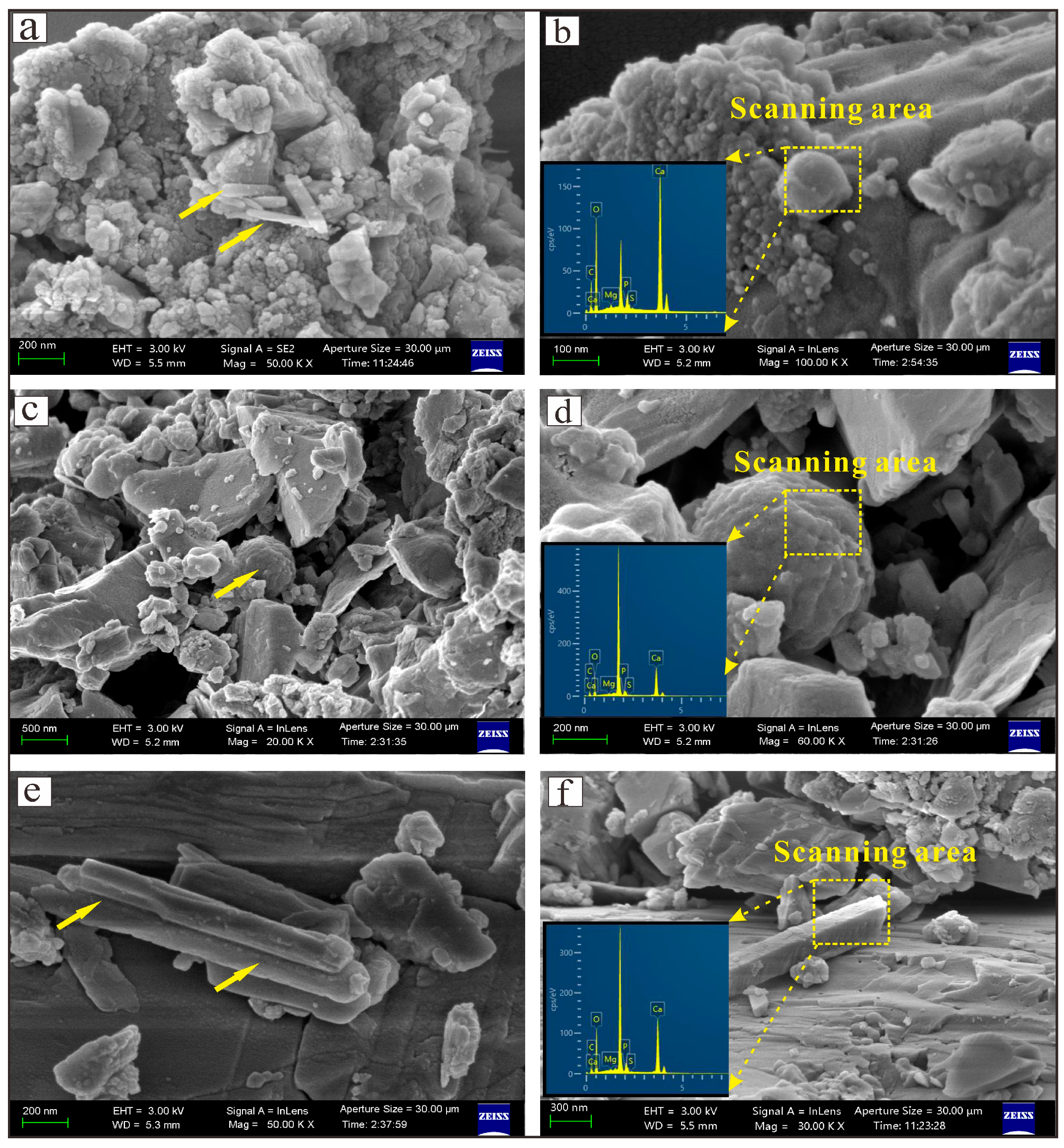
The morphology of the precipitates in the systems of different experimental groups is shown in Figure 4. Among the observations for the Bacillus sp. experimental group, it was found that the minerals generated in the SEM images exhibited diverse sizes and morphologies. Rod-shaped, ellipsoidal minerals can be observed, as well as aggregates composed of cauliflower-shaped particles arranged in close proximity (Figure 4a,b). Aggregates composed of cauliflower-shaped particles had a diameter of only about 50 nm, whereas individual rod and ellipsoidal mineral particles were relatively large, with diameters ranging from 100 to 200 nm. EDS spectroscopic analysis of these minerals showed elemental compositions containing C (16.9%), O (23.8%), Ca (46.3%), Mg (2.5%), P (7.6%), and S (2.9%). The results indicated that those individual spherical particles were mainly composed of calcium magnesium carbonate (proto-dolomite). The presence of the organic elements P and S suggested a close relationship between microorganisms and mineral formation. In addition, Na and Cl elements may be consumed through several pathways during biomineralization. For example, they may participate in metabolism as cofactors for enzymes (such as carbonic anhydrase) or compete with Ca2+ for adsorption sites through ion exchange. Therefore, they were not detected due to their low content in the experimental groups.
In the experimental group of Virgibacilus oceani, distinct mineral structures appeared in the SEM images. These minerals were mainly spherical and rhombic aggregates (Figure 4c,d), with particles ranging from 200 to 400 nm in diameter. These spherical structures were significantly more mature than those observed in the Bacillus sp. system. This suggested that the mineral growth and aggregation process had progressed significantly during this time. When these aggregates were fragmented, they exhibited radioactive structures, and this particular structure may be the result of a combination of microbial activity and the specific growth mechanism of the minerals [41,42]. EDS spectroscopic analysis of these minerals showed that the precipitates contain relatively high levels of both magnesium and calcium, suggesting that these minerals likely contain magnesium carbonate and calcium magnesium carbonate (proto-dolomite), which was consistent with the morphological and structural characteristics of the minerals observed previously.
The images of the co-precipitation system were mainly rod-like aggregates. These rod-like precipitates were adsorbed on the surface of microorganisms, the surface of EPS (extracellular polymers), and some inorganic materials, showing a complete precipitation state (Figure 4e,f). The comparison with the experimental group with a single microorganism revealed that more granular precipitates were adsorbed on the surfaces of both microorganisms. The SEM images showed that the generated mineral species were consistent with the experimental group inoculated with a single microorganism, but the morphology of the mineral aggregates was more regular and complete than that observed in the Bacillus sp. system, with particles ranging from 600 to 800 nm in diameter, indicating further development and maturation of the mineral precipitation process. The results of the EDS analyses showed that these rod-like aggregates consisted mainly of calcium magnesium carbonate (proto-dolomite).

3.4. XRD Analysis

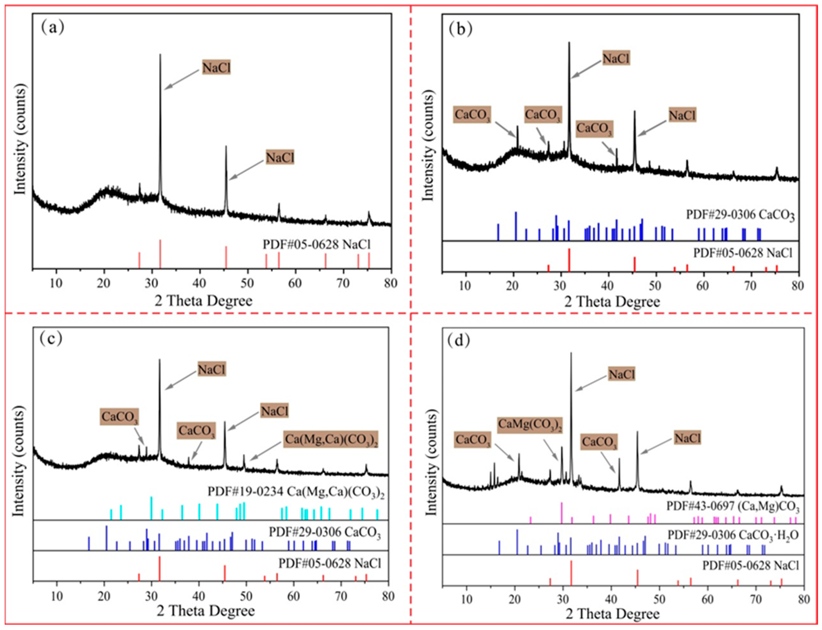
The mineralogical analysis of the precipitate is exhibited in Figure 5. Figure 5a shows the mineralogical analysis of the precipitate in the control group. By observing the XRD patterns, it was found that the precipitates in the control group appeared to be monolithic, and their precipitates were mainly sodium chloride. The presence of NaCl was related to the composition of the culture medium under experimental conditions, which originated from salts in the medium that eventually crystallized to form NaCl crystals in the absence of bacterial involvement. The minerals formed in the presence of Bacillus sp. were mainly sodium chloride and calcium carbonate (Figure 5b), and the characteristic peaks of this mineral were evident on the XRD pattern, demonstrating the efficiency of Bacillus sp. in contributing to the precipitation of this mineral. Based on XRD and EDS analysis, the phase of calcium carbonate (CaCO3) was calcite. Moreover, the precipitation of dolomite was mainly influenced and controlled by bacteria. In the control group, without the involvement of bacteria, the mineral morphology was uniform, and no dolomite precipitation occurred.
The precipitation of minerals under the influence of Virgibacilus oceani differed from that of the experimental group inoculated with Bacillus sp. In the spectrum of Figure 5c, a weak dolomite-like reflection peak is clearly seen in addition to sodium chloride and calcium carbonate. The appearance of dolomite-like peaks suggested that Virgibacilus oceani changed the chemical conditions of the environment through its unique metabolic activities and biological processes, thus promoting the formation and precipitation of minerals [43].
For co-precipitation systems, the XRD patterns have demonstrated a more complex and richer mineral composition. As shown in Figure 5d, the coexistence of calcium magnesium carbonate (i.e., proto-dolomite) and calcium carbonate was not a simple mixing of substances, but rather a visualization of the synergistic effect of Bacillus sp. and Virgibacilus oceani in the mineral precipitation process. The synergistic action of these two microorganisms resulted in a more diverse mineral composition of the precipitates. The only single peak appearing in the XRD measurements was due to the low content of the tested sample. However, as the content of the tested sample increased, peaks related to dolomite also appeared in other locations. The process of microbial mineralization also intensified.

3.5. Thermal Decomposition Characteristics and Kinetic Study in Different Systems

The results of thermal analysis of the precipitated minerals in different systems at a heating rate of 10 °C/min are shown in Figure 6. The results of the TG curves indicated that the ratio of mass loss in the control group amounted to 48%, and the ratio of residual mass was 52%. However, the mass loss ratio of minerals in different experimental groups was smaller than that of the control group, especially for the co-precipitation system of Bacillus sp. and Virgibacilus oceani, where the mass loss ratio of minerals was 44% and the remaining mass ratio was 56%.
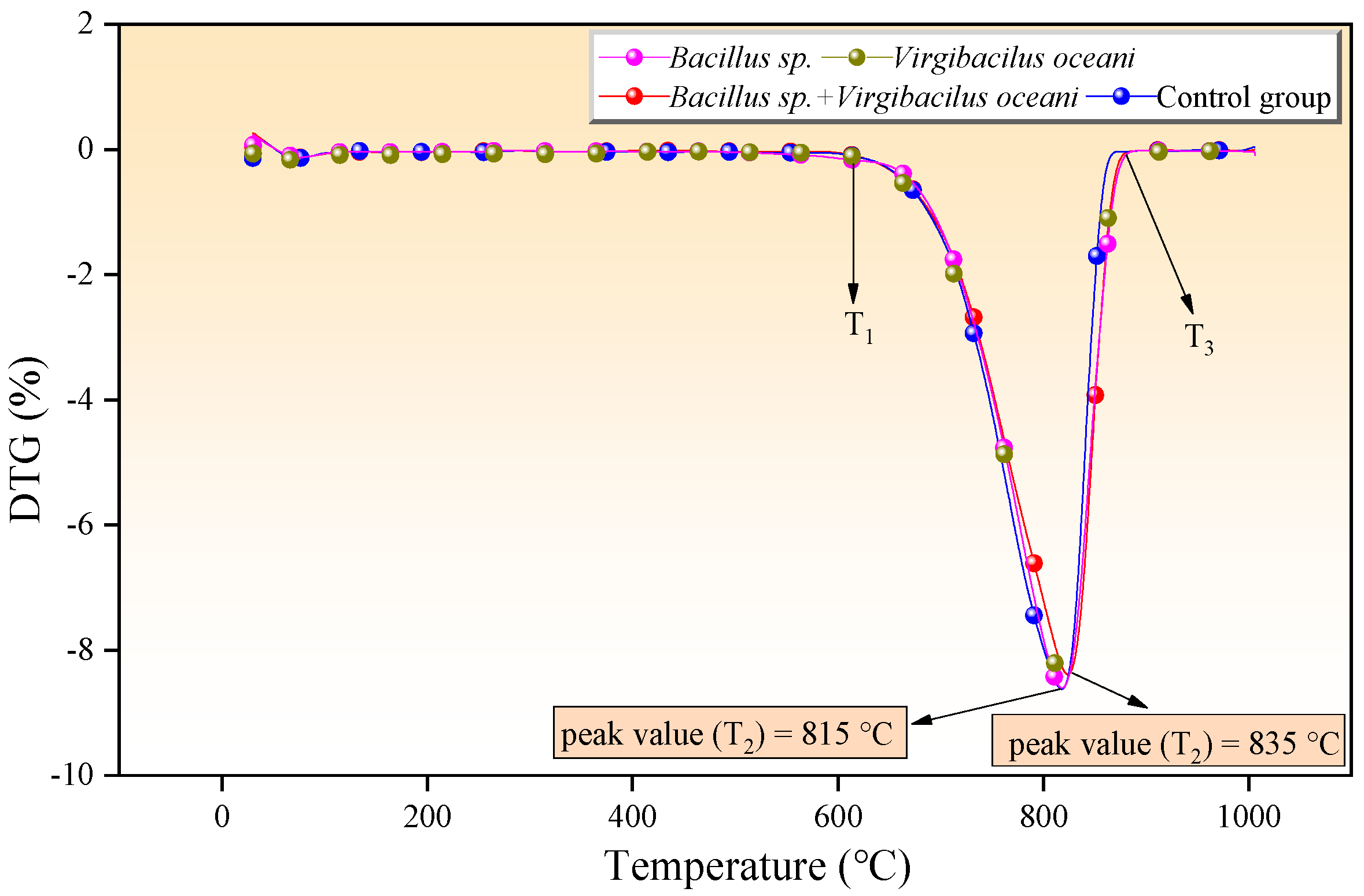
The DTG curves of the precipitated minerals in different systems at a 10 °C/min heating rate are shown in Figure 7. The results showed that the whole thermochemical decomposition process can be roughly divided into three stages. Among them, the first stage occurred at the initial setting temperature of 50 °C to T1. The second stage occurred from T1 to T3, and the second stage, in which weight loss of minerals occurred, was mainly due to the mass loss caused by the thermal decomposition. Meanwhile, at the temperature point of T2, the slope of the curve can be found to reach the maximum value from the TG curve, which represents the fastest rate of weight loss of the sample. In addition, the maximum thermal decomposition temperature of the minerals in the control group reached 815 °C, and the maximum thermal decomposition temperature of the minerals in the co-precipitation system reached 835 °C. The third stage occurred from T3 to 1000 °C, and at this stage, the quality of the minerals basically no longer changed and had stabilized. The results fully illustrate that the thermal stability and crystallinity of biogenic minerals were higher than those of inorganic minerals [44].
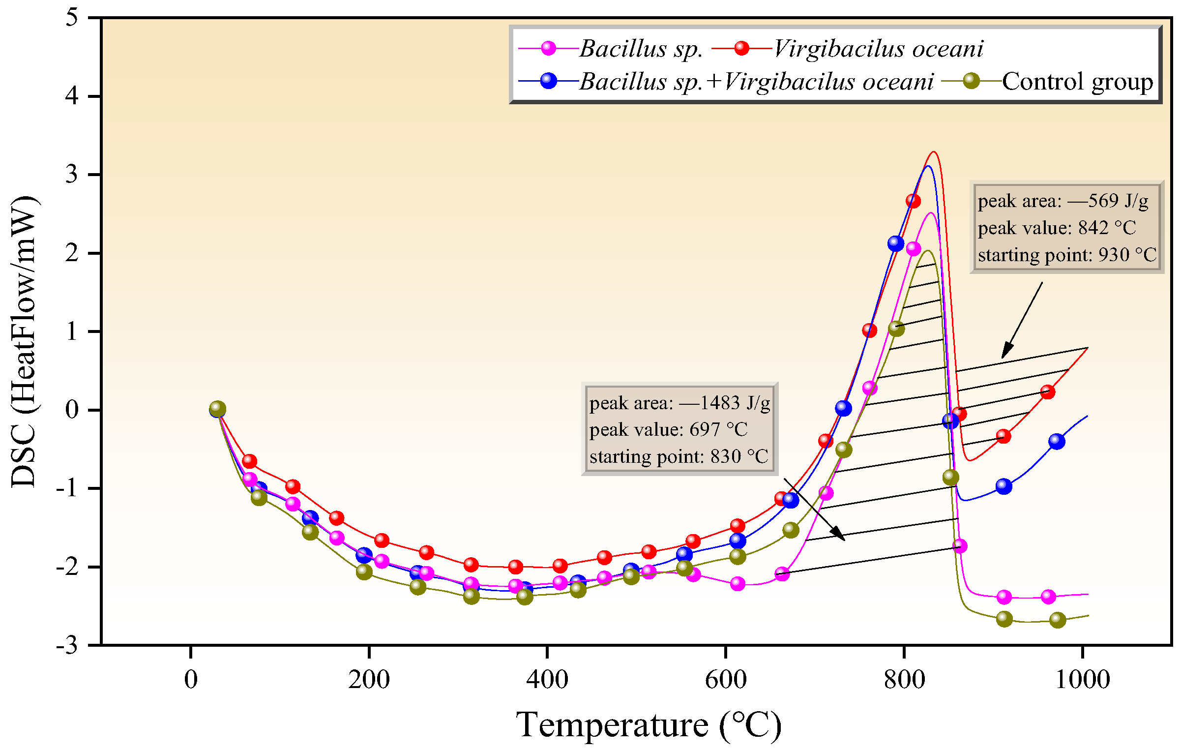
The results of heat flow change (ΔH) of precipitated minerals in different systems are shown in Figure 8. The peak area of the DSC curve was usually utilized to express the change in heat flow of the substance, i.e., the ΔH value. The results indicated that the ΔH value in the control group was 1246 J/g, which was lower than the ΔH value of 1483 J/g in the Bacillus sp. and 1663 J/g in the co-precipitation system. The peak area in the third stage appeared only in the Virgibacilus oceani and co-precipitation systems, which indicated that the heat flow changes occurred in this stage, which were 569 and 758 J/g, respectively.
According to Equations (11)–(13), the values of kinetic parameters E and lnA were calculated using the non-isothermal integration methods (Popescu, FWO, and KAS methods, respectively), as shown in Table 3.
The results in Figure 9 show that the E values of minerals in the control group were 221.6, 219.4, and 226.5 kJ/mol, with an average value of 222.5 kJ/mol, which were lower than those of the experimental group, which were 241.5, 243.9, and 257.7 kJ/mol, respectively. In addition, the correlation fitting coefficients (R) were all over 0.90, which indicated that the selected methods could better reflect the kinetic parameters of the minerals. In summary, the results of thermal analysis showed that the activation energy of biogenic minerals was higher than that of the control group, and their thermal stability was higher. Double asterisks represent p < 0.01 in Figure 9, indicating the results have statistical significance.

4. Discussion

4.1. Precipitation Mechanism of Dolomite

During the first two days of the experiment, the pH values of all experimental groups decreased dramatically. A large number of proteins were present in the culture medium, and their hydrolysis in water was catalyzed by acids, bases, or proteases. During hydrolysis, the proteins were gradually degraded into smaller and smaller peptide fragments and finally into a mixture of free amino acids, which directly affected the pH value of the solution. Subsequently, the growth and reproduction of microorganisms caused the pH value of the solution to change. During the three to five days of the experiment, the pH values of all the experimental groups first increased and then stabilized, and the pH values in the solution showed a similar trend. The influence of different types of microorganisms on the pH value of the solution depended mainly on their physiological characteristics and mechanisms of action. In addition to the metabolic mode and mechanism of action, the growth rate and the number of microorganisms in the solution may also affect the pH value [45,46].
Alkaline substances produced by microorganisms during growth and metabolism led to an increase in pH value, such as ammonia (NH3) through deamination, which then reacted with water to form ammonium ions (NH4+) and hydroxide ions (OH), thus increasing the alkalinity of the solution (Equation (14)).
  OH + NH 4 + NH 3 · H 2 O NH 3 + H 2 O
In addition, acid was consumed by microorganisms in the medium, resulting in an increase in pH value. In the experimental group, the metabolic activity of microorganisms was high, and the respiration and metabolism will change the water chemistry around themselves, including the concentration of HCO3 and CO32−. The main way to increase the CO32− concentration is the CO2 produced by microorganisms, which is dissolved in water to form a certain amount of HCO3 (Equation (15)).
  CO 2 + H 2 O H + + HCO 3 2 H + + CO 3 2
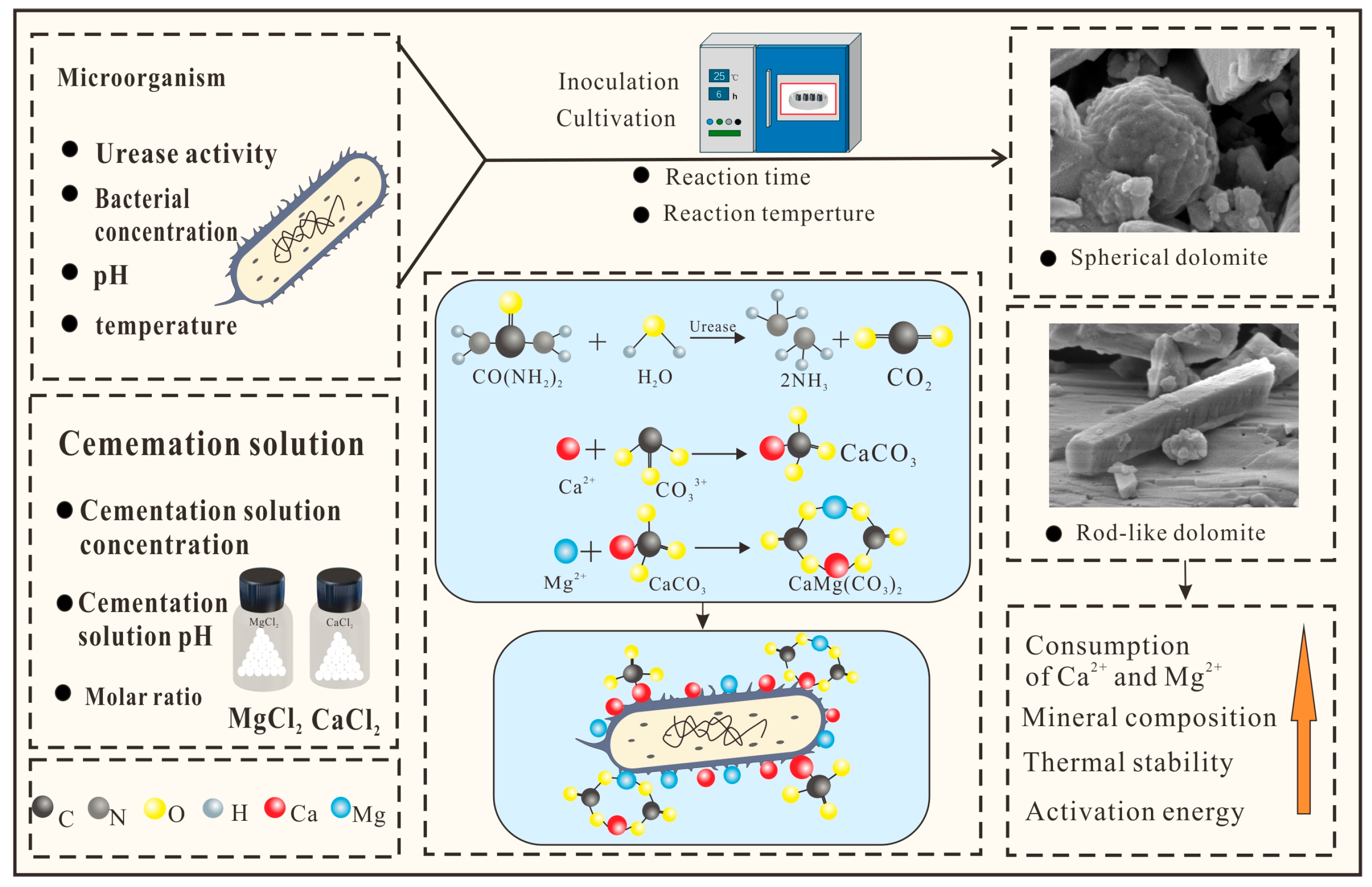
The formation of HCO3 and CO32− will be further consumed by the negatively charged EPS on the surface of the microorganisms and the calcium and magnesium ions in the solution (Equations (16)–(18)), which lead to a slow increase in the pH value of the solution and an increase in the amount of calcium and magnesium ions consumed in the solution. It was complexed with extracellular material or the cell membrane. That way, microorganisms can provide an alkaline environment for the precipitation of carbonate minerals, which is conducive to the formation of carbonate minerals. The precipitation mechanism of dolomite is shown in Figure 10.
C a 2 + + H C O 3 C a C O 3 + C O 2 + H 2 O
C a 2 + + C O 3 2 = C a C O 3
M g 2 + + C O 3 2 = M g C O 3
Bacillus sp. and Virgibacilus oceani elevated environmental pH and consumed Ca2+ and Mg2+ through synergistic metabolism. The results show that the increase in the pH value and consumption of Ca2+ and Mg2+ in the coexistence of the two microorganisms were greater than that of the single microorganism system, which provided a more suitable alkaline environment and a sufficient ion source for dolomite precipitation. In addition, the complementarity of the coexistence optimized the microbial adaptability to environmental changes. For example, in water bodies with imbalanced magnesium-to-calcium ratios, localized ion concentrations were regulated by metabolites to compensate for the limitations of single microorganisms, thus contributing to dolomite precipitation more efficiently.

4.2. Bioremediation of Adsorbed Ca2+ and Mg2+

In the early stage of the experiment, the ion consumption in the solution was relatively low and did not differ much. In the middle stage of the experiment, with the enhancement of microbial activities and the active chemical reaction in the solution, the consumption of Ca2+ and Mg2+ showed an obvious growth trend, in which Mg2+ dominated the reaction system, and its consumption rate exceeded that of Ca2+. The ability of cations in solution to be adsorbed was not only related to the ionic valence and ionic radius but also affected by the ionic concentration, whereby the higher the ionic concentration, the more ions were adsorbed. The higher concentration of Mg2+ compared to Ca2+ makes it easier for Mg2+ to occupy nucleation sites provided by exogenous factors. Ca2+ was involved in the formation of proto-dolomite, but Mg2+ was involved in the formation of calcium carbonate and proto-dolomite (which tended to have a higher Mg content and forms high-magnesium calcite) [47,48]. When the two microorganisms coexisted, their interactions significantly contributed to the consumption of Ca2+ and Mg2+, and the consumption significantly exceeded that of the single microorganism experimental group.
Microbially induced carbonate minerals (e.g., calcite, dolomite) can fix carbon dioxide in solid form for long periods of time, forming geological carbon sinks and mitigating the greenhouse effect. This process was particularly prominent in the metabolic activities of microorganisms, which converted CO2 to CO32− through photosynthesis or heterotrophic interactions, which in turn binds and precipitates Ca2+ and Mg2+. The negatively charged groups on the surface of microbial cells have a strong adsorption capacity for heavy metal ions, which combine with mineral precipitation to form a stable complex to remediate contaminated water bodies and soils. Therefore, microbial-mediated mineral precipitation was a key mechanism linking biological activities and geochemical processes, with far-reaching implications for ecological remediation, resource formation, and global climate change [49]. In addition, through the calcium carbonate coprecipitation mechanism, heavy metals such as As, Cu, Pb, and Cd in soil or slag can be effectively fixed, reducing their bioavailability and mobility. In addition, by injecting urease-containing microorganisms into deep saline aquifers or depleted oil and gas fields, urea is decomposed to produce CO32−, which combines with Ca2+ in the environment to form calcium carbonate precipitates, permanently sequestering carbon and reducing formation permeability.

5. Conclusions

This paper has mainly investigated the mineralogical and thermochemical characteristics of dolomite induced by microorganisms. The results of the study showed that the pH values in the early experimental groups decreased sharply, mainly due to the decomposition of proteins in the solution into a mixture of free amino acids. The pH value increased in the middle and late stages of the experiment, which was due to the decomposition of organic matter by microorganisms and the production of NH4+. In the co-precipitation system, the combined effect on the ions in the solution resulted in a greater change in pH value, which was more favorable to induce the formation of dolomite precipitates. The morphology of the mineral aggregates was more regular and complete, which was larger than that of the single-microorganism experimental group. XRD results and thermal analyses have shown an increase in the rate of precipitation and diversity of mineral species, as well as a higher proportion of proto-dolomite and an increase in thermal stability when Bacillus sp. and Virgibacilus oceani acted together. This suggests that they somehow promoted each other and jointly contributed to the mineral precipitation process. More importantly, this study has also indicated that the total consumption of Mg2+ was greater than Ca2+ in the solutions of each experimental group, which was due to the fact that Mg2+ was more easily adsorbed into the calcium carbonate lattice by organic matter such as proteins and amino acids. This suggests that microbial-induced precipitation of Ca2+ and Mg2+ immobilizes excess metal ions and improves water quality or soil salinity problems. This reflected the multidimensional value of biomineralization in pollution management, bioremediation, carbon sequestration, and geological resource formation.

Author Contributions

Conceptualization, S.F.; methodology, S.F.; software, D.Z.; validation, D.Z. and S.F.; formal analysis, D.Z.; investigation, S.F.; resources, D.Z.; data curation, D.Z.; writing—original draft preparation, S.F.; writing—review and editing, W.Y.; funding acquisition, S.F. All authors have read and agreed to the published version of the manuscript.

Funding

This work was supported by National Natural Science Foundation of China (42502105); the University Natural Science Research Project of Anhui Province (2023AH052223); the Academic Funding for Top-talents in Disciplines of Universities in Anhui Province (gxbjZD2022075); the Doctoral Research Start-up Fund of Suzhou University (2020BS008); the Postdoctoral Research Start-up Fund of Suzhou University (2023BSH002); the research on the integration of innovation and entrepreneurship education with professional education (szxy2023jyjf65); the National-level Undergraduate Innovation Training Program (202510379054); and the Project for Cultivating Outstanding Young Teachers (Study on the Kinetic and Thermodynamic Characteristics of Microbial Dolomite under Different Temperature Conditions Affected by SO42−).

Data Availability Statement

The article incorporates the original contributions of this study. For additional inquiries, please contact the corresponding author.

Acknowledgments

The authors extend their gratitude to Mr. Chao Wang from Scientific Compass (www.shiyanjia.com) for providing invaluable assistance with the TG-DSC analysis.

Conflicts of Interest

All authors have read and approved this version of the article, and due care has been taken to ensure the integrity of the work. No part of this paper has been published or submitted elsewhere. The authors declare no conflicts of interest.

References

  1. Cai, W.K.; Liu, J.H.; Zhou, C.H.; Keeling, J.; Glasmacher, U.A. Structure, genesis and resources efficiency of dolomite: New insights and remaining enigmas. Chem. Geol. 2021, 573, 120191. [Google Scholar] [CrossRef]
  2. Chen, C.; Zhong, H.; Wang, X.; Ning, M.; Wang, X.; Ge, Y.; Wang, H.; Tang, R.; Hou, M. Thermodynamic and kinetic studies of dolomite formation: A review. Minerals 2023, 13, 1479. [Google Scholar] [CrossRef]
  3. Agrawal, Y.; Gupta, T.; Siddique, S.; Sharma, R.K. Potential of dolomite industrial waste as construction material: A review. Innov. Infrastruct. Solut. 2021, 6, 205. [Google Scholar] [CrossRef]
  4. Wan, Y.; Lin, J.; Zhao, Z.; Wang, Z. Origin of the dolomite in the Buqu Formation (Mid-Jurassic) in the south depression of the Qiangtang Basin, Tibet: Evidence from petrographic and geochemical constraints. Front. Earth Sci. 2022, 10, 944701. [Google Scholar] [CrossRef]
  5. Zhang, J.; Zhang, B.; Zhang, S.; Wang, H. Autochthonous Dolomitization and Dissolution in the Microbial Carbonate Rocks of the Fengjiawan Formation in the Ordos Basin. Acta Geol. Sin.-Engl. Ed. 2022, 96, 1376–1387. [Google Scholar] [CrossRef]
  6. Jurado, I.V.; Paese, G.; Schneider, I.H.; Féris, L. Phosphate removal from aqueous solutions using natural and thermic treated dolomites: Equilibrium, kinetic, and thermodynamic. Int. J. Environ. Sci. Technol. 2021, 19, 1739–1752. [Google Scholar] [CrossRef]
  7. Resio, L.C. Dolomite thermal behaviour: A short review. Phys. Chem. Miner. 2024, 51, 19. [Google Scholar] [CrossRef]
  8. Meister, P.; Frisia, S.; Dódony, I.; Pekker, P.; Molnár, Z.; Neuhuber, S.; Gier, S.; Kovács, I.; Demény, A.; Pósfai, M. Nanoscale pathway of modern dolomite formation in a Shallow, Alkaline Lake. Cryst. Growth Des. 2023, 23, 3202–3212. [Google Scholar] [CrossRef] [PubMed]
  9. Rivers, J.M. Warm acidified seawater: A dolomite solution. J. Sediment. Res. 2023, 93, 187–201. [Google Scholar] [CrossRef]
  10. Fang, Y.; Xu, H. Dissolved silica-catalyzed disordered dolomite precipitation. Am. Mineral. 2022, 107, 443–452. [Google Scholar] [CrossRef]
  11. Santos, H.S.; Nguyen, H.; Venâncio, F.; Ramteke, D.; Zevenhoven, R.; Kinnunen, P. Mechanisms of Mg carbonates precipitation and implications for CO2 capture and utilization/storage. Inorg. Chem. Front. 2023, 10, 2507–2546. [Google Scholar] [CrossRef]
  12. Zhou, S.; Wang, Y.; Teng, H. Relative contributions of mg hydration and molecular structural restraints to the barrier of dolomite crystallization: A comparison of aqueous and non-aqueous crystallization in (BaMg)CO3 and (CaMg)CO3 systems. Minerals 2021, 11, 1214. [Google Scholar] [CrossRef]
  13. Aufort, J.; Raiteri, P.; Gale, J.D. Computational insights into Mg2+ dehydration in the presence of carbonate. ACS Earth Space Chem. 2022, 6, 733–745. [Google Scholar] [CrossRef]
  14. Zhang, F.; Xu, H.; Shelobolina, E.S.; Konishi, H.; Roden, E.E. Precipitation of low-temperature disordered dolomite induced by extracellular polymeric substances of methanogenic Archaea Methanosarcina barkeri: Implications for sedimentary dolomite formation. Am. Mineral. 2021, 106, 69–81. [Google Scholar] [CrossRef]
  15. Sun, Y.; Liu, F.; Liu, J.; Chen, L.; Li, Y.; Ding, H.; Lu, A. Microbial community response to photoelectrons and regulation on dolomite precipitation in marine sediments of Yellow Sea. Minerals 2023, 13, 753. [Google Scholar] [CrossRef]
  16. Sun, X.; Wai, O.W.H.; Xie, J.; Li, X. Biomineralization to prevent microbially induced corrosion on concrete for sustainable marine infrastructure. Environ. Sci. Technol. 2023, 58, 522–533. [Google Scholar] [CrossRef] [PubMed]
  17. Wang, Q.; Wang, M.; Feng, Z.; Zhang, Y.; Huang, X.; Xue, X. Efficient removal of Al (III) and P507 from high concentration MgCl2 solution based on in-situ reaction strategy. Trans. Nonferrous Met. Soc. China 2024, 34, 3042–3053. [Google Scholar] [CrossRef]
  18. Zhao, Y.; Li, N.; Shi, J.; Xia, Y.; Zhu, B.; Shao, R.; Min, C.; Xu, Z.; Deng, H. Extra-thin composite nanofiltration membranes tuned by γ-cyclodextrins containing amphipathic cavities for efficient separation of magnesium/lithium ions. Sep. Purif. Technol. 2022, 286, 120419. [Google Scholar] [CrossRef]
  19. Fan, Q.; Liu, D.; Papineau, D.; Qiu, X.; Wang, H.; She, Z.; Zhao, L. Precipitation of high Mg-calcite and protodolomite using dead biomass of aerobic halophilic bacteria. J. Earth Sci. 2023, 34, 456–466. [Google Scholar] [CrossRef]
  20. Teng, J.; Qiu, L.; Zhang, S.; Ma, C. Origin and diagenetic evolution of dolomites in paleogene Shahejie Formation lacustrine organic shale of Jiyang depression, Bohai Bay Basin, east China. Pet. Explor. Dev. 2022, 49, 1251–1265. [Google Scholar] [CrossRef]
  21. Zhuang, D.; Yao, W.; Guo, Y.; Chen, Z.; Gui, H.; Zhao, Y. Bioremediation of Heavy Metal-Contaminated Solution and Aged Refuse by Microbially Induced Calcium Carbonate Precipitation: Further Insights into Sporosarcina pasteurii. Microorganisms 2025, 13, 64. [Google Scholar] [CrossRef]
  22. Zhuang, D.; Yao, W.; Guo, Y.; Wu, C.; Cao, J.; Sun, B. Comparative study on geochemical and thermodynamic characteristics among wormkalk, micrite, thrombolite, and stromatolite. Carbonates Evaporites 2025, 40, 22. [Google Scholar] [CrossRef]
  23. Jarwar, M.A.; Dumontet, S.; Pasquale, V.; Chen, C. Microbial induced carbonate precipitation: Environments, applications, and mechanisms. Geomicrobiol. J. 2022, 39, 833–851. [Google Scholar] [CrossRef]
  24. Wang, J.; Zhao, Y.; Li, D.; Qi, P.; Gao, X.; Guo, N.; Meng, R.; Tucker, M.E.; Yan, H.; Han, Z. Extreme halophilic bacteria promote the surface dolomitization of calcite crystals in solutions with various magnesium concentrations. Chem. Geol. 2022, 606, 120998. [Google Scholar] [CrossRef]
  25. Li, J.; Sun, Y.; Chen, F.; Hu, X.; Dong, L. Pressure and temperature combined with microbial supernatant effectively inactivate Bacillus subtilis spores. Front. Microbiol. 2021, 12, 642501. [Google Scholar] [CrossRef]
  26. Xue, Z.F.; Cheng, W.C.; Wang, L.; Hu, W. Effects of bacterial inoculation and calcium source on microbial-induced carbonate precipitation for lead remediation. J. Hazard. Mater. 2022, 426, 128090. [Google Scholar] [CrossRef] [PubMed]
  27. Fatimah, S.; Ragadhita, R.; Al Husaeni, D.F.; Nandiyanto, A.B.D. How to calculate crystallite size from x-ray diffraction (XRD) using Scherrer method. ASEAN J. Sci. Eng. 2022, 2, 65–76. [Google Scholar] [CrossRef]
  28. Ali, A.; Chiang, Y.W.; Santos, R.M. X-ray diffraction techniques for mineral characterization: A review for engineers of the fundamentals, applications, and research directions. Minerals 2022, 12, 205. [Google Scholar] [CrossRef]
  29. Liu, A.; Liu, S.; Liu, Y.; Liu, B.; Liu, T. Characterizing mechanical heterogeneity of coal at nano-to-micro scale using combined nanoindentation and FESEM-EDS. Int. J. Coal Geol. 2022, 261, 104081. [Google Scholar] [CrossRef]
  30. Qureshi, F.; Yusuf, M.; Tahir, M.; Haq, M.; Mohamed, M.M.I.; Kamyab, H.; Nguyen, H.-H.T.; Vo, D.-V.N.; Ibrahim, H. Renewable hydrogen production via biological and thermochemical routes: Nanomaterials, economic analysis and challenges. Process Saf. Environ. Prot. 2023, 179, 68–88. [Google Scholar] [CrossRef]
  31. Zhuang, D.; Yang, F.; Xiang, Q.; Wei, Q.; Jiang, W.; Zhao, J.; Guo, Y. Differences in thermal decomposition and crystallinity of dark organic laminae and light mineral laminae in same stromatolite. Thermochim. Acta 2023, 728, 179576. [Google Scholar] [CrossRef]
  32. Zhuang, D.; Cao, A.; Pan, L.; Jiang, W.; Zhao, J. Comparative study on thermal decomposition of stromatolite and micrite using the technique of TG and DSC. J. Therm. Anal. Calorim. 2023, 148, 5529–5541. [Google Scholar] [CrossRef]
  33. Arumsari, A.G.; Hernowo, P.; Wibowo, J.; Fadlulloh, M.Y.; Setiawan, R. Study of Reaction Kinetics by Flynn Wall Ozawa Method on Sawdust Pyrolysis Process. Indones. J. Appl. Chem. 2022, 24, 9–14. [Google Scholar]
  34. Ivanov, V.B.; Kalugina, E.V.; Pomerantsev, A.L.; Samoryadov, A.; Shebanov, M. Kinetic features of nonisothermal degradation of polyarylene ketones. Thermochim. Acta 2023, 730, 179608. [Google Scholar] [CrossRef]
  35. Vyazovkin, S.; Burnham, A.K.; Criado, J.M.; Pérez-Maqueda, L.A.; Popescu, C.; Sbirrazzuoli, N. ICTAC Kinetics Committee recommendations for performing kinetic computations on thermal analysis data. Thermochim. Acta 2011, 520, 1–19. [Google Scholar] [CrossRef]
  36. Kohout, J. Modified Arrhenius equation in materials science, chemistry and biology. Molecules 2021, 26, 7162. [Google Scholar] [CrossRef]
  37. Michael, P.S. Positive and negative effects of addition of organic carbon and nitrogen for management of sulfuric soil material acidity under general soil use conditions. Pol. J. Soil Sci. 2021, 54, 71–87. [Google Scholar] [CrossRef]
  38. Bayazitova, Z.E.; Kurmanbayeva, A.S.; Tleuova, Z.O.; Temirbekova, N.G. Application of the thermophilic fermentation method to obtain environmentally friendly organic fertilizer. J. Ecol. Eng. 2023, 24, 202–216. [Google Scholar] [CrossRef] [PubMed]
  39. Huang, J.; Fisher, P.R.; Argo, W.R.; Jeong, K.Y.; Altland, J. Macronutrient solubility in response to the pH of soilless container substrates. J. Soil Sci. Plant Nutr. 2025, 25, 3224–3240. [Google Scholar] [CrossRef]
  40. Xu, X.; Xu, L.; Yang, Z.; Chen, L.; Wang, Y.; Ren, H.; Zhang, Z.; El-Kassaby, Y.A.; Wu, S. Identification of key gene networks controlling organic acid and sugar metabolism during star fruit (Averrhoa carambola) development. BMC Plant Biol. 2024, 24, 943. [Google Scholar] [CrossRef] [PubMed]
  41. Gupta, T.; Cho, J.; Prakash, J. Hydrothermal synthesis of TiO2 nanorods: Formation chemistry, growth mechanism, and tailoring of surface properties for photocatalytic activities. Mater. Today Chem. 2021, 20, 100428. [Google Scholar] [CrossRef]
  42. Vaou, N.; Stavropoulou, E.; Voidarou, C.; Tsigalou, C.; Bezirtzoglou, E. Towards advances in medicinal plant antimicrobial activity: A review study on challenges and future perspectives. Microorganisms 2021, 9, 2041. [Google Scholar] [CrossRef]
  43. Pan, Y.; Li, Y.; Chen, C.T.A.; Jiang, Z.-P.; Cai, W.-J.; Shen, Y.; Ding, Z.; Chen, Q.; Di, Y.; Fan, W.; et al. Unveiling the secrets of diatom-mediated calcification: Implications for the biological pump. Sci. China Earth Sci. 2024, 67, 2895–2909. [Google Scholar] [CrossRef]
  44. Klumpp, K.; Marcolli, C.; Peter, T. The impact of (bio-) organic substances on the ice nucleation activity of the K-feldspar microcline in aqueous solutions. Atmos. Chem. Phys. 2022, 22, 3655–3673. [Google Scholar] [CrossRef]
  45. Zhuang, D.; Wang, R.J.; Chen, S.; Li, X. The geochemical and thermodynamic characteristics of waste sand reinforced by microbially induced calcium carbonate precipitation. Environ. Technol. Innov. 2024, 36, 103828. [Google Scholar] [CrossRef]
  46. Zhuang, D.; Chen, S.; Li, J.; Han, S.; Guo, Y. Recycled coarse aggregate from waste concrete strengthened by microbially induced calcium carbonate precipitation. Environ. Technol. Innov. 2025, 37, 103981. [Google Scholar] [CrossRef]
  47. Han, Z.; Li, J.; Zhao, Y.; Chen, Q.; Gao, X.; Hu, K.; Guo, N.; Wei, X.; Meng, R.; Zhu, C.; et al. Dissolved Mn2+ promotes microbially-catalyzed protodolomite precipitation in brackish oxidized water. Chem. Geol. 2024, 650, 121986. [Google Scholar] [CrossRef]
  48. Liu, D.; Xu, Y.; Papineau, D.; Yu, N.; Fan, Q.; Qiu, X.; Wang, H. Experimental evidence for abiotic formation of low-temperature proto-dolomite facilitated by clay minerals. Geochim. Cosmochim. Acta 2019, 247, 83–95. [Google Scholar] [CrossRef]
  49. Zhuang, D.; Chen, Z.; Sun, B. Thermal decomposition of calcium carbonate at multiple heating rates in different atmospheres using the techniques of TG, DTG, and DSC. Crystals 2025, 15, 108. [Google Scholar] [CrossRef]
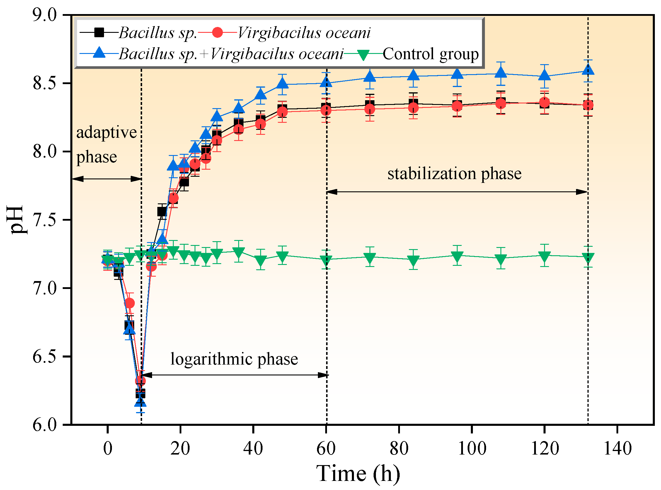
Figure 1. The changes in pH value in different systems within five days.
Crystals 15 00767 g001
Figure 2. Consumption of Ca2+ and Mg2+ in different systems. (a) The consumption of Ca2+ and Mg2+ in Bacillus sp. system; (b) the consumption of Ca2+ and Mg2+ in Virgibacilus oceani system; (c) the consumption of Ca2+ and Mg2+ in the coexistence of Bacillus sp. and Virgibacilus oceani systems; (d) the settlement difference in the consumption of Ca2+ and Mg2+.
Crystals 15 00767 g002
Figure 3. Morphological changes and elemental composition in control group. (a,b) The SEM results of the sample; (ch) the EDS results of the sample.
Crystals 15 00767 g003
Figure 4. Morphological changes and elemental composition in the experimental group, (a,b) Bacillus sp. system; (c,d) Virgibacilus oceani system; (e,f) the coexistence of Bacillus sp. and Virgibacilus oceani systems.
Crystals 15 00767 g004
Figure 5. XRD analyses of minerals: (a) control group; (b) Bacillus sp. system; (c) Virgibacilus oceani system; (d) the coexistence of Bacillus sp. and Virgibacilus oceani systems.
Crystals 15 00767 g005
Figure 6. TG analyses of minerals in different systems.
Crystals 15 00767 g006
Figure 7. DTG analyses of minerals in different systems.
Crystals 15 00767 g007
Figure 8. DSC analyses of minerals in different systems.
Crystals 15 00767 g008
Figure 9. Activation energy (E, kJ/mol) results of minerals in different systems. Double asterisk means p < 0.01, which indicating the results have statistical significance.
Crystals 15 00767 g009
Figure 10. The precipitation mechanism of dolomite.
Crystals 15 00767 g010
Table 1. Mass of corresponding substances per 1000 mL solution (g), Mg2+ = 0.05 mol/L, Ca2+ = 0.01 mol/L.
SystemMgCl2·6H2O (g)CaCl2 (g)NaCl (g)NB Medium (g)
Bacillus sp.10.151.113018
Virgibacilus oceani10.151.113018
Bacillus sp. + Virgibacilus oceani10.151.113018
Control group10.151.113018
Table 2. The components of the culture medium.
Material CompositionComposition Mass/Volume (g/L)
Ferric citrate0.1
Potassium chloride0.55
Sodium carbonate0.16
Potassium bromide0.08
Strontium chloride0.034
Boric acid0.022
Disodium hydrogen phosphate0.008
Sodium sulfate0.016
Tryptone10
Beef Powder3.0
Sodium chloride5.0
Table 3. The values of R, E, and lnA of minerals in different systems at different conversion rates.
MethodsFWOKASPopescu
Different SystemsConversion Rate/αRElnARElnARElnAAverage
Bacillus sp.0.10.988253.822.20.967228.420.240.967244.123.1
0.20.967248.921.00.958232.318.540.978249.823.9
0.30.945245.221.40.981224.826.240.927250.821.9_
0.40.964250.118.90.945258.010.940.951255.426.4_
0.50.938245.018.50.972223.226.440.990223.713.0_
0.60.974243.721.30.938243.610.340.944262.917.9_
0.70.991226.817.90.989264.115.240.972233.413.4_
0.80.923222.921.90.908242.89.140.926258.817.7_
0.90.919244.726.90.923226.417.140.937246.919.6_
242.3 ± 10.4238.1 ± 14.9247.3 ± 12.3241.5 ± 4.6
Virgibacilus oceani239.5 ± 15.2249.7 ± 12.5239.7 ± 14.5243.9 ± 5.9
Bacillus sp. + Virgibacilus oceani261.2 ± 13.2254.8 ± 10.2257.2 ± 13.1257.7 ± 3.2
Control group221.6 ± 12.7219.4 ± 14.2226.5 ± 12.6222.5 ± 3.6
Disclaimer/Publisher’s Note: The statements, opinions and data contained in all publications are solely those of the individual author(s) and contributor(s) and not of MDPI and/or the editor(s). MDPI and/or the editor(s) disclaim responsibility for any injury to people or property resulting from any ideas, methods, instructions or products referred to in the content.