Article

The Effect of Heat Treatment on Yellow-Green Beryl Color and Its Enhancement Mechanism

School of Gemology, China University of Geosciences, Beijing 100083, China
*
Author to whom correspondence should be addressed.
Crystals 2025, 15(8), 746; https://doi.org/10.3390/cryst15080746
Submission received: 29 July 2025 / Revised: 19 August 2025 / Accepted: 19 August 2025 / Published: 21 August 2025
(This article belongs to the Collection Topic Collection: Mineralogical Crystallography)

Abstract

Beryl is classified as a cyclosilicate mineral, and its color is primarily determined by the type and oxidation state of trace elements. In this study, natural yellow-green beryl was used as the research subject, and heat treatment experiments were performed at various temperatures under both oxidizing and reducing atmospheres. A combination of analytical techniques, including electron probe microanalysis (EPMA), X-ray diffraction (XRD), Fourier-transform infrared spectroscopy (FTIR), Raman spectroscopy, and ultraviolet-visible spectroscopy (UV-Vis), were employed to systematically investigate the composition, structure, and chromogenic mechanisms of beryl before and after heat treatment. The experimental results indicate that heat treatment under both atmospheres can lead to the transformation of yellow-green beryl into blue, with 500–600 °C under a reducing atmosphere identified as the optimal treatment condition. With increasing temperature, beryl gradually dehydrates, resulting in a faded blue color and reduced transparency. Even after treatment at 700 °C, no significant changes in unit cell parameters were observed, and both type I and type II water were retained, indicating that the color change is not attributed to crystal structure transformation or phase transitions. The study reveals that the essential mechanism of color modification through heat treatment lies in the valence change between Fe2+ and Fe3+ occupying channel and octahedral sites. The observed color variation is attributed to changes in absorption band intensity resulting from charge transfers of O2− → Fe3+ and Fe2+ → Fe3+. This study provides theoretical insights and technical references for the color enhancement of beryl through heat treatment.

1. Introduction

Beryl is a beryllium aluminum cyclosilicate mineral of significant gemological importance, with the chemical formula Be3Al2(Si6O18). It is highly valued for its vivid coloration, substantial hardness, and excellent optical properties. Variations in the types and valence states of chromophoric ions within its crystal structure result in a broad spectrum of colors, leading to the formation of different gem varieties.
The coloration of beryl is not only closely related to its chemical composition but is also significantly influenced by its internal crystal structure. Iron ions, serving as key chromophoric agents [1], influence beryl’s color based on their oxidation states, occupancy sites, and coordination environments; however, the specific mechanism remains controversial. In addition to chromophoric ions, the role of structural constituents in beryl also warrants further investigation [2]. Beryl is nominally anhydrous mineral, but vibrational results strongly indicate that H2O molecules exist in the structural channels. The number of vibrational bands and their frequencies revealed the presence of H2O type II, in which C2 symmetry axis of the water molecule is parallel to the structural channel (and to the c-axis of beryl) [3]. Its unique six-membered ring channels can host large-radius alkali metal ions (e.g., Cs+, Rb+, K+), water molecules, and other cations; the arrangement and concentration of these species also impact the gemstone’s color [4,5]. Heat treatment, as a widely used enhancement method, can alter the valence states of transition metal ions, thus modifying beryl’s color.
Generally, several mechanisms have been proposed for the color modification of gemstones by heat treatment, including changes in the valence states of chromophoric ions, destruction of original color centers, dehydration of water-bearing phases, diffusion of chromophoric ions, and alterations in the crystal lattice [6,7,8]. Although heat treatment has been widely applied to enhance the color of beryl, there is no consensus on the optimal temperature conditions. Proctor et al. reported that yellow to golden beryl can be converted to commercially valuable aquamarine by heating at 280–600 °C for 1–12 h. Yan found that yellow-green beryl can be efficiently transformed to blue at 500–550 °C for 1 h, regardless of atmospheric conditions. The color shift is mainly attributed to changes in Fe3+/Fe2+ intervalence charge transfer [9]. However, the underlying mechanism and optimal treatment parameters remain insufficiently understood. However, the color change mechanism of beryl after heat treatment has not been thoroughly examined, and the optimal thermal conditions for this transformation require further exploration.
Based on these considerations, beryl samples were subjected to heat treatment under both oxidizing and reducing atmospheres. Changes in composition, crystal structure, and spectral characteristics were further analyzed using Electron Probe Microanalysis (EPMA), powder X-ray Diffraction (XRD), Fourier Transform Infrared Spectroscopy (FTIR), Micro-Raman Spectroscopy, and UV–Visible Absorption Spectroscopy (UV–Vis), in combination with Differential Thermal Analysis (DTA), to investigate the water speciation in beryl, its thermal stability, and dehydration characteristics.
This study aims to determine the optimal heat treatment temperature for beryl and characterize its thermally induced changes, verify the feasibility of color enhancement through heat treatment, gain insight into the underlying chromogenic mechanisms, and enrich the experimental database on heat-treated beryl.

2. Materials and Methods

2.1. Materials

In this study, five yellow-green beryl samples were selected for this study (Figure 1). Among them, three samples (G1–G3) were used for heat treatment experiments. Samples G1 and G2 were each sliced into five pieces and subjected to thermal treatment under a single atmospheric condition. Sample G3 was cut into nine pieces, which were used separately under oxidizing and reducing atmospheres. Heat treatment was conducted using both a muffle furnace and a tubular furnace under oxidizing (air) and reducing (Ar/H2 mixed gas with a volume ratio of 95:5) atmospheres. For the oxidizing experiments, samples were heated directly in ambient air. In the reducing atmosphere, argon gas was first introduced for 30 min to remove residual oxygen from the furnace chamber. The samples were then heated at a constant rate of 5 °C/min to target temperatures of 400 °C, 500 °C, 600 °C, and 700 °C. Each sample was held at the designated temperature for 2 h, followed by natural cooling to room temperature. A controlled variable method was employed to ensure consistent experimental conditions across all treatments.

2.2. Methods

The major elements of the samples were quantitatively analyzed using an EPMA-1720 electron probe microanalyzer (Shimadzu, Kyoto, Japan). The analytical parameters were set as follows: accelerating voltage of 15 kV, beam current of 10 nA, and beam spot diameter of 5 μm. Data correction was performed using the ZAF3 method.
Differential thermal analysis (DTA) was carried out using a DTG-60 thermal analyzer (Shimadzu, Kyoto, Japan). Before testing, the samples were ground to 200 mesh. The analysis was conducted in air over a temperature range from 22 °C to 1000 °C at a heating rate of 10 °C/min.
X-ray diffraction (XRD) analysis was performed using a D8 Advance diffractometer (Bruker Scientific, Karlsruhe, Germany). The experimental settings included a Cu Kα radiation source (λ = 0.15418 nm), a graphite curved crystal monochromator, tube voltage of 40 kV, tube current of 100 mA, and both divergence and scattering slits set to 1°. The scanning was conducted over a 2θ range of 5° to 90° at a speed of 5°/min.
Fourier transform infrared spectroscopy (FTIR) was conducted using a Tensor 27 spectrometer (Bruker, Karlsruhe, Germany) in transmission mode. The Portions of the samples were ground into a fine powder with a particle size of 200 mesh and mixed with potassium bromide (KBr) to prepare pellets for infrared transmission measurements. The testing conditions were as follows: voltage of 220 V, room temperature, resolution of 4 cm−1, scan duration of 30 s, frequency of 50–60 Hz, and a spectral range of 400–4000 cm−1.
Raman spectroscopy was carried out using an HR-Evolution micro-Raman spectrometer (Horiba, Kyoto, Japan). The spectral range was set from 4000 to 100 cm−1, with an integration time of 3 s, one accumulation cycle, and an excitation wavelength of 532 nm. A 600 lines/mm grating centered at 500 nm was utilized, the laser output power was 50 mW, and the resolution was 1 cm−1.
UV-Vis diffuse reflectance spectra were recorded using a UV3600 spectrophotometer (Shimadzu, Kyoto, Japan). The measurements were performed using the reflection method at room temperature, with a spectral range of 300–800 nm, a sampling interval of 1.0 s, a slit width of 20 nm, and a resolution of 0.1 nm.

3. Results and Discussion

3.1. General Properties

The conventional gemological properties of beryl were examined. The refractive index ranged from 1.573 to 1.581, with a birefringence of approximately 0.005, and a specific gravity of about 2.70 g/cm3. Observations using a dichroscope revealed that the yellowish-green beryl samples exhibited weak pleochroism, with color variations ranging from yellowish-green to green or yellow. Additionally, no observable fluorescence was detected under either long-wave or short-wave ultraviolet light, indicating that the samples were fluorescently inert.
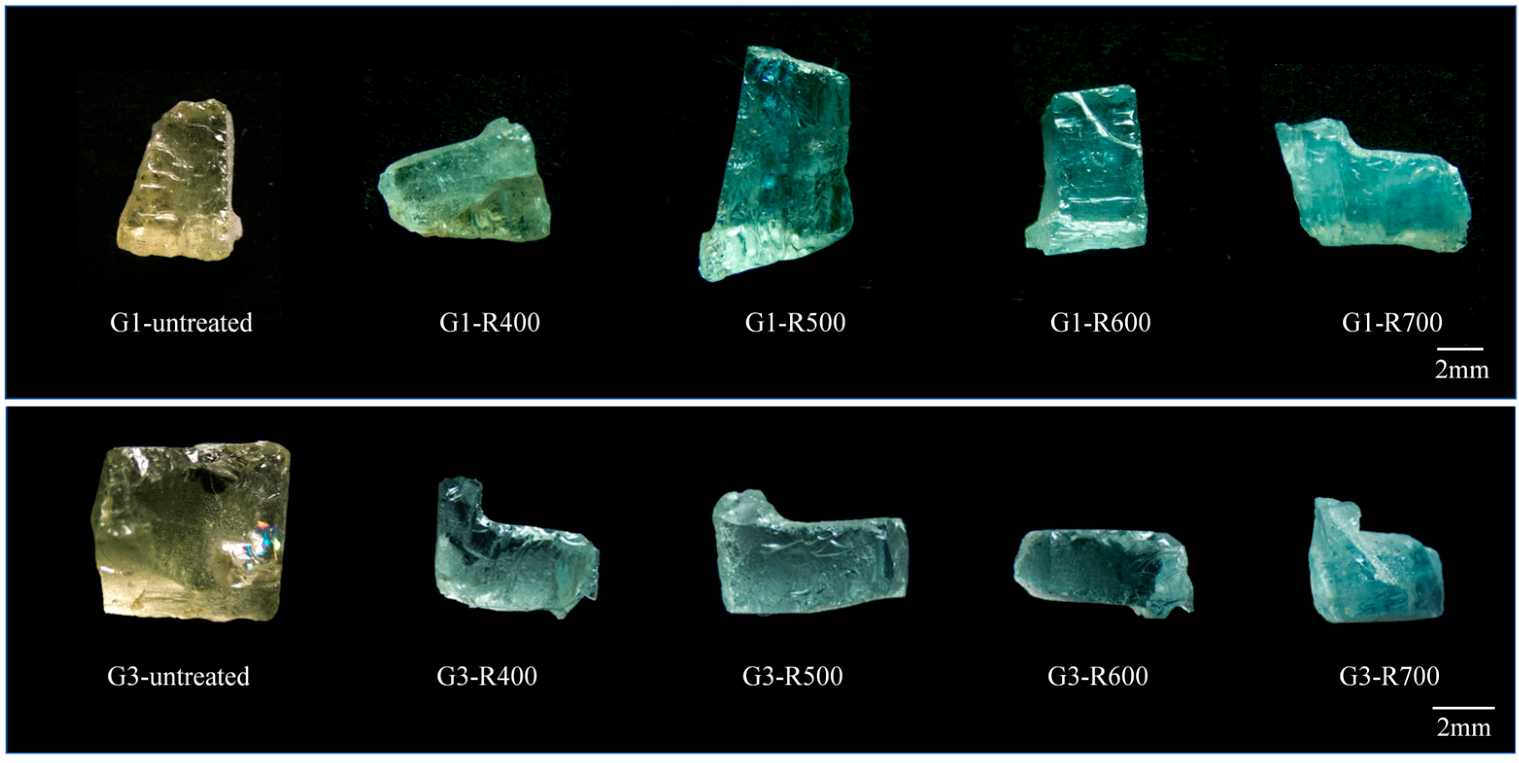
The color changes in the untreated and heat-treated beryl samples are presented in Figure 2 and Figure 3. Under oxidizing conditions (Figure 2), the blue hue of the samples gradually deepened with increasing temperature. A saturated blue color was achieved in the 500–600 °C range. When the temperature exceeded 600 °C, dehydration within the crystal structure caused a significant decrease in transparency, and the sample surfaces developed a hazy white appearance. Under reducing conditions (Figure 3), the yellowish-green beryl also transformed into a distinct blue color. At 400 °C, sample G1-R400 retained a slight yellow tint. However, after heating to 500 °C, the yellow component disappeared, resulting in a slightly less transparent but blue hue. The optimal blue color, with a vivid aquamarine appearance, was achieved between 500 and 600 °C. These results indicate that the ideal heat treatment temperature for converting yellow-green beryl to blue lies within the 500–600 °C range. Both oxidizing and reducing atmospheres can induce this color transformation, although the reducing atmosphere produces a more pronounced effect.

3.2. Composition and Crystal Structure

3.2.1. Chemical Composition

Composition analysis is an essential component of mineralogical research and a critical prerequisite for investigating the color-causing mechanism of mineral samples [10]. Major element analysis of the untreated beryl samples was performed using electron probe microanalysis (EPMA), and the results are presented in Table 1. The cation numbers were calculated using the oxygen atom normalization method, based on the equation Y = Y′ × X, where Y is the number of cations per unit cell, Y′ is the cation coefficient, and X is the oxygen normalization factor. For an oxide of the form YnOm, Y′ is calculated as n × (oxide weight percentage)/(molecular weight of the oxide), and X is determined by the number of oxygen atoms in the known formula divided by the sum of m × (oxide weight percentage/molecular weight of the oxide) [11]. The results are presented in Table 2.
According to the EPMA results, the total oxide content of all samples was generally less than 100 wt.%, typically ranging from 97 wt.% to 99 wt.%. This discrepancy may be attributed to the fact that Be and H cannot be directly detected by EPMA; their contents are typically estimated indirectly or calculated based on empirical formulas, which may introduce uncertainties in the stoichiometric calculations [12]. The primary oxide components of the yellowish-green beryl samples include SiO2 (approximately 63.9–65.3 wt.%), Al2O3 (17.95–20.63 wt.%), and BeO (11.43–13.01 wt.%). Minor amounts of Fe, Mn, and Mg were also detected, with FeO content being relatively high—a characteristic feature of yellowish-green beryl.
Based on the measured concentrations of Na and K (Li and Cs were not determined due to their very low contents), the samples can be classified as alkali-poor beryl, as the total alkali content is below 0.1 wt.%. [13]. In such samples, alkali ions enter the structural channels only to a limited extent, indicating a more stable crystal framework and suggesting that charge compensation is primarily achieved through internal cation substitution.
The presence of iron is strongly correlated with color, implying that Fe may serve as one of the key chromophores in yellowish-green beryl.

3.2.2. Differential Thermal Analysis

The differential thermal analysis (DTA) results are shown in Figure 4. The initial mass of sample G4 was 21.37 mg. From room temperature (23 °C) to 1000 °C, the sample exhibited a total mass loss of 0.41 mg, corresponding to 1.419% of the initial weight. The weight loss process can be broadly divided into four stages: (1) Between 22 and 270 °C, the sample mass remained nearly constant at approximately 21.36–21.37 mg, with no significant loss detected. (2) In the range of 270–600 °C, a gradual weight loss occurred, decreasing from 21.36 mg to 21.25 mg—approximately 0.11 mg or 0.51% of the total sample mass. (3) Between 600 and 800 °C, the rate of mass loss slowed, with a decrease from 21.25 mg to 21.22 mg—about 0.03 mg or 0.14% of the sample. (4) From 800 to 1000 °C, the mass loss rate increased significantly, dropping from 21.22 mg to 20.96 mg—approximately 0.26 mg or 1.22% of the sample mass.
Beryl possesses a hexagonal ring silicate structure, containing both channel water and structural water. Upon heating, dehydration reactions occur at specific temperature ranges, leading to observable mass loss. Based on the thermogravimetric data, mass loss begins gradually after 270 °C, which is attributed to the progressive release of channel water with minimal impact on the crystal framework. The DTA peak observed around 650 °C may correspond to dehydration or a structural rearrangement phase transition of beryl. When the temperature exceeds 800 °C, the weight loss rate increases markedly, indicating that the channel water has been largely removed, and the decomposition of structural hydroxyl groups (OH) begins and continues, ultimately leading to complete dehydration. This behavior is consistent with previous studies, which suggest that beryl typically undergoes dehydration at temperatures above 850 °C [14,15]. These patterns indicate that channel water begins to be released slowly after 270 °C, causing minimal disruption to the crystal structure. When the temperature exceeds 800 °C, the increased rate of weight loss suggests that structural hydroxyl groups (OH) start to decompose, leading to progressive and ultimately complete dehydration [16].

3.2.3. Crystal Structure and Phase Analysis

The crystal structure is also a key factor influencing the color of beryl. Beryl crystallizes in a hexagonal ring structure, with a general chemical formula of Be3Al2(Si6O18) [17]. The substitution relationships among the constituent elements are illustrated in Figure 5a,b. The [SiO4] tetrahedra form [Si6O18] six-membered rings parallel to the {0001} plane. These rings are interconnected by [BeO4] tetrahedra and [AlO6] octahedra to form a three-dimensional framework [18]. Channels aligned along the c-axis within the hexagonal rings can accommodate large-radius alkali metal ions (Cs+, Rb+, K+), water molecules, and other metal cations. Cationic substitutions can occur at octahedrally coordinated Al sites, tetrahedrally coordinated Be sites, and at the 2a and 2b channel positions. Specifically, Li+ can substitute for Be2+, while Al3+ can be replaced by other trivalent (Fe3+, Cr3+) or divalent (Fe2+, Mn2+) cations. To maintain charge balance, alkali metal ions such as Cs+ and Rb+ may enter the channel center (CH site) as charge compensators [19].
The X-ray diffraction (XRD) patterns of the samples before and after heat treatment are shown in Figure 5c,d and match well with the standard beryl reference pattern (PDF#74-2343). The results indicate that, despite undergoing heat treatments in both oxidizing and reducing atmospheres within the temperature range of 400–700 °C, the crystal structure of the samples remained stable without significant changes. All samples consistently exhibited strong diffraction peaks corresponding to the (100), (112), and (211) crystal planes, with average interplanar spacings (d-values) of 7.97 Å, 3.25 Å, and 2.86 Å, respectively—these are characteristic reflections of beryl. Using the reducing atmosphere as a representative example, XRD data of beryl samples before and after heat treatment were further tabulated and compared with the standard reference card. The complete dataset is provided in the Supplementary Material (Table S1). The analysis shows that the crystal plane indices, 2θ angles, and d-values remained essentially unchanged under different temperatures (400–700 °C) and atmospheric conditions, further confirming the structural stability of beryl during heat treatment.
To further evaluate the influence of thermal treatment on the crystal structure, the unit cell parameters (including their estimated standard deviations, as refined using Jade software) were determined from the XRD data of samples treated at different temperatures. The results are summarized in Table 3 According to the c/a ratio, beryl can be categorized into two structural types: one dominated by isomorphous substitution of Al3+ in octahedral sites, and the other by Li+ substitution for Be2+ in tetrahedral sites [20,21]. In the present study, all samples exhibited c/a ratios within 0.991 ± 0.001 to 0.998 ± 0.001, indicating that the structure is primarily controlled by [AlO6] octahedral substitution. A slight increase in unit cell volume(from 673.00 ± 0.02 Å3 to 676.00 ± 0.02 Å3) was detected after heat treatment, but this variation was minor and showed no systematic correlation with treatment temperature or atmosphere. These findings indicate that thermal treatments between 400 and 700 °C, whether in oxidizing or reducing environments, did not induce significant changes in the crystal structure or phase transitions of beryl.
Based on these results, we conclude that the color change observed in beryl during heat treatment is not attributable to alterations in crystal structure or phase transformation.

3.3. Spectroscopy Properties

3.3.1. Fourier Transform Infrared Spectroscopy (FTIR)

The infrared spectra of G1 and G2 samples before and after heat treatment are shown in Figure 6. The fingerprint region (400–1600 cm−1) displays consistent peak positions after heat treatment, and no clear linear relationship is observed between absorption intensity and temperature.
The absorption bands within 1219–680 cm−1 are primarily attributed to the stretching vibrations of Si–O–Si and O–Si–O. The band near 1200 cm−1 can be further resolved into peaks at 1205 and 1150 cm−1, while two sub-peaks at 1020 and 960 cm−1 are observed in the vicinity of 960 cm−1 [22]. The absorption peak near 650 cm−1 corresponds to the stretching vibration of the Be–O bond. The region between 590 and 450 cm−1 represents typical vibrational modes of ring silicates, while the double peaks at 525 and 495 cm−1 are associated with Al–O stretching vibrations. Additional lower-frequency bands are attributed to the bending vibrations of Si–O bonds [22,23].
Furthermore, the infrared spectra of the yellowish-green beryl samples exhibit distinct absorption features corresponding to CO2 and H2O molecules. A sharp absorption peak at 2348 cm−1 is assigned to the asymmetric stretching vibration of CO2 [24]. As the temperature increases, the intensity of this peak decreases and its width broadens, indicating that CO2 remains present even at 700 °C, without causing damage to the crystal structure of beryl.
To further investigate the effect of heat treatment on water molecules within the structure, infrared transmission spectra were collected for sample G3 treated under oxidizing conditions. Portions of the sample were ground into a fine powder with a particle size of 200 mesh and mixed with potassium bromide (KBr) to prepare pellets for measurement. The spectral range of 4000–400 cm−1 was scanned, and the 3800–3500 cm−1 region was extracted for analysis. As shown in Figure 7, all samples exhibit a strong absorption band near 3699 cm−1 corresponding to type I water (asymmetric stretching), and a weaker band near 3597 cm−1 corresponding to type II water (symmetric stretching), with type I water being dominant [25]. As temperature increases, notable changes are observed in the absorption behavior of water: at 400–500 °C, the intensities of both types remain stable, whereas at 600–700 °C, the absorption intensity of type II water significantly decreases. This reduction is likely related to structural rearrangements involving Na–O–H complexes within the channels. Upon the release of some doubly coordinated H2O molecules, an increase in structural symmetry may lead to reduced asymmetric vibrational absorption [26]. Simultaneously, the strengthening of Na–O bonds may suppress H–O vibrations, further contributing to the attenuation of the type II water absorption band.
In summary, heat treatment primarily affects the presence and behavior of volatile components within the beryl channels, rather than altering the crystal structure itself.

3.3.2. Raman Spectroscopy

Raman spectra of beryl samples before and after heat treatment under reducing and oxidizing atmospheres are shown in Figure 8. No significant shifts were observed in the characteristic Raman peaks after treatment, indicating that heat treatment below 700 °C did not induce phase transitions or structural damage, and the beryl crystal structure remained stable.
As shown in Figure 9, the intensity of the symmetric stretching vibration peak of the Si–O–Si bond at 1068 cm−1 varied significantly with the crystallographic orientation of the samples. Although all samples were cut from the same parent crystal, the cutting direction relative to the laser polarization influenced the measured intensity. When the measurement was taken parallel to the c-axis, the peak intensity was significantly higher than that measured perpendicular to the c-axis.
To investigate the behavior of water molecules after heat treatment, the Raman spectra in the range of 3500–3800 cm−1 were examined (Figure 8c,d). The results showed that the vibrational characteristics of water molecules were not dependent on crystal orientation. At 700 °C, both type-II (3598 cm−1) and type-I (3607 cm−1) water peaks were still detected under both oxidizing (G2-O700) and reducing (G1-R700) atmospheres. These observations confirm that type-I and type-II water molecules remain present at 700 °C, and the intensities of their associated peaks do not show a clear correlation with temperature. This finding is consistent with the previous infrared transmittance analysis. According to previous studies, dehydration of beryl slices occurs above 850 °C, while powdered samples release all structural water only in the 800–900 °C range [22,27]. Thus, the symmetric stretching bands of water molecules can still be detected at 700 °C.
In summary, Raman spectroscopy confirms the structural stability of beryl under heat treatment below 700 °C. Both type-I and type-II water molecules remain detectable, and their vibrational characteristics are independent of crystal orientation [28]. These findings are consistent with the infrared results and further demonstrate the ability of beryl’s channel structure to retain volatile components under moderate thermal conditions.

3.3.3. UV-Vis Spectroscopy

The UV-Vis absorption spectra of beryl samples after heat treatment at various temperatures are shown in Figure 10. Typical iron-related absorption features are observed in samples treated under both oxidizing and reducing atmospheres. After heat treatment, the absorption peaks are generally shifted toward shorter wavelengths, and the absorption bands become significantly broader. Notably, the peak intensity is substantially higher under reducing conditions, confirming the superior color-modifying effect of a reducing atmosphere.
In the UV region, two distinct absorption peaks near 372 nm and 427 nm are attributed to the d–d transitions of Fe3+: the 427 nm band corresponds to the spin-forbidden 6A1g → 4T2g transition, while the 372 nm band is assigned to the 6A1g → 4Eg + 4A1g transitions [29,30]. In the visible region, an additional absorption band at 500–550 nm is observed in yellow-green beryl, which is mainly attributed to d–d transitions of Fe2+/Fe3+ and ligand-to-metal charge transfer (LMCT) of Fe3+. Absorption in this region removes green–blue light, giving rise to the yellow-green coloration. Additionally, a broad and strong absorption band centered around 820 nm is associated with Fe2+ located in octahedral or channel sites, indicating a greater tendency for Fe3+ to be reduced to Fe2+ at elevated temperatures, especially under reducing conditions. Specifically, after heating to 600 °C in a reducing atmosphere, the absorption band beyond 800 nm is markedly enhanced, while the intensity around 425 nm is weakened, further suggesting a valence conversion from Fe3+ to Fe2+.
Before heat treatment, the samples exhibit a distinct yellow hue, primarily due to the [Fe2(OH)4] 2+ binuclear complex within the channels [31]. This complex contributes to enhanced absorption in the near-UV region through M–M charge transfer transitions, which are superimposed with d–d transitions of Fe3+ and O2− → Fe3+ charge transfer bands. When the concentration of this complex is high, the UV absorption is strengthened, and the absorption edge in the blue-violet region extends toward longer wavelengths, shifting the color tone toward yellow-green [23,32,33]. After heat treatment, partial dissociation of the binuclear complex occurs, particularly under reducing conditions, resulting in a lower [Fe2(OH)4]2+ concentration. Consequently, the d–d transition peak of Fe3+ at 372 nm becomes more prominent, the yellow hue fades, and the color gradually shifts toward blue. The coexistence of Fe2+ and Fe3+ leads to intervalence charge transfer transitions between them [34], giving rise to a broad absorption band near 650 nm. This band spans the yellow-orange-red region, and it is the primary optical mechanism responsible for the observed color transition of beryl from yellow-green to blue after heat treatment [35].
Thus, the combined presence of Fe3+ and Fe2+ is responsible for the color change of the samples.

4. Conclusions

In summary, the effects of heat treatment on beryl and the underlying mechanism of its color change have been systematically investigated. It has been demonstrated that heat treatment below 700 °C does not induce a phase transition in the crystal structure of beryl. The observed color variation is primarily attributed to changes in the valence state and site occupancy of iron ions. UV-Vis absorption spectra of samples treated under both oxidizing and reducing atmospheres exhibit typical iron-related features, with significantly stronger absorption observed under reducing conditions. Under oxidizing conditions, a portion of Fe2+ is oxidized to Fe3+, which predominantly occupies channel sites, whereas under reducing conditions, part of Fe3+ is reduced to Fe2+, which tends to occupy octahedral sites. Intervalence charge transfer between Fe2+ and Fe3+ leads to variations in the absorption band intensity, particularly the enhancement of a broad band near 650 nm, which plays a crucial role in the color change of beryl. Additionally, the CO2 absorption peak becomes weaker and broader with increasing temperature, while the intensity of the type-II water band gradually decreases after heat treatment. Overall, it is concluded that the thermally induced color modification of beryl is essentially governed by dynamic changes in the Fe2+/Fe3+ valence states, with variations in the absorption intensities of O2− → Fe3+ and Fe2+ → Fe3+ charge transfer bands being responsible for the observed color shift. This study not only elucidates the mechanism of beryl color change upon heat treatment but also identifies the appropriate temperature range for its thermal enhancement.

Supplementary Materials

The following supporting information can be downloaded at: https://www.mdpi.com/article/10.3390/cryst15080746/s1. Table S1: XRD Data of Beryl Samples Before and After Heat TreatmentCompared with Standard PDF Card (Reducing Atmosphere).

Author Contributions

B.H. and S.Z., performed the experiment, analysis, and writing—original draft; Q.G., performed review and editing. All authors have read and agreed to the published version of the manuscript.

Funding

This study was funded by the Ministry of Science and Technology of China (grant number 2013FY110900-3).

Data Availability Statement

The original contributions presented in this study are included in the article/supplementary material. Further inquiries can be directed to the corresponding author(s).

Acknowledgments

This study was supported by the National Science and Technology Infrastructure—the National Infrastructure of Mineral, Rock, and Fossil Resources for Science and Technology (http://www.nimrf.net.cn, (accessed on 25 December 2021)), as well as the Program for Data Integration and Standardization in Geological Science and Technology. We sincerely thank the Laboratory of the School of Gemmology, China University of Geosciences (Beijing), for their technical assistance during the experiment.

Conflicts of Interest

The authors declare no conflicts of interest.

References

  1. Thomazo, C.; Papineau, D. Biogeochemical Cycling of Nitrogen on the Early Earth. Elements 2013, 9, 345–351. [Google Scholar] [CrossRef]
  2. de Almeida Sampaio Filho, H.; Sighinolfi, G.P.; Galli, E. Contribution to the Crystal Chemistry of Beryl. Contrib. Mineral. Petrol. 1973, 38, 279–290. [Google Scholar] [CrossRef]
  3. Minerals from Macedonia: XXIII. Spectroscopic and Structural Characterization of Schorl and Beryl Cyclosilicates. Spectrochim. Acta Part A 2009, 73, 460–467. [Google Scholar] [CrossRef]
  4. Hanser, C.S.; Häger, T.; Botcharnikov, R. Incorporation and Substitution of Ions and H2O in the Structure of Beryl. Eur. J. Mineral. 2024, 36, 449–472. [Google Scholar] [CrossRef]
  5. Henry, R.E.; Groat, L.A.; Cempírek, J.; Škoda, R.; Holá, M. Predicting the Crystal Structure of Beryl from the Chemical Composition. Can. J. Mineral. Petrol. 2023, 61, 873–897. [Google Scholar] [CrossRef]
  6. Charalambous, F.A.; Ram, R.; Pownceby, M.I.; Tardio, J.; Bhargava, S.K. Chemical and Microstructural Characterisation Studies on Natural and Heat Treated Brannerite Samples. Miner. Eng. 2012, 39, 276–288. [Google Scholar] [CrossRef]
  7. Wang, Q.; Guo, Q.; Li, N.; Cui, L.; Liao, L. Study of the Mechanism of Color Change of Prehnite after Heat Treatment. RSC Adv. 2022, 12, 3044–3054. [Google Scholar] [CrossRef]
  8. Song, Z.-X.; Guo, Q.-F.; Liao, L.-B. Effect of Heat Treatment on the Color of Yellow-Green Sphene and Its Color-Causing Mechanism. JOM 2025, 77, 5182–5191. [Google Scholar] [CrossRef]
  9. Mathew, G.; Karanth, R.V.; Rao, T.K.G.; Deshpande, R.S. Channel Constituents of Alkali-Poor Orissan Beryls: An FT-IR Spectroscopic Study. Curr. Sci. 1997, 73, 1004–1011. [Google Scholar]
  10. Fan, Z.-W.; Xiong, Y.-Q.; Shao, Y.-J.; Wen, C.-H. Textural and Chemical Characteristics of Beryl from the Baishawo Be-Li-Nb-Ta Pegmatite Deposit, Jiangnan Orogen: Implication for Rare Metal Pegmatite Genesis. Ore Geol. Rev. 2022, 149, 105094. [Google Scholar] [CrossRef]
  11. Locock, A.J. An Excel Spreadsheet to Classify Chemical Analyses of Amphiboles Following the IMA 2012 Recommendations. Comput. Geosci. 2014, 62, 1–11. [Google Scholar] [CrossRef]
  12. Santiago, J.S.; Souza, V.d.S.; Filgueiras, B.d.C.; Jiménez, F.A.C. Emerald from the Fazenda Bonfim Deposit, Northeastern Brazil: Chemical, Fluid Inclusions and Oxygen Isotope Data. Braz. J. Geol. 2018, 48, 457–472. [Google Scholar] [CrossRef]
  13. Hawthorne, F.C.; Cerny, P. The Alkali-Metal Positions in Cs-Li Beryl. Can. Mineral. 1977, 15, 414–421. [Google Scholar]
  14. Fukuda, J.; Shinoda, K. Water Molecules in Beryl and Cordierite: High-Temperature Vibrational Behavior, Dehydration, and Coordination to Cations. Phys. Chem. Miner. 2011, 38, 469–481. [Google Scholar] [CrossRef]
  15. Fukuda, J.; Shinoda, K. Coordination of Water Molecules with Na+ Cations in a Beryl Channel as Determined by Polarized IR Spectroscopy. Phys. Chem. Miner. 2008, 35, 347–357. [Google Scholar] [CrossRef]
  16. Bharat, V.; Durga Prasad, B.; Venkateswarlu, K. Effect of Beryl on Wear and Thermal Expansion Behavior of Al-Beryl MMCs. Mater. Today Proc. 2018, 5, 7609–7614. [Google Scholar] [CrossRef]
  17. Franz, G.; Morteani, G. Be-Minerals: Synthesis, Stability, and Occurrence in Metamorphic Rocks. Rev. Mineral. Geochem. 2002, 50, 551–589. [Google Scholar] [CrossRef]
  18. Sripoonjan, T.; Seneewong Na Ayutthaya, M. Aquamarine from Southern Ethiopia: An Update. J. Gemmol. 2019, 36, 497–499. [Google Scholar] [CrossRef]
  19. Aurisicchio, C.; Fioravanti, G.; Grubessi, O.; Zanazzi, P.F. Reappraisal of the Crystal Chemistry of Beryl. Am. Mineral. 1988, 73, 826–837. [Google Scholar]
  20. Wang, P.; Gray, T.P.; Li, Z.; Anderson, E.J.D.; Allaz, J.; Smyth, J.R.; Koenig, A.E.; Qi, L.; Zhou, Y.; Raschke, M.B. Mineralogical Classification and Crystal Water Characterisation of Beryl from the W–Sn–Be Occurrence of Xuebaoding, Sichuan Province, Western China. Mineral. Mag. 2021, 85, 172–188. [Google Scholar] [CrossRef]
  21. Fan, Z.-W.; Xiong, Y.-Q.; Brzozowski, M.J.; Shao, Y.-J.; Gu, X.-P.; Anwar, M.; Ghoneim, S.M. Crystallographic Insights and Crystal Fractionation Simulations of Alkali- and Water-Bearing Beryl: Implications for Magmatic–Hydrothermal Evolution and Be Enrichment Mechanisms. Ore Geol. Rev. 2024, 174, 106278. [Google Scholar] [CrossRef]
  22. Łodziński, M.; Sitarz, M.; Stec, K.; Kozanecki, M.; Fojud, Z.; Jurga, S. ICP, IR, Raman, NMR Investigations of Beryls from Pegmatites of the Sudety Mts. Mol. Spectrosc. Mol. Struct. 2005, 744–747, 1005–1015. [Google Scholar] [CrossRef]
  23. Wang, H.; Guan, Q.; Liu, Y.; Guo, Y. Effects of Transition Metal Ions on the Colour of Blue-Green Beryl. Minerals 2022, 12, 86. [Google Scholar] [CrossRef]
  24. Chankhantha, C.; Thanasuthipitak, P.; Kidkhunthod, P. Iron K-Edge Xanes Study of Heated Green Beryl from Madagascar. Walailak J. Sci. Technol. (WJST) 2016, 13, 977–983. [Google Scholar]
  25. Li, J.; Zheng, Y.; Liu, X.; Li, G.; Yu, X.; Wang, Y.; Li, H.; Liu, H.; Shan, G.; Li, T.; et al. Thermal Process of Rock Crystal: Cause of Infrared Absorption Band at 3585 Cm−1. Crystals 2021, 11, 1083. [Google Scholar] [CrossRef]
  26. Fridrichová, J.; Bačík, P.; Rusinová, P.; Antal, P.; Škoda, R.; Bizovská, V.; Miglierini, M. Optical and Crystal-Chemical Changes in Aquamarines and Yellow Beryls from Thanh Hoa Province, Vietnam Induced by Heat Treatment. Phys. Chem. Miner. 2015, 42, 287–302. [Google Scholar] [CrossRef]
  27. Nguyen, P.Q.H.; Zhang, D.; Xu, J.; Downs, R.T.; Dera, P.K. Dehydration Kinetics of Nanoconfined Water in Beryl Probed by High Temperature Single Crystal Synchrotron X-Ray Diffraction. Sci. Rep. 2024, 14, 6079. [Google Scholar] [CrossRef] [PubMed]
  28. Bačík, P.; Fridrichová, J.; Uher, P.; Vaculovič, T.; Bizovská, V.; Škoda, R.; Dekan, J.; Miglierini, M.; Malíčková, I. Beryl Crystal Chemistry and Trace Elements: Indicators of Pegmatite Development and Fractionation (Damara Belt, Namibia). Lithos 2021, 404–405, 106441. [Google Scholar] [CrossRef]
  29. Gavrilenko, E.V.; Pérez, B.C.; Bolibar, R.C.; Amo, D.G. del Emeralds from the Delbegetey Deposit (Kazakhstan): Mineralogical Characteristics and Fluid-Inclusion Study. Mineral. Mag. 2006, 70, 159–173. [Google Scholar] [CrossRef]
  30. Romero Ordóñez, F.H.; González-Durán, A.F.; García-Toloza, J.; Rotlewicz Cohen, J.; Cedeño Ochoa, C.J.; Alvarado González, H.R.; Angarita Sarmiento, L.G.; Romero Ordóñez, F.H.; González-Durán, A.F.; García-Toloza, J.; et al. Mineralogy and Fluid Inclusions of the Cunas Emerald Mine, Maripí—Boyacá, Colombia. Earth Sci. Res. J. 2021, 25, 139–156. [Google Scholar] [CrossRef]
  31. Wang, H.; Shu, T.; Chen, J.; Guo, Y. Characteristics of Channel-Water in Blue-Green Beryl and Its Influence on Colour. Crystals 2022, 12, 435. [Google Scholar] [CrossRef]
  32. Hu, Y.; Lu, R. Color Characteristics of Blue to Yellow Beryl from Multiple Origins. Gems Gemol. 2020, 56, 54–65. [Google Scholar] [CrossRef]
  33. Yu, X.; Hu, D.; Niu, X.; Kang, W. Infrared Spectroscopic Characteristics and Ionic Occupations in Crystalline Tunneling System of Yellow Beryl. JOM 2017, 69, 704–712. [Google Scholar] [CrossRef]
  34. Taran, M.N.; Vyshnevskyi, O.A. Be, Fe2+-Substitution in Natural Beryl: An Optical Absorption Spectroscopy Study. Phys. Chem. Miner. 2019, 46, 795–806. [Google Scholar] [CrossRef]
  35. Shang, Y.; Guo, Y.; Tang, J. Spectroscopy and Chromaticity Characterization of Yellow to Light-Blue Iron-Containing Beryl. Sci. Rep. 2022, 12, 10765. [Google Scholar] [CrossRef]
Figure 1. Samples of yellow-green beryl.
Crystals 15 00746 g001
Figure 2. Color change of yellow-green beryl samples G2 and G3 after heat treatment in an oxidizing atmosphere.
Crystals 15 00746 g002
Figure 3. Color change of yellow-green beryl samples G1 and G3 after heat treatment in a reducing atmosphere.
Crystals 15 00746 g003
Figure 4. Differential thermal analysis (DTA) and Thermogravimetric analysis (TGA) curves of beryl sample G4.
Crystals 15 00746 g004
Figure 5. (a) Crystal structure of beryl viewed along (a) the a-axis and (b) the c-axis. The crystal structure visualization data were obtained using the software VESTA (Color figure online); (c,d) Beryl samples before and after heat treatment, the reducing atmosphere is set in H2 + Ar, and the oxidizing atmosphere is set in air.
Crystals 15 00746 g005
Figure 6. Infrared absorption spectra of beryl samples before and after heat treatment: (a) in oxidizing atmosphere; (b) in reducing atmosphere.
Crystals 15 00746 g006
Figure 7. Infrared absorption spectra of H2O in the G3 sample before and after heat treatment (transmission mode).
Crystals 15 00746 g007
Figure 8. Raman spectra of beryl samples before and after heat treatment: (a) 4000–100 cm−1 range in reducing atmosphere; (b) 4000–100 cm−1 range in oxidizing atmosphere. (c) 3500–3800 cm−1 range in reducing atmosphere; (d) 3500–3800 cm−1 range in oxidizing atmosphere.
Crystals 15 00746 g008
Figure 9. Raman spectra of the G1-R700 sample collected along two orientations: (a) 100–1200 cm−1 region; (b) 3500–3800 cm−1 region.
Crystals 15 00746 g009
Figure 10. UV–Vis reflectance spectra of beryl samples before and after heat treatment: (a) in reducing atmosphere; (b) in oxidizing atmosphere.
Crystals 15 00746 g010
Table 1. EPMA-derived oxide concentrations (wt.%) in beryl samples.
SampleSiO2Al2O3Cr2O3TiO2FeOMnOMgONa2OK2ONiOFClBeOH2OTotal
G265.2318.27000.660.030.330.370.010.020012.601.6599.17
G365.318.3000.9100.490.490.030.030012.241.8099.60
G564.7718.590.0200.650.010.330.420.020.010.040.0112.391.7298.93
Table 2. Calculated cation numbers in beryl samples.
SampleSiAlCrTiFeMnMgCaNaKNiBe
G26.0371.9930.0000.0000.0510.0020.0460.0000.0660.0010.0012.802
G36.0471.9970.0000.0000.0700.0000.0680.0000.0880.0040.0022.724
G56.0202.0360.0010.0000.0510.0010.0460.0000.0760.0020.0012.766
Table 3. Unit cell parameters of beryl samples under different heat treatment conditions.
Reducing AtmosphereOxidizing Atmosphere
G3400 °C500 °C600 °C700 °C400 °C500 °C600 °C700 °C
a (Å)9.209 (2)9.217 (2)9.219 (2)9.219 (2)9.217 (2)9.220 (2)9.216 (2)9.209 (2)9.202 (2)
c (Å)9.166 (2)9.193 (2)9.194 (2)9.189 (2)9.191 (2)9.194 (2)9.186 (2)9.195 (2)9.183 (2)
c/a0.995 (1)0.997 (1)0.997 (1)0.997 (1)0.997 (1)0.997 (1)0.997 (1)0.998 (1)0.998 (1)
Å3673.230 (2)676.280 (2)676.700 (2)676.320 (2)676.190 (2)676.840 (2)675.610 (2)675.370 (2)673.310 (2)
Disclaimer/Publisher’s Note: The statements, opinions and data contained in all publications are solely those of the individual author(s) and contributor(s) and not of MDPI and/or the editor(s). MDPI and/or the editor(s) disclaim responsibility for any injury to people or property resulting from any ideas, methods, instructions or products referred to in the content.