Article

Structure and Nonlinear Optical Characterization of a New Acentric Crystal of a 4-Hydroxybenzohydrazide Derivative

1
Department of Chemical Sciences, University of Naples Federico II, Via Cinthia, I-80126 Naples, Italy
2
Department of Chemistry, Katholieke Universiteit Leuven, 3000 Leuven, Belgium
3
Department of Applied Science and Technology (DISAT), Polytechnic of Turin, Corso Duca degli Abruzzi 24, I-10129 Torino, Italy
*
Author to whom correspondence should be addressed.
Crystals 2025, 15(8), 739; https://doi.org/10.3390/cryst15080739 (registering DOI)
Submission received: 24 July 2025 / Revised: 4 August 2025 / Accepted: 13 August 2025 / Published: 20 August 2025
(This article belongs to the Section Organic Crystalline Materials)

Abstract

We report the crystal structure and nonlinear optical (NLO) characterization of the monohydrate form of N′-[(E)-(2-fluorophenyl)methylidene]-4-hydroxybenzohydrazide (o-FHH), an organic compound showing strong potential for second-order nonlinear optical applications. The compound crystallizes in a non-centrosymmetric tetragonal space group. The supramolecular features of the novel crystal structure are strongly related to the role of the water molecule that stabilized columns of o-FHH through strong hydrogen bonding interactions. This structural feature is reflected in the high thermal stability of the compound, which is evidenced by its ability to withstand temperatures in excess of 100 °C without losing the water molecule. Second-harmonic generation (SHG) imaging confirms bulk nonlinearity throughout the entire volume of the crystal, consistent with the acentric class of the novel compound. The combination of a dense hydrogen-bonding network, structural robustness, and the ability to grow millimeter-sized single crystals makes o-FHH a good candidate for further development as an organic NLO material.

1. Introduction

Acentric and polar crystal structures have attracted considerable attention because of their remarkable physical properties that enable advanced applications such as ferroelectricity [1,2], pyro and piezoelectricity [3,4,5,6], second-harmonic and THz wave generation [7,8], and the linear electro-optic (Pockels) effect [9,10]. In optics, intense laser pulses can induce nonlinear effects such as second-harmonic generation (SHG), a nonlinear optical (NLO) phenomenon whereby a material generates coherent light at twice the frequency of the incident radiation. These properties are described by odd-rank tensors, which are non-zero only in acentric space groups and are significantly enhanced in polar space groups [11]. However, the discovery of new noncentrosymmetric materials is not trivial considering that 78.2% of all crystal structures deposited in the Cambridge Structural Database (CSD) [12] are centrosymmetric. Notably, the 21.8% of acentric structures in the CSD is likely an overestimate, as it includes different enantiopure chiral compounds (mainly natural products) that cannot crystallize in centrosymmetric space groups. The prevalence of centrosymmetric space groups over non-centrosymmetric ones has been a recurring theme since the early days of crystallographic science [13]. SHG has emerged as an important phenomenon in the field of NLO, with applications ranging from biological imaging to the development of advanced laser sources. SHG materials are essential for frequency conversion, especially in regions of the spectrum that are otherwise difficult to access, such as the blue, near-ultraviolet, and mid- to far-infrared ranges [14]. These capabilities are crucial for fields such as spectroscopy and information processing, particularly in the IR region, for the fabrication of devices designed for optical communications [15,16]. In order to perform effectively, SHG materials must fulfill a stringent set of criteria in addition to the crucial presence of a non-centrosymmetric crystal structure. These include high second-order molecular nonlinearity in terms of susceptibility (χ^((2))), a wide band gap (Eg), broad optical transparency windows, high laser-induced damage thresholds (LIDT), an appropriate birefringence (Δn) for phase matching, high chemical and physical stabilities, and easy-to-grow bulk single crystals [14,15,17,18].
While well-established inorganic crystals such as KH2PO4 (KDP), LiNbO3, KTiOPO4 (KTP), and β-BaB2O4 (BBO) are widely used for SHG in the visible and near-IR ranges, they often exhibit poor performance at longer wavelengths (1300–1550–2000 nm), which are now utilized in novel industrial lasers [18,19]. In order to address these limitations, substantial efforts have focused on organic NLO materials. These compounds often exhibit figures of merit that range from two to three orders of magnitude higher than their inorganic counterparts, largely due to their high molecular hyperpolarizability [7,19,20].
It has become increasingly evident that the bulk SHG response of acentric crystals is not primarily governed by their molecular properties, but rather by the supramolecular organization within the crystal lattice and the cooperative intermolecular interactions that occur between the constituent molecules. The assembly of molecules with optimal orientations is also a prerequisite to effectively exploit their tensor components. Consequently, the integration of hyperpolarizability prediction with crystal engineering methodologies has facilitated advancements in the synthesis and design of high-performance materials [21]. Strategies include the incorporation of chiral centers, steric hindrance, or tangled hydrogen-bonding networks [18]. Despite their advantages, there are challenges associated with the use of organic SHG materials, particularly in terms of crystal growth. The highly polar nature of these substances influences solvent–solute interactions and can complicate the formation of large, defect-free single crystals from solution [22]. Nevertheless, continued integration of computational predictions, synthetic chemistry, and materials engineering holds great promise for overcoming these limitations.
In a previous study, a series of imines produced by the condensation of 4-hydroxybenzohydrazide with aromatic aldehydes and ketones [23,24] consistently crystallize in acentric polar crystal structures, with a remarkable frequency of 47% when considering the overall structure containing the same moiety. In the present work, we report a crystal structure analysis of the monohydrate phase of N′-[(E)-(2-Fluorophenyl)methylidene]-4-hydroxybenzohydrazide (o-FHH, Scheme 1), along with an analysis of its optical properties and NLO imaging. This compound crystallizes in the tetragonal non-centrosymmetric space group, P 4 ¯ 2 1 c , in contrast to the non-hydrate phase that crystallizes in the orthorhombic centrosymmetric space group Pbca [23]. This compound has exhibited a marked propensity to yield large (millimetric) crystals that demonstrate stability over time—properties that are essential for the effective implementation of NLO applications. The stability of this hydrate is attributed to the formation of a dense network of hydrogen bonds that cage the water molecule inside the crystal lattice. This results in a packing arrangement that is fundamentally different from the centrosymmetric anhydrous structure, which exemplifies a case of crystal engineering.

2. Materials and Methods

2.1. General

The reagents used in this study were purchased from Fluorochem (Glossop, UK) and were used without further purification. The crystals were observed using a Zeiss Axioskop polarizing microscope and a Zeiss Stemi 305 stereo microscope (Turin, IT), NMR spectra were recorded with a Bruker spectrometer (Billerica, MA, USA) operating at 400 MHz in d6-DMSO.

2.2. Synthesis

The synthesis of N′-[(E)-(2-Fluorophenyl)methylidene]-4-hydroxybenzohydrazide was performed by following a procedure from the literature [17]. The product was obtained as an off-white powder; yield: 80%. 1H NMR (400 MHz, DMSO-d6): δ 11.75 (s, 1H), 10.13 (s, 1H), 8.68 (s, 1H), 7.93 (t, J = 7.5 Hz, 1H), 7.81 (d, J = 8.3 Hz, 2H), 7.48 (m, 1H), 7.29 (m, 2H), 6.86 (d, J = 8.5 Hz, 2H). 13C NMR (DMSO-d6): δ 115.16, 115.95, 116.16, 122.06, 122.16, 123.68, 125.01, 125.04, 126.36, 129.82, 131.83, 131.91, 139.59, 159.56, 160.89, 162.04, 162.90.

2.3. Crystal Growth

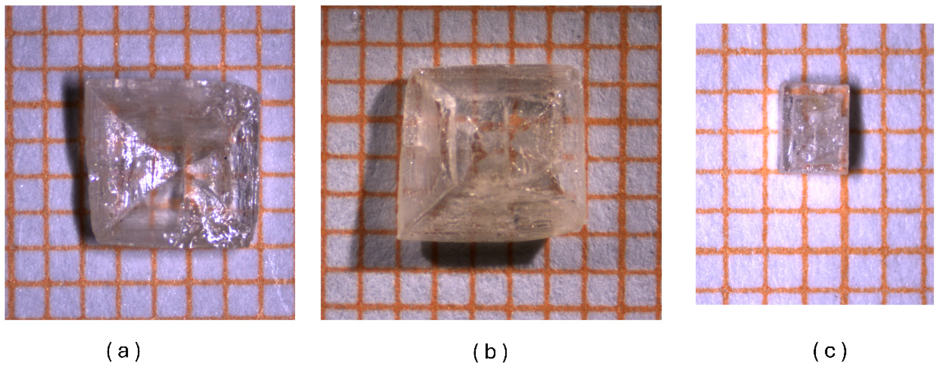
Colorless transparent crystals of the hydrate o-FHH were prepared by very slow evaporation (1 week), at room temperature, of a solution of N′-[(E)-(2-Fluorophenyl)methylidene]-4-hydroxybenzohydrazide in ethanol/water (90:10). In this way, specimens several millimeters long were obtained (Figure 1) in the form of square pyramids (also truncated) or prisms. The experimental PXRD pattern is in accordance with the simulated pattern obtained from the single-crystal X-ray structure solution (Figure S1). DSC analysis (Figure S2) shows that the hydrate crystals lose water between 120 and 160 °C, with melting in the anhydrous phase at 219 °C, as previously reported [23].

2.4. Crystal Structure Determination

Single-crystal X-ray diffraction data were obtained using a Kappa CCD diffractometer equipped with an Oxford Cryostream 700 apparatus (Oxford, UK) using graphite monochromated Mo Kα radiation (λ = 0.71073 A). Figure S3 shows the single crystal used for the data collection. Data reduction and semi-empirical absorption correction were performed with the SADABS program [25]. The structure was solved by direct methods with SIR97 [26] and was refined by the full-matrix least-squares method on F2 using SHELXL-2015 program [27] within the WinGX software v. 2023.1 [28]. Hydrogen atoms bonded to carbon were placed in calculated positions and refined with the riding model; those bonded to N and O were clearly found in different Fourier maps as the first maxima, and their coordinates were refined. For all H atoms, Uiso = 1.2 × Ueq of the carrier atom was assumed. The complete crystallographic and refinement parameters are listed in Tables S1 and S2 of the Supporting Information. The figures were created using the program Mercury [29]. The structural data have been deposited in the Cambridge Crystallographic Data Center (CCDC) with a deposition number of 2475743.

2.5. Differential Scanning Calorimetry

The thermal properties were studied using a NEXTA DSC-200 Hitachi calorimeter (Tokyo, Japan). The samples were heated from 25 to 300 °C at a heating rate of 10 °C/min. Nitrogen was used as the purge gas at 50 mL/min. The heat flow was measured in mW, and the melting point of the sample was determined through peak height.

2.6. NLO Microscopy Methodology

NLO spectra on the crystal were recorded by a wide-field illumination of the crystal at normal incidence with 1030 nm femtosecond IR pulses from a Pharos laser (Light Conversion, London, UK) The beam was directed into a Thorlabs (MM101, scanning multiphoton microscope, operating in reflection, Newton, NJ, USA) microscope with a 20× objective (Nikon, CFI Plan Fluor 20X CH, Tokyo, Japan) coupled with an imaging spectrometer (Andor, Kymera 328i, Oxford, UK) and an I-CCD camera (Andor, iStar 340, Oxford, UK). The spectrometer can be used for imaging and spectroscopy by switching between a mirror and a grating (150 l mm−1 groove density; blaze = 500 nm), The detailed experimental setup was described by Nicolas et al. [30].
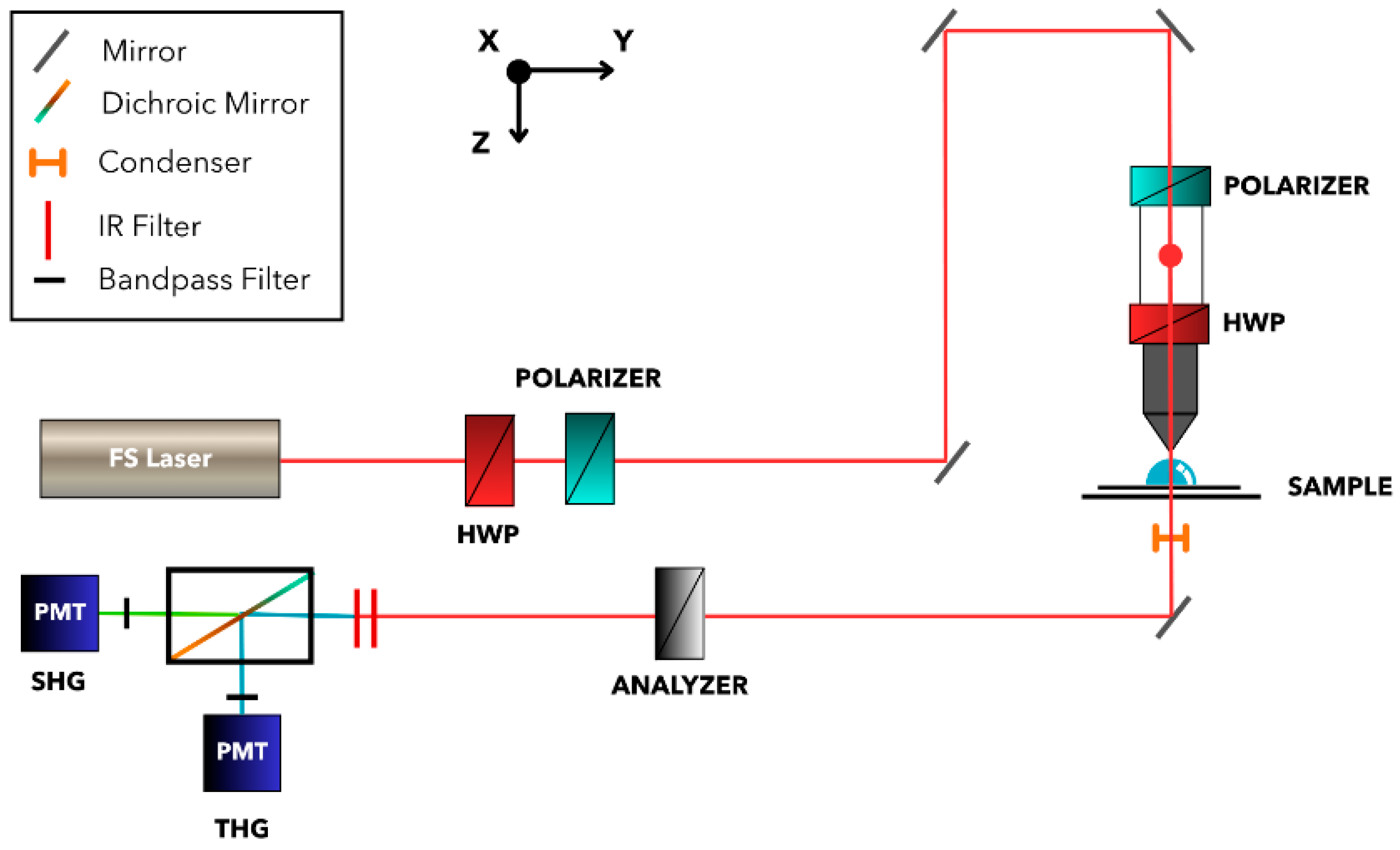
For the scanning microscopy setup, a mode-locked femtosecond pulsed Ti:Sapphire laser (80 MHz, 120 fs; Spectra-Physics, Insight DS+, Santa Clara, CA, USA) set at 1180 nm was used. A rotatable achromatic half-wave plate (HWP; Newport, 10RP52-4, Darmstadt, GE) was used in combination with a Glan-Laser polarizer (POL; Thorlabs, GL10) to tune to the intensity of the fundamental beam. The light is guided into the microscope (BX61WI, Olympus, Tokyo, Japan) through a series of mirrors (Thorlabs, F10-03-M01), where it enters polarized in the X-direction through a polarizer (Thorlabs, GL10). Inside the microscope, two Galvano scanners scan the light beam over the sample to scan the XY-plane. Subsequently, an achromatic half-wave plate (Thorlabs, SAHWP05M-1700) is mounted on a rotation stage, enabling control over the polarization state. A water immersion objective (40×, 0.80 NA, 3.5 mm WD, Nikon, Tokyo, Japan) was used. A condenser (OEM) with a numerical aperture of 0.9 was positioned after the sample to collimate the transmitted beam. The beam was then redirected by a mirror towards an analyzer (Thorlabs, WP25M) mounted on a rotation stage, enabling detection based on polarization. Two infrared filters are used to eliminate the fundamental beam, after which a dichroic mirror (Thorlabs, DMLP425R; 425 nm cutoff) separated the SHG and THG signals. The generated light was collected by photomultiplier tubes (PMT; Hamamatsu, R3896, Tokyo, Japan), each preceded by a bandpass filter: 590 ± 10 nm for SHG (Thorlabs, FBH590-10) and 390 ± 10 nm for THG (Thorlabs, FBH390-10). These were connected to a computer with software used to operate the shutter and HWPs. The setup was the same already described in the work by de Coene et al. [31] and is illustrated in Figure 2. The generation of SHG signals is contingent upon the concentration of a substantial number of photons at a singular point, due to the quadratic dependence of photon density. This phenomenon manifests exclusively at the laser’s focal point, yielding a highly resolved three-dimensional (3D) image exploiting this inherent confocal effect. The z-scan was performed with step size of 1 μm per slice with 150 slices, for a total z-distance of 150 μm. The image size was 360.56 × 360.56 μm, with a scanning speed of 12.5 μs/pixel and a Line Kalman averaging of 3. The total acquisition time for this z-scan experiment was ~50 min. SHG signals were collected in transmission mode. The 3D imaging consists of taking multiple images with incremental focal plane steps and recombining them using ImageJ software v. 1.54p.
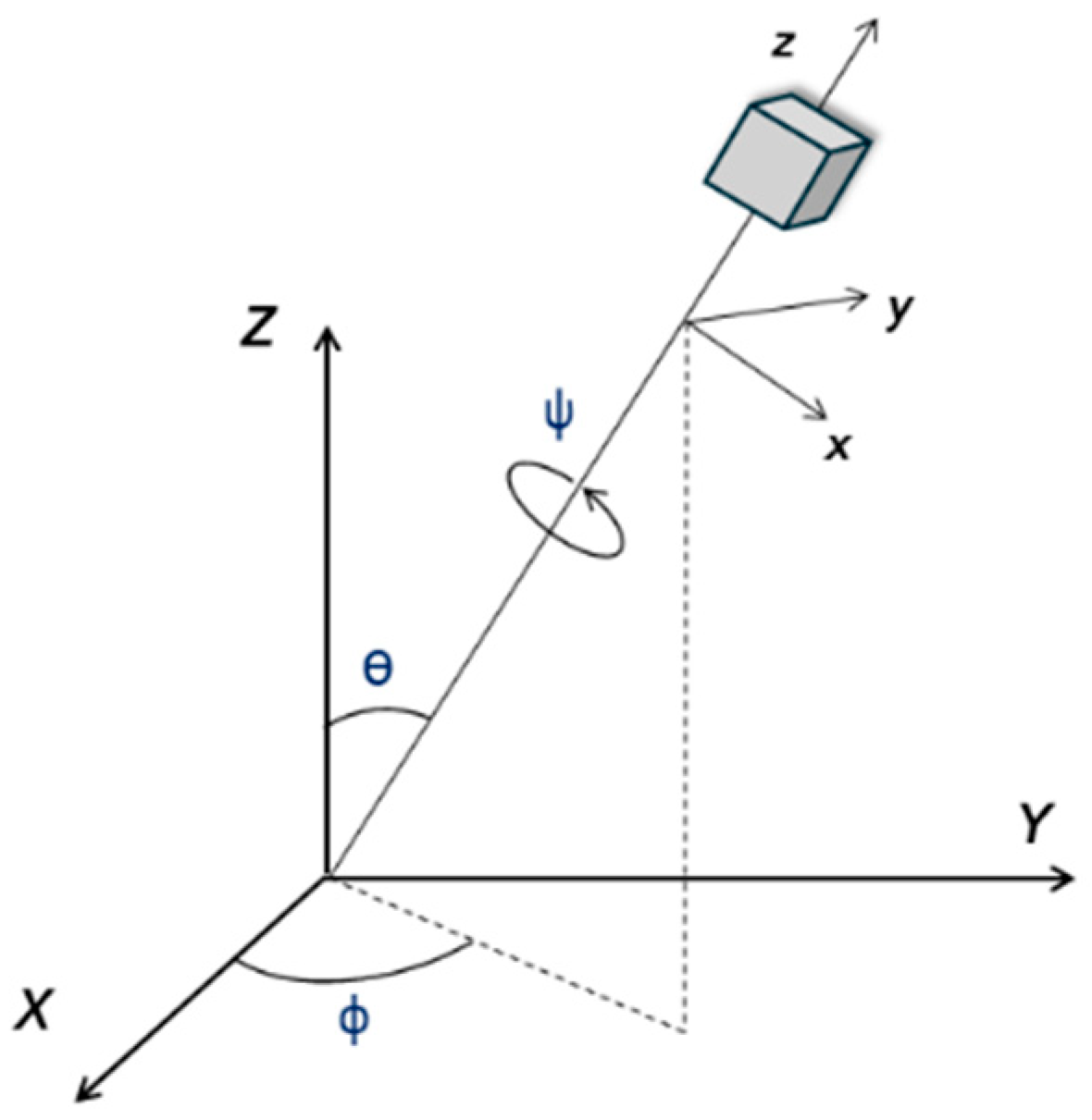
Polarized second-harmonic generation (SHG) imaging was used to determine the point group symmetry of the sample [32,33]. SHG polarimetry enables the identification of the structural origin of the second-order nonlinear response by analyzing the relative contributions of the χ i j k 2 tensor components. A rotational transformation matrix contaning the Euler angles Φ , ϴ , and ψ is used to describe the orientation of the microscopic coordinate system (xyz) operating in the macroscopic laboratory frame (XYZ). The in-plane xy angle for the linear polarization of the laser light is described by angle 2 α . The relation between the two coordinate systems can be visualized in Figure 3.
The electric field component of the laser beam along the propagation axis ( E Z ) can be neglected, allowing for the field to be considered as being confined to in-plane oscillations ( E X and E Y ). This assumption is valid when using a microscope objective with a numerical aperture (NA) of up to 0.8 [34]. In order to relate the laser fields to the fields generated within the sample, it is necessary to convert E X and E Y into E x , E y , and E z by using a rotational matrix over Euler angles Φ , ϴ , and ψ ( M Φ ,   ϴ , ψ   ). This allows us to define the induced second-order microscopic polarization as
P m i c r o ( 2 ) = χ ( 2 ) : M Φ ,     ϴ , ψ E m a c r o M Φ , ϴ , ψ E m a c r o  
with E m a c r o being the electric field strength expressed in the macroscopic frame and the Kronecker product. To simulate the detected SHG intensity, the microscopic polarization must be transformed back into the macroscopic reference frame using the inverse of the rotation matrix:
I S H G = P m a c r o 2 2 = M Φ , ϴ , ψ 1 P m i c r o 2 2
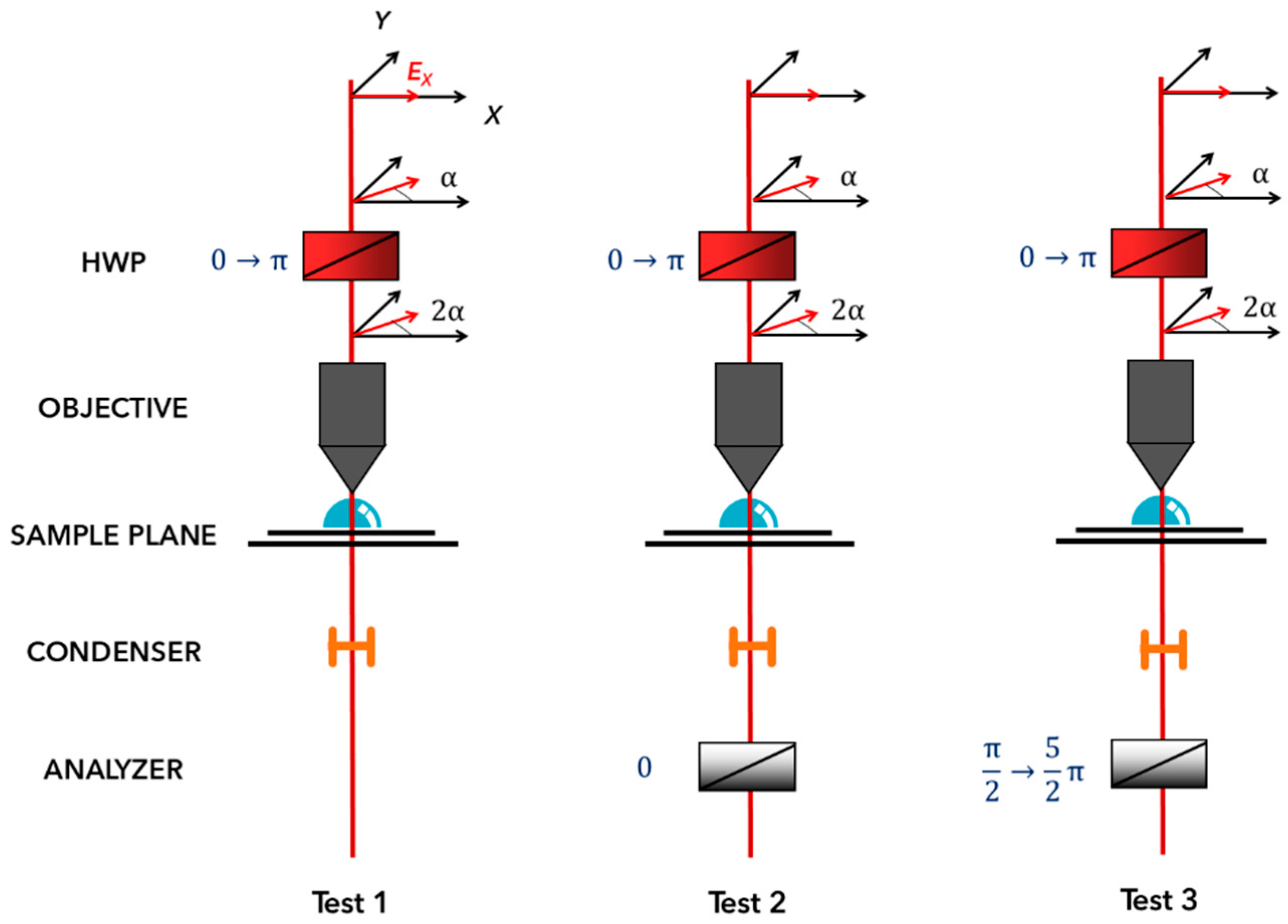
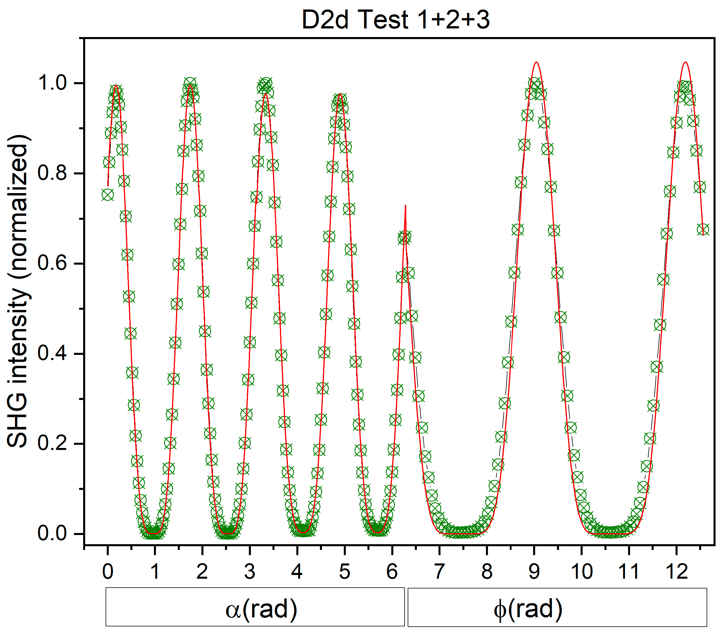
In order to be able to determine the point group symmetry, we defined three different polarization tests (see Figure 4). Test 1 is a rotation of the plane of linearly polarized incident light through 2 π   along the direction of light propagation while detecting all of the generated second-harmonic light. In Test 2, the plane of linearly polarized light is similarly rotated through 2 π , but detection is restricted to the second-harmonic light polarized along a specific, arbitrarily chosen direction using an analyzer. Test 3 consists of rotating both the incident and detecting linearly polarized light through 2 π , while maintaining an anti-parallel (out-of-phase) orientation between them throughout the rotation. The HWP is mounted in a rotation stage to control the angle of linear polarization.
During all polarimetry experiments, a kinetic series of 90 images was acquired consecutively, with no time delay between successive scans. The half-wave plate was rotated with increments of 2° successively in between each image acquisition. This procedure produced a full 2 π rotation of the polarization state, with the first image corresponding to α = 0 and the final image (image 90) corresponding to α = π . This is analogous to a full rotation of the ange Φ by 2 π for Test 3. The image size was 360.56 × 360.56, with a scan speed of 12.5 μs/pixel.
The 3D visualization software employed was Fiji ImageJ (available at https://rsb.info.nih.gov/ij (accessed on 1 March 2025); developed by Wayne Rasband, National Institutes of Health, Bethesda, MD, USA), importing the images as a sequence.

3. Results

3.1. Crystal Structure Description

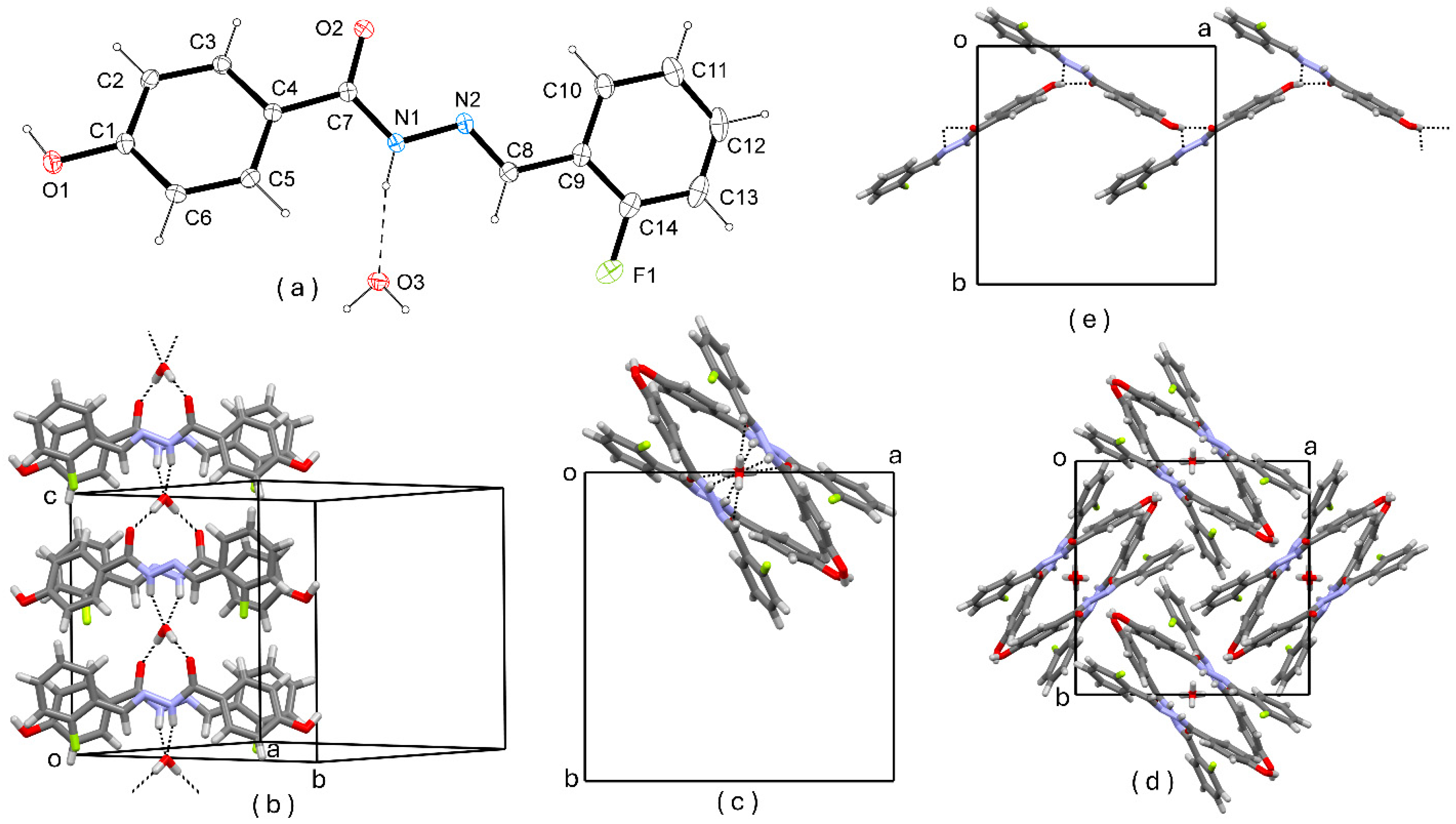
o-FHH crystallizes in the highly symmetric tetragonal space group P 4 ¯ 2 1 c (D2d point group) with half of a water molecule in the asymmetric unit (Figure 5a). The experimental PXRD pattern matches with the simulated pattern, as shown in Figure S1, of the supporting information file. The water molecule is in special position on a C2 axis, and then one hydrogen is generated by symmetry. The hydroxyphenyl and the fluorophenyl rings of the imine molecule are not coplanar, with their least squares planes making a dihedral angle of 25.26(3)°.
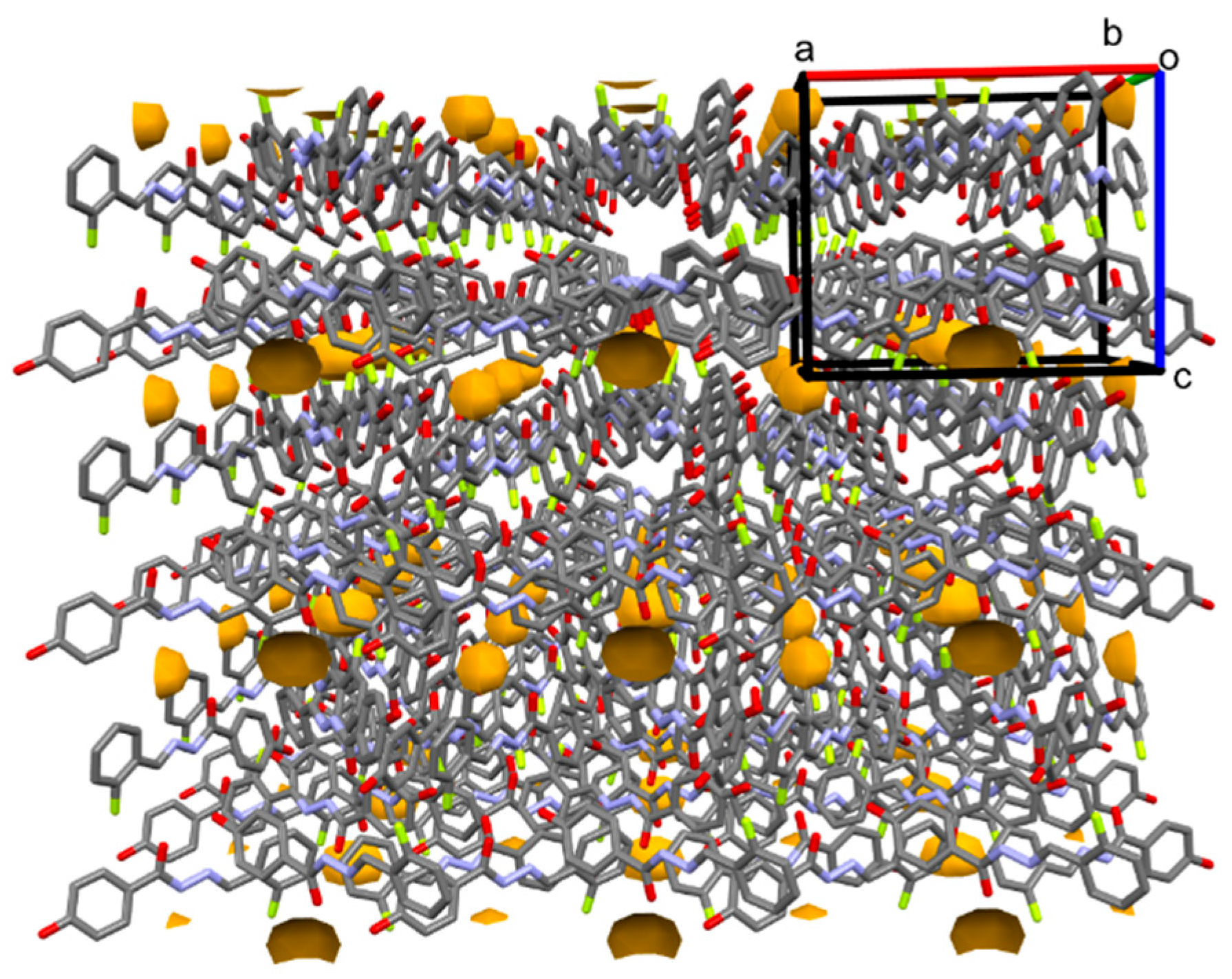
The basic supramolecular architecture in the crystal packing of o-FHH is represented by columns of H-bonded molecules that run parallel to the c axis (Figure 5b,c). These columns are formed by couples of C2 related imine molecules that are H-bonded to interposed water molecules. The columns are generated crystallographically by the c-glide planes. The crystal packing is characterized by a complex H-bonds pattern, as shown by compounds in which there is an a high number of donor and acceptor sites [35]. The interposed water molecules, which are placed within the hollow columns, act as H-bonding donors to two carbonyl O atoms (O3-H···O2i: 0.86(2), 1.93(2), 2.7805(17) Å, 172(2)°, i = −y + 1/2, −x + 1/2, z − 1/2) and as H-bonding acceptors from two N-H donors of imine molecules adjacent along the column (N1-H···O3 0.84(2), 2.18(2), 2.950(2) Å, 151(2)°). Therefore, water molecules are fully saturated in terms of H-bonds, and this accounts for the stability of the hydrate crystals [36] which exhibit a persistent water retention capacity, as clearly evident by the first endothermic peak of the differential scanning calorimetry reported in Figure S1 of the supporting information file, and the cavities in which the water molecules are present (Figure 6). Along the columns, ring patterns R 4 3 12 are observed. The colums fill the unit cell in the plane (a, b) through the 21 screw axes running parallel to the a and b axes (Figure 5d). In this way, additional H-bonds are formed between O-H donor and imine N and carbonyl O acceptors (O1-H···N2ii: 0.88(2), 2.63(2), 3.297(2), 133(2); O1-H···O2ii: 0.88(2), 1.92(3), 2.7448(19), 155(2), ii = x − 1/2, −y − 1/2, −z + 3/2) Figure 5e. Therefore, in the H-bonding patterns of o-FHH, phenolic O-H is a bifurcated donor and carbonyl O is a bifurcated acceptor.

3.2. NLO Microscopy

Preliminary to NLO microscopy experiments, we have checked the bulk SHG of powders of the sample, and the measured efficiency was equal to that of bulk powders of urea. Measurements were performed by following the procedure described in our previous paper [23]. In SHG microscopy, a NIR laser is directed through an objective lens and focused on the sample. The image acquisition is commonly achieved through point-by-point raster scanning of the laser beam across the sample, or by employing wide-field imaging. When the laser interacts with non-centrosymmetric structures in the sample, the specimen give rise to second-harmonic signals. In the electric dipole approximation, the electric field, oscillating at twice the excitation frequency ( E 2 ω ), can be related to the nonlinear second-order polarization P 2 ω ( 2 ) as follows:
E 2 ω P 2 ω 2 = Χ 2 E ω E ω
where E ω 2   = I ω is the intensity of the incoming light [37].
NLO microscopy provides high-resolution three-dimensional images due to its ability to achieve a significant imaging penetration depth without the means of external dyes. The capability of this technique to discern intricate details within the thickness of the samples has led to its utilization in the study of various biological samples and tissues, notably collagen. In nonlinear optical microscopy, laser excitation of the sample can induce a variety of nonlinear optical processes that are highly dependent on the material properties. These include phenomena such as the aforementioned SHG and THG, but also multi-photon fluorescence (MPF). As previously discussed, the use of multiple detection channels enables the discrimination of these signals. However, it should be noted that spectral overlap may occasionally occur, for example, in cases of partial leakage of fluorescence emission into the SHG detection channel. The integration of a spectrometer in the imaging setup enables a more precise spectral analysis.
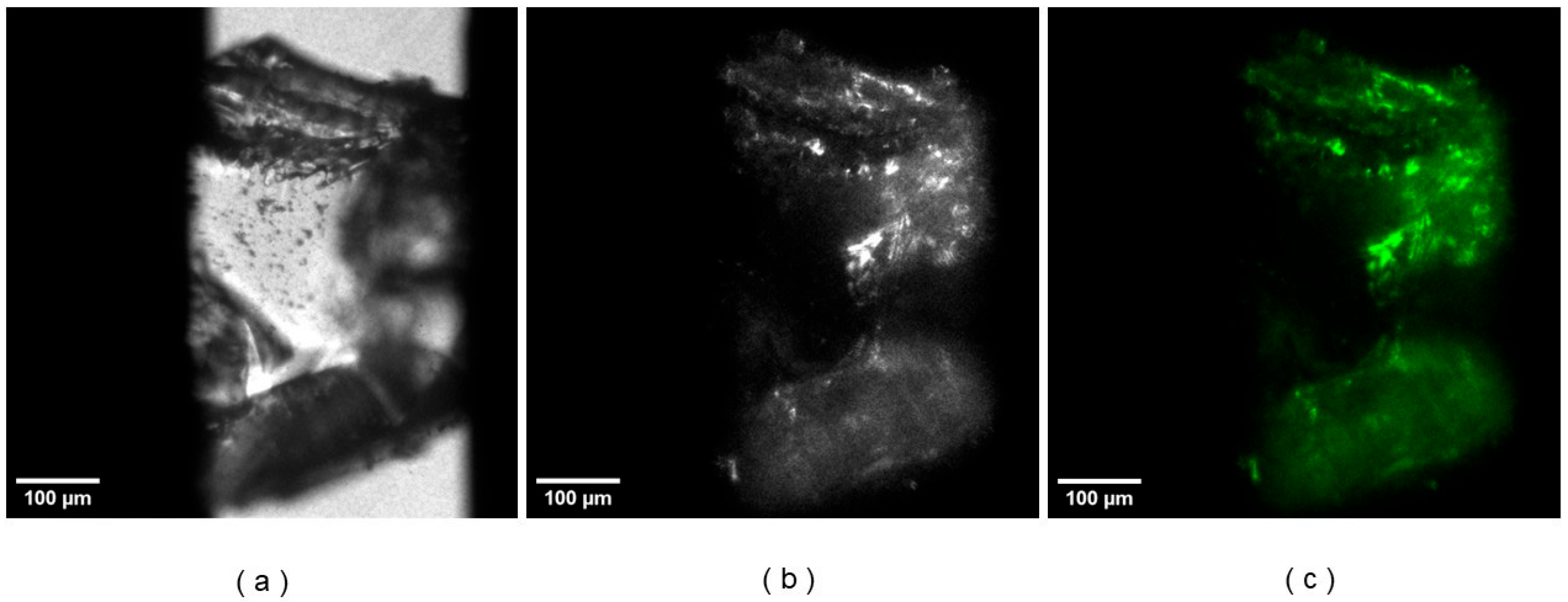
Nonlinear optical signals were observed during the imaging of o-FHH crystals. A comparison between images acquired with and without an SHG filter (Figure 7) using a wide-field microscope indicates that the detected signal seems to be due completely to second-harmonic generation.
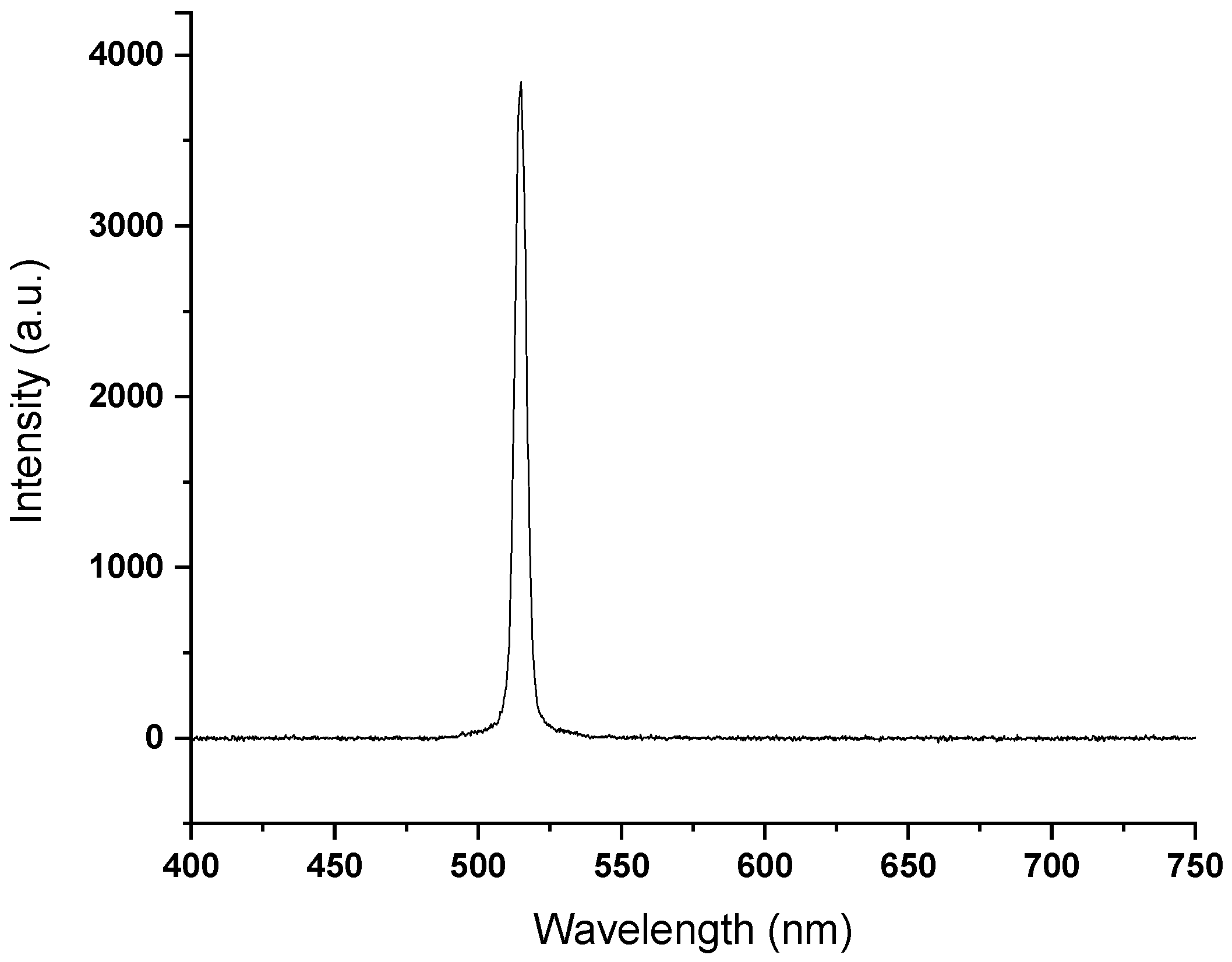
This interpretation is further supported by a spectral analysis performed on the same crystal. By narrowing the entrance slit of the spectrograph and employing full vertical binning, a specific line region of the image was analyzed with enhanced spectral resolution. The resulting spectrum reveals a single well-defined peak at 515 nm, Figure 8, consistent with SHG, and no significant contributions from other nonlinear processes.
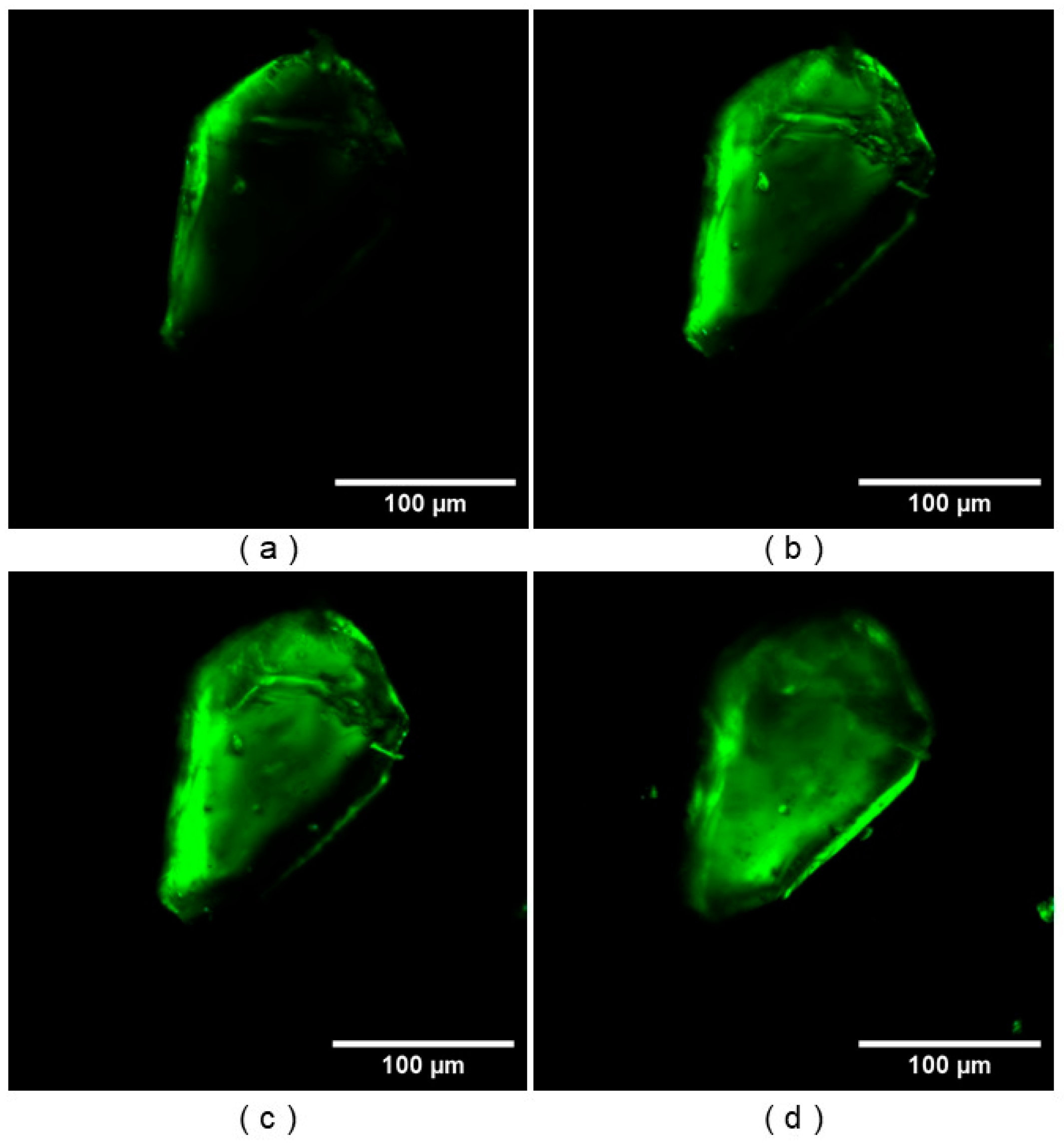
Figure 9 a–d illustrates frames from the scan at varying heights. The SHG signal is observed throughout the entire volume of the crystal, consistent with its non-centrosymmetric nature, which allows for a bulk second-order nonlinear optical response.

3.3. Symmetry Determination with NLO Polarimetry

By performing a series of polarization experiments, we can check if the SHG response is in accord with the D 2 d point group. For an SHG experiment, this point group has two independent and nonzero tensor components, χ x y z and χ z x y . The execution of three consequtive experiments, and fitting the experimental data to the theoretical intenstity dependence (see Figure 10), enables the extraction of the Euler angles and the ratio of the tensor components, as reported in Table 1.
It is remarkable that χ x y z and χ z x y are equal but opposite in sign. This is mainly because, under Kleinmann Symmetry conditions, which are valid far away from electronic resonance, χ x y z = χ z x y [38]. However, it has been shown that Kleinmann symmetry cannot always be assumed in practical applications, especially in chiral systems [39].

4. Conclusions

In this study, we crystallized a novel acentric organic crystal structure and investigated some NLO properties. In the crystal structure of o-FHH·H2O, the interstitial water acted as both a bifurcated hydrogen-bond donor and acceptor, creating a dense supramolecular network that caged the guest and stabilized a tetragonal framework. The arrangement of the transverse dipole in the crystal packing translated into a strong bulk SHG response that persists throughout the entire crystal volume, as confirmed by imaging measurements. The compound is highly stable in the presence of dehydration, and the crystal growth process demonstrates the potential to produce millimeter crystals from inexpensive solvents. It is clear from the nonlinear signature positions that it as a viable organic material for frequency conversion and other electro-optic applications. From a crystal engineering perspective, hydration can be viewed as a method for altering the supramolecular hierarchy, thereby directing crystallization towards acentric space groups without the need for complex synthetic modifications. Ongoing efforts will include the quantification of the χ2 tensor elements, the benchmarking of laser-damage thresholds, and the exploration of functionalized analogs within the hydrazide family to further enhance transparency in the near-infrared range and increase nonlinear coefficients.

Supplementary Materials

The following supporting information can be downloaded at: https://www.mdpi.com/article/10.3390/cryst15080739/s1, Table S1: Experimental details, Table S2: Geometric parameters (Å, °), Figure S1: PXRD patterns of o-FHH, Figure S2: DSC heating thermogram of the o-FHH imine. Heating rate is 10 K/min., Figure S3: Stereo microscope images of some crystal specimens of o-FHH placed on graph paper. Each square of the graph paper is 1 mm × 1 mm, NLO equations.

Author Contributions

This manuscript was written with contributions from all authors, and all authors have given approval to the final version of this manuscript. All authors have read and agreed to the published version of this manuscript.

Funding

This work was funded by the European Union-Next Generation EU, within the projects PRIN 2022 “Crystal Engineering of acentric and mechanically responsive smart crystals-ACME” (CUP E53D23009360006) and MUR PE14 “RESearch and innovation on future Telecommunications systems and networks, to make Italy more smart-RESTART” (CUP E63C22002040007). Y.d.C. acknowledges the Fund of Scientific Research Flanders (FWO) for a Senior Postdoctoral fellowship (1268825N). S.V.C. acknowledges support from ku leuven (c14/23/090, 3e230460), and fwo (g086522n,12r8218n).

Conflicts of Interest

The authors declare no conflicts of interest.

References

  1. Shkuratov, S.I.; Lynch, C.S. A review of ferroelectric materials for high power devices. J. Mater. 2022, 8, 739–752. [Google Scholar] [CrossRef]
  2. Gradauskaite, E.; Goossens, A.S.; Li, X.; Iglesias, L.; Gloter, A.; Meier, Q.N.; Bibes, M. Polarization Boost and Ferroelectricity Down to One Unit Cell in Layered Carpy-Galy La2Ti2O7 Thin Films. Adv. Mater. 2025, 37, 2416963. [Google Scholar] [CrossRef] [PubMed]
  3. Sekhar, M.C.; Veena, E.; Kumar, N.S.; Naidu, K.C.B.; Mallikarjuna, A.; Basha, D.B. A Review on Piezoelectric Materials and Their Applications. Cryst. Res. Technol. 2022, 58, 2200130. [Google Scholar] [CrossRef]
  4. Dishon Ben Ami, S.; Ehre, D.; Ushakov, A.; Mehlman, T.; Brandis, A.; Alikin, D.; Shur, V.; Kholkin, A.; Lahav, M.; Lubomirsky, I. Engineering of Pyroelectric Crystals Decoupled from Piezoelectricityas Illustrated by Doped α-Glycine. Angew. Chem. Int. Ed. 2022, 61, e202213955. [Google Scholar] [CrossRef]
  5. Venkataramudu, U.; Sahoo, C.; Leelashree, S.; Venkatesh, M.; Ganesh, D.; Naraharisetty, S.R.G.; Caudhary, A.K.; Srinath, S.; Chandrasekar, R. Terahertz radiation and second-harmonic generation from a single-component polar organic ferroelectric crystal. J. Mater. Chem. 2018, 6, 9330. [Google Scholar] [CrossRef]
  6. Das, K.; Ghosh, I.; Chakraborty, S.; Nambiar, A.G.; Maji, S.; Karan, S.K.; Kumar, D.; Bansal, A.K.; Reddy, C.M. Structural origin of fracture-induced surface charges in piezoelectric pharmaceutical crystals for engineering bulk properties. Nat. Commun. 2025, 16, 6858. [Google Scholar] [CrossRef] [PubMed]
  7. Kim, S.-J.; Kang, B.J.; Puc, U.; Kim, W.T.; Jazbinsek, M.; Rotermund, F.; Kwon, O.-P. Highly Nonlinear Optical Organic Crystals for Efficient Terahertz Wave Generation, Detection, and Applications. Adv. Opt. Mater. 2021, 9, 2101019. [Google Scholar] [CrossRef]
  8. Fang, Y.; Hao, J.; Sang, J.; Gao, J.; Song, L.; Tian, Y.; Li, R. Coherent manipulation of second-harmonic generation via terahertz-field mediated phonon-polariton in zinc oxide. Nat. Commun. 2025, 16, 5598. [Google Scholar] [CrossRef]
  9. Chelladurai, D.; Kohli, M.; Winiger, J.; Moor, D.; Messner, A.; Fedoryshyn, Y.; Eleraky, M.; Liu, Y.; Wang, H.; Leuthold, J. Barium titanate and lithium niobate permittivity and Pockels coefficients from megahertz to sub-terahertz frequencies. Nat. Mater. 2025, 24, 868. [Google Scholar] [CrossRef]
  10. Sun, M.-J.; Zheng, C.; Gao, Y.; Johnston, A.; Najarian, A.M.; Wang, P.-X.; Voznyy, O.; Hoogland, S.; Sargent, E.H. Linear Electro-Optic Modulation in Highly Polarizable Organic Perovskites. Adv. Mater. 2021, 33, 2006368. [Google Scholar] [CrossRef] [PubMed]
  11. Centore, R.; Roviello, A. Macromolecular Systems with Second Order Nonlinear Optical Properties. In Advances in Macromolecules: Perspectives and Applications, 1st ed.; Russo, M.V., Ed.; Springer: Dordrecht, The Netherlands, 2010; pp. 79–117. [Google Scholar]
  12. Groom, C.R.; Bruno, I.J.; Lightfoot, M.P.; Ward, S.C. The Cambridge Structural Database. Acta Crystallogr. Sect. B Struct. Sci. Cryst. Eng. Mater. 2016, 72, 171−179. [Google Scholar] [CrossRef]
  13. Brock, C.P.; Dunitz, J.D. Towards a Grammar of Crystal Packing. Chem. Mater. 1994, 6, 1118−1127. [Google Scholar] [CrossRef]
  14. Holland, K.M.; Aldair, A.; Ludlow, D.J.H.; Petersen, P.K.; Wright, M.A.; Chartrand, C.C.; Michaelis, S.J.; Johnson, J.A.; Patterson, J.E. Characterization of organic crystals for second-harmonic generation. Opt. Lett. 2023, 48, 5855–5858. [Google Scholar] [CrossRef]
  15. Debrus, S.; Ratajczak, H.; Venturini, J.; Pincon, N.; Baran, J.; Barycki, J.; Glowiak, T.; Pietraszko, A. Novel nonlinear optical crystals of noncentrosymmetric structure based on hydrogen bonds interactions between organic and inorganic molecules. Synth. Met. 2002, 127, 99–104. [Google Scholar] [CrossRef]
  16. Frazão, N.F.; Carvalho, P.J.; Leitão, M.M.; Lima, C.F.R.A.C. Organic–Inorganic Hybrid Materials with Enhanced Nonlinear Optical Response: Crystal Engineering Perspectives. J. Mater. Chem. C 2024, 12, 11215–11231. [Google Scholar]
  17. Radhakrishnan, T.P. Molecular structure, symmetry, and shape as design elements in the fabrication of molecular crystals for second harmonic generation and the role of molecules-in-materials. Acc. Chem. Res. 2008, 41, 367–376. [Google Scholar] [CrossRef]
  18. Chen, J.; Hu, C.L.; Kong, F.; Mao, J.G. High-performance second-harmonic-generation (SHG) materials: New developments and new strategies. Acc. Chem. Res. 2021, 54, 2775–2783. [Google Scholar] [CrossRef] [PubMed]
  19. Barhum, H.; McDonnell, C.; Alon, T.; Hammad, R.; Attrash, M.; Ellenbogen, T.; Ginzburg, P. Organic kainate single crystals for second-harmonic and broadband THz generation. ACS Appl. Mater. Interfaces 2023, 15, 8590–8600. [Google Scholar] [CrossRef] [PubMed]
  20. Yang, Y.; Zhang, X.; Hu, Z.; Wu, Y. Organic Nonlinear Optical Crystals for Highly Efficient Terahertz-Wave Generation. Crystals 2023, 13, 144. [Google Scholar] [CrossRef]
  21. Zhang, W.; Yao, J.; Yu, L. Organic Nonlinear Optical Crystals: Design Principles and Growth Techniques for High-Efficiency Frequency Conversion. Adv. Mater. 2020, 32, 1904083. [Google Scholar]
  22. Halfpenny, P.J.; Morrison, H.; Ristic, R.I.; Shepherd, E.E.A.; Sherwood, J.N.; Simpson, G.S.; Yoon, C.S. The growth of large single crystals of the organic nonlinear optical material 2-(α-methylbenzylamino)-5-nitropyridine. Proc. R. Soc. Lond. Ser. A Math. Phys. Sci. 1993, 440, 683–699. [Google Scholar]
  23. Parisi, E.; Borbone, F.; Carella, A.; Lettieri, S.; Capobianco, A.; Peluso, A.; Centore, R. Winning Strategy toward Acentric Crystals: Transverse Dipole Moment Molecules. Cryst. Growth Des. 2023, 23.6, 4538–4544. [Google Scholar] [CrossRef]
  24. Parisi, E.; Santagata, E.; Simone, E.; Borbone, F.; Centore, R. Frustration of H-Bonding and Frustrated Packings in a Hexamorphic Crystal System with Reversible Crystal–Crystal Transitions. J. Am. Chem. Soc. 2024, 146.28, 19405–19413. [Google Scholar] [CrossRef]
  25. Bruker-Nonius. SADABS; Bruker-Nonius: Delft, The Netherlands, 2002. [Google Scholar]
  26. Altomare, A.; Burla, M.C.; Camalli, M.; Cascarano, G.L.; Giacovazzo, C.; Guagliardi, A.; Moliterni, G.G.; Polidori, G.; Spagna, R. SIR97: A new tool for crystal structure determination and refinement. J. Appl. Crystallogr. 1999, 32, 115–119. [Google Scholar] [CrossRef]
  27. Sheldrick, G.M. Crystal structure refinement with SHELXL. Acta Crystallogr. Sect. C Struct. Chem. 2015, 71, 3–8. [Google Scholar] [CrossRef]
  28. Farrugia, L.J. WinGX and Ortep for Windows: An update. J. Appl. Crystallogr. 2012, 45, 849–854. [Google Scholar] [CrossRef]
  29. Macrae, C.F.; Bruno, I.J.; Chisholm, J.A.; Edgington, P.R.; McCabe, P.; Pidcock, E.; Rodriguez-Monge, L.; Taylor, R.; van de Streek, J.; Wood, P.A. Mercury CSD 2.0—New features for the visualization and investigation of crystal structures. J. Appl. Crystallogr. 2008, 41, 466–470. [Google Scholar] [CrossRef]
  30. Nicolas, P.; Minon, C.; Abdallah, S.; Chen, D.; Rizzi, G.; de Coene, Y.; Liu, W.; Jeannin, O.; Clays, K.; Bellec, N.; et al. Multiphoton-And SHG-Active Pyrimidine-Based Liquid Crystalline Thin Films Toward 3D Optical Data Storage. Adv. Opt. Mater. 2025, 13.2, 2402083. [Google Scholar] [CrossRef]
  31. de Coene, Y.; Jooken, S.; Deschaume, O.; Van Steenbergen, V.; Vanden Berghe, P.; Van den Haute, C.; Boon, V.; Callewaert, G.; Van Cleuvenbergen, S.; Verbiest, T.; et al. Label-Free Imaging of Membrane Potentials by Intramembrane Field Modulation, Assessed by Second Harmonic Generation Microscopy. Small 2022, 18.18, 2200205. [Google Scholar] [CrossRef]
  32. van der Veen, M.A.; Vermoortele, F.; De Vos, D.E.; Verbiest, T. Point-Group Symmetry Determination via Observables Revealed by Polarized Second-Harmonic Generation Microscopy: (1) Theory. Anal. Chem. 2012, 84, 6378. [Google Scholar] [CrossRef]
  33. Legat, T.; Grachev, V.; Kabus, D.; Lettinga, M.P.; Clays, K.; Verbiest, T.; de Coene, Y.; Thielemans, W.; Van Cleuvenbergen, S. Imaging with a Twist: Three-Dimensional Insights of the Chiral Nematic Phase of Cellulose Nanocrystals via SHG Microscopy. Sci. Adv. 2024, 10, eadp2384. [Google Scholar] [CrossRef]
  34. Dow, X.Y.; DeWalt, E.L.; Newman, J.A.; Dettmar, C.M.; Simpson, G.J. Unified Theory for Polarization Analysis in Second Harmonic and Sum Frequency Microscopy. Biophys. J. 2016, 111.7, 1553–1568. [Google Scholar] [CrossRef]
  35. Fusco, S.; Parisi, E.; Borbone, F.; Carella, A.; Capobianco, A.; Peluso, A.; Centore, R. Solid State Selection between Nearly Isoenergetic Tautomeric Forms Driven by Right Hydrogen-Bonding Pairing. Cryst. Growth Des. 2018, 18.10, 6293–6301. [Google Scholar] [CrossRef]
  36. Goswami, M.; Mukhopadhyay, S.; Kar, T. Role of Solvent Molecules in Directing Crystal Packing toward Noncentrosymmetric Structures: Insights from Hydrated Organic Crystals. CrystEngComm 2019, 21, 6731–6740. [Google Scholar]
  37. Verbiest, T.; Clays, K.; Rodriguez, V. General aspects of second-order nonlinear optics. In Second-Order Nonlinear Optical Characterization Techniques: An Introduction, 1st ed.; Taylor & Francis Group, CRC Press: Boca Raton, FL, USA, 2009; pp. 1–26. [Google Scholar]
  38. Kleinman, D.A. Nonlinear Dielectric Polarization in Optical Media. Phys. Rev. 1962, 126, 1977–1979. [Google Scholar] [CrossRef]
  39. Dailey, C.A.; Burke, B.J.; Simpson, G.J. The General Failure of Kleinman Symmetry in Practical Nonlinear Optical Applications. Chem. Phys. Lett. 2004, 390, 8–13. [Google Scholar] [CrossRef]
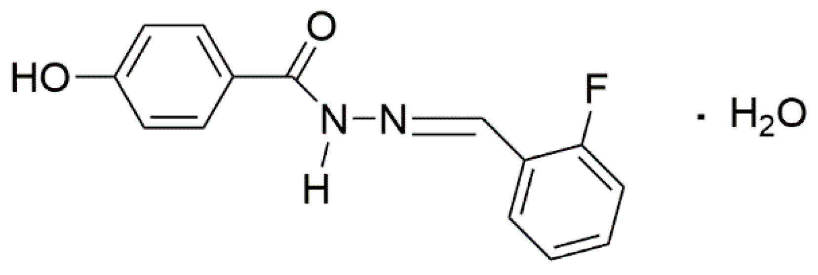
Scheme 1. Chemical diagram of N′-[(E)-(2-Fluorophenyl)methylidene]-4-hydroxybenzohydrazide monohydrate (o-FHH).
Crystals 15 00739 sch001
Figure 1. Stereo microscope images of some crystal specimens of o-FHH placed on graph paper. (a) square pyramid sample; (b) truncated square-pyramid sample; (c) prismatic sample. Each square of the graph paper is 1 mm × 1 mm.
Crystals 15 00739 g001
Figure 2. Schematic representation of the experimental setup, along with a laboratory coordinate system.
Crystals 15 00739 g002
Figure 3. The relative orientation of the microscopic frame (xyz) with regard to the laboratory frame (XYZ) expressed by means of the Euler angles Φ , ϴ , and ψ .
Crystals 15 00739 g003
Figure 4. Schematic representation of the 3 tests. The red arrow represents how the polarization of the laser ( E X ) is affected by rotating the HWP from 0 to π rads. The analyzer is fixed in Test 2, while it is rotated out-of-phase for Test 3.
Crystals 15 00739 g004
Figure 5. (a) Asymmetric unit of o-FHH with ellipsoids displayed at 30% of probability level; (b) skew view of a column of imine molecules H-bonded to interposed water molecules; (c) the same column of (b) viewed down the c axis; (d) crystal packing viewed down the c axis; (e) chain of H-bonded molecules running parallel to axis a. H-bonds are shown as dashed lines.
Crystals 15 00739 g005
Figure 6. Water molecules cavities (brown) in o-FHH. H atoms are omitted for clarity.
Crystals 15 00739 g006
Figure 7. (a) Bright-field image of a single crystal of o-FHH; (b) optical image of the same crystal acquired using laser excitation at 1030 nm without spectral filtering (power intensity of 206 mW); (c) optical image of the same crystal acquired under laser excitation with a filter only allowing for SHG centered at 515 nm (power intensity of 282 mW) in wide-field microscopy. The detection slit was fully open (~300 μm) during acquisition of the pictures.
Crystals 15 00739 g007
Figure 8. SHG spectrum of o-FHH crystal (using 1030 nm fundamental laser light and 76 mW power intensity). The detection slit was open of 100 μm during acquisition of the spectrum.
Crystals 15 00739 g008
Figure 9. Four slices from the SHG 3D scan of the lower (a), mid-low (b), mid-high (c), and the higher (d) depth of the total 121 slices for the z-scanning of o-FHH crystal. The step size in the Z direction is 1 μm. The step size between the four images is ~30 μm. Laser excitation wavelength is 1180 nm, with an intensity of 5.54 mW.
Crystals 15 00739 g009
Figure 10. Sequential fit of the experimental data (green squares) for the normalized SHG intensity (divided by the maximum within each test) for three consecutive tests. Test 1 comprises rotating the HWP from 0 to π (with alpha the HWP angle) and unpolarized detection; Test 2 ( π   t o   2 π ) describes the same, but only detecting the X-polarized component. The third test rotates the sample under crossed polarized detection, with φ from 4 π   t o   6 π . Each test is fitted to the intensity expression described in the SI.
Crystals 15 00739 g010
Table 1. Parameters extracted from the sequential fit.
χ x y z / χ z x y Φθψ
−0.97 ± 0.020.332 ± 0.0020.36 ± 0.011.589 ± 0.002
Disclaimer/Publisher’s Note: The statements, opinions and data contained in all publications are solely those of the individual author(s) and contributor(s) and not of MDPI and/or the editor(s). MDPI and/or the editor(s) disclaim responsibility for any injury to people or property resulting from any ideas, methods, instructions or products referred to in the content.