Article

Magnetic Behavior of Co2+-Doped NiFe2O4 Nanoparticles with Single-Phase Spinel Structure

Department of Materials Engineering, Isfahan University of Technology, Isfahan 84156-83111, Iran
*
Authors to whom correspondence should be addressed.
Crystals 2025, 15(7), 624; https://doi.org/10.3390/cryst15070624
Submission received: 27 May 2025 / Revised: 30 June 2025 / Accepted: 1 July 2025 / Published: 4 July 2025

Abstract

This study reports the synthesis and characterization of CoxNi1−xFe2O4 (x = 0, 0.2, 0.4, 0.6, 0.8, 1) nanoparticles using a co-precipitation method. In this approach, metal ions are precipitated in the presence of a stabilizing agent, which is a common and effective method for nanoparticle preparation. The microstructure and magnetic properties were studied after calcination at 600 °C and heat treatment at 1000 °C. X-ray diffraction (XRD) and Fourier transform infrared (FTIR) spectroscopy confirmed the formation of a single-phase spinel structure. The average crystallite size, calculated using the (311) diffraction peak and the Scherrer equation, ranged from 13 to 19 nm. Scanning electron microscopy (SEM) showed that the nanoparticles had a spherical morphology. Thermogravimetric and differential thermal analysis (TG-DTA) revealed a three-step weight loss process. Magnetic measurements, including remanent magnetization, saturation magnetization, and coercivity, were performed using a vibrating sample magnetometer (VSM) at room temperature. The replacement of Ni2+ with Co2+ enhanced the magnetic properties, resulting in increased magnetic moment and anisotropy. These effects are attributed to changes in cation distribution, exchange interactions, surface effects, and magnetocrystalline anisotropy. Overall, Co2+ doping improved the magnetic behavior of nickel ferrite, indicating its potential for application in memory devices and magnetic recording media.

1. Introduction

Investigating nanomaterials has spurred considerable interest from researchers because of their outstanding chemical and physical properties and potential for a range of technological applications in multiple fields [1,2,3]. Of these materials, ferrite nanoparticles are noteworthy for their broad range of fundamental properties and applications, including microwave devices, gas sensors, catalysis [4], controlled drug delivery as microwave absorption materials [5], transformer cores [6], radiographic circuits [7], magnets [8], high-quality filters, digital tapes, hybrid supercapacitors, and photocatalysts [9]. From a crystallographic perspective, ferrites are typically categorized into two primary groups: cubic (spinel) ferrites and hexagonal (hexaferrite) ferrites [10,11]. Cobalt ferrite (CoFe2O4) is a hard magnetic material known for its properties of high coercivity, excellent thermal stability, good electrical insulation, high mechanical hardness, high resistivity, non-toxicity, high Curie temperature of 870 K, good chemical stability, and low magnetization [12,13]. In contrast, nickel ferrite (NiFe2O4) represents a soft magnetic material with low magnetic anisotropy, low conductivity, and high electrochemical stability [14,15]. Spinel ferrites are generally denoted by the chemical expression MFe2O4, where M is a divalent metal cation and Fe is in a trivalent oxidation state. A spinel ferrite unit cell has 32 O ions arranged in a face-centered cubic (FCC) lattice with 64 tetrahedral and 32 octahedral sites to accommodate metal cations. Both divalent and trivalent metal cations occupy one-eighth of the tetrahedral sites (site A), and each cation occupies one-half of the octahedral sites (site B). Depending on the occupancy of the divalent or trivalent metal cation in the tetrahedral and octahedral sites, either a normal or inverse spinel may be generated. Doping nickel ferrite with Co2+ cations (larger ionic radii than Ni2+) causes structural distortions, as evidenced by changes in lattice spacing, lattice constants, and ionic radii of the cations at the tetrahedral and octahedral sites. Upon Co doping, redistribution of cations occurs within the nickel ferrite structure in which Co2+ ions preferentially occupy the octahedral sites (site B), resulting in the movement of Ni2+ ions from site B to the tetrahedral sites (site A). The movement of some Fe3+ ions from the tetrahedral sites to the octahedral sites also occurs. These structural alterations can have implications for a wide range of material properties, but most importantly magnetic properties. There is a direct relationship between structural parameters and magnetic properties. Subsequently, the magnetic properties exhibited by ferrites can be systematically fine-tuned by changing the cation distribution between tetrahedral and octahedral sites [16,17]. It is well-known that the properties of ferrites depend on the synthesis method, chemical composition, or cationic ordering. There are many effective methods for synthesizing ferrite nanoparticles, such as sol–gel [10], solution combustion [17], thermal plasma, high-energy ball milling [16], and co-precipitation [18]. Co-precipitation is an attractive option because it is inexpensive and quick and has many non-toxic alternatives to organic fuels, such as citric acid. The attraction of co-precipitation is in producing extremely uniform yields [19,20]. Firoz Khan et al. [21] demonstrated that the magnetic properties of Co ferrite increased when some of the cobalt in Co1−xNixFe2O4 (x = 0.1, 0.3, 0.5, 0.7, and 0.9) nanoparticles made using the co-precipitation method was substituted with nickel. Similarly, Annie Vinosha et al. [20] observed an enhancement in the magnetic properties of Co ferrite resulting from nickel substitution in Co1−xNixFe2O4 (x = 0.1, 0.3, 0.5, 0.7, and 0.9) nanoparticles, which were synthesized using the co-precipitation method. Chandekar et al. [22] also employed the co-precipitation method to prepare CoxNi1−xFe2O4 (x = 0–0.5) nanoparticles, reporting that the incorporation of Co2+ ions into the host lattice induced significant modifications in the magnetic properties. These modifications were a consequence of the exchange interactions at sites A and B and subsequently modified the magnetic anisotropy of the nanoparticles. This work will focus on the synthesis of Co2+-doped nickel ferrite nanostructures using the general formula CoxNi1−xFe2O4 (x = 0, 0.2, 0.4, 0.6, 0.8, 1). The focus will be on studying the effects that varying amounts of cobalt have on the magnetic, structural, and morphological properties of the cobalt-doped nickel ferrites. To minimize impurities and ensure that the desired structural phase is formed, a calcination and annealing process was utilized at temperatures of 600 °C and 1000 °C. The primary objective of this study was to refine the grain size of nanoparticles synthesized via the co-precipitation method, aiming to achieve particles with moderately high coercivity and magnetization, single magnetic domain behavior, and elevated magnetocrystalline anisotropy. The main innovation of this article lies in examining the effect of two distinct annealing temperatures on the final properties of the material. Annealing is a cost-effective technique for obtaining desirable material characteristics. By increasing the thermal energy of atoms, annealing facilitates atomic diffusion and rearrangement, allowing atoms to occupy more stable positions. This process results in a reduction of defects and an improvement in crystallinity, thereby enhancing the suitability of the material for diverse applications. The decrease in grain boundary spacing and dislocation density contributes to an increase in crystallite size and unit cell volume. This methodology leverages induced modifications during post-synthesis processing to precisely tailor the material’s structural and magnetic properties. Consequently, this work is significant due to its novel and advanced contributions to the field.

2. Experimental Section

2.1. Materials

The precursors utilized for the synthesis of CoxNi1−xFe2O4 nanoparticles included iron (III) nitrate hydrate (Fe(NO3)3·9H2O-98.5% purity-Merck), nickel nitrate (Ni(NO3)2·6H2O-98.5% purity-Merck), cobalt nitrate (Co(NO3)2·6H2O-99% purity-Merck), sodium hydroxide (NaOH-99% purity-Merck), and oleic acid as the surfactant.

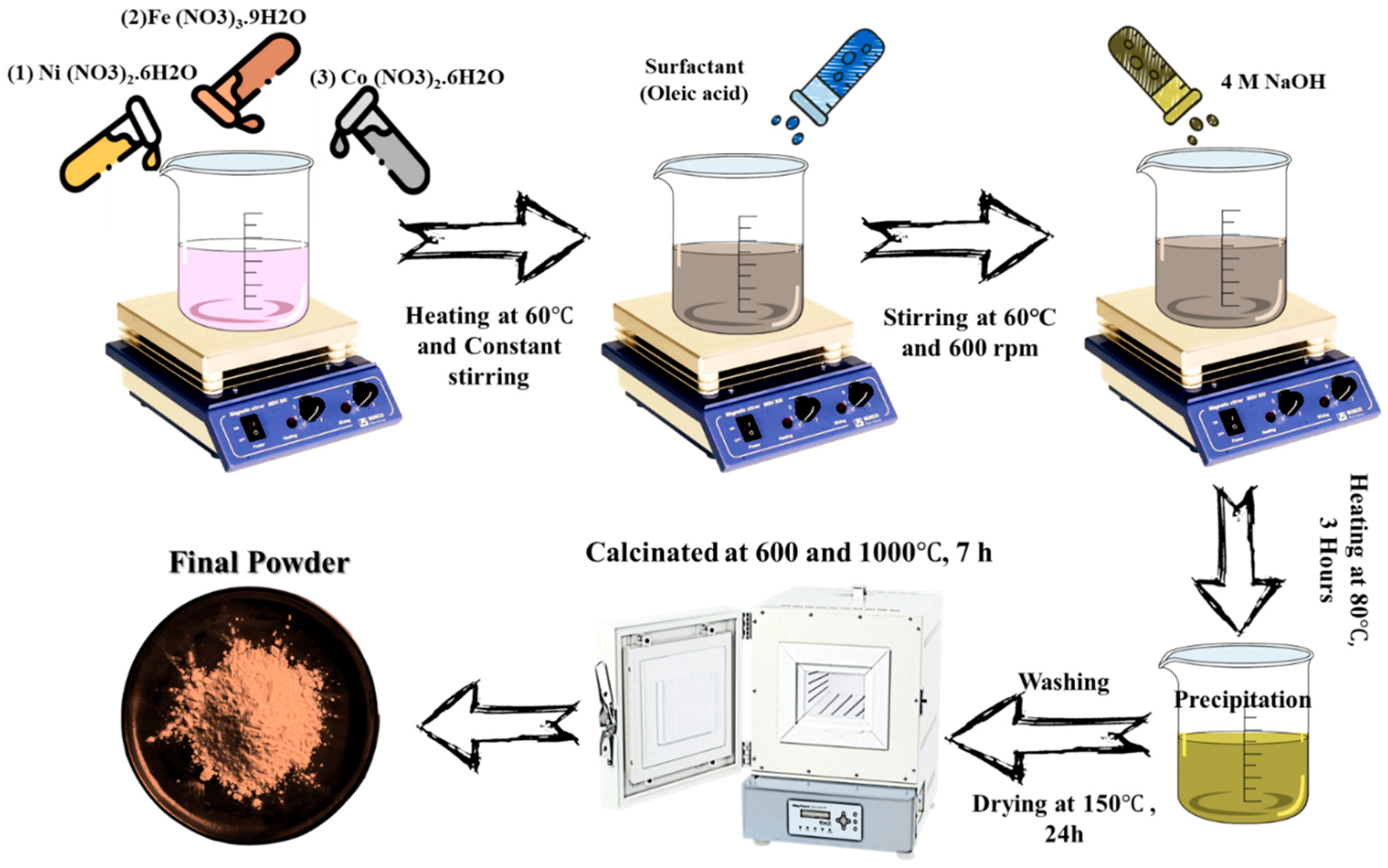
2.2. Synthesis of Co2+-Doped NiFe2O4 Nanocomposite

As shown in Figure 1, for the synthesis of CoxNi1−xFe2O4 nanoparticles, stoichiometric amounts of metal nitrates Fe(NO3)3·9H2O, Ni(NO3)2·6H2O, and Co(NO3)2·6H2O were dissolved in an appropriate volume of double-distilled water. The solution obtained was subsequently transferred to a hotplate and continuously stirred, while 2–3 drops of oleic acid were employed as a surfactant. Following this, sufficient amounts of 4 M sodium hydroxide solution were employed to maintain a consistent pH between the specified range of 12.5 and 13. The mixture was subjected to a continuous stirring process at 80 °C for a duration of 3 h. At this point, the precipitate was collected and separated using a centrifuge apparatus (Eppendorf 5810R, Eppendorf AG, Hamburg, Germany) at 2000 rpm, before being washed with acetone followed by deionized water until the wash-solution pH was optimized between 7 and 8. The precipitate was subsequently annealed in an oven at 150 °C overnight to let the dry precipitate yield CoNiFe2O4 powder. Finally, the powder produced was processed in an electric furnace at 600 °C, followed by a second exposure to heat at 1000 °C (a total of 7 h) to produce CoNiFe2O4 ferrite. A temperature of 600 °C was used to probe the effects of crystallization, as well as the successive development of the ferrite phase at a lower temperature.
This temperature assists in diminishing internal stresses in the nanoparticle structure and also limits excessive agglomeration of the particles, allowing for enhanced control of crystal size. In addition, heat treatment at 600 °C permits the observation of structural and magnetic changes associated with progressive crystallization at elevated temperatures compared to 1000 °C. The heat treatment speeded up ferrite formation, which could have occurred via simultaneous dissolution and crystallization; meanwhile, in the higher temperature treatment, extensive agglomeration occurred. The molar ratios of Co:Ni:Fe in the samples are shown in Table 1.

2.3. Characterization of Co2+-Doped NiFe2O4 Nanocomposites

An assortment of procedures was utilized to thoroughly assess and characterize Co2+-doped NiFe2O4 nanoparticles, including X-ray diffraction (XRD), field-emission checking electron microscopy (FE-SEM, model Ouanta 200 with drift 2017 EDAX, Cambridge, MA, USA), Fourier-transform infrared spectroscopy (FTIR), vibrating test magnetometry (VSM, MDKB, Kashan-Iran), thermogravimetric investigation (TG), and differential warm investigation (DTA). XRD was utilized to assess the stage arrangement of CoNiFe2O4 by analyzing the synthesized nanoparticles on a PANalytical X’Pert PRO MPD diffractometer (Malvern PANalytical, Almelo, The Netherlands), since it can assess crystallinity and stages. The XRD examination utilized copper-Kα radiation (wavelength λ = 1.540562 Å) within the 2θ run from 20° to 80°. For information collection, the check rate was 2° per diminutive, with 40 kV and 30 mA utilized for quickening the voltage and emanation current. FTIR spectra were collected within the extent of 400–4000 cm−1 employing a Tensor Spectrometer, after blending 1 mg of the ferrite test with 100 mg of potassium bromide (KBr) as the foundation.
To evaluate the formation of spinel ferrite phases and their thermal stability, the synthesized specimens were analyzed using thermogravimetric (TG) and differential thermal (DTA) analyses with a LABSYS EVO TG-DTA unit (Isfahan University, Iran) under an Ar atmosphere, with a heating rate of 5 °C/min. The morphological characteristics and composition of the cerium ferrites were examined using scanning electron microscopy (SEM, model XL30 Series). Particle size and distribution were examined using ImageJ 1.52V software. The magnetic properties of the samples were examined by a vibrating sample magnetometer (VSM, Magnetis Daqiq Kavir, Mashhad, Iran) at room temperature.

2.4. Statistical Analysis

The data were analyzed using one-way ANOVA and Tukey’s post hoc comparison test to gain a deeper understanding. The statistical significance was determined at p < 0.05 for the variances or conditions that differed between groups.

3. Results and Discussion

3.1. Thermogravimetric (TG) and Differential Thermal (DTA) Analysis

The thermal analysis curves of the Co0.4Ni0.6Fe2O4 samples heat-treated at 600 °C and 1000 °C are shown in Figure 2. Figure 2a (600 °C) shows three stages of weight loss during the course of 35–850 °C. The first weight loss, at around 114.42 °C, is attributed to the physical desorption of water molecules on the powder surface [23]. This stage primarily corresponds to the evaporation of residual moisture absorbed during the sample preparation process. The second mass loss, which takes place at 338 and 475 °C, relates to the thermal decomposition of organic residues, which encompasses both metal nitrate precursors and intermediate oxide species. During this step, the nitrate species and carbonaceous compounds spontaneously combust, which oxidizes and removes these compounds and causes a large loss of sample mass. The considerable loss of mass during this phase typically means that nearly all organic material has been lost during this temperature range.
The last stage of mass loss was observed from 460 °C to around 700 °C due to the crystallization of the final ferrite phase. There was some mass loss after 600 °C, leaving a nickel–cobalt ferrite that was thermally stable. Beyond this temperature, the sample mass remained unchanged, indicating the complete formation of all phases. Similarly, Figure 2b has a two-part mass loss profile for the 1000 °C treated sample. The first mass loss at 88 °C is due to the removal of adsorbed moisture, and overall, the trends are consistent with the profile for the sample treated at 600 °C. Yet, it is observed that the curve is shifted slightly to the right, indicating a greater temperature observed for the transition to the mass loss. This could be attributed to changes in the ferrite matrix composition that would affect the thermal performance of the material.

3.2. X-Ray Diffraction (XRD) Analysis

The X-ray diffraction (XRD) patterns are shown in Figure 3. As shown in Figure 3a, the XRD analysis indicates that the desired phases were produced after calcining the precursor at 600 °C. However, the additional peaks suggest the presence of an impurity phase, specifically a relatively small fraction of unreacted α-Fe2O3, which is within the acceptable proportion [24]. The XRD patterns of the sample calcined at 1000 °C (Figure 3b) reveal the growth of these impurity peaks, suggesting that the precipitate contains nickel, iron, cobalt, or some combination of these metals. Additionally, high temperatures play a role in the further development of these impurities, as well as the decomposition of phase and incorporation of structure and vacancies in the framework. The addition of cobalt doping demonstrates the capability of occupying the interstitial sites of the nanoparticle cubic spinel structure without changing the phase [25]. The findings additionally suggest that higher temperatures lead to sharper, more symmetric diffraction peaks, which indicates improved crystalline structures. The trend of peak narrowing versus temperature is a sign of an increase in crystallite size, which reflects increased crystallinity. The increase in crystallite size at higher temperatures is related to the stronger surface effects present in nanoscale samples. The agglomeration of particles accordingly increases their size and lowers the surface energy. The peak width at half maximum (FWHM) of the peaks is inversely correlated to crystallite size, according to the Scherrer equation. As such, each decrease in peak broadening is indicative of an increase in crystallite size. In addition, the decrease in energy also leads to an increase in size with increasing temperature, adding to the likelihood of crystallite growth. Heating also improves the crystallinity of the nanoparticles at higher temperatures while not changing their overall nature [26].
Grain size was calculated using the Scherrer equation. The grain size as a function of cobalt content is shown in Figure 3c. The results show that grain size increases with cobalt content. In addition, XRD data was analyzed to obtain lattice constant and unit cell volume at this temperature, as shown in Figure 3d. The results demonstrate that increasing cobalt content increases the lattice constant and therefore the unit cell volume. The increase in lattice constant, crystallite size, and unit cell volume with increasing cobalt content is due to the smaller ionic radius of nickel compared to cobalt [25]. In particular, cobalt, with its larger atomic radius (0.74 Å), substitutes for nickel atoms with a slightly smaller atomic radius (0.69 Å) in the octahedral sites, influenced by the atomic radius difference, which alters the 2θ range and reduces this range as well. Because 2θ is inversely related to the lattice constant, a smaller shift in the (311) peak leads to a larger lattice constant, which then leads to a larger interplanar spacing. An increase in the lattice constant could be attributed to point defects, e.g., ferric ion occupies normally unoccupies sites. In turn, it will cause a change in the degree of inversion of CoxNi1−xFe2O4 [27,28]. As shown in both Figure 3c,d, the grain size significantly increases with the rise in temperature above 1000 °C and cobalt content. With increased temperature, the particles aggregate to reduce surface energy, which leads to an overall increase in particle size, while the lattice constant and unit cell volume exhibit marked increases at this temperature compared to 600 °C. This is more or less the result of continuing larger surface effects.

3.3. Fourier Transform Infrared (FT-IR) Analysis

To further understand the crystallographic structure of the heat-treated CoxNi1−xFe2O4 samples, the Fourier transform infrared (FTIR) spectra of the heat-treated samples were obtained at room temperature, in the range of 400–4000 cm−1, and shown in Figure 4. In ferrites, metal ions occupy two separate sub-lattices: tetrahedral (A-site) and octahedral (B-site) sites according to spaces with neighboring oxygen atoms. The tetrahedral site shows stretching bands around 580–600 cm−1, while the octahedral stretching band generally appears around 375–450 cm−1. The absorption bands at about 1400 cm−1 belong to the characteristic band for the NO3 ion, the bands at 1600 cm−1 are due to carboxyl groups (COO), and the band at 3400 cm−1 is due to hydrogen-bonded hydroxyl groups (OH) [29]. The lower stretching frequency (ν_A) originates from the metal–oxygen bonds at the octahedral site, and the higher stretching frequency (ν_B) originates from the metal–oxygen bonds at the tetrahedral site.
When we examine the broadening of the tetrahedral stretching peaks in CoxNi1−xFe2O4, we find that the broadening becomes more severe as heat treatment and cobalt substitution increase. This broadening is attributed to the interaction between oxygen ions and Fe3+ ions in the case of Co2+ doping [30]. The observed increase in the stretching frequency of the metal–oxygen bonds is due to the greater atomic mass of Co2+ ions relative to Ni2+ ions [31]. Bulk nickel ferrite usually has an inverse spinel structure; however, cobalt substitution for nickel changes the structure from an inverse spinel to a partially inverse spinel. The change in the distribution of nearest-neighbor cations contributes to the broadening observed in the FTIR spectra [32].

3.4. Field Emission Scanning Electron Microscopy (FESEM) Analysis

Figure 5 shows the images obtained from the morphological examination of the shape, size, and distribution of CoxNi1−xFe2O4 ferrites synthesized at temperatures of 600 °C and 1000 °C, respectively, and the EDX analysis. Figure 5(aa–ae) confirm the observation that at a temperature of 600 °C, the synthesized nanoparticles have an almost spherical form, with a constant and uniform distribution of particle size and density. The average particle size was calculated using MIP software version 4.5, and it was determined that the range of particle size for the nanoparticles was between 13 and 19 nm, aligning fairly closely with the results from the X-ray diffraction (XRD) analysis. In addition, the particles appeared to be agglomerated owing to their relatively high surface-to-volume ratio, and their high surface energy attracted them into cluster formation. The magnetic character of these nanoparticles also caused them to cling to one another, producing larger particles [26].
Likewise, Figure 5(ba–be) depict the results for the samples synthesized at 1000 °C. The average particle size, calculated using MIP software, indicated that the nanoparticles ranged from 28 to 36 nm, demonstrating relative agreement between the size measures, suggesting a relatively uniform particle size distribution, though broader than that of the samples synthesized at 600 °C. The strong compatibility between Scherrer equation values and particle sizes presented in SEM can be attributed to the fact that the synthesized particles were effectively single-domain. As demonstrated in the images, particle size increased as a function of temperature and concentration of cobalt, but no change was observed in particle morphology, and no correlation was observed between increased crystallite size and increased cobalt concentration. EDX analysis nicely agrees with the result in Figure 5c for the Co0.8Ni0.2Fe2O4 sample, demonstrating the expected peaks for cobalt and nickel in addition to the peaks for iron that validate successful synthesis of the powders [21].

3.5. Vibrating Sample Magnetometer (VSM) Analysis

Figure 6 shows the hysteresis loop of cobalt-doped nickel ferrite nanoparticles (600 °C and 1000 °C). The saturation magnetization (MS), remanent magnetization (Mr), and coercivity (HC) values obtained from these loops are summarized in Table 2 and Table 3. The components of the cobalt-doped nickel ferrites clearly have a major effect on the magnetic properties of these nanoparticles. The samples show typical ferromagnetic behavior, as there is a transition between soft magnetism for pure NiFe2O4, to hard magnetism for pure CoFe2O4. Table 2 and Table 3 show that the saturation magnetization of NiFe2O4 (33.65 emu/g) and CoFe2O4 (61.17 emu/g) from the 600 °C sample and NiFe2O4 (22.37 emu/g) and CoFe2O4 (52.88 emu/g) from the 1000 °C sample, is lower than the literature bulk magnetization values of approximately 55 emu/g and 93 emu/g for NiFe2O4 and CoFe2O4, respectively [33,34]. The saturation magnetization corresponds with the previously established literature, affirming a notable relationship between magnetic properties and crystallization. The increase in MS is a result of the preeminence of organized magnetic moments in the core of the magnetic particles. As temperature increases, thermal agitation disrupts the spin-glass structure, resulting in an enhanced number of magnetic spins aligned with the applied magnetic field and consequently enhancing saturation magnetization [35]. Furthermore, particle size increases with prolonged thermal treatment due to nanoparticle fusion, leading to reduced core–shell interactions and consequently increasing saturation magnetization [36].
The S-shaped appearance of the M-H curve indicates that small magnetic particles demonstrate superparamagnetic behavior. The residual magnetization is also low at room temperature, which supports the likelihood of superparamagnetism. There are two possible reasons to explain the unsaturation hysteresis behavior commonly observed in ferites: (i) strong interactions between particles and (ii) small nanoparticles with a core–shell structure, endogenous to the core (ferrimagnetic or ferromagnetic state) and exogenous to the shell (a spin-glass state with large surface anisotropy effects on bulk behavior) [37]. One important property of superparamagnetic materials that is advantageous for applications in magnetic targeting is that superparamagnetic materials retain no magnetization after a steady external magnetic field is removed. Ferromagnetic and superparamagnetic phenomena are highly size-dependent and typically demonstrate superparamagnetic behavior for grains smaller than 30 nm. As illustrated in the figures, the saturation magnetization trend increased robustly, peaking at x = 0.6 relative to the overall mass when measured at 600 °C and x = 0.8 at 1000 °C, approaching a theoretical value that has reported megagauss values around 80 emu/g for cobalt ferrite. A decrease in saturation magnetization and remanent magnetization was also observed at x = 1.0 in two of the synthesized samples. This reduction can be attributed to the agglomeration of impurity phases, a diminished integrity of the spinel crystal structure, and an overall increase in crystallite size, as confirmed by X-ray diffraction analysis. The increase in crystallite size may be associated with a reduction in the surface-to-volume ratio; that is, as the nanoparticle size increases, the proportion of disordered surface spins relative to the total nanoparticle volume decreases [38].
The remanent magnetization results show that the slight increase in remanence with increasing cobalt content is due to the low remanent magnetization of soft nickel ferrites. The increase in Mr is mainly related to cobalt ions. The magnetic properties of ferrite nanoparticles are sensitive to both crystallinity and particle morphology. The figures show that the MS and Mr values are based on increases in cobalt content. The trend of increasing remanent and saturation magnetization could be a result of cobalt cations substituting for iron cations in tetrahedral sites to affect the tetrahedral site magnetic moment (MA), which also increases the overall magnetic moment. The changes in coercivity with composition are shown in Figure 7a,b. Cobalt ions are more anisotropic than nickel, so HC increases with cobalt doping. The increase in HC with cobalt content is due to an increase in the anisotropy field, which relates to an increase in domain wall energy [35]. In the structure of the crystal Ni2Fe2O4, the Ni cation is present in octahedral sites, and the Fe3+ cations have an ordered distribution in which they occupy both octahedral and tetrahedral sites with spins oriented antiparallel, giving a magnetization of 2 μB. The inclusion of another substitutional cation, cobalt, has a magnetization of 3 μB and is associated with the octahedral B site noted above [36].
The monotonic increase of HC (magnetocrystalline anisotropy measure) with rising cobalt content, as seen in Figure 7a,b, is related to the cobalt ferrite’s rigid nature and the cobalt ion’s contribution to magnetocrystalline anisotropy. Coercivity and saturation magnetization particle size dependence are a result of the domain structure, critical diameter, strain, shape anisotropy, and magnetocrystalline anisotropy of nanocrystals [37]. According to the core–shell model for grains, the saturation magnetization mostly arises from the core because the surface dipoles are defined completely with a random orientation; thus, they contribute little to the magnetization. As grain size increases, its surface-to-volume ratio decreases, accordingly increasing the contribution of the core, which has the effect of increasing saturation magnetization and coercivity. The model also presumes that the nanoparticles have a (nonmagnetic) layer surrounding them. In spite of this, the coercivity values at 1000 °C, shown in Figure 7b, do not show an increasing trend with cobalt concentration [39].
The observed differences in magnetic properties are attributed to the greater magnetocrystalline anisotropy and magnetic moment of Co2+ ions over Ni2+ ions. The differences in coercivity for particle size, however, may be described through parameters such as domain structure, critical diameter, strains, magnetocrystalline anisotropy, and shape anisotropy of the crystal. Thus, the magnetic behavior exhibited by nano-sized nickel–cobalt ferrites may be described as a collective result of these interactions. Also, according to Wohlfarth’s theory [40], the value of the anisotropy constant, K, can be calculated using the values of saturation magnetization and coercivity in the following Equation (1):
H C = 0.98 × K / M S
The Wohlfarth model defines the critical size of nanoparticles based on their single-domain magnetic behavior. This critical size is also affected by the intrinsic properties of the material. In the case of nickel cobalt ferrites, the critical size generally ranges from approximately 8 to 52 nm. Specifically, for cobalt ferrite (CoFe2O4), the critical size is around 60 nm [40]. This threshold represents the transition between single-domain and multi-domain magnetic behavior in nanoparticles. When the particle size is below this critical value, the nanoparticles exhibit single-domain characteristics, meaning that the magnetization is uniformly aligned throughout the entire particle. In contrast, when the size exceeds the critical threshold, the nanoparticles exhibit multi-domain behavior, characterized by the formation of multiple magnetic domains with differing magnetization directions within a single particle [41]. Figure 8a,b show the relationship of the anisotropy constant with Co content, and it can be seen that the value increases with increasing cobalt concentration. This is due to the cobalt ion replacement. Coercivity increases as a result, and this behavior is ascribed to strong grain-to-grain interactions and strong spin or orbital coupling at the B site. This is due to the Ni2+ ion possessing two unpaired electrons and the substitution of Co2+ at the octahedral B sites, with Co2+ having three unpaired electrons. Thus, the number of unpaired electrons at the octahedral sites in CoxNi1−xFe2O4 nanoparticles increases and results in an increasing anisotropy constant. In addition, the migration of Fe3+ ions from the A site to the B site also plays an important role in the increase of the anisotropy constant due to Co2+ doping. The migrated Co2+ and Fe3+ ions at the octahedral sites have magnetic moments of 3 and 5 μB, respectively. This results in an increase in the magnetic moment of the octahedral B sites in CoxNi1−xFe2O4 nanoparticles, and as a result, the value of the anisotropy constant increases overall.
As these parameters increase in value, coercivity increases. In the synthesized samples at both temperatures, an increase in cobalt can lead to greater magnetic anisotropy, which in turn can lead to increases in coercivity. Hence, the higher value is attributed to the strong resistance of magnetic dipoles to reversal under the influence of an external magnetic field. At the same time, the lower value of the anisotropy constant is presumed to result from magnetization relaxation in particles with lower anisotropy, a phenomenon influenced by inter-particle interactions [42]. The greater magnetic anisotropy of Co2+ in comparison to Ni2+ will increase the magnetocrystalline anisotropy constant. A reduction in particle size due to cobalt substitution may also enhance the domain wall contributions to magnetization loss, which may increase coercivity. Notably, the relatively high level of nanoparticle agglomeration from this synthesis method and strong magnetic interactions among super spins, on one hand, and spin disorder at the nanoparticle surface, on the other hand, can lead to increases in the energy barrier between equilibrium states. All of these factors and the intrinsic anisotropy from cobalt ions can collectively lead to the addition of an energy barrier, which ultimately can lead to the increased coercivity of the sample.
The hysteresis loop squareness is expressed by the Mr/MS ratio. The ratipers Mr/MS values are consolidated in Table 2 and Table 3. Hard ferrites typically exhibit a rectangular (square-shaped) magnetization–magnetic field (M–H) hysteresis loop. The squareness ratio is a critical parameter used to characterize the magnetic properties of ferrite materials. A squareness ratio equal to 1 is indicative of hard magnetic behavior, whereas a ratio less than 1 suggests soft magnetic characteristics. In contrast, superparamagnetic materials, which exhibit a squareness ratio significantly lower than unity, are characterized by an S-shaped hysteresis curve [43]. The squareness ratio was shown to increase substantially as the cobalt concentration increased. This effect is likely attributed to anisotropy, particle size, magnetic domain density, imperfections in crystals, and, of course, synthesis conditions [27,44,45,46]. In addition, this finding relates the squareness ratio to dependence on remanent magnetization or just remanent magnetization, as it depends on wall domain motion. For memory devices and magnetic recording, it is desirable to have a higher remanent ratio; conversely, a lower squareness ratio is preferred for optoelectronic devices and soft magnetics.

4. Conclusions

In this study, cobalt-doped nickel ferrite nanoparticles with varying cobalt concentrations (0.0, 0.2, 0.4, 0.6, 0.8, and 1.0) were successfully synthesized using an efficient chemical co-precipitation method. A key novelty of this work lies in the application of two distinct annealing temperatures (600 °C and 1000 °C), which allowed for a systematic investigation of the effect of thermal treatment on the physical characteristics of the samples. Unlike many previous studies that either neglected the annealing process or applied a single thermal condition, our work offers a comprehensive analysis of how annealing influences the structural, morphological, and magnetic properties of nanoparticles. The findings provide valuable insights into optimizing the performance of spinel ferrite materials through controlled thermal processing. The key results derived from this investigation are as follows:
  • Thermal analysis (TGA) revealed that no significant weight loss occurred beyond 600 °C, indicating the formation of a stable nickel–cobalt spinel phase.
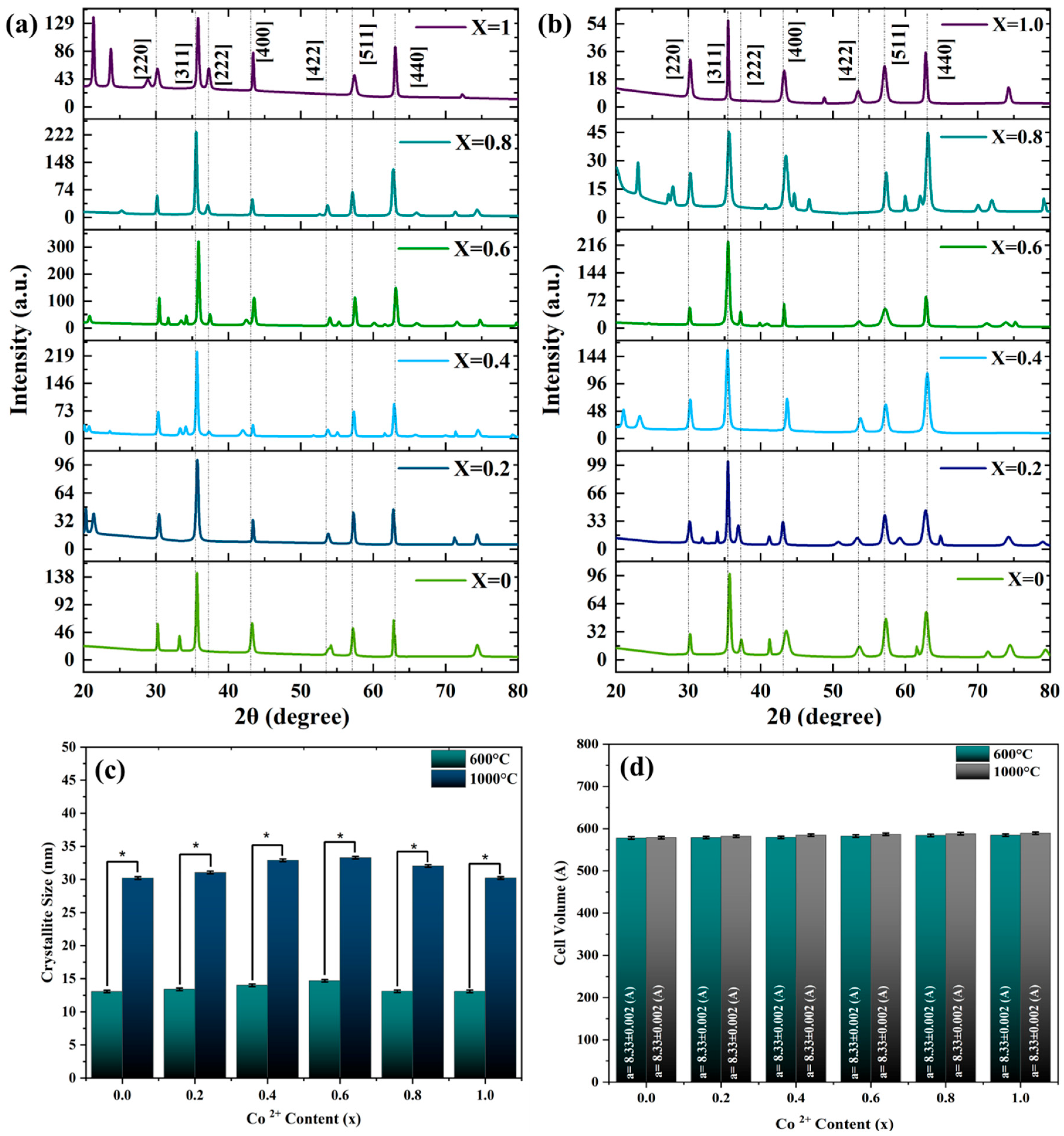
  • The X-ray diffraction (XRD) profiles confirmed the spinel structure of the powders obtained. Higher synthesis temperatures resulted in sharper, more symmetric peaks that reflected better crystallinity. Moreover, the broadening of peaks decreased with an increase in temperature, consistent with increased particle sizes and an improved crystal structure. In addition, increasing cobalt concentration also increased the grain size, unit cell volume, and lattice constant.
  • SEM images showed that the particle sizes ranged between 13–19 nm at 600 °C and 28–36 nm at 1000 °C, demonstrating good agreement with the XRD results.
  • The purity of the material was verified using Fourier transform infrared spectroscopy (FTIR), as no organic compounds were detected, while the presence of cations etched in octahedral and tetrahedral sites was verified. Furthermore, it can be seen from the FTIR results that the v1 band shifted to lower wave numbers with an increase in temperature and Co substitution. Shifts to lower wave numbers are explained by the inverse relationship between atomic mass and wave number in FTIR spectra.
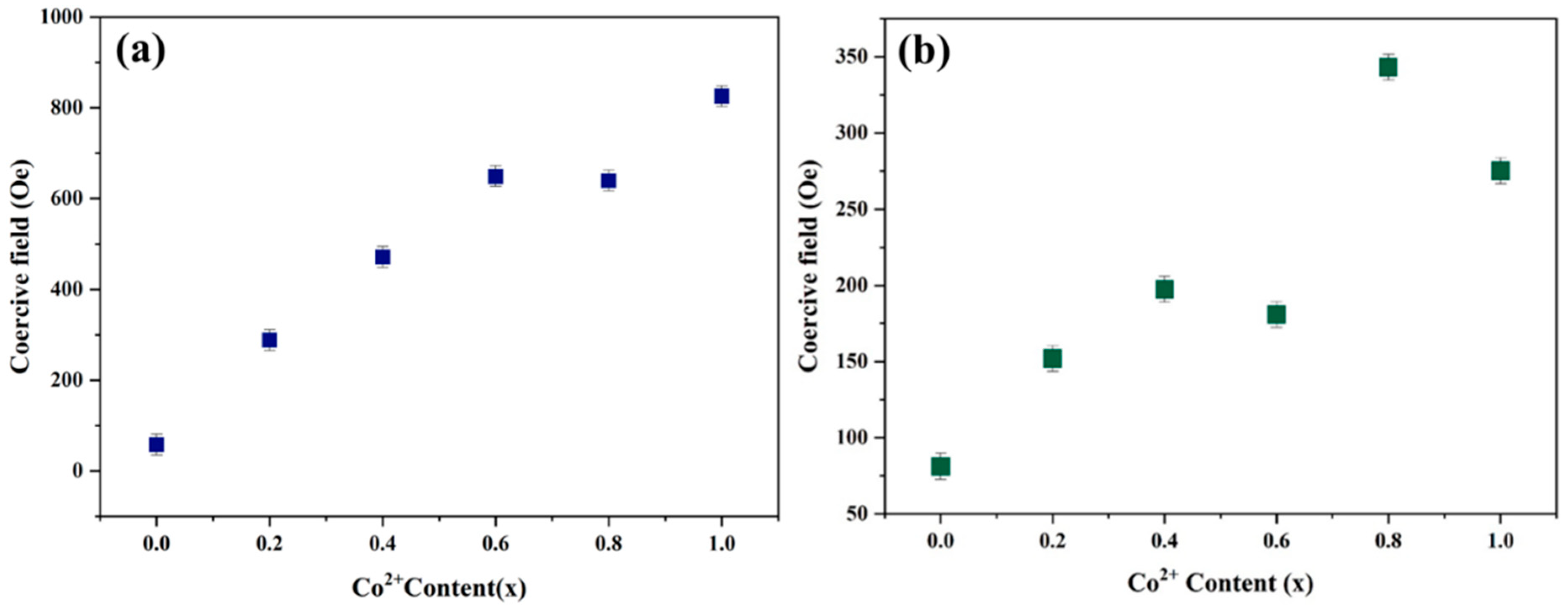
  • Vibrating sample magnetometry (VSM) analysis demonstrated that coercivity increased almost linearly with higher cobalt concentrations at both synthesis temperatures. The inherently higher anisotropic nature of cobalt ions compared to nickel ions resulted in an increase in coercivity with cobalt doping. This increase in coercivity can be attributed to the enhancement of the anisotropic field, which subsequently raises domain wall energy.
  • Saturation and remanent magnetization were shown to have random tendencies. This might be attributed to the presence of the low remanent magnetization of soft nickel ferrites. The observed random increasing trend in saturation magnetization and remanent magnetization may be due to the replacement of cobalt cations for iron cations in tetrahedral sites, which reduced MA and ultimately led to a greater overall magnetic moment.
These results will allow for a better understanding of the structural and magnetic properties of cobalt-doped nickel ferrite nanoparticles and their potential use in magnetic and electronic devices.

Author Contributions

Conceptualization, M.A. and F.H.L.; Methodology, F.V., M.A. and F.H.L.; Validation, A.B.; Formal analysis, F.V.; Investigation, F.V. and A.B.; Data curation; M.A.; Writing—original draft, F.V.; Writing—review and editing, M.A. and F.H.L.; Supervision, M.A. and A.B. All authors have read and agreed to the published version of the manuscript.

Funding

This research received no external funding.

Data Availability Statement

The original contributions presented in this study are included in the article. Further inquiries can be directed to the corresponding authors.

Acknowledgments

The authors wish to offer their thanks and appreciation to the Isfahan University of Technology for their support and involvement during this study.

Conflicts of Interest

The authors declare no conflicts of interest.

References

  1. Guo, P.; Wu, Y.; Ma, X.; Chen, R.; Zeng, Z.; Li, L.; Su, C.; Wang, S. Enhanced iodine capture by nano-copper particles modified benzimidazole-based molded porous carbon. Appl. Surf. Sci. 2025, 708, 163754. [Google Scholar] [CrossRef]
  2. Xu, R.; Liao, L.; Liang, W.; Wang, H.; Zhou, Q.; Liu, W.; Chen, M.; Fang, B.; Wu, D.; Jin, H.; et al. Fast Removing Ligands from Platinum-Based Nanocatalysts by a Square-Wave Potential Strategy. Angew. Chem. Int. Ed. 2025, e202509746. [Google Scholar]
  3. Li, S.; Zhao, S.; Hartmann, D.M.F.; Yang, W.; Lin, X.; Zhao, W.; Lavrijsen, R. Modulation of the Dzyaloshinskii–Moriya interaction in Pt/Co/Pt with electric field induced strain. Appl. Phys. Lett. 2025, 126, 162401. [Google Scholar] [CrossRef]
  4. Dippong, T.; Levei, E.A.; Cadar, O.; Mesaros, A.; Borodi, G. Sol-gel synthesis of CoFe2O4:SiO2 nanocomposites—Insights into the thermal decomposition process of precursors. J. Anal. Appl. Pyrolysis 2017, 125, 169–177. [Google Scholar] [CrossRef]
  5. Wang, G.; Ma, Y.; Wei, Z.; Qi, M. Development of multifunctional cobalt ferrite/graphene oxide nanocomposites for magnetic resonance imaging and controlled drug delivery. Chem. Eng. J. 2016, 289, 150–160. [Google Scholar] [CrossRef]
  6. Akhtar, M.N.; Khan, M.A.; Ahmad, M.; Nazir, M.S.; Imran, M.; Ali, A.; Sattar, A.; Murtaza, G. Evaluation of structural, morphological and magnetic properties of CuZnNi (Cu Zn0.5−Ni0.5Fe2O4) nanocrystalline ferrites for core, switching and MLCI’s applications. J. Magn. Magn. Mater. 2017, 421, 260–268. [Google Scholar] [CrossRef]
  7. Nawale, A.B.; Kanhe, N.S.; Raut, S.A.; Bhoraskar, S.V.; Das, A.K.; Mathe, V.L. Investigation of structural, optical and magnetic properties of thermal plasma synthesized Ni-Co spinel ferrite nanoparticles. Ceram. Int. 2017, 43, 6637–6647. [Google Scholar] [CrossRef]
  8. Cai, G.; Huang, Y.; Qing, Y.; Misra, R.D.K. Effects of Y and Hf addition on soft magnetic property and ductility improvement of Fe–6.9 wt% Si alloy. J. Mater. Res. Technol. 2025, 37, 1362–1378. [Google Scholar] [CrossRef]
  9. Cao, K.; Ge, X.; Li, S.; Tian, Z.; Cui, S.; Guo, G.; Yang, L.; Li, X.; Wang, Y.; Bai, S.; et al. Facile preparation of a 3D rGO/g-C3N4 nanocomposite loaded with Ag NPs for photocatalytic degradation. RSC Adv. 2025, 15, 17089–17101. [Google Scholar] [CrossRef]
  10. Gul, I.H.; Amin, F.; Abbasi, A.Z.; Anis-ur-Rehman, M.; Maqsood, A. Physical and magnetic characterization of co-precipitated nanosize Co–Ni ferrites. Scr. Mater. 2007, 56, 497–500. [Google Scholar] [CrossRef]
  11. Raj, K.; Moskowitz, B.; Casciari, R. Advances in ferrofluid technology. J. Magn. Magn. Mater. 1995, 149, 174–180. [Google Scholar] [CrossRef]
  12. Adeela, N.; Maaz, K.; Khan, U.; Karim, S.; Nisar, A.; Ahmad, M.; Ali, G.; Han, X.F.; Duan, J.L.; Liu, J. Influence of manganese substitution on structural and magnetic properties of CoFe2O4 nanoparticles. J. Alloys Compd. 2015, 639, 533–540. [Google Scholar] [CrossRef]
  13. Meenakshisundaram, A.; Gunasekaran, N.; Srinivasan, V. Distribution of Metal Ions in Transition Metal Manganites AMn2O4 (A: Co, Ni, Cu, or Zn); De Gruyter: Berlin, Germany, 1982. [Google Scholar]
  14. Mozaffari, M.; Amighian, J.; Darsheshdar, E. Magnetic and structural studies of nickel-substituted cobalt ferrite nanoparticles, synthesized by the sol–gel method. J. Magn. Magn. Mater. 2014, 350, 19–22. [Google Scholar] [CrossRef]
  15. Kambale, R.C.; Shaikh, P.A.; Kamble, S.S.; Kolekar, Y.D. Effect of cobalt substitution on structural, magnetic and electric properties of nickel ferrite. J. Alloys Compd. 2009, 478, 599–603. [Google Scholar] [CrossRef]
  16. Torkian, S.; Ghasemi, A.; Razavi, R.S. Cation distribution and magnetic analysis of wideband microwave absorptive CoxNi1−xFe2O4 ferrites. Ceram. Int. 2017, 43, 6987–6995. [Google Scholar] [CrossRef]
  17. Castro, J.I.; Astudillo, S.; Hernandez, J.H.M.; Saavedra, M.; Zapata, P.A.; Valencia-Llano, C.H.; Chaur, M.N.; Grande-Tovar, C.D. Synthesis, Characterization, and Optimization Studies of Polycaprolactone/Polylactic Acid/Titanium Dioxide Nanoparticle/Orange Essential Oil Membranes for Biomedical Applications. Polymers 2022, 15, 135. [Google Scholar] [CrossRef]
  18. Ati, A.A.; Othaman, Z.; Samavati, A. Influence of cobalt on structural and magnetic properties of nickel ferrite nanoparticles. J. Mol. Struct. 2013, 1052, 177–182. [Google Scholar] [CrossRef]
  19. Balaji, S.; Selvan, R.K.; Berchmans, L.J.; Angappan, S.; Subramanian, K.; Augustin, C.O. Combustion synthesis and characterization of Sn4+ substituted nanocrystalline NiFe2O4. Mater. Sci. Eng. B 2005, 119, 119–124. [Google Scholar] [CrossRef]
  20. Vinosha, P.A.; Manikandan, A.; Ragu, R.; Dinesh, A.; Thanrasu, K.; Slimani, Y.; Baykal, A.; Xavier, B. Impact of nickel substitution on structure, magneto-optical, electrical and acoustical properties of cobalt ferrite nanoparticles. J. Alloys Compd. 2021, 857, 157517. [Google Scholar] [CrossRef]
  21. Khan, T.F.; Naresh, U.; Ramprasad, T.; Kumar, R.J. Structural, Morphological, and Magnetic Properties of Cobalt-Doped Nickel Ferrite Nanoparticles. J. Supercond. Nov. Magn. 2021, 34, 797–803. [Google Scholar] [CrossRef]
  22. Chandekar, K.V.; Yadav, S.P.; Chinke, S.; Shkir, M. Impact of Co-doped NiFe2O4 (CoxNi1−xFe2O4) nanostructures prepared by co-precipitation route on the structural, morphological, surface, and magnetic properties. J. Alloys Compd. 2023, 966, 171556. [Google Scholar] [CrossRef]
  23. Kumar, G.S.; Raguram, T.; Rajni, K.S. Synthesis and Characterization of Nickel-Substituted Cobalt Ferrite Nanoparticles Using Sol–Gel Auto-combustion Method. J. Supercond. Nov. Magn. 2019, 32, 1715–1723. [Google Scholar] [CrossRef]
  24. Sumalatha, M.; Reddy, S.S.K.; Reddy, M.S.; Sripada, S.; Reddy, P.V.; Ch, G.R.; Reddy, P.Y.; Reddy, V.R. Effect of indium substitution on structural and hyperfine parameters of CoFe2O4. AIP Conf. Proc. 2020, 2220, 110038. [Google Scholar]
  25. Debnath, S.; Das, A.; Das, R. Effect of cobalt doping on structural parameters, cation distribution and magnetic properties of nickel ferrite nanocrystals. Ceram. Int. 2021, 47, 16467–16482. [Google Scholar] [CrossRef]
  26. Kiani, M.N.; Butt, M.S.; Gul, I.H.; Saleem, M.; Irfan, M.; Baluch, A.H.; Akram, M.A.; Raza, M.A. Synthesis and Characterization of Cobalt-Doped Ferrites for Biomedical Applications. ACS Omega 2023, 8, 3755–3761. [Google Scholar] [CrossRef]
  27. Seeman, V.; Feldbach, E.; Kärner, T.; Maaroos, A.; Mironova-Ulmane, N.; Popov, A.I.; Shablonin, E.; Vasil’chenko, E.; Lushchik, A. Fast-neutron-induced and as-grown structural defects in magnesium aluminate spinel crystals with different stoichiometry. Opt. Mater. 2019, 91, 42–49. [Google Scholar] [CrossRef]
  28. Tatarchuk, T. Studying the Defects in Spinel Compounds: Discovery, Formation Mechanisms, Classification, and Influence on Catalytic Properties. Nanomaterials 2024, 14, 1640. [Google Scholar] [CrossRef]
  29. Shafiq, K.; Aadil, M.; Hassan, W.; Choudhry, Q.; Gul, S.; Rais, A.; Fattah, A.A.; Mahmoud, K.H.; Ansari, M.Z.; Mohd, Z. Cobalt and holmium co-doped nickel ferrite nanoparticles: Synthesis, characterization and photocatalytic application studies. Z. Für Phys. Chem. 2023, 237, 1325–1344. [Google Scholar] [CrossRef]
  30. Kumar, E.R.; Srinivas, C.; Deepty, M.; Pradeep, I.; Mehar, M.V.K.; Prajapat, C.L.; Rao, T.V.C.; Mohan, N.K.; Sastry, D.L. Synergistic effect of heat treatment on structural, magnetic and dielectric properties of spinel ferrite nanoparticles. J. Mater. Sci. Mater. Electron. 2018, 29, 20968–20977. [Google Scholar] [CrossRef]
  31. Nejati, K.; Zabihi, R. Preparation and magnetic properties of nano size nickel ferrite particles using hydrothermal method. Chem. Cent. J. 2012, 6, 23. [Google Scholar] [CrossRef] [PubMed]
  32. Majid, F.; Dildar, S.; Ata, S.; Bibi, I.; Mohsin, I.U.; Ali, A.; Almoneef, M.M.; Iqbal, M.; Irshad, S.; Nazir, A.; et al. Cobalt doping of nickel ferrites via sol gel approach: Effect of doping on the structural and dielectric properties. Z. Für Phys. Chem. 2021, 235, 1811–1829. [Google Scholar] [CrossRef]
  33. Milutinović, A.; Lazarević, Z.Ž.; Šuljagić, M.; Andjelković, L. Synthesis-Dependent Structural and Magnetic Properties of Monodomain Cobalt Ferrite Nanoparticles. Metals 2024, 14, 833. [Google Scholar] [CrossRef]
  34. Sertkol, M.; Köseoǧlu, Y.; Baykal, A.; Kavas, H.; Toprak, M.S. Synthesis and magnetic characterization of Zn0.7Ni0.3Fe2O4 nanoparticles via microwave-assisted combustion route. J. Magn. Magn. Mater. 2010, 322, 866–871. [Google Scholar] [CrossRef]
  35. Zhou, X.; Zhou, K.; Zhang, T.; Fan, L.; Zhang, H.; Zheng, H. Static magnetic, complex dielectric and complex permeability properties of aluminum substituted hexagonal barium ferrites based on doping concentration. J. Ceram. Soc. Jpn. 2021, 129, 21037. [Google Scholar] [CrossRef]
  36. Šuljagić, M.; Vulić, P.; Jeremić, D.; Pavlović, V.; Filipović, S.; Kilanski, L.; Lewinska, S.; Slawska-Waniewska, A.; Milenković, M.R.; Nikolić, A.S.; et al. The influence of the starch coating on the magnetic properties of nanosized cobalt ferrites obtained by different synthetic methods. Mater. Res. Bull. 2021, 134, 111117. [Google Scholar] [CrossRef]
  37. Sardar, I.; Hossain, M.D.; Sikder, M.S.; Khan, M.N.I.; Rahman, M.R. Enhancement of coercivity, permeability, and dielectric properties of Co0.2Zn0.3Ni0.5Eu Fe2–O4 ferrites for memory and high frequency devices. J. Rare Earths 2024, 42, 1128–1135. [Google Scholar] [CrossRef]
  38. Nugraha, A.R.; Arrasyid, E.N.; Dedi, D.; Nugroho, A.A. The Distorted Octahedral and Magnetocrystalline Anisotropy of FeMnO3 by Stoner-Wohlfarth Model. Mater. Sci. Forum 2023, 1080, 139–145. [Google Scholar] [CrossRef]
  39. Gerina, M.; Angotzi, M.S.; Mameli, V.; Gajdošová, V.; Rainer, D.N.; Dopita, M.; Steinke, N.-J.; Aurélio, D.; Vejpravová, J.; Zákutná, D. Size dependence of the surface spin disorder and surface anisotropy constant in ferrite nanoparticles. Nanoscale Adv. 2023, 5, 4563–4570. [Google Scholar] [CrossRef]
  40. Jian, G.; Fu, Q.; Zhou, D. Particles size effects of single domain CoFe2O4 on suspensions stability. J. Magn. Magn. Mater. 2012, 324, 671–676. [Google Scholar] [CrossRef]
  41. Maaz, K.; Kim, G.H. Single domain limit for NixCo1−xFe2O4 (0 ≤ x ≤ 1) nanoparticles synthesized by coprecipitation route. Mater. Chem. Phys. 2012, 137, 359–364. [Google Scholar] [CrossRef]
  42. Tahir, W.; Zeeshan, T.; Waseem, S.; Ali, M.D.; Kayani, Z.; Aftab, Z.-E.-H.; Mehtab, S.M.T.; Ezzine, S. Impact of silver substitution on the structural, magnetic, optical, and antibacterial properties of cobalt ferrite. Sci. Rep. 2023, 13, 1–23. [Google Scholar] [CrossRef] [PubMed]
  43. Majid, F.; Shahin, A.; Ata, S.; Bibi, I.; Malik, A.; Ali, A.; Laref, A.; Iqbal, M.; Nazir, A. The effect of temperature on the structural, dielectric and magnetic properties of cobalt ferrites synthesized via hydrothermal method. Z. Fur Phys. Chem. 2021, 235, 1279–1296. [Google Scholar] [CrossRef]
  44. Brik, M.G.; Ma, C.G.; Yamamoto, T.; Piasecki, M.; Popov, A.I. First-Principles Methods as a Powerful Tool for Fundamental and Applied Research in the Field of Optical Materials. In Phosphor Handbook: Experimental Methods for Phosphor Evaluation and Characterization; CRC Press: Boca Raton, FL, USA, 2022; pp. 1–26. [Google Scholar]
  45. Long, X.; Chong, K.; Su, Y.; Du, L.; Zhang, G. Connecting the macroscopic and mesoscopic properties of sintered silver nanoparticles by crystal plasticity finite element method. Eng. Fract. Mech. 2023, 281, 109137. [Google Scholar] [CrossRef]
  46. Wang, X.; Chen, X.; Wang, B.; He, Q.; Cao, J.; Zhu, Y.; Su, K.; Yan, H.; Sun, P.; Li, R.; et al. Ultra-Bandwidth Microwave Absorption and Low Angle Sensitivity in Dual-Network Aerogels with Dual-Scale Pores. Small 2025, 2412744. [Google Scholar] [CrossRef]
Figure 1. A schematic of the synthesis of composite powders.
Crystals 15 00624 g001
Figure 2. TG/DT analysis of Co0.6Ni0.4Fe2O4 at (a) 600 °C and (b) 1000 °C.
Crystals 15 00624 g002
Figure 3. (a) XRD spectra for CoxNi1−xFe2O4 nanoparticles at 600 °C. (b) XRD spectra for CoxNi1−xFe2O4 nanoparticles at 1000 °C. (c) Crystallite size vs. concentration of cobalt substituted nickel ferrites (p < 0.05). (d) Variations in unit cell volume with cobalt concentration.
Crystals 15 00624 g003
Figure 4. Fourier transform analysis of CoxNi1−xFe2O4 at (a) 600 °C and (b) 1000 °C.
Crystals 15 00624 g004
Figure 5. (a) SEM images of nanoparticles synthesized at 600 °C (b) and at 1000 °C (c). EDS analysis for (c-1) Co0.8Ni0.2Fe2O4 at 600 °C and (c-2) Co0.8Ni0.2Fe2O4 at 1000 °C.
Crystals 15 00624 g005
Figure 6. Magnetization curves of as-synthesized CoxNi1−xFe2O4, with 0 ≤ x ≤1 at (a) 600 °C and (b) 1000 °C.
Crystals 15 00624 g006
Figure 7. Compositional variation in coercivity of CoxNi1−xFe2O4 nanoparticles at (a) 600 °C and (b) 1000 °C.
Crystals 15 00624 g007
Figure 8. Variation in the anisotropy constant, K, of CoxNi1−xFe2O4 nanoparticles at (a) 600 °C and (b) 1000 °C.
Crystals 15 00624 g008
Table 1. Different molar ratios of Co:Ni:Fe with the expected composition.
No.Molar Ratio of Co:Ni:FeNominal Composition
10.0:1.0:2.0NiFe2O4
20.2:0.8:2.0Co0.2Ni0.8Fe2O4
30.4:0.6:2.0Co0.4Ni0.6Fe2O4
40.6:0.4:2.0Co0.6Ni0.4Fe2O4
50.8:0.2:2.0Co0.8Ni0.2Fe2O4
61.0:0.0:2.0CoFe2O4
Table 2. The room temperature characteristics and magnetic properties for the samples heat-treated at 600 °C.
Sample M S   ( ± 1 )
(emu/g)
M r   ( ± 1 )
(emu/g)
H C   ( ± 8 )
(Oe)
Mr/MS
NiFe2O433.656.3081.20.18
Co0.2Ni0.8Fe2O443.010.49152.00.24
Co0.4Ni0.6Fe2O457.8813.44197.550.23
Co0.6Ni0.4Fe2O468.6011.90180.910.17
Co0.8Ni0.2Fe2O462.4316.04343.220.25
CoFe2O461.1715.70275.1820.25
Table 3. The room temperature characteristics and magnetic properties for the samples heat-treated at 1000 °C.
Sample M S   ( ± 1 )
(emu/g)
M r   ( ± 0.5 )
(emu/g)
H C   ( ± 5.5 )
(Oe)
Mr/MS
NiFe2O422.371.6258.120.07
Co0.2Ni0.8Fe2O432.587.71288.460.23
Co0.4Ni0.6Fe2O448.7719.28471.400.39
Co0.6Ni0.4Fe2O453.1120.15649.270.37
Co0.8Ni0.2Fe2O459.4921.06639.270.35
CoFe2O452.8819.97852.700.37
Disclaimer/Publisher’s Note: The statements, opinions and data contained in all publications are solely those of the individual author(s) and contributor(s) and not of MDPI and/or the editor(s). MDPI and/or the editor(s) disclaim responsibility for any injury to people or property resulting from any ideas, methods, instructions or products referred to in the content.

