Article

Experimental Evaluation of GAGG:Ce Crystalline Scintillator Properties Under X-Ray Radiation

by
Anastasios Dimitrakopoulos
,
Christos Michail
*,
Ioannis Valais
,
George Fountos
,
Ioannis Kandarakis
and
Nektarios Kalyvas
Radiation Physics, Materials Technology and Biomedical Imaging Laboratory, Department of Biomedical Engineering, University of West Attica, Ag. Spyridonos, 12210 Athens, Greece
*
Author to whom correspondence should be addressed.
Crystals 2025, 15(7), 590; https://doi.org/10.3390/cryst15070590
Submission received: 26 May 2025 / Revised: 19 June 2025 / Accepted: 21 June 2025 / Published: 23 June 2025
(This article belongs to the Special Issue Exploring New Materials for the Transition to Sustainable Energy)

Abstract

The scope of this study was to evaluate the response of Ce-doped gadolinium aluminum gallium garnet (GAGG:Ce) crystalline scintillator under medical X-ray irradiation for medical imaging applications. A 10 × 10 × 10 mm3 crystal was irradiated at X-ray tube voltages ranging from 50 kVp to 150 kVp. The crystal’s compatibility with several commercially available optical photon detectors was evaluated using the spectral matching factor (SMF) along with the absolute efficiency (AE) and the effective efficiency (EE). In addition, the energy-absorption efficiency (EAE), the quantum-detection efficiency (QDE) as well as the zero-frequency detective quantum detection efficiency DQE(0) were determined. The crystal demonstrated satisfactory AE values as high as 26.3 E.U. (where 1 E.U. = 1 μW∙m−2/(mR∙s−1)) at 150 kVp, similar, or in some cases, even superior to other cerium-doped scintillator materials. It also exhibits adequate DQE(0) performance ranging from 0.99 to 0.95 across all the examined X-ray tube voltages. Moreover, it showed high spectral compatibility with commonly used photoreceptors in modern day such as complementary metal–oxide–semiconductors (CMOS) and charge-coupled-devices (CCD) with SMF values of 0.95 for CCD with broadband anti-reflection coating and 0.99 for hybrid CMOS blue. The aforementioned properties of this scintillator material were indicative of its superior efficiency in the examined medical energy range, compared to other commonly used scintillators.

1. Introduction

The importance of scintillator materials in diagnostic imaging systems is vital, since their light energy efficiency influences the radiation exposure levels to the person under X-ray examination. The development of new scintillating materials focuses towards the improvement of the detectors used in imaging diagnostics [1]. An efficient material leads in the production of a high-quality image that can be obtained with less radiation burden to the patient. For computed tomography (CT) scintillators a low afterglow of less than 1% at 3 ms is necessary along with high chemical stability, small temperature dependence and radiation damage [2]. A high density of >6 g/cm3 is required as well as satisfactory spectral matching with the readout photosensor circuit [3,4]. A high luminous efficiency is also desirable, since it contributes in reducing the number of X-rays needed for optimal SNR. For that reason, the evaluation of scintillator properties is of outmost importance to potentially improve imaging performance. A relatively new developed and promising material is gadolinium aluminum gallium garnet doped with cerium, Gd3Al2Ga3O12:Ce (GAGG:Ce) which has excellent properties that makes it ideal for use in medical imaging. First reported in 2011 by Kamada [5] and produced in a single crystal using the Czochralski method one year later [6], it has its emission peak around 520 nm [7,8,9,10] which is well suited with the wavelength of various commercially available photomultiplier tubes (PMT) and silicon photodiodes (SiPD), making it suitable for CT [11,12,13,14]. Ce-activated materials are reported to have fast decay time of under 100 ns [3,15] and high light output [12] from 8000 photons/Mev (GSO) [3] up to 63,000 photons/MeV (LaBr3) [4]. In addition, many of these materials show good stopping power. Research has shown that single crystal GAGG:Ce exhibits faster decay time than its ceramic counterpart [8]. Ce is an exceptional dopant as a result of the intense, quick and highly efficient 5d → 4f transition with the consequent Ce3+ emission peak at 500–550 nm [6,10,16]. GAGG:Ce has a relatively fast decay time of 50–150 ns [7,8,17], but an afterglow of several hours has been reported after high radiation exposure conditions [18]. Its light yield (LY) is 46,000 photons/MeV [6,7,10,17,19,20]. Its energy resolution is 4.9% at 662 keV [6,7,21] and has a refractive index of 1.8 to 2.2 [13,21]. Its density is 6.63 g/cm3 [7,9,14,22,23], it is robust, mechanically hard (8 Mho) [19,21], non-hygroscopic [7,9,19,21] with an effective atomic number (Zeff) of 54.4 [9,14,21,22] and melting point of 1850 °C [14,19]. It has no intrinsic radioactivity, like that found in lutetium, (Lu). Recent studies by many groups indicate excellent energy resolution [7,10,11,24,25,26,27,28] of up to 8% especially when coupled with S10362-33–050 SiPM [9]. Also, it shows better response as temperature decreases [10,29]. The crystal is efficiently used in numerous applications such as space applications [18,30,31], positron emission tomography (PET) [11,32,33], single-photon emission computed tomography (SPECT) [34,35], CT, gamma-ray spectroscopy and Compton scattering detection [7,12,23,24,36]. Moreover, luminescent ions or dopants are added to the scintillating material. In some cases, it is possible for more than one dopant to be used in order to reduce the afterglow [37,38,39]. Several tests have been conducted with different dopants such as Cr ions [20], Ca2+ and Zr4+ [40]. Additionally, GAGG:Ce efficiency properties of crystals with thicknesses up to 4 cm have also been studied either through Monte Carlo simulation or under ionizing energy irradiation [12,41,42].
This study acts complementary to the published literature, since it performs a thorough investigation of a 10 × 10 × 10 mm3 GAGG:Ce crystalline scintillator response under X-ray radiation, in the energy range 50 to 150 kVp. More specifically the scintillator’s compatibility with several optical photon detectors was calculated via the spectral matching factor (SMF). Its response in terms of efficiency was examined by experimentally determining the absolute luminescence efficiency (AE) and the effective efficiency (EE). Additionally, the energy-absorption efficiency (EAE) and the quantum-detection efficiency (QDE) were calculated. Finally, in order to study in depth, the noise transfer characteristics of the scintillator, the zero-frequency detective quantum detection efficiency (DQE(0)) was also determined by measured and theoretically calculated data.
Our results suggest that the 10 × 10 × 10 mm3 scintillator shows excellent efficiency over 130 kVp, with DQE(0) values above 0.94. In addition, its excellent performance matched with various photodetectors makes it a promising candidate as a radiation detector in the range of 100–150 kVp.

2. Materials and Methods

A 10 × 10 × 10 mm3 single crystal GAGG:Ce scintillator with polished surfaces was commercially procured [19]. The crystal was irradiated with 20 mm Al filtered X-rays which were generated by radiographic X-ray tube with tube voltages ranging from 50 kV to 150 kV in steps of 10 kV [4]. The crystal was carefully wrapped in Teflon tape except its entrance and the exit surfaces to ensure high reflectivity [43]. The incident radiation dose was kept as low as medical X-ray radiographic levels suggest, ranging from 0.04 mGy to 4.03 mGy. No kind of degradation was observed on the material for these exposure conditions. The performance of GAGG crystal has been examined in literature under extreme exposure conditions under proton irradiation [44] and a fluence of charged hadrons of 5 × 10¹⁴ p/cm2 was not found to impose any significant change in the optical transmittance of crystalline GAGG scintillators. Similarly, the irradiation with electron beams delivering doses of 300 Mrad, where 1 rad = 1 cGy [45], did not seem to affect GAGG:Ce optical transmission properties, although a 10% light yield degradation has been reported after irradiation with 100 krad by a 60Co source [18]. Every photodetector can detect a different percent of the optical photons produced by a scintillator. A scintillator’s compatibility with the optical sensor as well as how efficient a sensor is at detecting photons emitted from the crystal is given by a factor called spectral-matching factor (SMF) [46]:
S M F = S p λ S D λ d λ S p λ d λ
where S P ( λ ) denotes the spectrum of the emitted light and S D λ is the spectral sensitivity of the optical photon detector per wavelength λ . The crystal’s emission spectrum was measured using a grating spectrometer HR2000+ (Ocean Optics Inc., Largo, FL, USA) under UV excitation.
The absolute luminescence efficiency (AE) defined as the ratio of the crystal’s light flux Ψ λ ˙ , for a given X-ray exposure rate Χ ˙ , [4] was experimentally determined, that is:
A E = Ψ λ ˙ Χ ˙
The experimental optical photon measurement setup comprised a light integration sphere (Newport Corp., Irvine, CA, USA, Oriel model 70451), an EMI photomultiplier tube (PMT) (EMI, London, UK, EMI model 9798) and a Cary 401 (Cary instruments/Varian, Palo Alto, CA, USA) electrometer as described in literature [4]. The schematic diagram of the experimental measurement setup is shown in Figure 1.
The experimental AE values were expressed in efficiency units (E.U.) where 1 E.U. = 1 μW∙m−2/(mR∙s−1). The radiation detection efficiency of GAGG:Ce was theoretically determined by calculated energy absorption efficiency (EAE), giving the fraction of the incident energy deposited in the material and quantum-detection efficiency (QDE), giving the fraction of the incident X-ray photons interacting with the material. The EAE and QDE were calculated with the use of Equations (3) and (4) respectively as [4,47]:
E A E = 0 E 0 Φ 0 E E μ e n ( E ) / ρ μ a t t ( E ) / ρ 1 e μ a t t ( E ) / ρ ρ T d E 0 E 0 Φ 0 E E d E
Q D E = 0 E 0 Φ 0 E 1 e μ a t t ( E ) / ρ ρ T d E 0 E 0 Φ 0 E d E
where Φ 0 E indicates the X-ray photon fluence, E is the photon energy, μ e n ( E ) / ρ is the X-ray total mass attenuation coefficient of the scintillator and μ a t t ( E ) / ρ is the total mass energy absorption coefficient. T is the crystal’s thickness (equal to 10 mm) and ρ is the density (in g/cm3) [46,48]. μ e n ( E ) / ρ and μ a t t ( E ) / ρ were determined by the XMuDat software version 1.0.1 [49].
As a means to evaluate the overall efficiency of the scintillator when used in combination with a photodetector, the effective efficiency (EE) is used given by [4]:
E E = A E · S M F
The detective quantum efficiency (DQE) is utilized as a measurement of system performance. First described in the mid-seventies [50,51] it is indicative of how efficiently a system transfers all the input quanta incident within the detector. It is often defined by the ratio [4,52]:
D Q E = S N R O u t S N R I n 2
where S N R O u t denotes the output signal to noise ratio of the imaging system and S N R I n is the input signal to noise ratio. The DQE(0) can be calculated by AE, QDE and radiation exposure measurements as [4]:
D Q E 0 , k V = A E k V · X ˙ · Q D E k V A E k V · X ˙ + Q ¯ · Q D E k V · h c λ
The tungsten anode spectral model using interpolating polynomials (TASMIP) spectra calculator was then used for each tube voltage value to obtain the spectrum Φ ο E and calculate Q ¯ [53], where Q ¯ corresponds to the X-ray fluence of our specific experimental conditions. Φ ο expresses the amount of photons/mm2 for a specific X-ray spectrum at a specific exposure. The TASMIP model utilized is based on X-ray spectra measured at 1 keV intervals over the range from 40 kVp to 140 kVp. Subsequently, we determined the exposure X ( m R ) T as follows [4,54]:
X m R T = 1.83 × 10 6 0 E 0 Φ ο E · E · μ e n ρ a i r E d E
where energy should be considered and Φ ο E in photons/mm2 in keV. Additionally, the exposure was obtained as, X m R = X ˙ · t , where t was taken equal to the irradiation duration that is 1 s. Q ¯ was calculated by the following relation [4]:
Q ¯ = X m R 1.83 × 10 6 0 E 0 Φ ο E · E · μ e n ρ a i r E d E
where the constant 1.83 × 10 6 is used for converting to mR and is expressed in m R · m m 2 / [ k e V · ( c m 2 / g ) ] [54].

3. Results

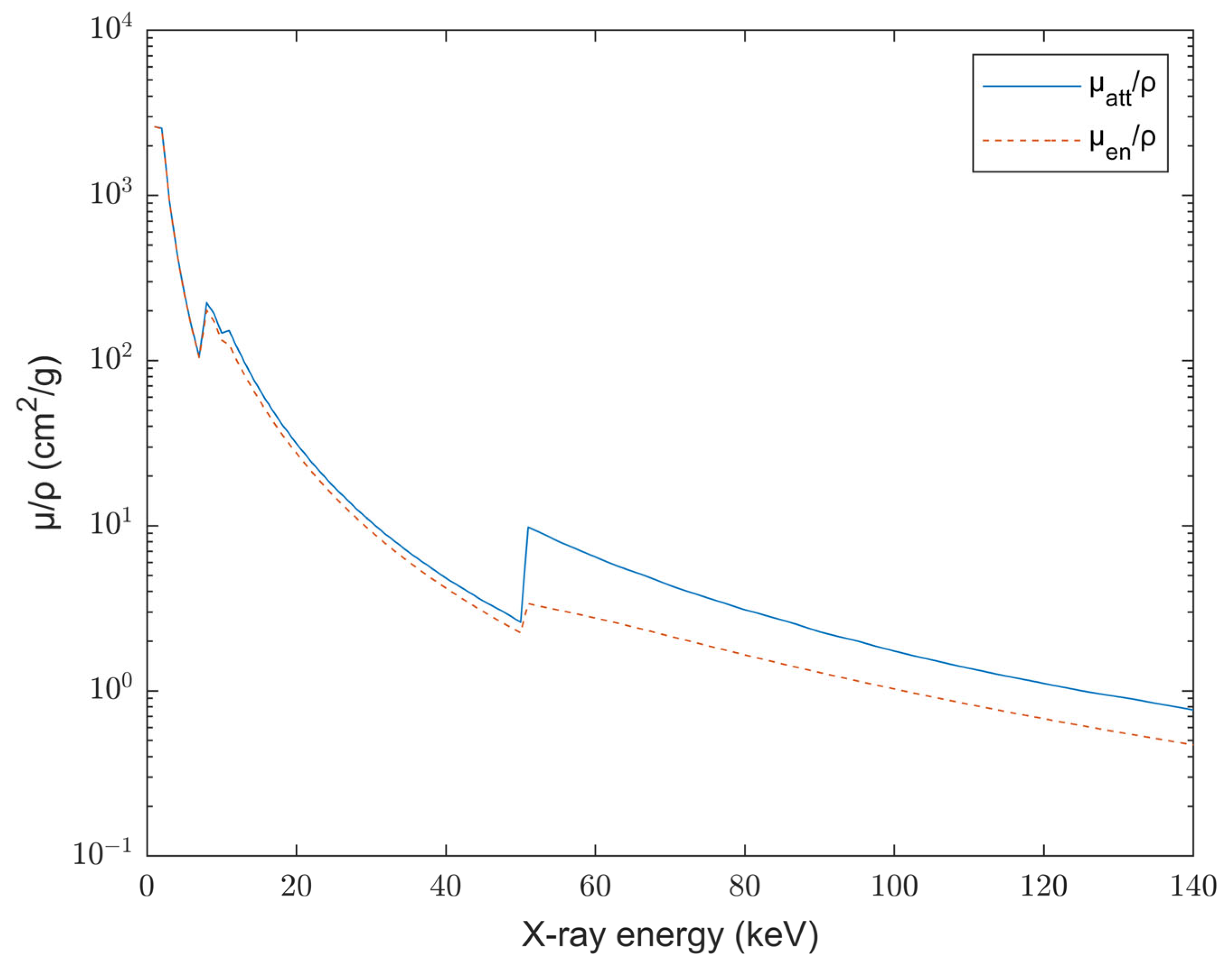
Figure 2 shows the attenuation coefficients of GAGG:Ce given by XMuDat software indicating the interaction of X-rays with the material’s mass. The fluctuation of the values in Figure 2 up to 100 keV is attributed to the photoelectric effect that is highly probable to occur in this energy range. The discontinuity of the values observed also called “absorption edges” correspond to the excitation energies for ionization of a particular electron shell. For instance, the discontinuity found at about 50 keV appears when the energy of the photon particles equals to the ionization energies of the K-shell [55] which increases the absorption of the photons resulting in the sudden spike presented in Figure 2. After 50 keV the attenuation coefficients gradually decrease.
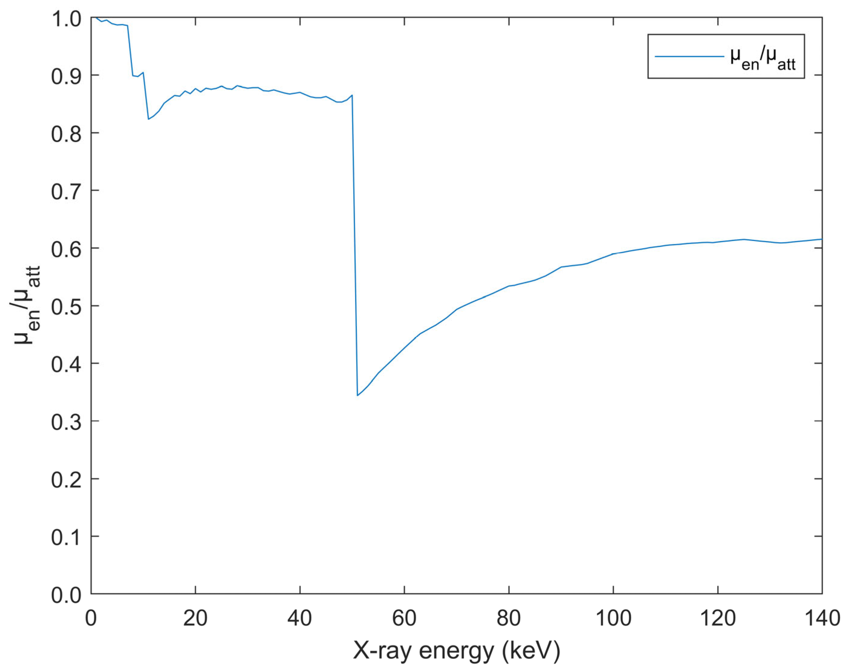
μ a t t expresses how probable is for the photons to interact with the single crystal and be absorbed while μ e n expresses how probable is for the energy to be transferred and be absorbed by the scintillator matter. Both are indicative of how the X-rays interact with the crystal. In Figure 3 the ratio of μ e n / μ a t t used in Equation (3) is presented. The ratio may be considered as an expression providing the effectiveness of energy absorption per photon interaction. The vertical drop at 50.23 keV corresponds to the GAGG:Ce K-edge of Gd in correlation with the discontinuity found at the same energy range in Figure 2. After 50 keV the ratio presents an increase until 110 keV where it stabilizes (~0.6). This affects EAE and explains the slight increase in EAE values after 110 kVp.
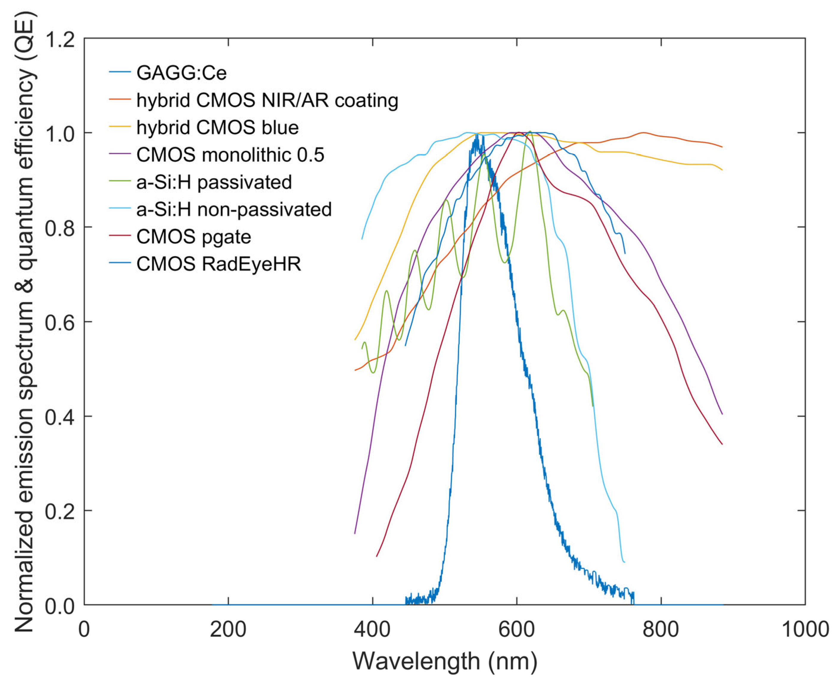
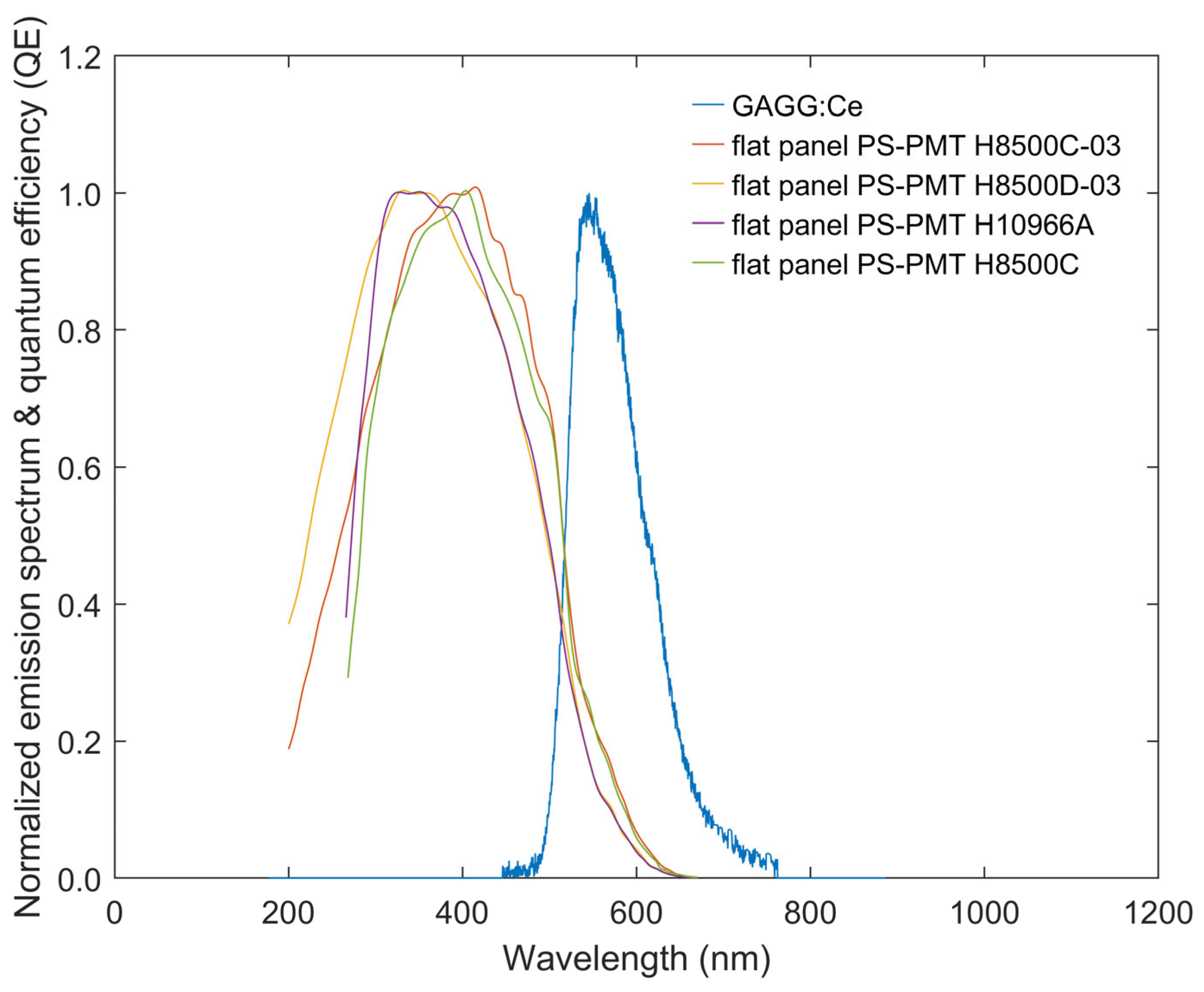
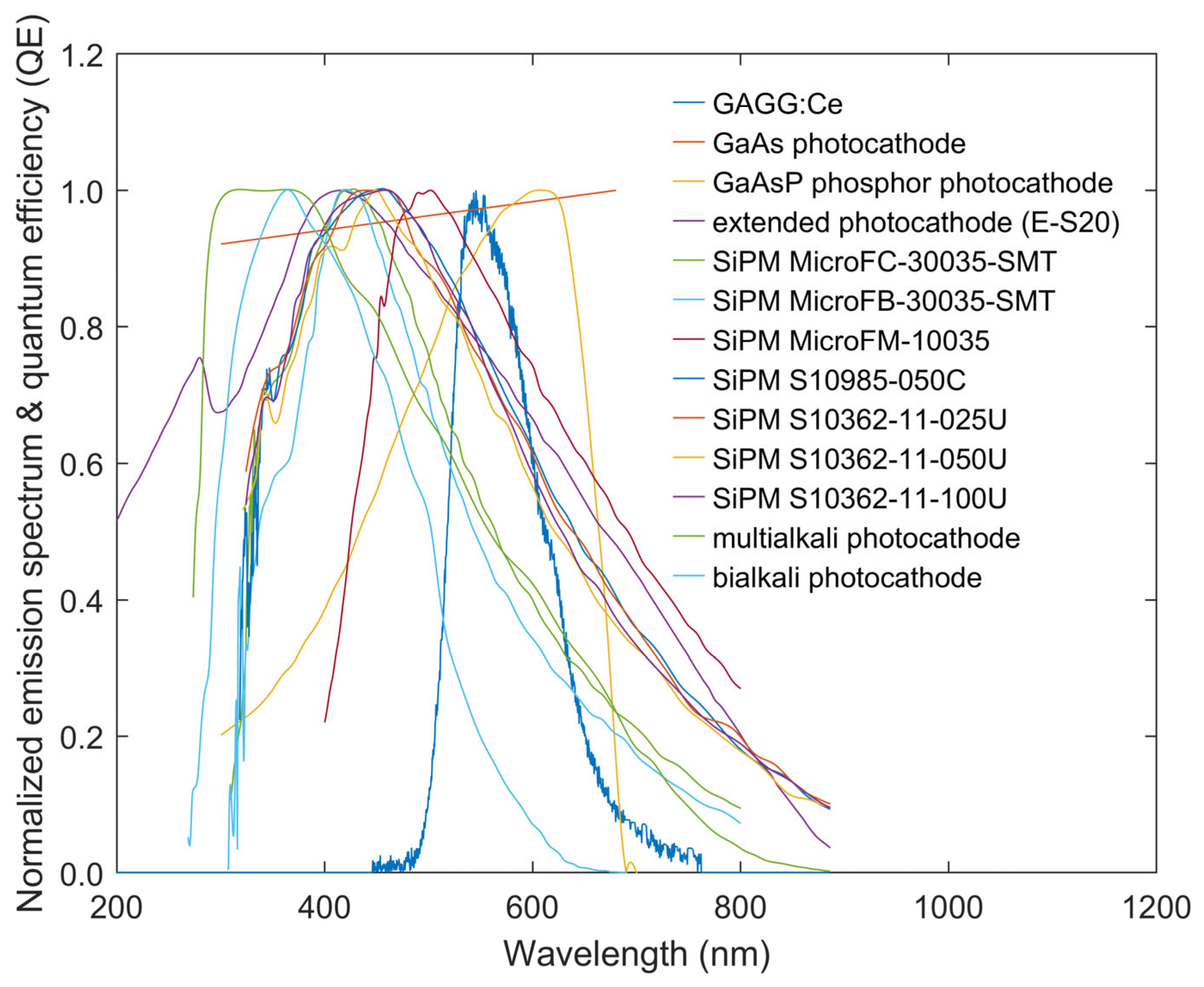
In Figure 4, Figure 5, Figure 6 and Figure 7, the optical emission spectrum of GAGG:Ce in conjunction with the spectral sensitivity of several commercially available sensors are presented. The spectral width of GAGG:Ce crystal was measured to be 315 nm in approximation, from 445 to 760 nm and its emission peak was measured at 545 nm. The normalized emission spectrum was used in Equation (1) to calculate the SMF of GAGG:Ce with the aforementioned sensors. While it can be seen from Figure 4, Figure 5, Figure 6 and Figure 7 that GAGG:Ce emission spectrum is suitable with most charge-coupled devices (CCDs), metal-oxide-semiconductors (CMOS) but not flat panels the SMF was used in order to calculate its compatibility.
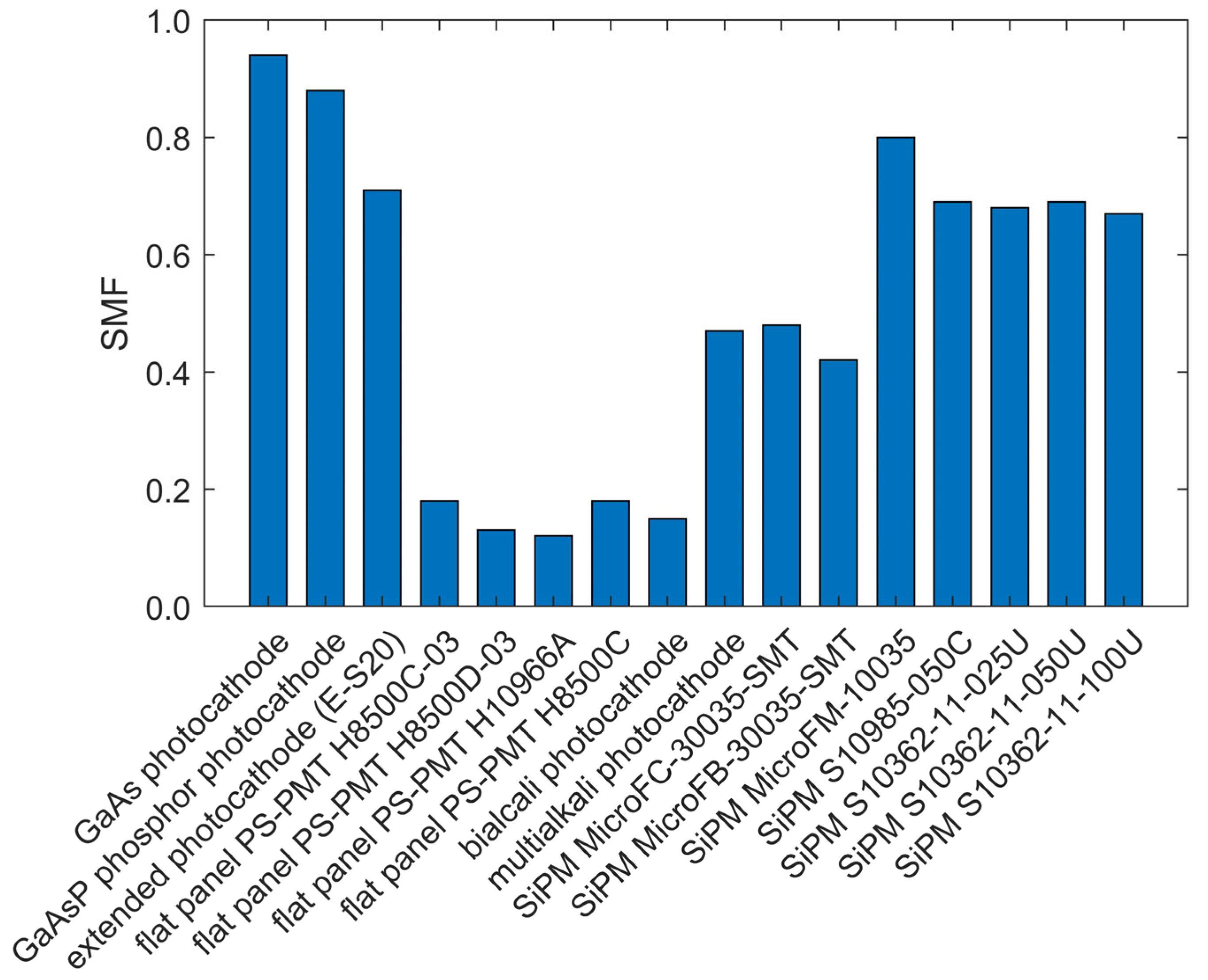
Figure 8 and Figure 9 illustrate the aforementioned compatibility of GAGG:Ce with optical photon detectors. The calculated SMF for each crystal-detector pair is shown. GAGG:Ce exhibits excellent compatibility with all the CCDs examined with the SMF values fluctuating from 0.69 to 0.95. Also, it was deduced that it was particularly suitable with CMOS detectors as demonstrated in Figure 8 with SMF values fluctuating from 0.79 to 0.99 for hybrid CMOS blue which was the highest overall SMF value of all the examined detectors. For different photocathodes and silicon photomultipliers (SiPMs) the crystal’s compatibility was found to vary, as seen in Figure 9. While the crystal is appropriate for applications with optical detectors such as gallium arsenide (GaAs), gallium arsenide phosphide (GaAsP) and the extended-S20 with SMF values of 0.94, 0.88 and 0.71 respectively, it was incompatible with other photocathodes demonstrating SMF values fluctuating from 0.12 to 0.47. Considering SiPMs, compatibility with GAGG:Ce, as shown in Figure 9, it was found to be mostly acceptable except for microFC-30035-SMT (0.48) and microFb-30035-SMT (0.42) that demonstrated especially low SMF values. The material was highly suitable with most of the modern detectors such as CCD and CMOS. This renders it an excellent candidate for systems where the high resolution and low noise levels of the CCD are needed, with potential future applications even in SPECT [11,34,35]. This is illustrated in Figure 8 where the corresponding SMF values were particularly high demonstrating that the material can be appropriately paired with CMOS in applications such as micro-CT imaging, endoscopy, dental applications (like panoramic and cone beam CT) along with other mammographic applications namely positron emission mammography (PEM). It can also be applied in tomography when combined with flat panel and amorphous silicon (a-Si) detectors. Contrarily, its matching factor was inconsistent for various photocathodes and SiPMs. While it showed compatibility with GaAs and GaAsP that find uses in PET it was inadequate with various SiPMs with applications in fast time-of-flight (ToF) PET.
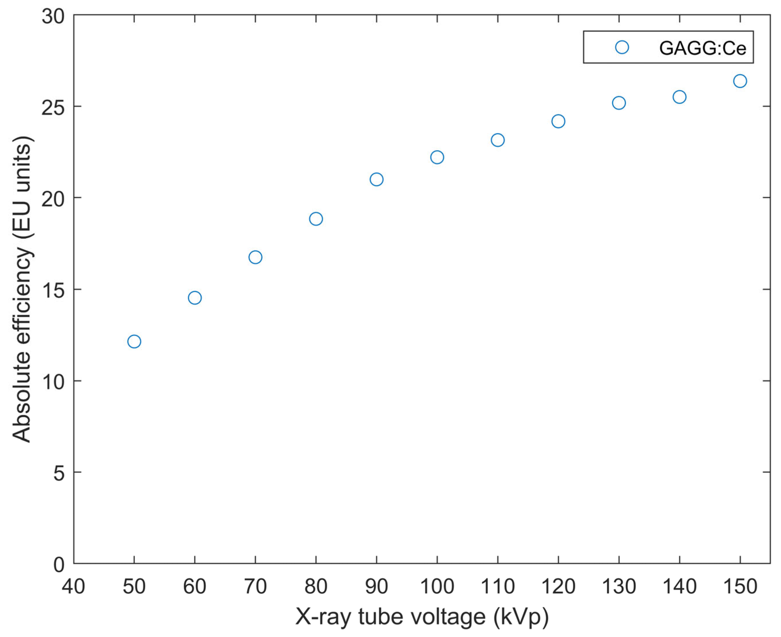
Figure 10 demonstrates the AE values of GAGG:Ce for the tube voltages under investigation. It can be seen that the AE of the crystal is increasing continuously reaching values of 26.3 E.U. for 150 kVp. Under the same exposure conditions, this value is comparable with the AE of cadmium tungstate (CdWO4) [3,4,46] and is higher than the AE of bismuth germanate (BGO) and lutetium yttrium orthosilicate (LYSO:Ce) [3]. AE is increasing in all of the energy range studied as at higher energies photons are more penetrating, thus reaching the detector. AE does not depend on the absorbed energy in the crystal matter but considers all the interactions taking place so it increases even after 100 kVp where Compton scattering is more dominant than photoelectric effect. The high AE values of Figure 10 can also be attributed to the density (6.63 g/cm3) of the crystal as a high density affects the probability of interactions within the crystal matter.
The crystal’s EAE values are presented in Figure 11. From 50 to 70 kVp the EAE values of GAGG:Ce decrease gradually with a maximum of 0.864 at 50 kVp, reaching the minimum point of 0.523 at 100 kVp. At higher energies the probability of photoelectric effect occurring and consequently the chance of the particles depositing all their energy in the crystal matter dwindles, thus the EAE is decreasing. After 100 keV it is more probable for Compton scattering to take place. Additionally, as seen in Figure 3 the μ e n / μ a t t ratio stabilizes around (~0.6) after 110 keV. Thus, after 110 kVp the EAE curve present a slight increase in Figure 11.
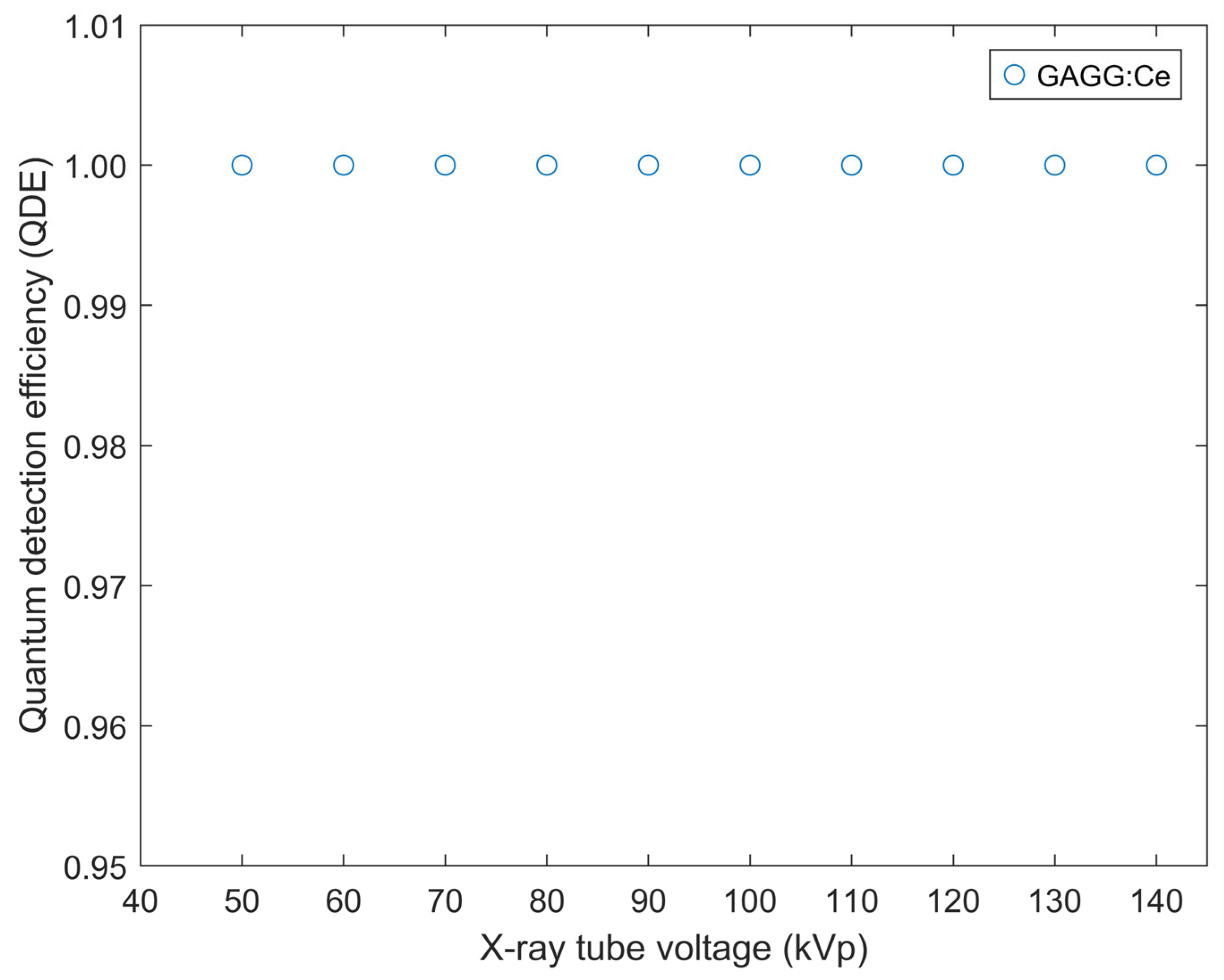
The crystal’s quantum detection efficiency for the tube voltages under investigation is shown in Figure 12. The crystal presents perfect efficiency in detecting the photons for tube voltages up to 140 kVp. The photons generated in the crystal surface, traveling in the material, transfer and consequently, deposit a portion of their energy as secondary electrons which is why the EAE in Figure 11 is highly affected by the energy that is absorbed locally and the photon fluence used in Equation (3). The photon energy spectrum was generated with the TASMIP algorithm which is commonly used to calculate the X-ray spectra in applications such as CT, thus the maximum tube voltage in EAE and QDE calculations was up to 140 kVp, albeit the experimental irradiation of the crystal, in our study, had a tube voltage range of 50 kVp to 150 kVp. The slight increase of the EAE values after 100 kVp hints the crystal’s suitability in applications like CT imaging and radiography that require high energy levels. The incline of EAE is also ascribed to the μ e n / μ a t t ratio presented in Figure 3.
QDE indicates the X-ray photons that interact with the scintillator, thus denotes the efficiency of a detection system. Ideally for scintillators used in medical imaging it should be as close as possible to 1.0, ensuring low noise levels and lower radiation dose to the patient. In our case, GAGG:Ce exhibited QDE values equal to 1.0. For our testing tube voltage range the quantum efficiency was higher than the energy absorption efficiency due to the fact that QDE factors in all the possible X-ray interactions that occur in the crystal length such as Compton scattering, braking radiation and the K characteristic radiation.
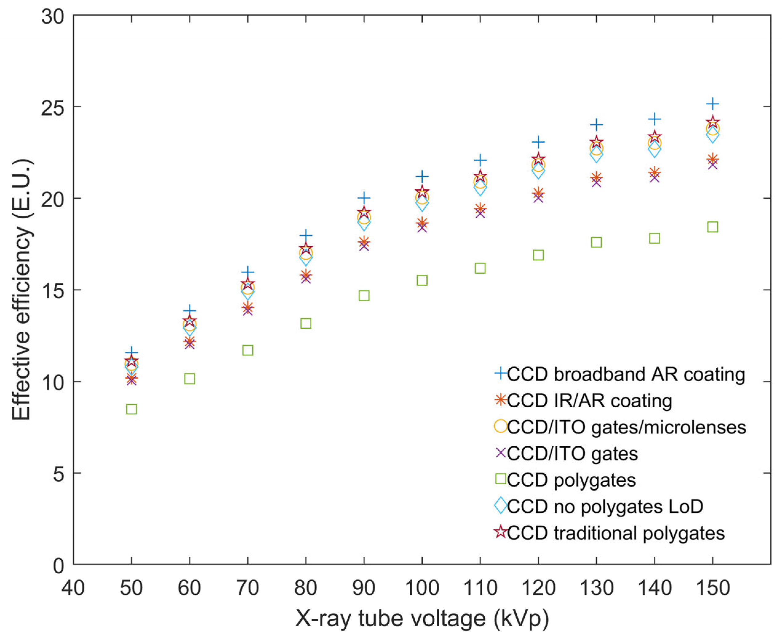
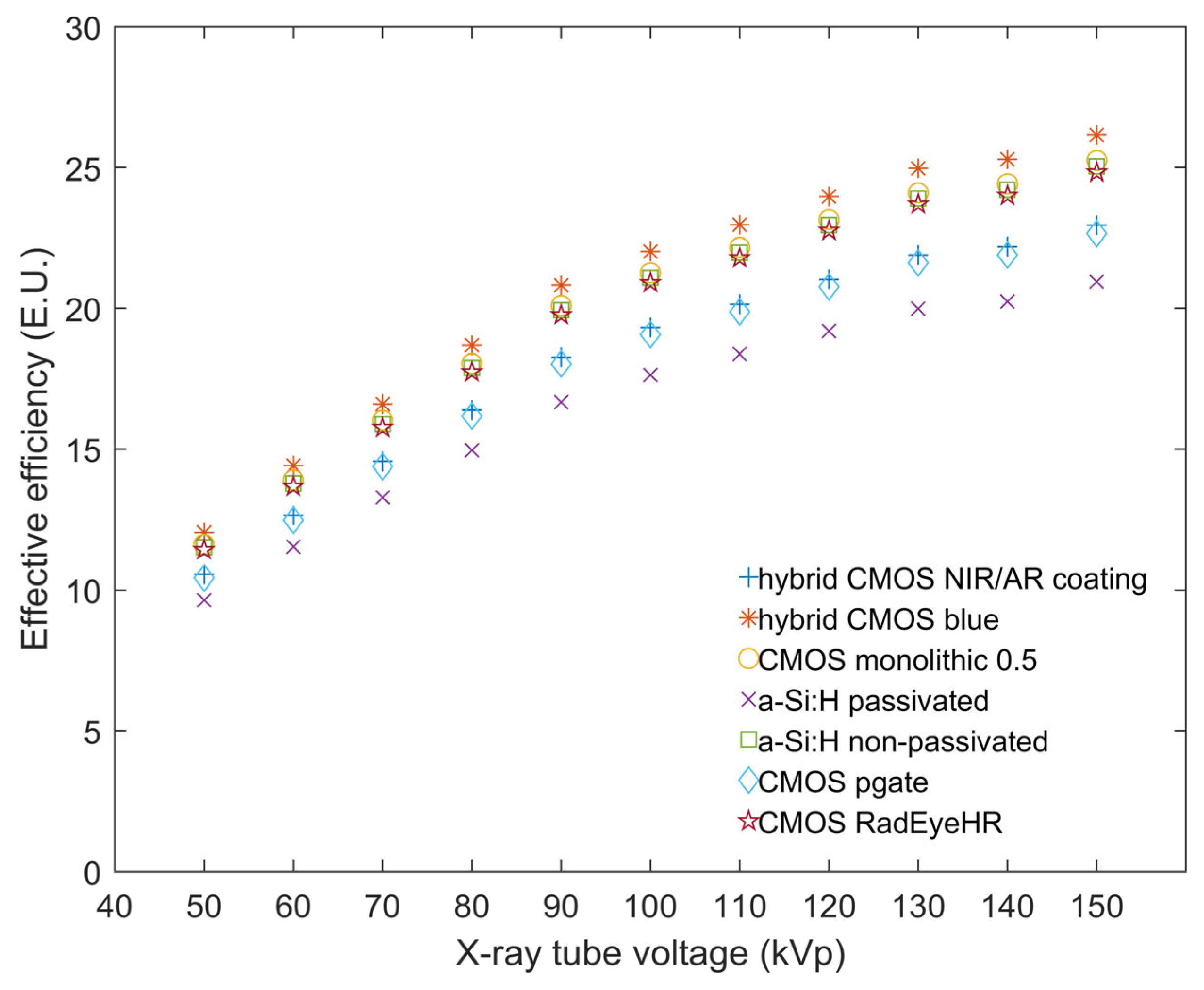
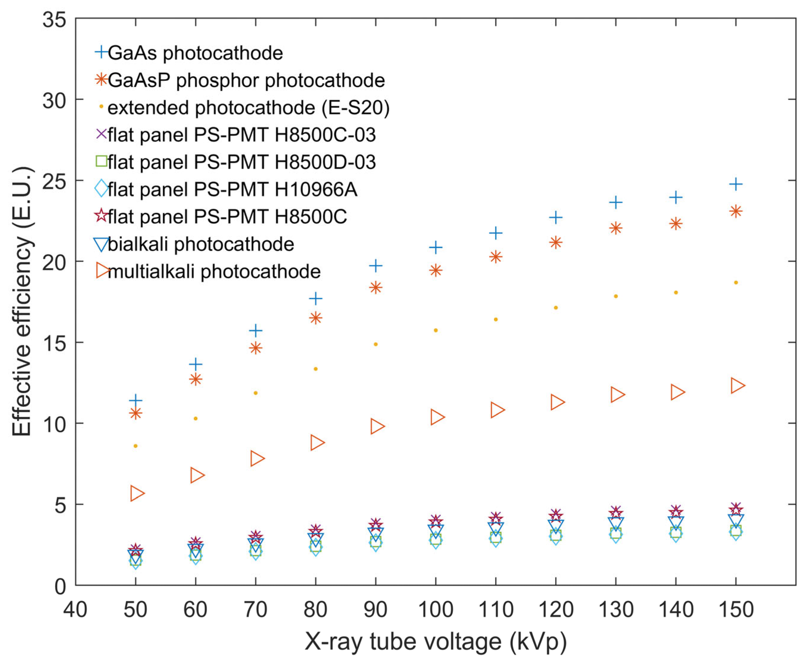
Figure 13, Figure 14, Figure 15 and Figure 16 showcase the calculated EE of every aforementioned optical photon detector. The excellent performance of GAGG:Ce with CCD and CMOS is indicated from Figure 13 and Figure 14, where EE values of 25.1 E.U. for CCD broadband with AR coating and 26.1 E.U. for hybrid CMOS blue, were achieved. However, in accordance with the SMF calculated values illustrated in Figure 15 and Figure 16, GAGG:Ce demonstrated poor EE with most photocathodes and SiPMs. It presented values of 4.0 E.U. for bialcali photocathode and 11.0 E.U. for MicroFB-30035-SMT. The lowest EE value of 1.5 E.U. was calculated, for flat panel detector PS-PMT H10966A, as seen in Figure 15. The most adequate of all the photocathodes studied were the GaAs and GaAsP photocathodes with EE values of 24.7 E.U. and 23.0 E.U. respectively. Also, the most promising SiPM was MicroFM-10035 with EE values as high as 21.1 E.U.
In Figure 17 the values of DQE(0) are illustrated. DQE(0) is affected by the absorbed X-ray photons and by the statistical fluctuations in the number of emitted light photons per absorbed incident photon, described by the statistical, or Swank, factor [3]. For a perfect detector the swank factor equals 1. The DQE, defined in this way, is referred to as zero-frequency DQE and takes into account the fluctuation in the detector signal per incident photon. Such fluctuations appear, although these incident photons may be of equal detected energy [3]. All the DQE(0) values measured were above 0.947. GAGG:Ce showed adequate performance as in accordance with the system’s high QDE and AE values for all the examined X-ray tube voltage range but it’s DQE(0) values were slightly inferior to the recently studied [4] cerium-doped lanthanum bromide (LaBr3:Ce) and lanthanum chloride (LaCl3:Ce) crystals. This is mainly due to the aforementioned crystals having higher light yield and absolute efficiency.

4. Discussion

GAGG:Ce is a bright scintillator material with high light yield [6,7,10,17,19,20], robust and durable [19,21] with steady energy resolution for −10 °C to +50 °C [10]. It has fast decay times [30] and is non-hygroscopic [7,9,19], a trait that further denotes its stability. Literature review evidence showed that GAGG:Ce exhibits no significant degradation or afterglow when tested under 70 MeV proton irradiation indicating its suitability for space applications [30,45]. It has a broad energy range of 10 keV to 2 MeV [56] so it can be used as a gamma-ray detector. Also, scintillators with no intrinsic radioactivity like GAGG:Ce are desirable in medical applications due to their reduced level of background noise. The aforementioned technological capabilities of the crystal also find use in applications such as nuclear security [41], neutron detection [41,57], Compton detection [58] and, as our results indicate, X-ray detection in medical imaging.
The fluctuation of the AE values shown in Figure 10, offers some insight into GAGG:Ce crystals properties. It is evident that the AE values increase steadily for all the examined voltage range. Based on the findings for the AE values of other scintillators from literature we deduced that GAGG:Ce presents almost similar AE with CdWO4 [3,4,46], LGSO:Ce ((Lu,Gd)2SiO5:Ce) [3] and LuAG:Ce (Lu3Al5O12:Ce) [3,46]. Specifically, GAGG:Ce AE was found 25.18 E.U. for 130 kVp when CdWO4, LGSO:Ce and LuAg:Ce AE values for the same tube voltage of 130 kVp are 26.9 E.U., ~25 E.U. and ~30 E.U. respectively. GAGG:Ce also appears to have better AE than BGO which exhibits for 130 kVp AE lower than 5 E.U. [4]. Also, when compared with other cerium-doped scintillator materials it exhibits higher AE values. For comparison’s sake, at 130 kVp the values in literature were lower than 15 E.U. for LYSO:Ce ((Lu0.9,Y0.1)2SiO5:Ce) [3,4,15], less than 25 E.U. for LSO:Ce (Lu2SiO5:Ce) [3,15,16] and under 10 E.U. for GSO:Ce (Gd2SiO5:Ce) [3,15]. In terms of AE at 130 kVp it is superior to LuYAP:Ce ((Lu0.7, Y0.3)AlO3:Ce) whose values don’t surpass 20 E.U. [15]. Similarly, it is better than YAlO3:Ce (YAP:Ce) and GdAlO3:Ce (GAP:Ce) that exhibit values under 15 E.U. [15] and less than 20 E.U. [16], respectively. Additionally, GAGG:Ce showed lower AE values than LaCl3:Ce (38.7 E.U. for 130 kVp studied under the same conditions) and LaBr3:Ce (higher than 55 E.U. for 130 kVp) due to its lower light yield compared to these crystals [3,4]. The overall absolute efficiency is dependent on the geometry of the scintillator. The light photons propagate by optical diffusion to the crystal’s surface in accordance with the pathlength they travel within the scintillator matter. The AE values increase with tube voltage as seen in Figure 10 reaching the maximum at 150 kVp as the X-ray beam travels longer distance and penetrates deep within the crystal and closer to the optical detector. This allows fewer of the light photons generated to interact and propagate with the matter thus increasing the light output and the efficiency of the scintillator to transform the X-rays into measurable light photons.
An important factor is the crystal’s high QDE values that remain equal to 1.0. GAGG:Ce exhibits optimal efficiency to detect photons and its QDE values are comparable with those of LaCl3:Ce, BGO, LSO:Ce and CdWO4 [4]. On thick crystals X-ray photons travel longer propagation pathlengths resulting in more X-ray interactions in the scintillator material hence depositing a fraction of their energy locally generating secondary electrons. All these interactions affect the photon transfer inside the crystal leading to lower spatial resolution. As a consequence, the thinner the crystal used the higher the QDE. The crystal’s dimensions used in this study, were 10 × 10 × 10 mm3 in order to ensure satisfactory absorption [41,59] in the X-ray energy range (keV). The optimum crystal thickness depends upon the light yield and the optical transmission properties. For La based scintillators a useful thickness ranges from 0.2 cm up to 1.0 cm has been reported for SPECT/CT applications [60]. Another factor of the crystal’s response is its light yield of 46,000 photons/MeV which in accordance with the high QDE makes GAGG:Ce a rather efficient scintillator material.
In terms of EAE GAGG:Ce has better performance than GdAlO3:Ce [16], CaF2:Eu [46] and LaCl3:Ce [3,4] based on previously published data on crystals of same thickness (10 × 10 × 10 mm3) under the same energy range. The said crystals have densities of 6.63 g/cm3 for GdAlO3:Ce [16], 3.18 g/cm3 for CaF2:Eu [46] and 3.86 g/cm3 for LaCl3:Ce [3,4]. When compared to crystals of similar or higher density such as LGSO:Ce (7.3 g/cm3), GSO:Ce (6.7 g/cm3), LSO:Ce (7.4 g/cm3), [3] CdWO4 (7.9 g/cm3) and LuAG:Ce (6.73 g/cm3) [46] it shows almost similar EAE values. However, the crystal showed slight worse EAE values than BGO as expected due to the fact that BGO has a higher density of 8.71 g/cm3 and an effective atomic number Zeff = 74 [4] in comparison with the 6.63 g/cm3 and Zeff = 54.4 of GAGG:Ce [19]. This intrinsic energy is used for the creation of the output signal in the detector system thus the decrease of EAE shown after 50 kVp is not indicative of the signal detection efficiency.
Regarding the SMF values GAGG:Ce showed high compatibility with CCDs and CMOS. However, the GAGG:Ce crystal’s compatibility with various photocathodes and SiPMs wasn’t coherent for all the test sample of the detectors studied. Concerning EE while LaCl3:Ce [4] and CdWO4 [46] are better coupled with photocathodes and SiPMs, GAGG:Ce has overall better compatibility with CMOS and CCDs.
According to Equation (7), DQE(0) is dependent, upon the output signal and the X-ray absorption properties of the scintillator. For higher tube voltages AE and X-ray exposure are increased, while the X-ray absorption efficiency in the scintillator is decreased. The shape of the DQE(0) curve is due to the combined effect of these properties. Its high value, over 0.94, demonstrates that DQE in the energy range under investigation is more affected by QDE than the output signal variations. In the medical energy range in this study, GAGG:Ce showed better efficiency in comparison with other commonly used cerium-doped scintillators which along with its high light yield (LY) (46,000 photons/MeV) and high density (6.63 g/cm3) renders it as a promising material for further study.
Significantly, the aforementioned characteristics of GAGG:Ce along with the fact that it is non-hygroscopic with a relatively fast decay time of 50–150 ns and no intrinsic radiation make it a good candidate as a part of a digital detector for energies up to 150 keV in applications like imaging detectors, radiation safety and security applications.

5. Conclusions

The response and properties of GAGG:Ce were experimentally evaluated in the present study under medical X-ray excitation and tube voltages ranging from 50–150 kVp. The maximum absolute efficiency was measured at 150 kVp (26.4 E.U.). GAGG:Ce emission spectrum was found to be highly compatible with the spectral sensitivities of several optical photon detectors especially CCDs and CMOS. Moreover, it demonstrated excellent detective quantum efficiency for zero-spatial frequency. GAGG:Ce showed adequately efficient AE values and is non-hygroscopic. Taking into consideration its spectral compatibility with modern photon detectors and the very good DQE(0) values, GAGG:Ce is already considered as a suitable scintillator material for X-ray medical imaging systems. Notably, our results indicated a superior response in terms of absolute luminescence efficiency (AE), energy absorption efficiency (EAE) and quantum-detection efficiency (QDE) in comparison with other cerium-doped scintillators usually employed in X-ray imaging applications. In addition, the scintillator under investigation was determined to be a promising candidate as a radiation detector in the X-ray tube voltage range of 100–150 kVp.

Author Contributions

Conceptualization, N.K. and A.D.; methodology, N.K., A.D., C.M., I.V., G.F. and I.K.; software, A.D.; validation, C.M. and I.V.; formal analysis, N.K., C.M. and A.D.; investigation, N.K., C.M., A.D., I.V., G.F. and I.K.; data curation, C.M., N.K., A.D., I.V., G.F. and I.K.; writing—original draft preparation, A.D. and N.K.; writing—review and editing, N.K., C.M., I.V., G.F. and I.K.; visualization, N.K.; supervision, C.M.; project administration, C.M. and I.V. All authors have read and agreed to the published version of the manuscript.

Funding

This research received no external funding.

Data Availability Statement

The original contributions presented in this study are included in this article. Further inquiries can be directed to the corresponding author.

Conflicts of Interest

The authors declare no conflicts of interest.

Abbreviations

GAGGGadolinium aluminum gallium garnet
SMFSpectral matching factor
AEAbsolute luminescence efficiency
EEEffective efficiency
EAEEnergy absorption efficiency
QDEQuantum-detection efficiency
DQEDetective quantum efficiency
CMOSComplementary metal-oxide semiconductors
CCDCharged-coupled devices
PETPositron emission tomography
CTComputed tomography
SPECTSingle-photon emission computed tomography
PMTPhotomultiplier tube
SiPDSilicon photodiodes
LYLight yield
Lulutetium
GdGadolinium
AlAluminum
GaGallium
CeCerium
OOxygen
CrChromium
E.U.Efficiency units
TASMIPTungsten anode spectral model using interpolating polynomials
ToFTime-of-flight
QEQuantum efficiency
a-SiAmorphous silicon
GaAsGallium arsenide
GaAsPGallium arsenide phosphide
LaBr3Lanthanum bromide
LaCl3Lanthanum chloride
CdWO4Cadmium tungsten
LGSOLutetium gadolinium oxyorthosilicate
LuAgLutetium aluminum garnet
BGOBismuth germanate
LYSOLutetium-yttrium oxyorthosilicate
LSOLutetium oxyorthosilicate
GSOGadolinium oxyorthosilicate
LuYAPLutetium yttrium orthosilicate perovskite
YAlO3Yttrium aluminum perovskite
GdAlO3Gadolinium aluminum perovskite

References

  1. Dorenbos, P. Light Output and Energy Resolution of Ce3+-Doped Scintillators. Nucl. Instrum. Methods Phys. Res. A Accel. Spectrom. Detect. Assoc. Equip. 2002, 486, 208–213. [Google Scholar] [CrossRef]
  2. Moses, W.W. Scintillator Requirements for Medical Imaging; Lawrence Berkeley National Lab. (LBNL): Berkeley, CA, USA, 1999. [Google Scholar]
  3. Michail, C.; Liaparinos, P.; Kalyvas, N.; Kandarakis, I.; Fountos, G.; Valais, I. Phosphors and Scintillators in Biomedical Imaging. Crystals 2024, 14, 169. [Google Scholar] [CrossRef]
  4. Tseremoglou, S.; Michail, C.; Valais, I.; Ninos, K.; Bakas, A.; Kandarakis, I.; Fountos, G.; Kalyvas, N. Evaluation of Cerium-Doped Lanthanum Bromide (LaBr3:Ce) Single-Crystal Scintillator’s Luminescence Properties under X-Ray Radiographic Conditions. Appl. Sci. 2023, 13, 419. [Google Scholar] [CrossRef]
  5. Kamada, K.; Yanagida, T.; Endo, T.; Tsutumi, K.; Usuki, Y.; Nikl, M.; Fujimoto, Y.; Yoshikawa, A. 2-Inch Size Single Crystal Growth and Scintillation Properties of New Scintillator; Ce:Gd3Al2Ga3O12. In Proceedings of the 2011 IEEE Nuclear Science Symposium and Medical Imaging Conference, Valencia, Spain, 23–29 October 2011; Institute of Electrical and Electronics Engineers Inc.: Piscataway, NJ, USA, 2011; pp. 1927–1929. [Google Scholar]
  6. Kamada, K.; Yanagida, T.; Endo, T.; Tsutumi, K.; Usuki, Y.; Nikl, M.; Fujimoto, Y.; Fukabori, A.; Yoshikawa, A. 2inch Diameter Single Crystal Growth and Scintillation Properties of Ce:Gd3Al2Ga3O12. J. Cryst. Growth 2012, 352, 88–90. [Google Scholar] [CrossRef]
  7. Yeom, J.Y.; Yamamoto, S.; Derenzo, S.E.; Spanoudaki, V.C.; Kamada, K.; Endo, T.; Levin, C.S. First Performance Results of Ce:GAGG Scintillation Crystals with Silicon Photomultipliers. IEEE Trans. Nucl. Sci. 2013, 60, 988–992. [Google Scholar] [CrossRef]
  8. Zhu, Y.; Qian, S.; Wang, Z.; Guo, H.; Ma, L.; Wang, Z.; Wu, Q. Scintillation Properties of GAGG:Ce Ceramic and Single Crystal. Opt. Mater. 2020, 105, 109964. [Google Scholar] [CrossRef]
  9. Potiriadis, N.; Skouroliakou, A.; Liaparinos, P.; David, S. Energy Resolution Values of GAGG:Ce Crystals Coupled to Various SiPMs. Eur. Phys. J. Spec. Top. 2025, 1–8. [Google Scholar] [CrossRef]
  10. Seitz, B.; Campos Rivera, N.; Stewart, A.G. Energy Resolution and Temperature Dependence of Ce:GAGG Coupled to 3 mm × 3 mm Silicon Photomultipliers. IEEE Trans. Nucl. Sci. 2016, 63, 503–508. [Google Scholar] [CrossRef]
  11. Yamamoto, S.; Yeom, J.Y.; Kamada, K.; Endo, T.; Levin, C. Development of an Ultrahigh Resolution Block Detector Based on 0.4 Mm Pixel Ce:GAGG Scintillators and a Silicon Photomultiplier Array. IEEE Trans. Nucl. Sci. 2013, 60, 4582–4587. [Google Scholar] [CrossRef]
  12. David, S.L.; Valais, I.G.; Michail, C.M.; Kandarakis, I.S. X-Ray Luminescence Efficiency of GAGG:Ce Single Crystal Scintillators for Use in Tomographic Medical Imaging Systems. J. Phys. Conf. Ser. 2015, 637, 012004. [Google Scholar] [CrossRef]
  13. Yu, H.; Meng, X.; Yang, S.; Zhao, J.; Zhen, X.; Tai, R. Photonic-Crystals-Based GAGG:Ce Scintillator with High Light Output and Fast Decay Time for Soft X-Ray Detection. Nucl. Instrum. Methods Phys. Res. A Accel. Spectrom. Detect. Assoc. Equip. 2022, 1032, 166653. [Google Scholar] [CrossRef]
  14. Gerasymov, I.; Nepokupnaya, T.; Boyarintsev, A.; Sidletskiy, O.; Kurtsev, D.; Voloshyna, O.; Trubaieva, O.; Boyarintseva, Y.; Sibilieva, T.; Shaposhnyk, A.; et al. GAGG:Ce Composite Scintillator for X-Ray Imaging. Opt. Mater. 2020, 109, 110305. [Google Scholar] [CrossRef]
  15. Valais, I.G.; Michail, C.M.; David, S.L.; Liaparinos, P.F.; Fountos, G.P.; Paschalis, T.V.; Kandarakis, I.S.; Panayiotakis, G.S. Comparative Investigation of Ce3+ Doped Scintillators in a Wide Range of Photon Energies Covering X-Ray CT, Nuclear Medicine and Megavoltage Radiation Therapy Portal Imaging Applications. IEEE Trans. Nucl. Sci. 2010, 57, 3–7. [Google Scholar] [CrossRef]
  16. Michail, C.; Kalyvas, N.; Valais, I.; David, S.; Seferis, I.; Toutountzis, A.; Karabotsos, A.; Liaparinos, P.; Fountos, G.; Kandarakis, I. On the Response of GdAlO3:Ce Powder Scintillators. J. Lumin. 2013, 144, 45–52. [Google Scholar] [CrossRef]
  17. Kamada, K.; Endo, T.; Tsutumi, K.; Yanagida, T.; Fujimoto, Y.; Fukabori, A.; Yoshikawa, A.; Pejchal, J.; Nikl, M. Composition Engineering in Cerium-Doped (Lu,Gd)3(Ga,Al)5O12 Single-Crystal Scintillators. Cryst. Growth Des. 2011, 11, 4484–4490. [Google Scholar] [CrossRef]
  18. Yoneyama, M.; Kataoka, J.; Arimoto, M.; Masuda, T.; Yoshino, M.; Kamada, K.; Yoshikawa, A.; Sato, H.; Usuki, Y. Evaluation of GAGG:Ce Scintillators for Future Space Applications. J. Inst. 2018, 13, P02023. [Google Scholar] [CrossRef]
  19. GAGG(Ce)—Scintillator Crystal Advatech UK. Available online: https://www.advatech-uk.co.uk/gagg_ce.html (accessed on 15 January 2025).
  20. Inkrataite, G.; Laurinavicius, G.; Enseling, D.; Zarkov, A.; Jüstel, T.; Skaudzius, R. Characterization of GAGG Doped with Extremely Low Levels of Chromium and Exhibiting Exceptional Intensity of Emission in NIR Region. Crystals 2021, 11, 673. [Google Scholar] [CrossRef]
  21. Gray, T.J.; Allmond, J.M.; Dowling, D.T.; Febbraro, M.; King, T.T.; Pain, S.D.; Stracener, D.W.; Ajayi, S.; Aragon, J.; Baby, L.; et al. CLARION2-TRINITY: A Compton-Suppressed HPGe and GAGG:Ce-Si-Si Array for Absolute Cross-Section Measurements with Heavy Ions. Nucl. Instrum. Methods Phys. Res. A Accel. Spectrom. Detect. Assoc. Equip. 2022, 1041, 167392. [Google Scholar] [CrossRef]
  22. Kobayashi, M.; Tamagawa, Y.; Tomita, S.; Yamamoto, A.; Ogawa, I.; Usuki, Y. Significantly Different Pulse Shapes for γ- and α-Rays in Gd3Al2Ga3O12:Ce3+ Scintillating Crystals. Nucl. Instrum. Methods Phys. Res. A Accel. Spectrom. Detect. Assoc. Equip. 2012, 694, 91–94. [Google Scholar] [CrossRef]
  23. Iwanowska, J.; Swiderski, L.; Szczesniak, T.; Sibczynski, P.; Moszynski, M.; Grodzicka, M.; Kamada, K.; Tsutsumi, K.; Usuki, Y.; Yanagida, T.; et al. Performance of Cerium-Doped Gd3Al2Ga3O12 (GAGG:Ce) Scintillator in Gamma-Ray Spectrometry. Nucl. Instrum. Methods Phys. Res. A Accel. Spectrom. Detect. Assoc. Equip. 2013, 712, 34–40. [Google Scholar] [CrossRef]
  24. Makek, M.; Bosnar, D.; Kožuljević, A.M.; Pavelić, L. Investigation of GaGG:Ce with TOFPET2 ASIC Readout for Applications in Gamma Imaging Systems. Crystals 2020, 10, 1073. [Google Scholar] [CrossRef]
  25. Xie, S.; Zhang, X.; Zhang, Y.; Ying, G.; Huang, Q.; Xu, J.; Peng, Q. Evaluation of Various Scintillator Materials in Radiation Detector Design for Positron Emission Tomography (PET). Crystals 2020, 10, 869. [Google Scholar] [CrossRef]
  26. Yoshino, M.; Yamamoto, S.; Nakanishi, K.; Yogo, K.; Kamada, K.; Koshikawa, N.; Kataoka, J.; Yoshikawa, A. Development and Performance Evaluation of a Thin GAGG:Ce Scintillator Plate for High Resolution Synchrotron Radiation X-Ray Imaging. J. Instrum. 2024, 19, P10030. [Google Scholar] [CrossRef]
  27. Bogomolov, V.V.; Dosovitskiy, G.A.; Iyudin, A.F.; Korzhik, M.V.; Tikhomirov, S.A.; Svertilov, S.I.; Kozlov, D.Y.; Yashin, I.V. The Timing and Spectral Characteristics of Detectors Based on a Ce:GAGG Inorganic Scintillator Using Photomultiplier Tubes and Silicon Photodetectors. Instrum. Exp. Tech. 2020, 63, 633–640. [Google Scholar] [CrossRef]
  28. Furuno, T.; Koshikawa, A.; Kawabata, T.; Itoh, M.; Kurosawa, S.; Morimoto, T.; Murata, M.; Sakanashi, K.; Tsumura, M.; Yamaji, A. Response of the GAGG(Ce) Scintillator to Charged Particles Compared with the CsI(Tl) Scintillator. J. Inst. 2021, 16, P10012. [Google Scholar] [CrossRef]
  29. Korjik, M.; Alenkov, V.; Borisevich, A.; Buzanov, O.; Dormenev, V.; Dosovitskiy, G.; Dosovitskiy, A.; Fedorov, A.; Kozlov, D.; Mechinsky, V.; et al. Significant Improvement of GAGG:Ce Based Scintillation Detector Performance with Temperature Decrease. Nucl. Instrum. Methods Phys. Res. A Accel. Spectrom. Detect. Assoc. Equip. 2017, 871, 42–46. [Google Scholar] [CrossRef]
  30. Dilillo, G.; Zampa, N.; Campana, R.; Fuschino, F.; Pauletta, G.; Rashevskaya, I.; Ambrosino, F.; Baruzzo, M.; Cauz, D.; Cirrincione, D.; et al. Space Applications of GAGG:Ce Scintillators: A Study of Afterglow Emission by Proton Irradiation. Nucl. Instrum. Methods Phys. Res. B Beam Interact. Mater. At. 2022, 513, 33–43. [Google Scholar] [CrossRef]
  31. Pallu, M.; Pailot, D.; Bréelle, E.; Laurent, P.; Carron, J.; Lebrun, F.; Koumeir, C.; Chapron; Biernacki, K. Studies of GAGG:Ce Scintillators for Space Missions Dedicated to Terrestrial Gamma-Ray Flashes and Gamma-Ray Bursts Observation. Nucl. Instrum. Methods Phys. Res. A Accel. Spectrom. Detect. Assoc. Equip. 2024, 1069, 169997. [Google Scholar] [CrossRef]
  32. Yu, X.; Zhang, X.; Zhang, H.; Peng, H.; Ren, Q.; Xu, J.; Peng, Q.; Xie, S. Requirements of Scintillation Crystals with the Development of PET Scanners. Crystals 2022, 12, 1302. [Google Scholar] [CrossRef]
  33. Lee, S.; Kim, K.Y.; Lee, M.S.; Lee, J.S. Recovery of Inter-Detector and Inter-Crystal Scattering in Brain PET Based on LSO and GAGG Crystals. Phys. Med. Biol. 2020, 65, 195005. [Google Scholar] [CrossRef]
  34. Xu, S.; Yan, Z.; Wei, Q. A Survey of Crystals for SPECT Imaging. Crystals 2024, 14, 1039. [Google Scholar] [CrossRef]
  35. Yu, Z.; Lyu, Z.; Fan, P.; Wu, J.; Liu, Y.; Ma, T. Sub-Millimeter Pixelated SPECT Detector Using GAGG:Ce and Light Guide With Optical Barrier Slits. IEEE Trans. Radiat. Plasma Med. Sci. 2025. [Google Scholar] [CrossRef]
  36. Hua, J.; Wang, H.; Li, C.; Dong, Y.; Yuan, Z.; Yang, H.; Liu, Y.; Jiang, J. Performance Evaluation of a GAGG-SiPM Based Compton Camera for Gamma-Ray Astronomy. Nucl. Instrum. Methods Phys. Res. A Accel. Spectrom. Detect. Assoc. Equip. 2024, 1068, 169765. [Google Scholar] [CrossRef]
  37. Tamulatis, G.; Dosovitskiy, G.; Gola, A.; Korjik, M.; Mazzi, A.; Nargelas, S.; Sokolov, P.; Vaitkevičius, A. Improvement of Response Time in GAGG:Ce Scintillation Crystals by Magnesium Codoping. J. Appl. Phys. 2018, 124, 215907. [Google Scholar] [CrossRef]
  38. Omuro, K.; Yoshino, M.; Bartosiewicz, K.; Horiai, T.; Murakami, R.; Kim, K.J.; Kamada, K.; Kucerkova, R.; Babin, V.; Nikl, M.; et al. Tailoring Scintillation and Luminescence through Co-Doping Engineering: A Comparative Study of Ce,Tb Co-Doped YAGG and GAGG Garnet Crystals. J. Alloys Compd. 2024, 1008, 176550. [Google Scholar] [CrossRef]
  39. Qi, Q.; Li, M.; Zhao, S.; Xue, Z.; Ding, D.; Shi, J. Luminescence and Scintillation Properties of Yb—Co-Doped GAGG:Ce Single Crystal. Opt. Mater. 2022, 123, 111904. [Google Scholar] [CrossRef]
  40. Kasimova, V.M.; Kozlova, N.S.; Buzanov, O.A.; Zabelina, E.V. Effect of Ca2+ and Zr4+ Co-Doping on the Optical Properties of Gd3Al2Ga3O12: Ce Single Crystals. Mod. Electron. Mater. 2019, 5, 101–105. [Google Scholar] [CrossRef]
  41. Karmakar, A.; Kumar, G.A.; Tyagi, M.; Pal, A. Thickness Dependent Sensitivity of GAGG:Ce Scintillation Detectors for Thermal Neutrons: GEANT4 Simulations and Experimental Measurements. J. Radioanal. Nucl. Chem. 2025, 334, 2203–2210. [Google Scholar] [CrossRef]
  42. Sidletskiy, O.; Gorbenko, V.; Zorenko, T.; Syrotych, Y.; Witkiwicz-Łukaszek, S.; Mares, J.A.; Kucerkova, R.; Nikl, M.; Gerasymov, I.; Kurtsev, D.; et al. Composition Engineering of (Lu,Gd,Tb)3(Al,Ga)5O12:Ce Film/Gd3(Al,Ga)5O12:Ce Substrate Scintillators. Crystals 2022, 12, 1366. [Google Scholar] [CrossRef]
  43. Stuhl, L.; Krasznahorkay, A.; Csatlós, M.; Algora, A.; Gulyás, J.; Kalinka, G.; Kertész, Z.I.; Timár, J. A Newly Developed Wrapping Method for Scintillator Detectors. J. Phys. Conf. Ser. 2016, 665, 012050. [Google Scholar] [CrossRef]
  44. Auffray, E.; Dosovitskiy, G.; Fedorov, A.; Guz, I.; Korjik, M.; Kratochwill, N.; Lucchini, M.; Nargelas, S.; Kozlov, D.; Mechinsky, V.; et al. Irradiation Effects on Gd3Al2Ga3O12 Scintillators Prospective for Application in Harsh Irradiation Environments. Radiat. Phys. Chem. 2019, 164, 108365. [Google Scholar] [CrossRef]
  45. Kasimova, V.M.; Kozlova, N.S.; Buzanov, O.A.; Zabelina, E.V.; Lagov, P.B.; Pavlov, Y.S. Effect of Electron Irradiation on the Optical Properties of Gadolinium-Aluminum-Gallium Garnet Crystals. J. Surf. Investig. 2021, 15, 1259–1263. [Google Scholar] [CrossRef]
  46. Michail, C.; Koukou, V.; Martini, N.; Saatsakis, G.; Kalyvas, N.; Bakas, A.; Kandarakis, I.; Fountos, G.; Panayiotakis, G.; Valais, I. Luminescence Efficiency of Cadmium Tungstate (CdWO4) Single Crystal for Medical Imaging Applications. Crystals 2020, 10, 429. [Google Scholar] [CrossRef]
  47. Boone, J. X-Ray Production, Interaction, and Detection in Diagnostic Imaging. In Handbook of Medical Imaging; SPIE Press: Bellingham, WA, USA, 2000; Volume 1: Physics and Psychophysics, pp. 36–57. ISBN 978-0-8194-7772-9. [Google Scholar]
  48. Evans, R.D. The Atomic Nucleus; McGraw-Hill, Inc.: New York, NY, USA, 1955. [Google Scholar]
  49. Report IAEA-NDS-195, XMuDat. Available online: https://www-nds.iaea.org/publications/iaea-nds/iaea-nds-0195.htm (accessed on 15 January 2025).
  50. Shaw, R. Some Fundamental Properties of Xeroradiographic Images. In Proceedings of the Application of Optical Instrumentation in Medicine IV, Atlanta, GA, USA, 25 March 1976; SPIE: Bellingham, WA, USA, 1976; Volume 0070, pp. 359–363. [Google Scholar]
  51. Wagner, R.F.; Muntz, E.P. Detective Quantum Efficiency (DQE) Analysis of Electrostatic Imaging and Screen-Film Imaging in Mammography. In Proceedings of the Application of Optical Instrumentation in Medicine VII, Toronto, ON, Canada, 6 July 1979; SPIE: Bellingham, WA, USA, 1979; Volume 173, pp. 162–167. [Google Scholar]
  52. Dick, C.E.; Motz, J.W. Image Information Transfer Properties of X-Ray Fluorescent Screens. Med. Phys. 1981, 8, 337–346. [Google Scholar] [CrossRef]
  53. TASMIP Spectra Calculator—Calculate X-Ray Imaging Spectra. Available online: https://solutioinsilico.com/medical-physics/applications/tasmip-app.php (accessed on 15 January 2025).
  54. Abbene, L.; Manna, A.; Fauci, F.; Gerardi, G.; Stumbo, S.; Raso, G. X-Ray Spectroscopy and Dosimetry with a Portable CdTe Device. Nucl. Instrum. Methods Phys. Res. Accel. Spectrom. Detect. Assoc. Equip. 2007, 571, 373–377. [Google Scholar] [CrossRef]
  55. Campana, R.; Evola, C.; Labanti, C.; Ferro, L.; Moita, M.; Virgilli, E.; Marchesini, E.J.; Frontera, F.; Rosati, P. Measurement of the Non-Linearity in the γ-Ray Response of the GAGG:Ce Inorganic Scintillator. Nucl. Instrum. Methods Phys. Res. A Accel. Spectrom. Detect. Assoc. Equip. 2023, 1056, 168587. [Google Scholar] [CrossRef]
  56. Wen, J.-X.; Zheng, X.-T.; Yu, J.-D.; Che, Y.-P.; Yang, D.-X.; Gao, H.-Z.; Jin, Y.-F.; Long, X.-Y.; Liu, Y.-H.; Xu, D.-C.; et al. Compact CubeSat Gamma-Ray Detector for GRID Mission. Nucl. Sci. Tech. 2021, 32, 99. [Google Scholar] [CrossRef]
  57. Taggart, M.P.; Nakhostin, M.; Sellin, P.J. Investigation into the Potential of GAGG:Ce as a Neutron Detector. Nucl. Instrum. Methods Phys. Res. Sect. A Accel. Spectrometers Detect. Assoc. Equip. 2019, 931, 121–126. [Google Scholar] [CrossRef]
  58. Zarei, H.; Razaghi, S.; Nagao, Y.; Itoh, M.; Yamaguchi, M.; Kawachi, N.; Ay, M.R.; Watabe, H. Evaluation and Optimization of Geometry Parameters of GAGG Scintillator-Based Compton Camera for Medical Imaging by Monte Carlo Simulation. J. Inst. 2023, 18, P01035. [Google Scholar] [CrossRef]
  59. Ronda, C. Scintillators for Medical Imaging. Opt. Mater. X 2024, 22, 100293. [Google Scholar] [CrossRef]
  60. Tseremoglou, S.; Michail, C.; Valais, I.; Ninos, K.; Bakas, A.; Kandarakis, I.; Fountos, G.; Kalyvas, N. Optical Photon Propagation Characteristics and Thickness Optimization of LaCl3:Ce and LaBr3:Ce Crystal Scintillators for Nuclear Medicine Imaging. Crystals 2024, 14, 24. [Google Scholar] [CrossRef]
Figure 1. Experimental measurement setup.
Crystals 15 00590 g001
Figure 2. Attenuation coefficients of GAGG:Ce single crystal.
Crystals 15 00590 g002
Figure 3. The fraction μenatt of GAGG:Ce scintillation crystal.
Crystals 15 00590 g003
Figure 4. Emission spectrum of GAGG:Ce crystal and responsiveness of CCD photoreceptors.
Crystals 15 00590 g004
Figure 5. Emission spectrum of GAGG:Ce crystal with the responsiveness of CMOS and a-Si photoreceptors.
Crystals 15 00590 g005
Figure 6. Emission spectrum of GAGG:Ce crystal and responsiveness of flat panel systems.
Crystals 15 00590 g006
Figure 7. Emission spectrum of GAGG:Ce crystal with the responsiveness of photocathodes and SiPMs.
Crystals 15 00590 g007
Figure 8. The spectral matching factors of several CCDs, CMOS and a-Si with GAGG:Ce.
Crystals 15 00590 g008
Figure 9. The spectral matching factors of several photocathodes, flat panel systems and SiPMs with GAGG:Ce.
Crystals 15 00590 g009
Figure 10. The absolute efficiency of GAGG:Ce scintillation crystal in E.U. units (1 E.U. = 1 μW m−2/(mR s−1)).
Crystals 15 00590 g010
Figure 11. EAE of the 10 × 10 × 10 mm3 GAGG:Ce scintillation crystal.
Crystals 15 00590 g011
Figure 12. Quantum detection efficiency values of the GAGG:Ce scintillation crystal.
Crystals 15 00590 g012
Figure 13. Effective efficiency of GAGG:Ce paired with several CCDs.
Crystals 15 00590 g013
Figure 14. Effective efficiency of GAGG:Ce paired with several CMOS.
Crystals 15 00590 g014
Figure 15. Effective efficiency of GAGG:Ce paired with several photocathodes.
Crystals 15 00590 g015
Figure 16. Effective efficiency of GAGG:Ce paired with several SiPMs.
Crystals 15 00590 g016
Figure 17. Detective quantum efficiency DQE(0) of GAGG:Ce.
Crystals 15 00590 g017
Disclaimer/Publisher’s Note: The statements, opinions and data contained in all publications are solely those of the individual author(s) and contributor(s) and not of MDPI and/or the editor(s). MDPI and/or the editor(s) disclaim responsibility for any injury to people or property resulting from any ideas, methods, instructions or products referred to in the content.