Review

An Insight into Synthesis, Optical Properties, and Applications of Green Fluorescent Carbon Dots

1
Department of Physics, Chandigarh University, Gharuan, Mohali 140413, India
2
School of Basic and Applied Sciences, RIMT University, Mandi Gobindgarh 147301, India
3
Regional Institute of Education, NCERT, Ajmer 305004, Rajasthan, India
4
Department of Physics Rajdhani College, University of Delhi, Delhi 110015, India
5
Mechatronics Engineering Department, School of Auto Mobile, Mechanical and Mechatronics, Manipal University Jaipur, Jaipur 303007, Rajasthan, India
6
Amity School of Applied Sciences, Amity University, Gurugram 122412, Haryana, India
7
Department of Physics, Sri Guru Granth Sahib World University, Fatehgarh Sahib 140406, India
8
Department of Applied Physics, Amity Institute of Applied Sciences, Amity University Uttar Pradesh, Noida 201313, India
9
Department of Electrical and Electronics and Communication Engineering, DIT University, Dehradun 248009, India
*
Authors to whom correspondence should be addressed.
Crystals 2025, 15(4), 320; https://doi.org/10.3390/cryst15040320
Submission received: 24 December 2024 / Revised: 18 January 2025 / Accepted: 21 January 2025 / Published: 28 March 2025
(This article belongs to the Special Issue Research Progress of Photoluminescent Materials)

Abstract

:
In the ever-advancing field of nanotechnology and nanoscience, luminescent carbon dots, or carbon quantum dots, have emerged as one of the most up-and-coming carbon-based nanomaterials in recent years due to their diverse physicochemical properties, which include low toxicity, ease of synthesis, superior photostability, excellent water solubility, high specific surface areas with ease of surface functionalization, and unique electronic and optical properties. They exhibit two-photon absorption and unique tunable fluorescence emission across a wide range of wavelengths, which can be precisely controlled by surface modifications and particle size. These characteristics have led to their widespread usage in a variety of applications, including optical/fluorescent sensing, electrochemical sensing, and energy-related fields, such as light-emitting diodes, photovoltaic supercapacitors, bioimaging, drug delivery, and antimicrobial research. Recently, focus has shifted to the green synthesis of carbon dots, with significant success achieved in this area, opening a plethora of opportunities in both basic and applied sciences. This review is a comprehensive study of milestones achieved in the area of green carbon dots. This review starts with the historical background of luminescent materials and how carbon dots/carbon quantum dots have been emerging as the stars among all luminescent nanomaterials. The challenges of conventional synthesis methods for nanoparticles are also discussed, with a detailed review of the various green synthesis processes reported to date. This section provides insights into widely accepted formation mechanisms and their influence on the shapes and sizes of CDs. In the next section, various physical properties of CDs are discussed, highlighting characteristics such as high quantum yield, photoluminescence stability, and chemical inertness, which make them exceptional nanomaterials. Last but not least, various CD-related challenges and future prospects are highlighted. Overall, this review provides details of recent developments in the area of green CDs.

1. Introduction

Over the last few years, the term luminescence (sometimes called “cold light”) has considerably impacted the field of research and device application. The term “phosphor”, derived from the Greek meaning “light bearer”, was coined in the 17th century by an Italian alchemist, Vincentinus Casciarolo of Bologna. He found a stone, probably barite (BaSO4), and fired it with the intention of turning it into a metal. Although he did not obtain any metal, he discovered a material that glowed in the dark after exposure to sunlight. Later, in 1866, Theodore Sidot designed zinc sulfide (ZnS), a prototype of phosphorus that is currently used in cathode ray tubes [1]. The term “luminescence” was first used in 1888 by a German physicist, Eilhardt Weidmann. ‘Lumen’ is a Latin word which means ‘light’. Luminescence is the phenomenon of the emission of light that is produced when an electron from an excited state of a chemical species returns to its ground state [2]. The materials exhibiting this phenomenon are known as “luminescent materials” or “phosphors”. Wiedemann and Schmidt (1895) were probably the first to report the thermoluminescence (TL) of at least two of the modern materials (fluorite and CaF2:Mn) that are widely used [3]. Luminescence is an intrinsic property of substances in any of their aggregate states, be they gases, liquids, solids (crystalline or amorphous), polymers, glasses, and organic or inorganic substances.
In 1933, the Polish physicist Aleksander Jablonski proposed an energy-level diagram to describe the luminescence phenomenon. He depicted the possible radiative and non-radiative transitions in organic compounds, providing a theoretical framework that remains foundational to luminescence studies. However, the real boost to the research came from the pioneering work of Farrington Daniel and his colleagues at the University of Wisconsin (USA) during the 1940s and 1950s. They demonstrated luminescence in a wide range of organic and inorganic substances, including alkali and alkaline earth halides, quartz (SiO2), oxides, phosphates, borates, and sulfates [4]. The properties of materials change drastically when the bulk material is reduced to dimensions in the range of 1–100 nm, at which point they are referred to as nanoparticles (NPs). NPs are usually classified into various categories based on their properties, shapes, or sizes, including fullerenes, metal NPs, ceramic NPs, and polymeric NPs. NPs exhibit unique optical properties, which are highly size-dependent and result in different colors due to their absorption in the visible region. Additionally, their chemical and physical properties, such as reactivity, toughness, and conductivity, are influenced by their distinct size, shape, and structure. Nanoparticles made from semiconductor materials are called quantum dots (QDs). In QDs, the quantum confinement effect plays a significant role, causing the absorbance and emission of light to strongly depend on particle size and shape. This effect allows the emission wavelengths of QDs to be tuned across the spectrum, from ultraviolet to visible to near-infrared, by simply altering their size and composition. These characteristics make QDs highly suitable as luminescent materials. However, most QDs with desirable luminescence properties are composed of heavy metals, raising environmental and health concerns. In recent years, the eco-friendly synthesis of fluorescent carbon dots (CDs) has gained significant attention as an alternative to heavy metal-based QDs. CDs are known for their low toxicity, resistance to photobleaching, and remarkable optical properties, making them a safer and more sustainable luminescent material. Fluorescence carbon quantum dots (CDs) are a new class of carbon-based nanomaterials that are <10 nm in size and have sp2 and sp3 hybridization. These CDs were discovered accidentally while working in the laboratory. In 1991, Iijima synthesized single-walled carbon nanotubes (SWCNTs) using the arc discharge method in which an arc is generated between two graphite rods (one is of cathode and another of anode), which are kept at a certain distance of a few nanometers. It was observed that, between two electrodes, the anode is sublimated, whereas carbon is deposited on the cathode electrode [5,6]. Later, Xu et al. (2004) named this carbon at the cathode electrode as a CD [7]. In 2006, Sun et al. synthesized stable photoluminescent carbon nanoparticles of different sizes, demonstrated the photophysical properties that depend on the nature of the surface of carbon dots, and named them carbon quantum dots (CDs) [8]. Owing to their high stability, good biocompatibility, low toxicity, environmental friendliness, cost-effectiveness, and easy functionalization, they found a wide range of applications, which proved to be fruitful for researchers in every field, including physics, chemistry, biology, nanotechnology, etc. CDs can exhibit π-electronic conjugation in the presence of –OH, –COOH, –OR, C=O, etc., groups on their surface. Sometimes, the surfaces of CDs are doped with nitrogen, sulfur, and other dopants, such as the NH2, –NO2, –CN, –SH, and –SOH groups, for modified optical properties [9,10,11].
CDs have been gaining importance in nanochemistry because of their distinct physical attributes, particularly their strong luminescence. CDs have intensely adjustable fluorescence properties that resemble inorganic luminous semiconductor nanoparticles [12]. Therefore, CDs with this characteristic property assist scientists in identifying cutting-edge uses and products associated with CDs in a variety of fields, particularly biomedical ones like drug delivery, disease detection, bioimaging, biosensing, photocatalysis, electrochemical luminescence, therapeutic genes, photosensitizers, and optronics [13]. The plentiful element carbon, which is non-hazardous, as well as one of a number of the building blocks of life itself, is used to make CDs. The development of CDs has so far been approached in some ways. Nonetheless, experts from all around the world are interested in green CDs that are synthesized from natural green materials. Green CDs are carbon dots made from sustainable resources, including vegetables, fruits, agro-waste, and renewable green sources, like biomass. Due to their affordability, ease of acquisition, high stability, straightforward technique, safety, and abundance of carbon sources, natural green resources are a great source for the synthesis of CDs. Furthermore, in contrast to CDs made from chemical precursors, green CDs exhibit environmental friendliness. As CDs can turn low-value biowaste into lucrative goods, the environmentally friendly production of CDs has emerged as the most popular sustainable green strategy. These unique advantages also make green CD uses possible in physical, chemical, and biological domains, such as solar cells, biological imaging, and photonic device manufacture [14]. Furthermore, compared with organic dyes and conventional semiconductor quantum dots, green-synthesized CDs have many advantageous properties, such as small size, high water dissolution, highly adjustable photoluminescence (PL), biocompatibility, simplicity of alterations, nontoxic multi-photon excitation (up-conversion), economical scale-up production, distinct fluorescence behavior, electrochemiluminescence, and versatility when combined with other nanoparticles [12]. Green CDs can be rapidly coupled with biomolecules owing to these advantageous characteristics. Compared with carbon, which is often a dark substance with weak fluorescence and little water dissolution, they are less hazardous and chemically inert. As a result, CDs are now preferred for use as an efficient medication delivery and biological imaging carrier [15]. The exceptional electrical features of CDs, such as their ability to absorb or donate electrons, are primarily responsible for their electrochemical luminescence (ECL) and chemical luminescence qualities, which allow for uses in the fields of catalytic activity, optronic, and sensors [16,17]. This review focuses on three major areas of carbon dots (CDs): the green techniques used for their fabrication, their unique electronic and optical properties, and the recent advances in their environmental and biomedical applications. It highlights the significant contributions that CDs can make in these fields. Furthermore, this review provides a comprehensive outline of ongoing research on CDs, aiming to explore their potential for future development and innovation.

2. Challenges of Conventional Synthesis Methods for Nanoparticles

With advancements in nanoscience and nanotechnology, various categories of NPs with specific applications—ranging from technology and medicine to environmental remediation—have been developed. In essence, NPs have become an integral part of modern life. However, the synthesis of NPs presents significant environmental challenges due to issues such as instability in hostile environments, toxicity, limited recyclability, reusability, and regeneration. These limitations hinder their widespread application in environmental remediation, medical therapeutics, and other fields [18].
The release of toxic chemical species during the chemical synthesis of NPs, as well as the introduction of these species into the environment, has contributed to the degradation of soil, water, air quality, and ultimately human health [19]. These chemical species are often unsustainable and not easily degradable, further exacerbating environmental concerns. Consequently, there is a pressing need to develop sustainable approaches that minimize the production of toxic waste. In this context, the green synthesis of NPs has garnered significant attention in nanotechnology research. Utilizing natural resources, this novel approach offers a more sustainable alternative by producing fewer harmful by-products compared with conventional NP synthesis methods.
Additionally, biomass-derived nanomaterials and nanocomposites exhibit a wide range of applications, including energy conversion, photocatalysis, sensing, water purification, and storage. Agricultural waste biomass, particularly from seeds, vegetable peels, shells, leaves, and similar sources, serves as a plentiful and sustainable resource for the production of carbonaceous nanomaterials. Depending on the size and structure of the generated nanomaterials, these bioderived carbon-based nanomaterials often display diverse optical and photoluminescent properties. It has been discovered that these biomass-derived nanoparticles are highly water-soluble, biocompatible, less toxic, and thermally stable. Due to their remarkable optical and emission characteristics, photostability, chemical inertness, and excellent biocompatibility, carbon nanomaterials have been extensively utilized across various sectors. These include applications in biomedicine, environmental remediation, and energy-related technologies, making them highly versatile and valuable materials [20,21].

3. Green Synthesis Method of Carbon Dots

Global demand for a safer environment and sustainable economy has impacted several industries, including the nanotechnology and materials science research fields. The “green” synthesis of NPs has become a hot topic of research to produce NPs that are reliable, sustainable, and eco-friendly. However, the question arises: what is green synthesis? This technology, which is environmentally friendly, cost-effective, easy to handle, and uses green precursors like plants and their extracts, microorganisms, algae, enzymes, and biomolecules for the synthesis of different nanoparticles, is referred to as “green synthesis”. The goal of green NP synthesis is to reduce chemical waste while also promoting environmental sustainability. The main principles of green chemistry are to achieve: (a) atom economy, (b) energy efficiency, (c) safer chemicals, (d) prevention, (e) renewable feedstocks, (f) design for degradation, (g) less hazardous chemical synthesis, (h) reduce derivatives, (i) pollution prevention, (j) safer solvents and auxiliaries, (k) catalysis, and (l) accident prevention [19]. Green technology has had a great impact during the last decade due to its inherent advantages, such as the use of non-toxic reagents, enhanced stability, biocompatibility, sustainability, expediency, and low energy requirements. Typically, the green mode of nanoparticle synthesis is preferred over the physical and chemical routes of nanoparticle synthesis. Biological agents, such as bacteria, fungi, yeasts, and plants, may be employed as bio-nano factories for eco-friendly, one-step rapid synthesis of nanoparticles. These methods are being extensively studied because of their wide application in all areas of science. Furthermore, in green synthesis methods, nanoparticles are not only synthesized, but also stabilized by various proteins, carbohydrates, and other organic molecules [22,23].
The synthesis of any NPs can be divided into two categories: top-down and bottom-up. In the top-down approach, heavy materials are broken into nanosized structures or particles according to their intended composition and suitable properties. The top-down approach involves arc discharge, laser ablation/passivation, electrochemical synthesis, and chemical oxidation. The bottom-up approach is a technique in which a material is built up atom-by-atom, molecule-by-molecule, or cluster-by-cluster [24]. The bottom-up approach includes combustion, thermal, hydrothermal pyrolysis, and microwave irradiation. In this review, we focus on the green synthesis methods of CDs, mainly bottom-up approaches, because these techniques are the most commonly used to synthesize green CDs.
Bio-waste-derived CDs have attracted a lot of interest due to their special features and wide range of uses. Their exceptional photostability and minimal cytotoxicity make them useful as sustained fluorescent probes in biomedical imaging. Furthermore, by effectively delivering therapeutic chemicals, they provide ecologically friendly drug delivery systems. CDs made from bio-waste make it possible to create inexpensive, environmentally friendly sensor devices for biological molecule identification, heavy metal detection, and environmental monitoring. Their adaptability includes water purification, offering a sustainable solution to address water contamination through adsorption and pollutant-degrading capabilities. CDs made from biowaste also exhibit potential as sustainable alternatives for batteries and supercapacitors in energy storage systems [25].

3.1. Hydrothermal Assisted Synthesis of CDs

Over the years, the hydrothermal method has become one of the most commonly used methods for the synthesis of advanced nanomaterials, as well as composites exhibiting tailored properties [26]. In the green hydrothermal method, CDs are prepared using extracts of various carbon-based green precursors that are derived from various parts of plants, such as leaves, roots, stems, fruits, seeds, vegetables, and waste materials. In this method, the synthesis of CDs depends on the solubility of carbon-based precursors in hot water under high pressure. The main steps involved in the hydrothermal synthesis of CDs are the dehydration, carbonization, and surface deactivation of various green carbon-based precursors under high pressure and high temperatures. Hydrothermal synthesis is thought to be more intriguing and useful because of its excellent quantum yield, inexpensiveness, nontoxic nature, and environmental friendliness [27,28].
The synthesis of CDs by the green hydrothermal method has been reported by many researchers. Several parameters are optimized by these researchers, such as the source of carbon, reaction temperature, hydrothermal treatment temperature, pH of the reaction solution, centrifugation, and dialysis before hydrothermal treatment. It has been observed that different green precursors require different temperatures, times, and reaction conditions in hydrothermal treatment (Table 1). Also, to increase the quantum yield and tune the various intrinsic properties like surface reactivity, optical and electronic properties, etc., CDs are doped with nitrogen, sulfur, or other heteroatoms [29]. In a typical hydrothermal synthesis of CDs, Sachdev et al. chose coriander leaves as a carbon source [30]. A certain amount of it was taken and dissolved in a certain amount of solvent. Without establishing its reaction pH, temperature, and stirring, they transferred this solution to an 80 mL internal Teflon vessel, which was further enclosed in the SS body, and subjected it to hydrothermal treatment at 240 °C for 4 h. After that, the autoclave was cooled, and particles were washed and filtered. Figure 1 shows schematically the variation in various steps for the synthesis of CDs. In general, hydrothermal processes involve many steps. The first stage is the selection of a carbon source. Generally, the raw materials used for the synthesis of biomass-derived CDs include the micro-molecules derived from biomass, such as citric acid, glucose, sucrose, ascorbic acid, etc.; biomass or plant waste components; and natural biomass, such as peanut shells, rice husks, onion peels, etc. After the carbon source is finalized, different molar concentrations of it can be used.
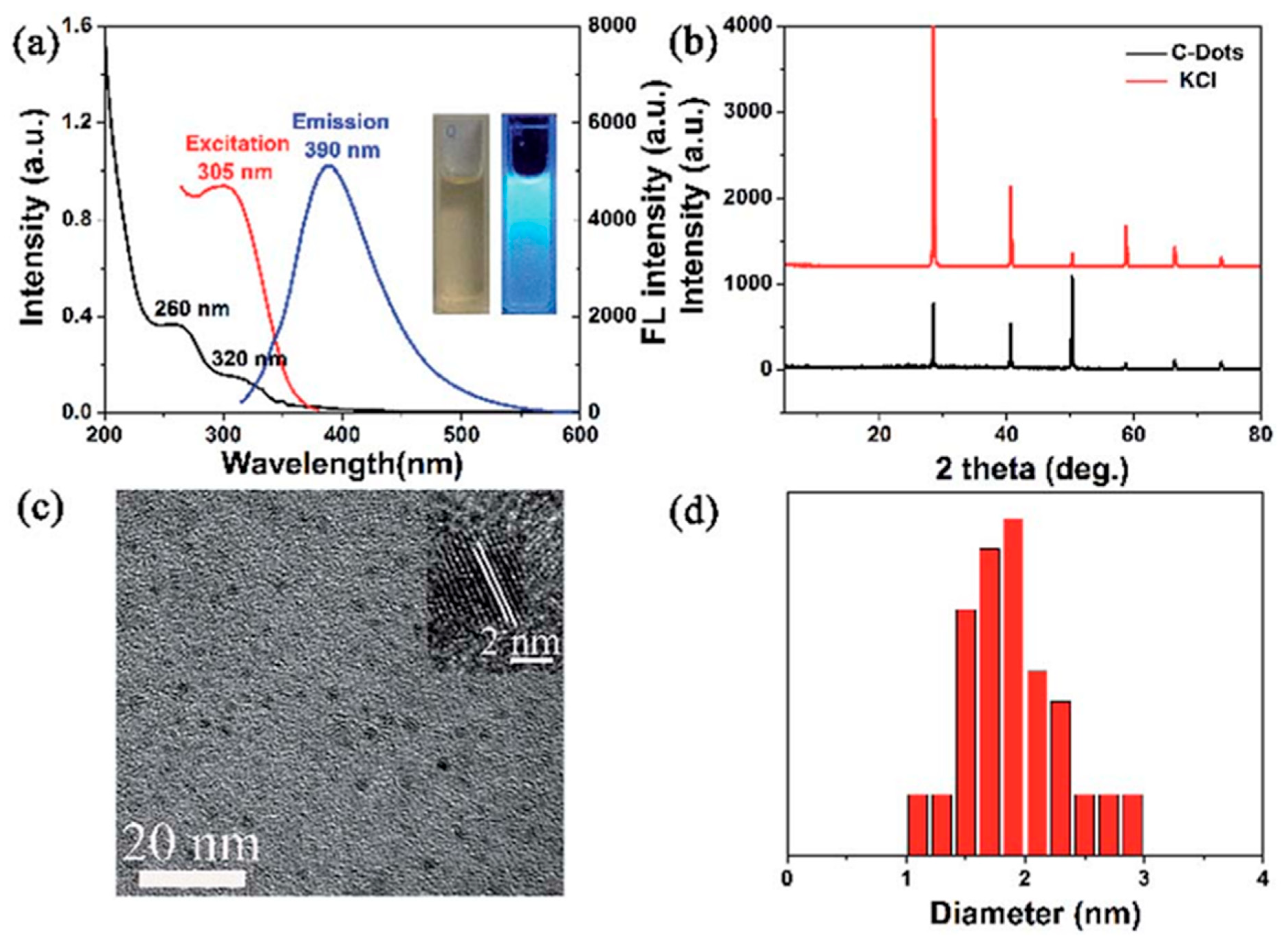
Other parameters of the reaction, i.e., the pH of the reaction (stage 2, Figure 1), can be optimized within the range of 2 to 11. Adjusting these pH values has been shown to influence the morphology of the resulting CDs. In the third stage (Figure 1), the transfer of the extract to the autoclave and optimization of the hydrothermal treatment temperature have been widely explored by researchers. The filling of the inner Teflon cylindrical vessel with the extract, also referred to as the autoclave filling factor, has been reported to range from 50% to 80%, with hydrothermal treatment temperatures in the range of 120–280 °C and reaction times of 2 to 36 h. At the end of the process, the autoclave is allowed to cool naturally to room temperature. The change in the solution’s color indicates the successful formation of CDs. The as-synthesized CDs are purified using centrifugation at speeds of 1000–16,000 rpm for 10–20 min, effectively removing larger or agglomerated particles (residues). Further purification can be achieved by dialyzing the obtained solution with distilled water using a dialysis membrane, with durations ranging from 10 min to 48 h. With these modifications and optimization steps, researchers have successfully synthesized various CDs (Figure 1). In one study, Huang et al. synthesized CDs using sugarcane molasses as a carbon source via a simple and environmentally friendly hydrothermal method. The UV-vis absorption spectra, fluorescence spectra, XRD patterns, and HRTEM images are presented in Figure 2. In the UV-vis absorption spectrum, two distinct absorption peaks at 260 nm and 320 nm were observed, along with an emission peak at 390 nm. The HRTEM results confirmed the formation of spherical C-dots with an average diameter of 1.9 nm. The XRD analysis revealed that the CDs possessed a low-carbon lattice structure. The XPS results confirmed that carbon was the major component, along with oxygen and trace amounts of potassium, calcium, and silicon. Furthermore, these CDs were utilized to sense Fe³⁺ ions by detecting changes in fluorescence intensity in the presence of Fe³⁺ ions.

3.2. Microwave-Assisted Synthesis of CDs

Microwave-assisted green synthesis of CDs is an inexpensive and one-step method that has been widely described by many researchers. In this approach, green precursors derived from various plant parts, such as fruit, juice, seeds, roots, and stems, are utilized to eliminate the need for high-acidity reaction precursors, elevated temperatures, and prolonged reaction times (Figure 3). The primary stages involved in microwave-assisted green synthesis include the selection of green carbon sources, preparation of plant extracts, microwave treatment, and subsequent purification steps, such as centrifugation, dialysis, and sonication. This method is highly energy-efficient and operates under high-temperature and high-pressure conditions [75]. Microwave-assisted fabrication is quick, easy, economical, and environmentally friendly. Moreover, it provides high yields, minimal impurities, precise size control, enhanced safety, greater reproducibility, and superior experimental parameter control. These benefits are attributed to the uniform and selective distribution of microwave radiation throughout the sample during the manufacturing of CDs [76]. A significant advantage of microwave heating is its contactless heat transfer mechanism, which enables reactions to occur rapidly and uniformly [77].
Many researchers have used microwave-assisted green synthesis of CDs by optimizing various synthesis parameters, such as the type of green precursor, reaction temperature and pH, microwave irradiation temperature and time, and centrifugation conditions (Table 2). In 2021, Hu et al. reported a simple and effective method for the synthesis of CDs using orange peel as the carbon source. Fully cleaned orange peels were naturally dried and ground into a fine powder [78]. The orange peel was mixed in 10 mL of ethylene glycol. Without establishing the pH of the solution, the mixture was heated in a household microwave oven for 1 min. The resulting brown solution was purified by centrifugation at 15,000 rpm, yielding CDs with sizes ranging from 3 to 5 nm. Similar work, with greater detail, has been reported by other researchers using various green precursors, such as mushrooms, tapioca flour, tissue paper, silk fibroin, fresh ripe pomegranate, and watermelon peels. Different molar ratios of these precursors were used. Following this, several researchers adjusted the pH of the reaction solution within the range of 1 to 10, finding this step to be critical for controlling the morphology of the CDs. In subsequent steps, different microwave treatment temperatures and time intervals were optimized to synthesize CDs with enhanced properties. Once the CDs were synthesized, purification was performed to remove impurities. In 2019, Basoglu et al. reported the synthesis of CDs using roasted chickpeas, where they employed centrifugation to remove aggregated particles, further refining the quality of the CDs [79]. Similarly, in 2020, Suhail et al. synthesized CDs using carrageenan as a carbon precursor. Before centrifugation, the synthesized CDs were subjected to sonication for 15 min to improve the uniformity and dispersion of the particles [80]. Various researchers have subsequently purified CDs through dialysis treatment, lasting 8 to 24 h, using a dialysis membrane. In 2020, Dager et al. synthesized CDs using fenugreek seeds as a carbon-based green precursor via a single-step microwave plasma-enhanced decomposition (MPED) process [81]. They introduced hydrogen into the MPED chamber at a constant flow rate of 30 standard cubic centimeters per minute (sccm) for five minutes while maintaining the chamber pressure at thirty Pascals. It was observed that the synthesis of CDs via MPED was 97.2% faster than the conventional thermal decomposition process. These process modifications resulted in CDs with varying morphologies and sizes.
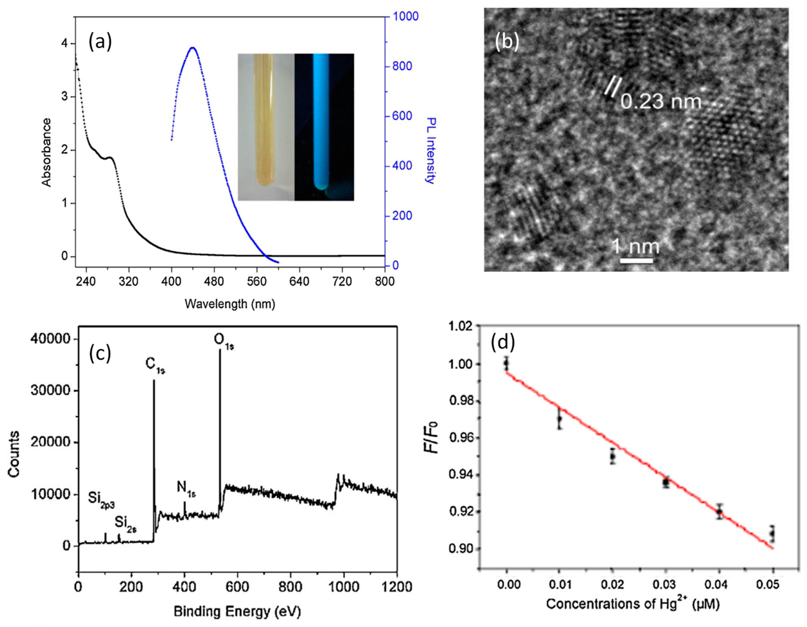
Qin et al. [86] prepared CDs using flour as carbon source with the help of a microwave-assisted rapid green synthesis method. CDs were successfully synthesized with diameters in the range of 1–4 nm. CDs exhibited high sensitivity and selectivity toward Hg2+ with a detection limit as low as 0.5 nm and a linear range of 0.0005–0.01 M [86]. The absorbance and emission spectra of the prepared CDs, HRTEM images, XPS spectra, and changes in the relative PL intensity (F/F0) of CDs with Hg2+ ions under the same conditions are shown in Figure 4.

3.3. Pyrolysis Treatment Assisted Synthesis of CDs

The term pyrolysis comes from two Greek words: ‘pyro’, meaning fire, and ‘lysis’, which means disintegration into integral parts. This process was discovered by Jayme Navarro, founder of Poly-Green Technology and Resources, when he was converting plastic waste into fuel. Pyrolysis is an endothermic process in which a raw material is exposed to high temperatures, and in the absence of oxygen, goes through chemical and physical separation into different molecules [99]. Pyrolysis treatment is a robust and low-cost approach to synthesizing CDs using green carbon-based precursors. The following are some of the main advantages of pyrolysis: It is a straightforward, low-cost technique that can handle a large range of feedstocks. It lowers greenhouse gas emissions and garbage that ends up in landfills. Finally, it lowers the possibility of water contamination [100].
In general, the carbon source is gradually transformed into CDs through processes such as heating, dehydration, degradation, and carbonization under high temperatures, either in a vacuum or inert atmospheres. This is followed by purification steps involving ultrasonication, centrifugation, and dialysis. However, this process often requires high-concentration acids or alkalis to break down carbon precursors into nanoparticles. Additionally, by altering the conditions of pyrolysis, such as the temperature, duration, and pH of the reaction system, the properties of the resulting CDs can be regulated [101].
An advantageous feature of this process is that it typically does not involve other reagents, such as oxygen (O2; combustion process) or water (H2O; hydrolysis process), and it can withstand high pressures.
The synthesis of CDs using pyrolysis treatment has been reported by many researchers employing green carbon-based precursors. In 2014, Teng et al. described the pyrolysis-assisted synthesis of CDs using a green extract of konjac flour. In this process, konjac flour was pyrolyzed at 470 °C for 90 min under air [102]. The resulting carbonized black solid was ground into a fine powder and mixed with 20 mL of ethanol. After continuous stirring overnight, the CDs were extracted. These CDs were then dispersed in water and filtered through a filter membrane to remove residual inorganic salts. The obtained CDs were found to have an average size of 3.37 nm.
Similarly, various researchers have synthesized CDs using different carbon-based precursors and optimized curing temperatures and times (Table 3). Further modifications involved optimizing the pH of the reaction and applying sonication to achieve a homogeneous dark brown solution after pyrolysis treatment, followed by purification through centrifugation. In some cases, ultra-purification was performed by dialyzing the solution using a dialysis membrane to remove agglomerated particles. Using pyrolysis treatment (Figure 5), researchers can control the size of the CDs by varying synthesis parameters.
In one report, Tripathi et al. synthesized water-soluble carbon dots (wsCDs) from kidney beans as a green precursor via a facile pyrolysis technique. These wsCDs exhibited good stability, high fluorescence, and tunable photoluminescence. Moreover, the wsCDs demonstrated excellent photostability, even in high-ionic-strength environments. These wsCDs were further utilized as a fluorescent probe for the multicolor imaging of HeLa cells. Under a fluorescence microscope, the HeLa cells showed bright green and red fluorescence when observed under bandpass filters of 488 nm and 561 nm, respectively (Figure 6), indicating the good cell permeability of the wsCDs in HeLa cells [103].
Table 3. Pyrolysis-assisted synthesis of CDs.
Precursor TypePrecursors UsedMolar Mass/Molar RatioCrystal Size/Particle Size (nm)Optimum ParametersApplicationsRef.
Waste polyolefinsMM 0.033 g/mLPS 70PT120 °C for 12 h,
S-700 W for 2 h and
Dr-72 h
Sensing and live cell imaging[104]
Tea and peanut shellsMR 0.6PS 7–9PT 200 °C for 4 h,
S-15 min, and
Dr-24 h
Biomarkers, ion detection, and photocatalysis[105]
Peanut shellsMM 0.00025 g/mLPS 4.26PT 200 °C for 15 min,
Dr-24 h, and
P-65 °C for 22 h
Sensing[106]
Tea leaf residueMM 0.2 g/mLPS 2CT 350 °C for 2 h,
Cen-4000 rpm, and
pH-7
Bioimaging[107]
Biomass residueMM 0.2 g/mL PT 300 °C to 500 °C for 5 °C/min, and
Cen-19,000 rpm
Efficient surfactants[108]
Durian peel wasteMM 0.1 g/mL PT-250 °C for 5 h,
S-30 min, and
C-10,000 rpm for 15 min
Supercapacitor[109]
Gram peel--5.5 for C1
20 nm for C2
PT 200 °C and 450 °C for 8 h Ultrafast response humidity sensor[110]
Mango peelsMM 0.03 g/mLPS 3CT 300 °C for 2 h Detection of ferrous succinate,
biological imaging
[111]
VegetablesAllium sativum (garlic)MM 0.1 g/mLPS 2PT 315 °C for 3 h
and S-15 min
Photobleaching, solar conversion, and in vitro cell imaging[112]
Zingiberis rhizome--CS 0.2PT 300, 350, and 400 °C for 0.5, 1, and 1.5 h,
Dr-72 h, and
Cen-11,000 rpm for 30 min
Drug delivery[113]
BiomassKonjac flourMM 0.05 g/mLPS 3.37PT 470 °C for 1.5 hBioimaging[102]
Finger millet ragi (Eleusine Coracana)MM 0.01 g/mLPS 6PT 80 °C for 5 h
T-300 °C for 5 °C /min
Detection of Cu2+ ions[114]
SeedKidney beans--PS 20–30PT 450 °C for 2 h
and pH-7
Biological cell imaging[103]
Fennel seeds--PS 0.22PT500 °C for 3 h,
S-5 min, and
Cen-15,000 rpm for 10 min
LED, bio-sensing, and cellular imaging[115]
LeafPlant leafMM 001 g/mLPS 3.7PT 250, 300, 350 and 400 °C for 2 h at 5 °C/min and
Cen-12,000 rpm for 10 min
Coding, bioimaging, and drug delivery[116]
Prosopis juliflora leaves--PS 5.8 PT 200 °C for 30 min followed by grinding to powder and heating at 200 °C for about 1 h.Sensitive, selective, label-free, and reproducible off–on sensing assay[117]
PlantCottonMM 0.1 g/mLPS 4.9PT 300 °C for 2 h,
Cen-4000 rpm for 10 min, and
Dr-2 days
Multi-color imaging, patterning, and sensing[118]
FruitMalus domestica (apple)--PS 3PT 300 °C for 1 h,
S-10 min,
Cen-8000 rpm for 5 min, and
Dr-24 h
Biosensing and cell imaging[119]
FoodChicken egg--PS 2.15PT 230 °C for 19 minPrinting ink[120]
Raw materialRaw whey20 mLPS 4PT180–225 °C for 10 to 40 minPhotocatalysis, biosensing, and drug delivery[121]
MM—molar mass, MR—molar ratio, PT—pyrolysis treatment, Dr—drying temperature, Cen—centrifugation.

3.4. Carbonization-Assisted Synthesis of CDs

The process of converting complex organic materials, such as carbon-rich plants and residues of dead animals, into carbon through destructive distillation is known as carbonization. This is a complex process in which multiple reactions, including dehydration, condensation, hydrogen transfer, and isomerization, occur simultaneously. It is an efficient, time-saving, and environmentally friendly method commonly used for the synthesis of CDs.
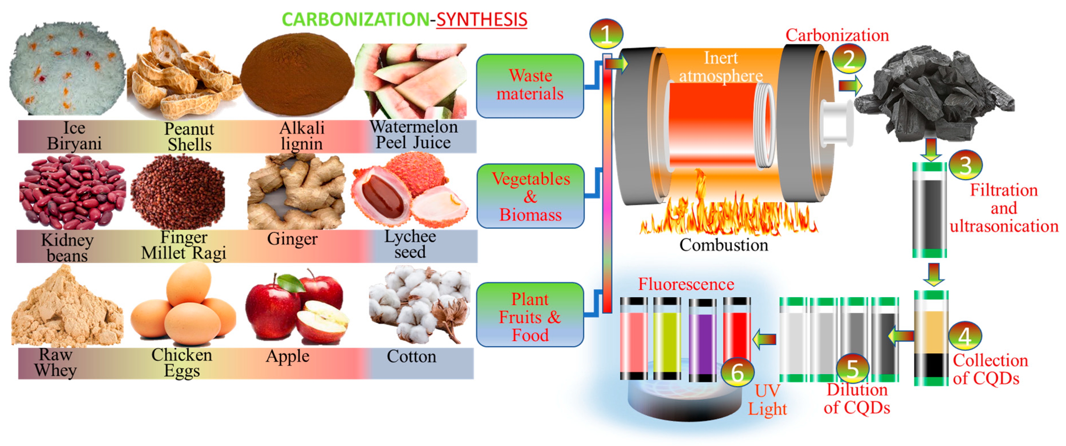
The main phases of this process include washing, drying, carbonization at controlled temperatures, adjusting the pH of the reaction, sonication, centrifugation, and dialysis. Various carbon-based green precursors have been utilized for the synthesis of CDs, including lychee seeds, alkali lignin, rice biryani, peanut peel, melon peel, water hyacinth, mango, and lychee peel (Table 4).
The synthesis of CDs using carbonization treatment (Figure 7) has been reported by various researchers employing carbon-based green precursors. In 2015, Xue et al. utilized lychee seeds as a green carbon source for CD synthesis. The lychee seeds were placed in a ceramic crucible and carbonized at 300 °C. The resulting dark product was cooled to room temperature and ground into a fine powder. Subsequently, the powder was mixed with pure water, and a black solution was obtained after ultrasonic treatment. This solution was then purified using a filtration membrane to remove larger and aggregated particles. The obtained CDs were 1.12 nm in size and exhibited a spherical morphology [122].
Similar studies have been conducted by various researchers using different optimization parameters and carbon-based green precursors for CD synthesis. In 2018, Jiang et al. used alkali lignin as a precursor for CD synthesis. Following carbonization, the resulting yellow solution was evaporated using a rotary evaporator at 55 °C. In 2019, Deka et al. synthesized CDs from water hyacinth. The washed precursor was first dried in a microwave oven for 48 h and then carbonized in a furnace, gradually heating from room temperature to 160 °C. The carbonized material was subjected to sonication for 30 min and then centrifuged to remove impurities.
To achieve ultra-purification, dialysis was performed for 24–72 h by many researchers using various carbon-based precursors. These optimized protocols and purification steps significantly enhanced the quality and characteristics of CDs.
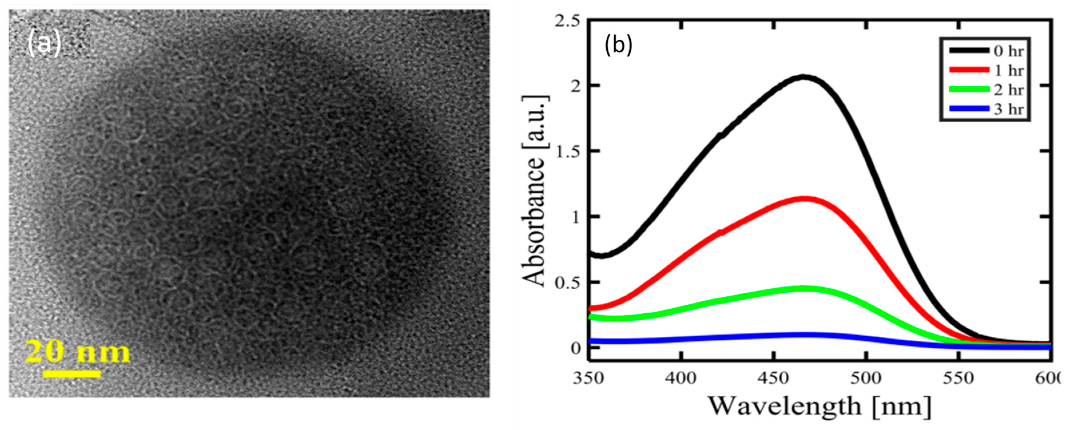
Kavitha et al. synthesized mesoporous CDs from date palm fronds using a simple, green, one-step carbonization process. These CDs exhibited excellent excitation wavelength-independent photoluminescence (PL), along with high photo- and storage-stability, superior biocompatibility, and remarkable thermal and electrical conductivity.
Moreover, the CDs demonstrated light-activated biocidal functions, as evidenced by their enhanced photocatalytic activity toward methyl orange (MO) degradation under sunlight. The HRTEM images of the as-synthesized CDs and UV–visible spectra of the MO solution in the presence of CDs at different UV irradiation times are shown in Figure 8 [123].
Table 4. Carbonization-assisted synthesis of CDs.
Precursor TypePrecursorsM Mass/M RatioCS, PS Size (nm)Optimum ParametersApplicationsReference
SeedLychee seedMM 0.01 g/mLPS 1.12CT 300 °C for 2 h at 10 °C min−1Microbiology, surgery, and in the diagnostic field[122]
Waste materialAlkali lignin11.8 gPS 8CT 300 °C for 30 min
and RE-55 °C
Biomedicine[124]
Ice-biryani1 gPS 41CT 250 °C for 24 h,
ST-1200 rpm for 36 h, and
pH 1–10
Bioimaging[125]
Peanut shellMM-0.1 g/mLPS 1.62CT 250 °C for 2 h at 10 °C min−1, and pH 3–12Cell imaging[126]
Lorhange--PS 5.72CT 220 °C for 2 h,
Cen-18,000 rpm for 20 min, and
Dr-48 h
Cell imaging[127]
Fresh oranges and lemons peels--PS 6.5 and 4.5 nm for citrus sinensis and citrus limonCT 180 °C for 2 h,
Cen-8000 rpm for 5 min
Iron and tartrazine sensing and cell imaging[128]
LeafWater hyacinth--PS 5.22MT-48 h
CT 160 °C at 10 °C min−1, S-30 min, and
pH 7
Sensors[129]
FruitsDate palm --PS 50 nmCT 300 °C for couple of hours at a heating rate of 10 °C/min--[123]
MangoMM-0.1 g/mL S-5 min
CT 100 °C for 60 min, and
72 h
Bioimaging[130]
Litchi peelMM-0.0033 g/mLPS 3.1CT 140 °C for 12 h,
24 h, and
Cen-16,000 rpm for 15 min
Colorimetric determination of ascorbic acid[131]
MM—molar mass, CT—carbonization treatment, Dr—drying temperature, Cen—centrifugation.

3.5. Laser Ablation-Assisted Synthesis of CDs

Laser ablation is a novel and promising synthesis method that has been used to create CDs because of its quick operating time and ease of use. Sun et al. initially showed how to synthesize GQDs from graphite using laser ablation [8]. Li et al. used quick laser passivation of carbon particles to create GQDs with visible, stable, and adjustable PL performance. They also showed that passivation by laser irradiation significantly affected the origin of PL [132]. Among the many benefits of laser ablation are its versatility in creating different types of nanostructures and its ease of use. However, this method requires a significant amount of carbon materials to satisfy the carbon mark [133]. There are many different sizes of carbon nanostructures created by laser radiation, and large particles may be effortlessly separated by centrifugation, resulting in the efficient use of carbon materials and carbon nanoparticles [10]. Calabro et al. generated graphene quantum dots by chemically oxidizing carbon nano-onions and synthesizing quantum carbon. The laser ablation approach appeared to have a blue-shifted emission in comparison with the chemical oxidation approach that they mentioned in the particle size and surface functional group effects, which the researchers found after comparing the photoluminescence spectra of the two quantum dots. The scientists came to the conclusion that liquid-form laser ablation develops quantum dots in a single step that is safer, quicker, and requires fewer starting chemicals and residues than chemical oxidation [134].

3.6. Ultrasonic Assisted Synthesis of CDs

A lot of literature has linked the fabrication of carbon dots to using ultrasonic technology as it was discovered to be an effective way to generate different kinds of carbon dots. This method breaks down carbon particles into extremely small nanoparticles by subjecting carbon precursors, acid, alkali, and other oxidants to strong ultrasonic vibrations. The molecules continuously cavitate. The simple synthesis of CDs with a small size is made possible by the application of high-intensity ultrasonic waves, which circumvent the difficult post-treatment procedure [135]. Li et al. synthesized a fluorescent carbon dot and demonstrated its water-solubility in an extremely thorough study. In the same year, they employed activated carbon and an ultrasonic treatment method with one-step H2O2 assistance. The TEM results revealed an observed size range of 5–10 nm and a surface rich in hydroxyl groups on the produced carbon dots [136]. The sonochemical production of carbon quantum dots (CQDs) and a full description of their electrical applications were published by Kumar et al. Employing the sonochemical approach, Pan et al. synthesized fluorescent CDs and used them for nutritional monitoring [137]. Fluorescent CDs functionalized with thiol-terminated polyethylene glycol were synthesized using the ultrasonication process, as stated by Huang et al. In this instance, the dispersibility of the CDs in the water phase was enhanced as a result of the integration of a hydrophobic PEG group. Additionally, it made the produced CDS more biocompatible [138]. It is beneficial to scale up the manner by using a straightforward and eco-friendly technique instead of using powerful acids. Employing H2O2 as an oxidant and anthracite and bituminous coal as a precursor, Saikia et al. constructed CQD using a one-pot ultrasonication process [139]. Because of the many oxygen-containing surface functions, the produced CQDs, which ranged in diameter from 2 to 12 nm, had hydrophilic particles. This process offers a green synthesis method for the mass production of CDs using plentiful precursor coal.

4. Structure and Properties of Carbon Dots

4.1. Structure of Carbon Dots

CDs are regarded as 0-D nanomaterials with sizes smaller than 10 nm. Their chemical structure is typically governed by the synthesis route employed. In general, CDs possess an amorphous carbonaceous core. However, recent studies indicate that selective starting materials and specific reaction conditions can result in partial graphitic crystallinity within the CD structures [140,141,142]. These crystalline CDs exhibit broad D- and G-bands in Raman spectrometry, with a lower D/G ratio signifying the vibration of sp2 carbon atoms. Raman spectra of CDs typically show two first-order bands: the D-band and the G-band. The D-band represents the vibration of carbon atoms in disordered graphite or glassy carbon, while the G-band indicates the vibration of sp2-hybridized carbon atoms.
The ratio of the intensities of the D-band to the G-band (D/G ratio) serves as an indicator of the degree of purity or graphitization of CDs. A high D/G ratio indicates an amorphous structure, whereas a lower D/G ratio reflects a higher degree of graphitization. The crystalline nature of CDs can be broadly classified into two categories: (i) a graphitic crystalline core and (ii) a high degree of non-graphitic crystallinity.

4.2. Luminescence Properties

The distinctive and remarkable fluorescence properties of CDs have garnered significant interest. However, the mechanism underlying carbon dot fluorescence remains a topic of debate among scientists. Currently, there are three primary hypotheses that explain the luminescence mechanism of CDs [143]. The first hypothesis attributes the fluorescence of CDs to the quantum size effect, which is linked to the size-dependent behavior of their carbon nuclei. Fluorescence arises due to changes in quantum confinement as the particle size decreases. The second hypothesis focuses on the surface defect states, suggesting that the presence of various functional groups on the surface of CDs creates defect states that influence their optical properties. The third hypothesis proposes that the fluorescence is induced by the extended π-bond conjugated structure within the CDs, which facilitates efficient light absorption and emission.

4.3. Optical Properties

4.3.1. UV-Absorption Properties

CDs exhibit strong and broad absorption bands in the ultraviolet (UV) region within the wavelength range of 220–280 nm. These peaks are generally attributed to the π-π transitions arising from the aromatic C=C framework. Additionally, peaks observed in the wavelength range of 280–350 nm are associated with n-π transitions of C-O and C=O bonds. Occasionally, an extended absorption tail into the visible and near-infrared (NIR) regions is observed, which can be attributed to the transitions of functional groups on the surfaces of CDs, resulting in smaller electronic bandgaps. Furthermore, the position of these absorption peaks is influenced by the degree of surface oxidation of the CDs [144,145,146].

4.3.2. Emission Property

In recent times, CDs have been emerging as a new class of “nano lights” due to their fascinating optical properties, wavelength-tunable emission, and excellent photostability. As a result, studying the photoluminescence (PL) properties of CDs and their diverse applications has become a prominent area of interest. CDs exhibit highly attractive PL properties, and it has been observed that CDs synthesized through different methods and materials display diverse compositions and chemical structures, resulting in varied emission properties. Typically, the PL of CDs is attributed to the quantum size effect, surface states, and molecular states. However, the exact PL mechanism of CDs remains unclear and requires further detailed investigation. The most widely accepted models for explaining the PL of CDs are core emission, originating from the conjugated π-domains of the carbon core, or the quantum confinement effect. It is well known that the energy gap of CDs is influenced by particle size and shape, which ultimately impacts their PL properties. Another critical factor for analyzing the PL mechanism of CDs is the surface state. Due to the diversity and uncertainty of molecular structures, accurately defining surface states is challenging. The surface of CDs is populated with various oxygen-related groups (e.g., –COOH, –OH, and C–O–C), hanging bonds, and hybrid carbon atoms (sp2 or sp3), which act as trap states and significantly influence their optical properties. In addition to surface states, the molecular state, where emissions originate from free or bonded fluorescent molecules, also plays a crucial role in determining the emission properties of CDs.
It is interesting to note that CDs exhibit excitation-dependent emissions in terms of both wavelength and intensity, which violates the Kasha–Vavilov rule. Jiang et al. reported that, upon excitation, carbon dots typically display two trends in their fluorescence spectra depending on the excitation wavelength range: an excitation-independent component in the range of 275–400 nm and an excitation-dependent component in the range of 425–550 nm [147]. These findings clearly indicate the presence of two distinct photoluminescence (PL) mechanisms in CDs [148]. The observed PL emission spectra of CDs are usually broad and symmetrical on the wavelength scale. Moreover, factors such as excitation wavelength, metal ions, solvent, pH, size, heteroatom doping, and other surface modifications significantly influence the emission spectrum of CDs. In addition to their characteristic PL emission, some CDs exhibit up-converted photoluminescence (UCPL). UCPL refers to a process in which low-energy photons are converted into high-energy photons, resulting in the emission of shorter wavelengths compared with the excitation wavelength through consecutive interactions within the medium. This unique up-converted PL property makes CDs promising candidates for applications in sensing, catalysis, and bioimaging fields.

4.3.3. Toxicity

CDs have a lot of promise for use in a variety of bio-applications because of their distinctive optical characteristics, high surface area, and surface functionality. As potential industrial and biological uses for CDs have been investigated, CD biosafety concerns have progressively come to light. To assess the toxicity of CDs, there are two primary approaches. The first is in vitro assessment, which often uses specific assays like MTT, CCK-8, WST-1, etc., to evaluate for cell viability [149]. Both the control and experimental populations are cells that have been grown with or without CDs. Then, by contrasting these two groups, the toxicity of CDs may be evaluated. These investigations often include an additional blank group connected to the external environment to exclude the influence of the culture dish and the chosen assays. The second kind is called in vivo evaluation, and it involves injecting a CD solution directly into mice and zebrafish through their veins or tails [150]. The toxicity and distribution of carboxylate CDs in mice were initially investigated by Lee et al. Blood biochemical, hematological, and inflammatory analyses for the liver, kidney, spleen, heart, or lung were carried out a certain amount of time after treatment. The findings indicated that neither organisms nor bodily tissues were negatively impacted by CDs. Fluorescence imaging may be used to investigate how CDs circulate throughout the body, first entering the bloodstream, then moving on to the kidneys and spleen, and finally exiting the body through the kidney filter [151].

4.3.4. Biocatalyst

Apart from its exceptional optical qualities and little toxicity, catalysis has also grown in popularity among chemists. Nowadays, CDs are a well-established instrument that are frequently used to design and develop new catalytic processes, such as bio-catalysis, artificial photosynthesis, organic transformations, etc. [152]. Amongst them, the enzyme-assisted biocatalytic transformation has drawn increasing interest and is a crucial regulating mechanism in human life activities and medical intervention. Disease prevention, detection, and treatment can be aided by tracking the biocatalytic transition between various enzymes and chemicals. CDs are believed to offer sensitive driving of enzyme-assisted biocatalytic processes by monitoring the expression levels of pertinent metabolites due to their special characteristics [153]. Meng et al. combined CDs with Au nanoclusters, taking advantage of the easy-to-modify characteristics of CDs. This enhanced the horseradish peroxidase activity of Au nanoclusters while preserving the superoxide dismutase (SOD)-like activity of CDs [154].

5. Applications

5.1. Catalysis

5.1.1. Photocatalysis

CDs have been recorded as agile materials in photocatalytic applications due to their absorption and electron transfer properties and ease of coupling with other materials [155]. The advantageous feature of CDs is their ability to be used alone in a pure form to deteriorate environmental pollutants [156]. Typically, the operation of CD-based photocatalysis goes through three stages, which are as follows: (a.) production of electron/proton pairs by absorption of light, (b.) formation of reactive species for the separation and transfer of electron/proton pairs, and (c.) the production of reactive species, giving rise to suitable photocatalytic reactions [157].
In 2016, Tyagi et al. [44] synthesized CDs with sizes of 1–3 nm through hydrothermal treatment, which can be used for the photocatalytic degradation of MB under UV light irradiation. In their study, they compared TiO2-wsCDs composite with TiO2 nanofibers and stated that, due to better charge separation at the interface, the photocatalytic activity of the TiO2-wsCDs composite was ~2.5 times higher than that of TiO2 nanofibers. Furthermore, the load ability of both was tested in an air environment from room temperature to 600 °C and it was found that TiO2 nanofibers lost ~2% weight when submerged in water while TiO2-wsCDs lost ~5.7% of their total weight overall. From their study, they found an effective loading of 3.7% of TiO2-wsCDs composite on TiO2 nanofibers. They also recorded the emission peak upon excitation with 370 nm light for both samples. From this, they concluded that the photocatalytic degradation of MB with the TiO2-wsCDs composite enhanced the photocatalytic activity by increasing the catalytic location at the interface of the TiO2-wsCMDs composite.
In 2019, Sargin et al. [90] synthesized CDs with a size smaller than 20 nm through microwave treatment. These as-synthesized CDs with sensitized TiO2 were used as a photocatalytic hydrogen experiment, with a total volume of 135 mL in Pyrex flasks. They could be radiated using a 300 W Xe lamp with a wavelength of ≥420 nm that was filtered under constant stirring. They further tested the pH of the reaction between 7 and 10 and also found an optimum pH value of 9 for hydrogen evolution. Comparing the hydrogen performance in the presence of a Pt co-catalyst, the hydrogen amount was recorded to be 1458 μmol g−1 and in the absence of a Pt co-catalyst, it was 472 μmol g−1, from which they observed that the use of Pt co-catalyst resulted in an increase in the rate of hydrogen evolution. TiO2/CD/Pt composite screening was also reusable for photocatalytic hydrogen development. In 2020, Genc et al. [97] reported an unambiguous synthesis of CDs with sizes of 15–20 nm using microwave irradiation of Ginkgo biloba. They performed the photocatalytic hydrogen evolution reactions (HERs) in triethanolamine (TEOA) under visible light. They used TiO2 as a proton reduction photocatalyst, Pt as a co-catalyst, and CDs as absorbers. For the best photocatalytic activity, they optimized the ratio to 4 mL CDs/0.4 gm TiO2 and pH to 9. It was observed that the amount of hydrogen decreased under a basic pH due to the protonation of the catalyst and also under an acidic pH due to the protonation of TEOA. They then studied the HER quality of the CDs/TiO2 and CDs/TiO2/Pt composites from 1 to 8 h and observed that the photocatalytic hydrogen production increased linearly under visible light irradiation. Furthermore, they also tested the reusability of the CDs/TiO2 composite up to the fourth cycle and found that it can be reused without any reducing agent. The hydrogen production mechanism is related to the conduction band levels of TiO2, CODs, and Pt. In this process, CDs first absorb visible light and transfer the photoexcited electron to the lowest molecule atomic orbital (LUMO) level. These photoexcited electrons can be easily transferred from the LUMO level to the conduction level due to the presence of more negative conduction band levels of TiO2. Water is reduced to photoexcited electrons to evolve the hydrogen gas on the conduction band level of TiO2 without the addition of any co-catalyst to it. Upon adding the co-catalyst (Pt), the photoexcited electrons are transferred from the TiO2 surface to the Pt surface and hydrogen gas evolves on the Pt surface. The regeneration of the systems is achieved by the TEOA (known as the whole scavenger).

5.1.2. Other Catalysis

In 2015, a specific green and environmentally friendly synthesis approach for CDs was reported by Zhang et al. [62] using bee pollen as a precursor, with hydrothermal treatment and a size of 1–2 nm. These as-synthesized CDs had a high quantum yield, due to which they were used as homogeneous catalysts toward the reduction of noble metal ions in aqueous solution. They also recognized the role of the catalysis of CDs in controlling the set of sunshine-irradiated AgNO3/HAuCl4 solutions in the sole presence of CDs or sodium citrate, which led to a reduction in the emission intensity of CDs. This catalytic effect is based on the discovery of a turn-on fluorescent sensor for cyanide ions that is still under investigation.

5.2. Sensors

Sensors are revolutionary devices widely used to detect and respond to electrical or chemical signals. The term sensor was introduced by an American physicist and engineer, Eric R. Fossum, to sense physical parameters (temperature, blood pressure, humidity, speed, etc.) and convert them into electrical signals [58]. CDs have immense sensor applications due to their very small size, high surface-to-volume ratio, and high reactivity.

5.2.1. Metal Ion Sensors

Due to high sensitivity and selectivity, metal ions such as Ag+, Ba2+, Ca2+, Cd2+, Co2+, Fe3+, Hg2+, Mg2+, Mn2+, Ni2+, Pb2+, and Zn2+ are used for sensor applications [10]. Various metal ion sensors using CDs are described in this section.
(i) Iron (Fe3+) ion sensors: In 2019, Carvalho et al. [35] synthesized CDs with different sizes using hydrothermal treatment of acerola fruit, which were used for the detection of Fe3+ ions. The phenomenon of fluorescence quenching was used for the detection of Fe3+ ions. To indicate that sensing was sensitive to Fe3+ ions, there was a decrease in the fluorescence intensity with an increase in the concentration of Fe3+ ions. In 2016, Bandi et al. [41] synthesized CDs with a size of 15 nm using hydrothermal treatment of onion waste, which were also used for the detection of Fe3+ ions. With the drastic decrease in the fluorescence intensity of CDs due to Fe3+ ions, they are used as highly selective fluorescent probes for Fe3+ ions. Furthermore, there was a steady decrease in fluorescence intensity with increasing concentrations of Fe3+ ions, which suggests that they were sensitive to Fe3+ ions. These CDs also had a good response to Fe3+ ions in the pH range of 4–8.
In 2017, Atchudan et al. synthesized CDs with a size of 5 nm using hydrothermal treatment of Chionanthus retusus fruit extract as a carbon source [32]. It is incredible to note that, among all the metal ions, Fe3+ ions have quenching fluorescence intensity at concentrations between 0 and 500 μM. They also have outstanding selectivity, which is not the case for other metal ions. If the plot is not a straight line at a lower concentration and the lifetime of the free probe and its complex are the same, then it is static quenching, and if the plot is curved at a high concentration and the complexes are different, then energy transfer occurs through collision and/or close interactions of π-π overlap in the excited state via resonance energy transfer, which is known as dynamic quenching. Due to this property, they are widely used for the selective quenching of Fe3+ ions from industrial waste. These Fe3+ ions have high sensitivity and high selectivity, which makes the synthesized CDs ideal fluorescent probes for the highly selective detection of Fe3+ ions. They also demonstrated the effect of Fe3+ on the UV-vis spectra of synthesized CDs, which were different before and after the addition of Fe3+. In the presence of Fe3+, the absorption peaks disappeared at 269 and 301 nm, which could influence the surface state of CDs. This result reveals that the present sensor has outstanding selectivity toward the Fe3+ sensing. In 2017, Huang et al. used sugarcane molasses for the synthesis of CDs with sizes of 1.2–3.8 nm [43]. In their study, the phenolic hydroxyl groups present on the surface of CDs were interacted with Fe3+ ions, which resulted in a decrease in the fluorescence intensity of CDs. It was observed that, at 700 μM of Fe3+ ions, the fluorescence intensity was almost quenched. There was a good linear relationship observed in the range of 0–100 μM. They also calculated the lowest detection limit (LOD), which was 1.46 μM based on the standard deviation (σ) of the blank signal (n = 3).
Similarly, in 2015, Sachdev et al. reported the green synthesis of CDs with a size of 2.387 nm using hydrothermal treatment of coriander leaves for the detection of highly selective and sensitive Fe3+ ions [30]. They observed the sensitivity of CDs toward Fe3+ ions with different concentrations in the range of 0–60 μM, which showed that, with increasing Fe3+ concentration, there was a steady decrease in the fluorescence intensity. They also described the quenching of fluorescence of CDs in the presence of Fe3+ ions, providing a platform for their quantitative sensing by monitoring the fluorescence emission intensity of CDs.
(ii) Copper (Cu2+) ion sensors: Although copper ions play an important role in oxidative metabolism, their high levels have high toxicity in some organisms [158]. The most advantageous feature of copper is that they are highly important for the functioning of the brain due to the high consumption of oxygen in the body relative to its size [159]. In 2019, Murugan et al. reported the facile, rapid, and frugal synthesis of CDs using pyrolysis of Finger millet ragi (Eleusine coracana) which is 6 nm in size [114]. In their study, they illustrated the fact that all metal ions describe remarkable properties of selective fluorescence intensities in Cu2+ ions by electron or energy transfer. Also, as-synthesized fluorescent CDs can be used as a selective indicator sensor for Cu2+ ions. The sensitivity of CDs to different concentrations of Cu2+ ions from 0 to 100 μM is also evaluated. In addition, they also detect Cu2+ ions in real water samples by fluorescence spectroscopy.
(iii) Arsenic (As3+) ion sensors: Arsenic is a toxic and ubiquitous element found in the environment that is mobilized through a combination of natural processes, such as changes in weather, biological activities, and reactions arising due to volcanic eruptions. In 2018, Ramezani et al. synthesized the CDs using microwave treatment of quince fruit as a carbon source with a size of 4.85 nm. The CDs were used for sensing As3+, along with permanganate ions, due to the potential reactions of As3+ [92]. These synthesized CDs were also used for the determination of As3+ ions using calibration methods, such as extrinsic and tap water.

5.2.2. Biosensors

Biosensors are biological devices widely used to monitor biological or biochemical processes. They were first introduced in 1964 as a glucose oxidase biosensor [160]. In 2016, Yang et al. reported the facile synthesis of CDs with an average size of 4.44 nm using microwave treatment of xylan as a carbon source [95]. The as-synthesized CDs exhibited fluorescence quenching upon interaction with tetracycline (TC) without requiring any further chemical modification. When 50 μM TC was injected into the CD solution, the fluorescence intensity decreased sharply, indicating that the fluorescence of CDs was highly sensitive to TC. Yang et al. also analyzed the quenching efficiency (F0/F) as a function of pH. Here, F0 and F represent the PL intensities of CDs in the absence and presence of TC, respectively. The quenching efficiency remained constant across a pH range of 3–11, suggesting that the fluorescence quenching was influenced by the different hydrion concentrations of CDs, rather than by TC itself. Compared with other biological molecules, tetracycline-based antibiotics such, as tetracycline, chlortetracycline, and oxytetracycline, had a stronger impact on the fluorescence properties of CDs. These findings demonstrate that the as-synthesized CDs, without further chemical modifications, can serve as effective fluorescence probes for detecting TC under optimal conditions. A further practical application was performed under mild conditions, with an incubation temperature of 30 °C in a water bath and an incubation time of 30 min. The results revealed that the PL intensities steadily decreased until reaching equilibrium at 30 min. Under optimum conditions, as the TC concentration increased from 0 to 200 μM, the maximum PL intensity of CDs decreased gradually. A linear relationship was observed between the quenching efficiency (F0/F) and TC concentration in the range of 0.05–20 μM. Additionally, the limit of detection (LOD) was calculated as 3σ/S, where σ represents the slope of the regression equation and S is the standard deviation of the blank solution. For TC, the LOD was determined to be approximately 6.49 nM.
Furthermore, CDs stored for three months were retested using ultrasound treatment under the same conditions as the initial detection experiment. The relative standard deviation (RSD) for five repeated determinations of 5 μM TC was 2.78%. Various advanced techniques, including high-performance liquid chromatography (HPLC), enzyme-linked immunosorbent assay (ELISA), capillary electrophoresis (CE), and liquid chromatography–tandem mass spectrometry (LC-MS), have also been employed for TC detection. The results underscore that CDs are promising fluorescence probes for the detection of TC, combining sensitivity, stability, and ease of use.

5.3. Bioimaging

Analyzing the composition and physiological processes of cells and organisms is made possible by bioimaging. As a result, it is a very dependable instrument in the medical field for diagnosing human illnesses. CD-based optical bioimaging is becoming more and more popular among several bioimaging techniques [161,162]. The method makes use of optical contrast, or the difference in optical characteristics within the imaged spot and the backdrop [163]. In addition to providing rapid screening, diagnosis, and image-guided therapy of critical illnesses like cancer, this technology enables imaging at the cellular and even single-molecule levels. The distinctive optical characteristics of CDs or the functional agents integrated onto their surface or core provide imaging capabilities [164]. The low diffusion of light in cell walls at relatively short wavelengths causes significant background interference in imaging using conventional UV-absorbing and blue/green-emitting CDs. For bioimaging, it is crucial to create CDs with red/near-infrared emission characteristics in order to guarantee sufficient light penetration depth and reduce photodamage to cells [165]. For instance, Gedda et al. studied bioimaging applications by conjugating with folic acid [17]. Employing conventional EDC and Sulpho-NHS coupling chemicals, an amide linkage is made possible by the carboxylic acid group on the surface of CDs being linked with an amino group of folic acid. Water-soluble folic acid specifically targets folate receptors, which are mostly present in the cells of different types of tumors and malignancies. Using fluorescence spectroscopy, zeta potential estimation, and UV-visible absorption, effective FA conjugation with CDs (FACDs) was assessed. Natural compounds with unique light-absorbing qualities can be employed as endogenous contrast agents for PA imaging in organisms [166]. Additionally, by injecting a CD–silica complex into the subcutaneous region of the mice’s dorsal area, good afterglow imaging was obtained [167]. Although autofluorescence interference is eliminated by afterglow imaging, the advancement of this imaging technique is severely constrained by the difficulty of making liquid-phase phosphorescent CDs [168,169].

5.4. Drug Delivery

Drugs are incorporated into nanoparticle carriers by encasing, adsorption, or binding in nanomaterial-based drug delivery, enabling reliable and secure injection throughout the body [170]. Numerous antibacterial or anticancer medications have been effectively administered thus far. CDs are useful carriers for the specific administration of anti-cancer medications for cancer therapy because they are simple to modify [171,172]. As the majority of CD–drug complexes have cleavable chemical bonds, medicines can be released specifically to respond to various triggers or in the acidic environment of tumor locations. CDs can assist cancer cells and other sick cells in escaping their medication tolerance, along with improving delivery and regulating emission at the target spot [173]. The guided diffusion of DOX in the tumor microenvironment via biocompatible CDs has garnered a lot of attention in recent years. In order to promote more tumor accumulation and boost drug-loading capability, the majority of these ground-breaking studies have concentrated on improving the EPR (enhanced penetration and retention) impact. Composites of CDs and DOX have been created by building CDs with a hydrophilic shell and a hydrophobic carbon core. These composites exhibit improved anti-tumor efficacy and considerable tumor targeting, with fewer side effects [174,175]. Consistently releasing the therapeutic agent during the treatment time and preventing the medication from being degraded throughout the release phase are equally essential [176].
A thorough understanding of controlled release processes is necessary to better develop and build CD-based drug delivery systems with desired features. Currently, there are three broad categories into which controlled release systems may be placed. Reservoir-controlled release systems limit the release of trapped therapeutic agents through a membrane that controls the rate at which the therapeutic agent diffuses out of the reservoir; (1) therapeutic molecules diffuse through a tortuous network of interconnected pores formed during the phase separation of the drug/excipient and the polymer; and (2) the mesh size of the swelling polymer network controls the rate of drug release from hydrogel-based release systems.

5.5. Hybridization of CDs with Other Functional Materials like Liquid Crystals

Liquid crystals (LCs) are considered smart functional materials that possess properties between those of liquid and crystalline materials. They possess unique physical and optical properties, which have been exploited in the display device sector and other electro-optoelectronic devices over the last few decades [177,178]. Over time, various physical properties of liquid crystals have been tuned by dispersing a minute quantity of nanomaterials in a liquid crystal matrix. With the discovery of eco-friendly and green CDs, focus has shifted from heavy metal-based quantum dots to green CDs because of the outstanding physical and optical properties of CDs. Results have shown that these green CDs are capable of replacing heavy metal-based QDs to improve the properties of LCs. Hasan Eskalen reported the dispersion of water-soluble fluorescent CDs derived from mandarin juice in nematic liquid crystal mixture E7. By dispersing CDs in LC, the threshold voltage and splay constant increase, with a minor decrease in the transition temperature of LC [179].
Similarly, Rastogi et al. studied the electro-optical properties of oil palm leaf-based CDs dispersed in nematic liquid crystal E 48 at different concentrations termed as MIX 1, MIX 2, and MIX 3. The electro-optical results showed that the total response time for MIX 2 and MIX 3 increased as compared with pure nematic. Additionally, these composites exhibited a memory effect, making them applicable in phase shifters, security systems, memory devices, etc. [180]. In another report, Rastogi et al. (2020) investigated the dielectric properties, morphology, and zeta potential of E48 nanocomposites formed by dispersing oil palm leaf-based carbonaceous quantum dots (OPL QDs) into E48 [181]. The obtained results collectively lead to the conclusion that electrical capacitors with low power consumption, extended life cycles, and quick charging and discharging rates perform better overall.

6. Outlook and Summary

CDs, a novel class of carbon-based nanomaterials, can be prepared through numerous synthesis methods. CDs derived from green sources have attracted significant attention from scientists worldwide due to their tunable fluorescence behavior, biocompatibility, and eco-friendliness compared with CDs synthesized from chemical precursors. Therefore, in this review, we focused on green synthesis routes employed to prepare CDs from sustainable sources. A study of the overall methods used for the green synthesis of CDs revealed that hydrothermal (or solvothermal), microwave, and dry heating methods are the most preferred techniques. Following synthesis, CDs are purified through a combination of filtration, centrifugation, and occasionally dialysis. The subsequent sections explored their structural and optical properties in detail. Furthermore, recent advancements in the applications of CDs in catalysis, sensors, and as dopants in liquid crystal matrices to tune the properties of liquid crystals (LCs) are systematically highlighted. Despite their outstanding properties, several challenges remain to further diversify the applications of CDs. The field of green CD synthesis is advancing rapidly, but challenges persist in ensuring uniformity in their properties (e.g., fluorescence quantum yield) relative to conventional synthesis approaches. Although numerous methods for CD synthesis have been reported, from the perspective of real-world applications, there is a pressing need for controllable and sustainable synthetic strategies that can produce high-quality CDs at a large scale. This requires a deeper understanding of the structure–performance relationship. CDs are widely recognized for their fascinating luminescence properties, particularly their photoluminescence (PL) in the blue and green fluorescence range. These properties have been extensively studied; however, the exact mechanism of PL in CDs remains under debate. Moreover, as discussed earlier, CDs synthesized via different methods and green sources exhibit inconsistent optical properties, with significant variations in their PL characteristics. Extensive studies on the PL mechanism of CDs suggest that quantum confinement effects, surface states, and molecular states are primarily responsible for their fluorescence. Nevertheless, there is a critical need for more theoretical and experimental research to elucidate the precise PL mechanism. This would not only advance our understanding of CDs’ fluorescence properties, but also pave the way for exploring their other optical characteristics more effectively.
The potential applications of CDs in optoelectronics, bioimaging, sensing, energy storage, and catalysis have garnered significant attention in recent years. Nonetheless, several challenges and potential directions for carbon dots remain. One of the primary challenges lies in the synthesis methods. Developing effective and scalable carbon dot synthesis techniques is essential for their broader application. Despite the use of technologies such as pyrolysis, microwave-assisted synthesis, and hydrothermal/solvothermal processes, achieving precise control over the size, shape, and surface chemistry of carbon dots remain a critical need. The synthesis conditions and surface functionalization of CDs play a pivotal role in determining their varied properties. Standardizing synthesis procedures and characterization techniques, such as elemental analysis, transmission electron microscopy, and spectroscopy (UV-Vis and fluorescence), is essential to ensuring repeatability and reliability. Moreover, the exact mechanism of photoluminescence in CDs is not yet fully understood. Surface states, functional groups on the surface, and quantum confinement effects are known to contribute to their emission characteristics, but further investigation is required to elucidate the underlying mechanisms and enhance photoluminescence for specific applications. CDs also face stability challenges when exposed to external factors such as heat, light, and moisture. To enable extended operation and real-world applications, improving their stability and photostability is crucial. Additionally, although CDs show promise for biomedical applications such as drug delivery and bioimaging, a comprehensive evaluation of their potential toxicity and biocompatibility is necessary to ensure their safe use in biological systems. Scaling up production and integrating carbon dots into electronic devices are essential steps in realizing their full potential. This endeavor requires addressing issues related to cost-effective large-scale synthesis, device manufacturing, and compatibility with existing technologies. CDs hold immense potential for advanced applications in solar cells, sensors, and catalysts. Ongoing research and development will facilitate the exploration of novel applications and optimization of CDs for specific purposes. In energy-related applications, CDs offer clear advantages by combining the electrical properties of carbon materials with the unique optical properties of semiconductor quantum dots. CDs can simultaneously function as light absorbers and sensitizers in solar energy capture, making them a promising candidate for photovoltaic and photocatalytic systems.

Funding

This research received no external funding.

Data Availability Statement

The data presented in this study are available on request from the corresponding author due to privacy.

Conflicts of Interest

The authors declare no conflict of interest.

References

  1. Cohen, I.B. A History of Luminescence From the Earliest Times Until 1900. By E. Newton Harvey. [Memoirs of the American Philosophical Society, Volume 44]. (Philadelphia: The Society. 1957. Pp. xxiv, 692. $6.00.). Am. Hist. Rev. 1958, 63, 937–939. [Google Scholar] [CrossRef]
  2. Rahman, A. Solid State Luminescent Materials: Applications. In Reference Module in Materials Science and Materials Engineering; Elsevier: Amsterdam, The Netherlands, 2014. [Google Scholar]
  3. Francis, P.S.; Hogan, C.F. Chapter 13—Luminescence. In Comprehensive Analytical Chemistry; Kolev, S.D., McKelvie, I.D., Eds.; Elsevier: Amsterdam, The Netherlands, 2008; Volume 54, pp. 343–373. [Google Scholar]
  4. Murthy, K.V.R.; Virk, H. Luminescence Phenomena: An Introduction. Defect. Diffus. Forum 2013, 347, 1–34. [Google Scholar] [CrossRef]
  5. Iijima, S. Helical microtubules of graphitic carbon. Nature 1991, 354, 56–58. [Google Scholar] [CrossRef]
  6. Iijima, S.; Ichihashi, T. Single-shell carbon nanotubes of 1-nm diameter. Nature 1993, 363, 603–605. [Google Scholar] [CrossRef]
  7. Xu, X.; Ray, R.; Gu, Y.; Ploehn, H.J.; Gearheart, L.; Raker, K.; Scrivens, W.A. Electrophoretic Analysis and Purification of Fluorescent Single-Walled Carbon Nanotube Fragments. J. Am. Chem. Soc. 2004, 126, 12736–12737. [Google Scholar] [CrossRef] [PubMed]
  8. Sun, Y.-P.; Zhou, B.; Lin, Y.; Wang, W.; Fernando, K.A.S.; Pathak, P.; Meziani, M.J.; Harruff, B.A.; Wang, X.; Wang, H.; et al. Quantum-Sized Carbon Dots for Bright and Colorful Photoluminescence. J. Am. Chem. Soc. 2006, 128, 7756–7757. [Google Scholar] [CrossRef] [PubMed]
  9. Gayen, B.; Palchoudhury, S.; Chowdhury, J. Carbon Dots: A Mystic Star in the World of Nanoscience. J. Nanomater. 2019, 2019, 3451307. [Google Scholar] [CrossRef]
  10. Liu, J.; Li, R.; Yang, B. Carbon Dots: A New Type of Carbon-Based Nanomaterial with Wide Applications. ACS Cent. Sci. 2020, 6, 2179–2195. [Google Scholar] [CrossRef] [PubMed]
  11. Yan, F.; Jiang, Y.; Sun, X.; Bai, Z.; Zhang, Y.; Zhou, X. Surface modification and chemical functionalization of carbon dots: A review. Microchim. Acta 2018, 185, 424. [Google Scholar] [CrossRef] [PubMed]
  12. Manikandan, V.; Lee, N.Y. Green synthesis of carbon quantum dots and their environmental applications. Environ. Res. 2022, 212, 113283. [Google Scholar] [CrossRef]
  13. Ganesh, S.S.; Anushikaa, R.; Swetha Victoria, V.S.; Lavanya, K.; Shanmugavadivu, A.; Selvamurugan, N. Recent Advancements in Electrospun Chitin and Chitosan Nanofibers for Bone Tissue Engineering Applications. J. Funct. Biomater. 2023, 14, 288. [Google Scholar] [CrossRef] [PubMed]
  14. Yan, H.; Li, P.; Wen, F.; Xu, Q.; Guo, Q.; Su, W. Green synthesis of carbon quantum dots from plant turmeric holds promise as novel photosensitizer for in vitro photodynamic antimicrobial activity. J. Mater. Res. Technol. 2023, 22, 17–34. [Google Scholar] [CrossRef]
  15. Portela, C.I.; Vieira, N.C.S.; Brazil, T.R.; Giroto, A.S.; Gabriel Filho, J.B.; Gonçalves, M. Utilization of biodiesel residue through efficient microwave-assisted synthesis of carbon quantum dots: A versatile nanomaterial for environmental remediation. Environ. Res. 2025, 264, 120311. [Google Scholar] [CrossRef] [PubMed]
  16. Parambil, A.M.; Rajan, S.; Huang, P.-C.; Shashikumar, U.; Tsai, P.-C.; Rajamani, P.; Lin, Y.-C.; Ponnusamy, V.K. Carbon and graphene quantum dots based architectonics for efficient aqueous decontamination by adsorption chromatography technique—Current state and prospects. Environ. Res. 2024, 251, 118541. [Google Scholar] [CrossRef] [PubMed]
  17. Gedda, G.; Sankaranarayanan, S.A.; Putta, C.L.; Gudimella, K.K.; Rengan, A.K.; Girma, W.M. Green synthesis of multi-functional carbon dots from medicinal plant leaves for antimicrobial, antioxidant, and bioimaging applications. Sci. Rep. 2023, 13, 6371. [Google Scholar] [CrossRef]
  18. Ray, P.C.; Yu, H.; Fu, P.P. Toxicity and environmental risks of nanomaterials: Challenges and future needs. J. Environ. Sci. Health. Part C Environ. Carcinog. Ecotoxicol. Rev. 2009, 27, 1–35. [Google Scholar] [CrossRef] [PubMed]
  19. Hegde, K.; Brar, S.K.; Verma, M.; Surampalli, R.Y. Current understandings of toxicity, risks and regulations of engineered nanoparticles with respect to environmental microorganisms. Nanotechnol. Environ. Eng. 2016, 1, 5. [Google Scholar] [CrossRef]
  20. Yang, H.-L.; Bai, L.-F.; Geng, Z.-R.; Chen, H.; Xu, L.-T.; Xie, Y.-C.; Wang, D.-J.; Gu, H.-W.; Wang, X.-M. Carbon quantum dots: Preparation, optical properties, and biomedical applications. Mater. Today Adv. 2023, 18, 100376. [Google Scholar] [CrossRef]
  21. Kaushal, S.; Kumari, V.; Singh, P.P. Sunlight-driven photocatalytic degradation of ciprofloxacin and organic dyes by biosynthesized rGO–ZrO2 nanocomposites. Environ. Sci. Pollut. Res. 2023, 30, 65602–65617. [Google Scholar] [CrossRef]
  22. Bandeira, M.; Giovanela, M.; Roesch-Ely, M.; Devine, D.; da Silva Crespo, J. Green synthesis of zinc oxide nanoparticles: A review of the synthesis methodology and mechanism of formation. Sustain. Chem. Pharm. 2020, 15, 100223. [Google Scholar] [CrossRef]
  23. Devatha, C.P.; Thalla, A.K. Green Synthesis of Nanomaterials. In Synthesis of Inorganic Nanomaterials; Elsevier: Amsterdam, The Netherlands, 2018; pp. 169–184. [Google Scholar]
  24. Sharma, D.; Kanchi, S.; Bisetty, K. Biogenic synthesis of nanoparticles: A review. Arab. J. Chem. 2019, 12, 3576–3600. [Google Scholar] [CrossRef]
  25. Sharma, S.; Kumar, R.; Kumar, K.; Thakur, N. Sustainable applications of biowaste-derived carbon dots in eco-friendly technological advancements: A review. Mater. Sci. Eng. B 2024, 305, 117414. [Google Scholar] [CrossRef]
  26. Byrappa, K.; Yoshimura, M. Handbook of Hydrothermal Technology; Elsevier: Amsterdam, The Netherlands, 2013. [Google Scholar]
  27. Shabbir, H.; Tokarski, T.; Ungor, D.; Wojnicki, M. Eco Friendly Synthesis of Carbon Dot by Hydrothermal Method for Metal Ions Salt Identification. Materials 2021, 14, 7604. [Google Scholar] [CrossRef]
  28. Ferjani, H.; Abdalla, S.; Oyewo, O.A.; Onwudiwe, D.C. Facile synthesis of carbon dots by the hydrothermal carbonization of avocado peels and evaluation of the photocatalytic property. Inorg. Chem. Commun. 2024, 160, 111866. [Google Scholar] [CrossRef]
  29. Atchudan, R.; Edison, T.; Aseer, K.R.; Perumal, S.; Karthik, N.; Lee, Y.R. Highly fluorescent nitrogen-doped carbon dots derived from Phyllanthus acidus utilized as a fluorescent probe for label-free selective detection of Fe3+ ions, live cell imaging and fluorescent ink. Biosens. Bioelectron. 2018, 99, 303–311. [Google Scholar] [CrossRef]
  30. Sachdev, A.; Gopinath, P. Green synthesis of multifunctional carbon dots from coriander leaves and their potential application as antioxidants, sensors and bioimaging agents. Analyst 2015, 140, 4260–4269. [Google Scholar] [CrossRef] [PubMed]
  31. Atchudan, R.; Edison, T.N.J.I.; Sethuraman, M.G.; Lee, Y.R. Efficient synthesis of highly fluorescent nitrogen-doped carbon dots for cell imaging using unripe fruit extract of Prunus mume. Appl. Surf. Sci. 2016, 384, 432–441. [Google Scholar] [CrossRef]
  32. Atchudan, R.; Edison, T.N.J.I.; Chakradhar, D.; Perumal, S.; Shim, J.-J.; Lee, Y.R. Facile green synthesis of nitrogen-doped carbon dots using Chionanthus retusus fruit extract and investigation of their suitability for metal ion sensing and biological applications. Sens. Actuators B Chem. 2017, 246, 497–509. [Google Scholar] [CrossRef]
  33. Atchudan, R.; Edison, T.N.J.I.; Lee, Y.R. Nitrogen-doped carbon dots originating from unripe peach for fluorescent bioimaging and electrocatalytic oxygen reduction reaction. J. Colloid Interface Sci. 2016, 482, 8–18. [Google Scholar] [CrossRef]
  34. Lai, Z.; Guo, X.; Cheng, Z.; Ruan, G.; Du, F. Green Synthesis of Fluorescent Carbon Dots from Cherry Tomatoes for Highly Effective Detection of Trifluralin Herbicide in Soil Samples. ChemistrySelect 2020, 5, 1956–1960. [Google Scholar] [CrossRef]
  35. Carvalho, J.; Santos, L.; Germino, J.; Terezo, A.; Moreto, J.; Quites, F.; Freitas, R. Hydrothermal Synthesis to Water-stable Luminescent Carbon Dots from Acerola Fruit for Photoluminescent Composites Preparation and its Application as Sensors. Mater. Res. 2019, 22, e20180920. [Google Scholar] [CrossRef]
  36. Hoan, B.T.; Tam, P.D.; Pham, V.-H. Green Synthesis of Highly Luminescent Carbon Quantum Dots from Lemon Juice. J. Nanotechnol. 2019, 2019, 2852816. [Google Scholar] [CrossRef]
  37. Kasibabu, B.S.; D’Souza, S.L.; Jha, S.; Kailasa, S.K. Imaging of Bacterial and Fungal Cells Using Fluorescent Carbon Dots Prepared from Carica papaya Juice. J. Fluoresc. 2015, 25, 803–810. [Google Scholar] [CrossRef] [PubMed]
  38. Mehta, V.N.; Jha, S.; Kailasa, S.K. One-pot green synthesis of carbon dots by using Saccharum officinarum juice for fluorescent imaging of bacteria (Escherichia coli) and yeast (Saccharomyces cerevisiae) cells. Mater. Sci. Engineering. C Mater. Biol. Appl. 2014, 38, 20–27. [Google Scholar] [CrossRef]
  39. Mehta, V.N.; Jha, S.; Basu, H.; Singhal, R.K.; Kailasa, S.K. One-step hydrothermal approach to fabricate carbon dots from apple juice for imaging of mycobacterium and fungal cells. Sens. Actuators B Chem. 2015, 213, 434–443. [Google Scholar] [CrossRef]
  40. Fatahi, Z.; Esfandiari, N.; Ehtesabi, H.; Bagheri, Z.; Tavana, H.; Ranjbar, Z.; Latifi, H. Physicochemical and cytotoxicity analysis of green synthesis carbon dots for cell imaging. EXCLI J. 2019, 18, 454–466. [Google Scholar] [CrossRef]
  41. Bandi, R.; Gangapuram, B.R.; Dadigala, R.; Eslavath, R.; Singh, S.S.; Guttena, V. Facile and green synthesis of fluorescent carbon dots from onion waste and their potential applications as sensor and multicolour imaging agents. RSC Adv. 2016, 6, 28633–28639. [Google Scholar] [CrossRef]
  42. Cheng, C.; Shi, Y.; Li, M.; Xing, M.; Wu, Q. Carbon quantum dots from carbonized walnut shells: Structural evolution, fluorescence characteristics, and intracellular bioimaging. Mater. Sci. Eng. C 2017, 79, 473–480. [Google Scholar] [CrossRef]
  43. Huang, G.; Chen, X.; Wang, C.; Zheng, H.; Huang, Z.; Chen, D.; Xie, H. Photoluminescent carbon dots derived from sugarcane molasses: Synthesis, properties, and applications. RSC Adv. 2017, 7, 47840–47847. [Google Scholar] [CrossRef]
  44. Tyagi, A.; Tripathi, K.M.; Singh, N.; Choudhary, S.; Gupta, R.K. Green synthesis of carbon quantum dots from lemon peel waste: Applications in sensing and photocatalysis. RSC Adv. 2016, 6, 72423–72432. [Google Scholar] [CrossRef]
  45. Atchudan, R.; Edison, T.N.J.I.; Shanmugam, M.; Perumal, S.; Somanathan, T.; Lee, Y.R. Sustainable synthesis of carbon quantum dots from banana peel waste using hydrothermal process for in vivo bioimaging. Phys. E Low-Dimens. Syst. Nanostructures 2021, 126, 114417. [Google Scholar] [CrossRef]
  46. Zhang, D.; Zhang, F.; Liao, Y.; Wang, F.; Liu, H. Carbon Quantum Dots from Pomelo Peel as Fluorescence Probes for “Turn-Off-On” High-Sensitivity Detection of Fe3+ and L-Cysteine. Molecules 2022, 27, 4099. [Google Scholar] [CrossRef]
  47. Vandarkuzhali, S.A.A.; Natarajan, S.; Jeyabalan, S.; Sivaraman, G.; Singaravadivel, S.; Muthusubramanian, S.; Viswanathan, B. Pineapple Peel-Derived Carbon Dots: Applications as Sensor, Molecular Keypad Lock, and Memory Device. ACS Omega 2018, 3, 12584–12592. [Google Scholar] [CrossRef]
  48. Qin, X.; Lu, W.; Asiri, A.M.; Al-youbi, A.; Sun, X. Green, low-cost synthesis of photoluminescent carbon dots by hydrothermal treatment of willow bark and their application as an effective photocatalyst for fabricating Au nanoparticles–reduced graphene oxide nanocomposites for glucose detection. Catal. Sci. Technol. 2013, 3, 1027–1035. [Google Scholar] [CrossRef]
  49. Raja, D.; Sundaramurthy, D. Facile synthesis of fluorescent carbon quantum dots from Betel leafs (Piper betle) for Fe3+sensing. Mater. Today Proc. 2020, 34, 488–492. [Google Scholar] [CrossRef]
  50. Atchudan, R.; Gangadaran, P.; Edison, T.N.J.I.; Perumal, S.; Sundramoorthy, A.K.; Vinodh, R.; Rajendran, R.L.; Ahn, B.-C.; Lee, Y.R. Betel leaf derived multicolor emitting carbon dots as a fluorescent probe for imaging mouse normal fibroblast and human thyroid cancer cells. Phys. E Low-Dimens. Syst. Nanostructures 2022, 136, 115010. [Google Scholar] [CrossRef]
  51. Amer, W.A.; Rehab, A.F.; Abdelghafar, M.E.; Torad, N.L.; Atlam, A.S.; Ayad, M.M. Green synthesis of carbon quantum dots from purslane leaves for the detection of formaldehyde using quartz crystal microbalance. Carbon 2021, 179, 159–171. [Google Scholar] [CrossRef]
  52. Chellasamy, G.; Arumugasamy, S.K.; Govindaraju, S.; Yun, K. Green synthesized carbon quantum dots from maple tree leaves for biosensing of Cesium and electrocatalytic oxidation of glycerol. Chemosphere 2022, 287, 131915. [Google Scholar] [CrossRef] [PubMed]
  53. Arumugham, T.; Alagumuthu, M.; Amimodu, R.G.; Munusamy, S.; Iyer, S.K. A sustainable synthesis of green carbon quantum dot (CQD) from Catharanthus roseus (white flowering plant) leaves and investigation of its dual fluorescence responsive behavior in multi-ion detection and biological applications. Sustain. Mater. Technol. 2020, 23, e00138. [Google Scholar] [CrossRef]
  54. Bano, D.; Kumar, V.; Singh, V.K.; Hasan, S.H. Green synthesis of fluorescent carbon quantum dots for the detection of mercury(ii) and glutathione. N. J. Chem. 2018, 42, 5814–5821. [Google Scholar] [CrossRef]
  55. Kumar, A.; Chowdhuri, A.R.; Laha, D.; Mahto, T.K.; Karmakar, P.; Sahu, S.K. Green synthesis of carbon dots from Ocimum sanctum for effective fluorescent sensing of Pb2+ ions and live cell imaging. Sens. Actuators B Chem. 2017, 242, 679–686. [Google Scholar] [CrossRef]
  56. Bhatt, S.; Bhatt, M.; Kumar, A.; Vyas, G.; Gajaria, T.; Paul, P. Green route for synthesis of multifunctional fluorescent carbon dots from Tulsi leaves and its application as Cr(VI) sensors, bio-imaging and patterning agents. Colloids Surf. B Biointerfaces 2018, 167, 126–133. [Google Scholar] [CrossRef] [PubMed]
  57. Asha Jhonsi, M.; Kathiravan, A. Photoinduced interaction of arylamine dye with carbon quantum dots ensued from Centella asiatica. J. Lumin. 2017, 192, 321–327. [Google Scholar] [CrossRef]
  58. Shahshahanipour, M.; Rezaei, B.; Ensafi, A.A.; Etemadifar, Z. An ancient plant for the synthesis of a novel carbon dot and its applications as an antibacterial agent and probe for sensing of an anti-cancer drug. Mater. Sci. Eng. C 2019, 98, 826–833. [Google Scholar] [CrossRef]
  59. Komalavalli, L.; Amutha, P.; Monisha, S. A facile approach for the synthesis of carbon dots from Hibiscus sabdariffa & its application as bio-imaging agent and Cr (VI) sensor. Mater. Today Proc. 2020, 33, 2279–2285. [Google Scholar] [CrossRef]
  60. Feng, X.; Jiang, Y.; Zhao, J.; Miao, M.; Cao, S.; Fang, J.; Shi, L. Easy synthesis of photoluminescent N-doped carbon dots from winter melon for bio-imaging. RSC Adv. 2015, 5, 31250–31254. [Google Scholar] [CrossRef]
  61. Zhao, S.; Lan, M.; Zhu, X.; Xue, H.; Ng, T.-W.; Meng, X.; Lee, C.-S.; Wang, P.; Zhang, W. Green Synthesis of Bifunctional Fluorescent Carbon Dots from Garlic for Cellular Imaging and Free Radical Scavenging. ACS Appl. Mater. Interfaces 2015, 7, 17054–17060. [Google Scholar] [CrossRef] [PubMed]
  62. Zhang, J.; Yuan, Y.; Liang, G.; Yu, S.-H. Scale-Up Synthesis of Fragrant Nitrogen-Doped Carbon Dots from Bee Pollens for Bioimaging and Catalysis. Adv. Sci. 2015, 2, 1500002. [Google Scholar] [CrossRef] [PubMed]
  63. Amin, N.; Afkhami, A.; Hosseinzadeh, L.; Madrakian, T. Green and cost-effective synthesis of carbon dots from date kernel and their application as a novel switchable fluorescence probe for sensitive assay of Zoledronic acid drug in human serum and cellular imaging. Anal. Chim. Acta 2018, 1030, 183–193. [Google Scholar] [CrossRef]
  64. Kaur, N.; Sharma, V.; Tiwari, P.; Saini, A.K.; Mobin, S.M. “Vigna radiata” based green C-dots: Photo-triggered theranostics, fluorescent sensor for extracellular and intracellular iron (III) and multicolor live cell imaging probe. Sens. Actuators B Chem. 2019, 291, 275–286. [Google Scholar] [CrossRef]
  65. Vandarkuzhali, S.A.A.; Jeyalakshmi, V.; Sivaraman, G.; Singaravadivel, S.; Krishnamurthy, K.R.; Viswanathan, B. Highly fluorescent carbon dots from Pseudo-stem of banana plant: Applications as nanosensor and bio-imaging agents. Sens. Actuators B Chem. 2017, 252, 894–900. [Google Scholar] [CrossRef]
  66. Shen, J.; Shang, S.; Chen, X.; Wang, D.; Cai, Y. Facile synthesis of fluorescence carbon dots from sweet potato for Fe3+ sensing and cell imaging. Mater. Sci. Eng. C 2017, 76, 856–864. [Google Scholar] [CrossRef] [PubMed]
  67. Yang, R.; Guo, X.; Jia, L.; Zhang, Y.; Zhao, Z.; Lonshakov, F. Green preparation of carbon dots with mangosteen pulp for the selective detection of Fe3+ ions and cell imaging. Appl. Surf. Sci. 2017, 423, 426–432. [Google Scholar] [CrossRef]
  68. D’souza, S.L.; Chettiar, S.S.; Koduru, J.R.; Kailasa, S.K. Synthesis of fluorescent carbon dots using Daucus carota subsp. sativus roots for mitomycin drug delivery. Optik 2018, 158, 893–900. [Google Scholar] [CrossRef]
  69. Wang, Z.; Liu, Q.; Leng, J.; Liu, H.; Zhang, Y.; Wang, C.; An, W.; Bao, C.; Lei, H. The green synthesis of carbon quantum dots and applications for sulcotrione detection and anti-pathogen activities. J. Saudi Chem. Soc. 2021, 25, 101373. [Google Scholar] [CrossRef]
  70. Hu, Y.; Zhang, L.; Li, X.; Liu, R.; Lin, L.; Zhao, S. Green Preparation of S and N Co-Doped Carbon Dots from Water Chestnut and Onion as Well as Their Use as an Off–On Fluorescent Probe for the Quantification and Imaging of Coenzyme A. ACS Sustain. Chem. Eng. 2017, 5, 4992–5000. [Google Scholar] [CrossRef]
  71. Tai, J.Y.; Leong, K.H.; Saravanan, P.; Tan, S.T.; Chong, W.C.; Sim, L.C. Facile green synthesis of fingernails derived carbon quantum dots for Cu2+ sensing and photodegradation of 2,4-dichlorophenol. J. Environ. Chem. Eng. 2021, 9, 104622. [Google Scholar] [CrossRef]
  72. Zhao, P.; Zhang, Q.; Cao, J.; Qian, C.; Ye, J.; Xu, S.; Zhang, Y.; Li, Y. Facile and Green Synthesis of Highly Fluorescent Carbon Quantum Dots from Water Hyacinth for the Detection of Ferric Iron and Cellular Imaging. Nanomaterials 2022, 12, 1528. [Google Scholar] [CrossRef] [PubMed]
  73. Xu, N.; Gao, S.; Xu, C.; Fang, Y.; Xu, L.; Zhang, W. Carbon quantum dots derived from waste acorn cups and its application as an ultraviolet absorbent for polyvinyl alcohol film. Appl. Surf. Sci. 2021, 556, 149774. [Google Scholar] [CrossRef]
  74. Wang, C.; Shi, H.; Yang, M.; Yan, Y.; Liu, E.; Ji, Z.; Fan, J. Facile synthesis of novel carbon quantum dots from biomass waste for highly sensitive detection of iron ions. Mater. Res. Bull. 2020, 124, 110730. [Google Scholar] [CrossRef]
  75. Bilecka, I.; Niederberger, M. Microwave chemistry for inorganic nanomaterials synthesis. Nanoscale 2010, 2, 1358–1374. [Google Scholar] [CrossRef] [PubMed]
  76. Architha, N.; Ragupathi, M.; Shobana, C.; Selvankumar, T.; Kumar, P.; Lee, Y.S.; Kalai Selvan, R. Microwave-assisted green synthesis of fluorescent carbon quantum dots from Mexican Mint extract for Fe3+ detection and bio-imaging applications. Environ. Res. 2021, 199, 111263. [Google Scholar] [CrossRef]
  77. de Medeiros, T.V.; Manioudakis, J.; Noun, F.; Macairan, J.-R.; Victoria, F.; Naccache, R. Microwave-assisted synthesis of carbon dots and their applications. J. Mater. Chem. C 2019, 7, 7175–7195. [Google Scholar] [CrossRef]
  78. Hu, X.; Li, Y.; Xu, Y.; Gan, Z.; Zou, X.; Shi, J.; Huang, X.; Li, Z.; Li, Y. Green one-step synthesis of carbon quantum dots from orange peel for fluorescent detection of Escherichia coli in milk. Food Chem. 2021, 339, 127775. [Google Scholar] [CrossRef] [PubMed]
  79. Başoğlu, A.; Ocak, Ü.; Gümrükçüoğlu, A. Synthesis of Microwave-Assisted Fluorescence Carbon Quantum Dots Using Roasted-Chickpeas and its Applications for Sensitive and Selective Detection of Fe3+ Ions. J. Fluoresc. 2020, 30, 515–526. [Google Scholar] [CrossRef] [PubMed]
  80. Suhail, B.; Bajpai, S.K.; Souza, A.D. ‘Microwave assisted facile green synthesis of carrageenan carbon dots(CDs) and their interaction with Hisbiscus Rosa sinensis leaf cells’. Int. J. Environ. Anal. Chem. 2022, 102, 2697–2713. [Google Scholar] [CrossRef]
  81. Dager, A.; Baliyan, A.; Kurosu, S.; Maekawa, T.; Tachibana, M. Ultrafast synthesis of carbon quantum dots from fenugreek seeds using microwave plasma enhanced decomposition: Application of C-QDs to grow fluorescent protein crystals. Sci. Rep. 2020, 10, 12333. [Google Scholar] [CrossRef]
  82. Chatzimitakos, T.G.; Kasouni, A.I.; Troganis, A.N.; Stalikas, C.D. Carbonization of Human Fingernails: Toward the Sustainable Production of Multifunctional Nitrogen and Sulfur Codoped Carbon Nanodots with Highly Luminescent Probing and Cell Proliferative/Migration Properties. ACS Appl. Mater. Interfaces 2018, 10, 16024–16032. [Google Scholar] [CrossRef]
  83. Sivasankaran, U.; Jesny, S.; Jose, A.R.; Girish Kumar, K. Fluorescence Determination of Glutathione Using Tissue Paper-derived Carbon Dots as Fluorophores. Anal. Sci. 2017, 33, 281–285. [Google Scholar] [CrossRef]
  84. Ko, N.R.; Nafiujjaman, M.; Cherukula, K.; Lee, S.J.; Hong, S.-J.; Lim, H.-N.; Park, C.H.; Park, I.-K.; Lee, Y.-K.; Kwon, I.K. Microwave-Assisted Synthesis of Biocompatible Silk Fibroin-Based Carbon Quantum Dots. Part. Part. Syst. Charact. 2018, 35, 1700300. [Google Scholar] [CrossRef]
  85. Asghar, K.; Qasim, M.; Das, D. One-pot green synthesis of carbon quantum dot for biological application. AIP Conf. Proc. 2017, 1832, 050117. [Google Scholar] [CrossRef]
  86. Qin, X.; Lu, W.; Asiri, A.M.; Al-Youbi, A.O.; Sun, X. Microwave-assisted rapid green synthesis of photoluminescent carbon nanodots from flour and their applications for sensitive and selective detection of mercury(II) ions. Sens. Actuators B Chem. 2013, 184, 156–162. [Google Scholar] [CrossRef]
  87. Yahaya Pudza, M.; Zainal Abidin, Z.; Abdul Rashid, S.; Md Yasin, F.; Noor, A.S.M.; Issa, M.A. Eco-Friendly Sustainable Fluorescent Carbon Dots for the Adsorption of Heavy Metal Ions in Aqueous Environment. Nanomaterials 2020, 10, 315. [Google Scholar] [CrossRef]
  88. Monte-Filho, S.S.; Andrade, S.I.E.; Lima, M.B.; Araujo, M.C.U. Synthesis of highly fluorescent carbon dots from lemon and onion juices for determination of riboflavin in multivitamin/mineral supplements. J. Pharm. Anal. 2019, 9, 209–216. [Google Scholar] [CrossRef] [PubMed]
  89. Das, B.; Dadhich, P.; Pal, P.; Srivas, P.K.; Bankoti, K.; Dhara, S. Carbon nanodots from date molasses: New nanolights for the in vitro scavenging of reactive oxygen species. J. Mater. Chem. B 2014, 2, 6839–6847. [Google Scholar] [CrossRef] [PubMed]
  90. Sargin, I.; Yanalak, G.; Arslan, G.; Patir, I.H. Green synthesized carbon quantum dots as TiO2 sensitizers for photocatalytic hydrogen evolution. Int. J. Hydrogen Energy 2019, 44, 21781–21789. [Google Scholar] [CrossRef]
  91. Muktha, H.; Sharath, R.; Kottam, N.; Smrithi, S.P.; Samrat, K.; Ankitha, P. Green Synthesis of Carbon Dots and Evaluation of Its Pharmacological Activities. BioNanoScience 2020, 10, 731–744. [Google Scholar] [CrossRef]
  92. Ramezani, Z.; Qorbanpour, M.; Rahbar, N. Green synthesis of carbon quantum dots using quince fruit (Cydonia oblonga) powder as carbon precursor: Application in cell imaging and As3+ determination. Colloids Surf. A Physicochem. Eng. Asp. 2018, 549, 58–66. [Google Scholar] [CrossRef]
  93. Liu, H.; Wang, Q.; Shen, G.; Zhang, C.; Li, C.; Ji, W.; Wang, C.; Cui, D. A multifunctional ribonuclease A-conjugated carbon dot cluster nanosystem for synchronous cancer imaging and therapy. Nanoscale Res. Lett. 2014, 9, 397. [Google Scholar] [CrossRef]
  94. Ramanan, V.; Thiyagarajan, S.K.; Raji, K.; Suresh, R.; Sekar, R.; Ramamurthy, P. Outright Green Synthesis of Fluorescent Carbon Dots from Eutrophic Algal Blooms for In Vitro Imaging. ACS Sustain. Chem. Eng. 2016, 4, 4724–4731. [Google Scholar] [CrossRef]
  95. Yang, P.; Zhu, Z.; Chen, M.; Chen, W.; Zhou, X. Microwave-assisted synthesis of xylan-derived carbon quantum dots for tetracycline sensing. Opt. Mater. 2018, 85, 329–336. [Google Scholar] [CrossRef]
  96. Gu, D.; Shang, S.; Yu, Q.; Shen, J. Green synthesis of nitrogen-doped carbon dots from lotus root for Hg(II) ions detection and cell imaging. Appl. Surf. Sci. 2016, 390, 38–42. [Google Scholar] [CrossRef]
  97. Genc, M.T.; Yanalak, G.; Arslan, G.; Patir, I.H. Green preparation of Carbon Quantum dots using Gingko biloba to sensitize TiO2 for the photohydrogen production. Mater. Sci. Semicond. Process. 2020, 109, 104945. [Google Scholar] [CrossRef]
  98. Sharma, N.; Sharma, I.; Bera, M.K. Microwave-Assisted Green Synthesis of Carbon Quantum Dots Derived from Calotropis gigantea as a Fluorescent Probe for Bioimaging. J. Fluoresc. 2022, 32, 1039–1049. [Google Scholar] [CrossRef] [PubMed]
  99. Chowdhury, Z.; Pal, K.; Yehye, W.; Suresh, S.; Shah, S.T.; Adebisi, A.; Marliana, E.; Rafique, R.; Johan, R. Pyrolysis: A Sustainable Way to Generate Energy from Waste. Pyrolysis 2017, 1, 3–6. [Google Scholar]
  100. Al-Rumaihi, A.; Shahbaz, M.; McKay, G.; Mackey, H.; Al-Ansari, T. A review of pyrolysis technologies and feedstock: A blending approach for plastic and biomass towards optimum biochar yield. Renew. Sustain. Energy Rev. 2022, 167, 112715. [Google Scholar] [CrossRef]
  101. Zuo, P.; Lu, X.; Sun, Z.; Guo, Y.; He, H. A review on syntheses, properties, characterization and bioanalytical applications of fluorescent carbon dots. Microchim. Acta 2016, 183, 519–542. [Google Scholar] [CrossRef]
  102. Teng, X.; Ma, C.; Ge, C.; Yan, M.; Yang, J.; Zhang, Y.; Morais, P.C.; Bi, H. Green synthesis of nitrogen-doped carbon dots from konjac flour with “off–on” fluorescence by Fe3+ and l-lysine for bioimaging. J. Mater. Chem. B 2014, 2, 4631–4639. [Google Scholar] [CrossRef]
  103. Tripathi, K.M.; Tran, T.S.; Tung, T.T.; Losic, D.; Kim, T. Water Soluble Fluorescent Carbon Nanodots from Biosource for Cells Imaging. J. Nanomater. 2017, 2017, 7029731. [Google Scholar] [CrossRef]
  104. Kumari, A.; Kumar, A.; Sahu, S.K.; Kumar, S. Synthesis of green fluorescent carbon quantum dots using waste polyolefins residue for Cu2+ ion sensing and live cell imaging. Sens. Actuators B Chem. 2018, 254, 197–205. [Google Scholar] [CrossRef]
  105. Zhu, J.; Zhu, F.; Yue, X.; Chen, P.; Sun, Y.; Zhang, L.; Mu, D.; Ke, F. Waste Utilization of Synthetic Carbon Quantum Dots Based on Tea and Peanut Shell. J. Nanomater. 2019, 2019, 7965756. [Google Scholar] [CrossRef]
  106. Singh, L.; Sharma, T.; Singh, V. Study of structural and functional properties of fluorescent EDTA@CQDs synthesized from peanut shells via pyrolysis technique. Mater. Today Proc. 2021, 44, 192–198. [Google Scholar] [CrossRef]
  107. Hu, Z.; Jiao, X.-Y.; Xu, L. The N,S co-doped carbon dots with excellent luminescent properties from green tea leaf residue and its sensing of gefitinib. Microchem. J. 2020, 154, 104588. [Google Scholar] [CrossRef]
  108. Ren, R.; Zhang, Z.; Zhao, P.; Shi, J.; Han, K.; Yang, Z.; Gao, D.; Bi, F. Facile and one-step preparation carbon quantum dots from biomass residue and their applications as efficient surfactants. J. Dispers. Sci. Technol. 2019, 40, 627–633. [Google Scholar] [CrossRef]
  109. Praneerad, J.; Neungnoraj, K.; In, I.; Paoprasert, P. Environmentally Friendly Supercapacitor Based on Carbon Dots from Durian Peel as an Electrode. Key Eng. Mater. 2019, 803, 115–119. [Google Scholar] [CrossRef]
  110. Chaudhary, P.; Maurya, D.K.; Yadav, S.; Pandey, A.; Tripathi, R.K.; Yadav, B.C. Ultrafast responsive humidity sensor based on roasted gram derived carbon quantum dots: Experimental and theoretical study. Sens. Actuators B Chem. 2021, 329, 129116. [Google Scholar] [CrossRef]
  111. Jiao, X.-Y.; Li, L.-s.; Qin, S.; Zhang, Y.; Huang, K.; Xu, L. The synthesis of fluorescent carbon dots from mango peel and their multiple applications. Colloids Surf. A Physicochem. Eng. Asp. 2019, 577, 306–314. [Google Scholar] [CrossRef]
  112. Das, P.; Ganguly, S.; Maity, P.P.; Srivastava, H.K.; Bose, M.; Dhara, S.; Bandyopadhyay, S.; Das, A.K.; Banerjee, S.; Das, N.C. Converting waste Allium sativum peel to nitrogen and sulphur co-doped photoluminescence carbon dots for solar conversion, cell labeling, and photobleaching diligences: A path from discarded waste to value-added products. J. Photochem. Photobiol. B Biol. 2019, 197, 111545. [Google Scholar] [CrossRef] [PubMed]
  113. Zhang, M.; Cheng, J.; Zhang, Y.; Kong, H.; Wang, S.; Luo, J.; Qu, H.; Zhao, Y. Green synthesis of Zingiberis rhizoma-based carbon dots attenuates chemical and thermal stimulus pain in mice. Nanomedicine 2020, 15, 851–869. [Google Scholar] [CrossRef]
  114. Murugan, N.; Prakash, M.; Jayakumar, M.; Sundaramurthy, A.; Sundramoorthy, A.K. Green synthesis of fluorescent carbon quantum dots from Eleusine coracana and their application as a fluorescence ‘turn-off’ sensor probe for selective detection of Cu2+. Appl. Surf. Sci. 2019, 476, 468–480. [Google Scholar] [CrossRef]
  115. Dager, A.; Uchida, T.; Maekawa, T.; Tachibana, M. Synthesis and characterization of Mono-disperse Carbon Quantum Dots from Fennel Seeds: Photoluminescence analysis using Machine Learning. Sci. Rep. 2019, 9, 14004. [Google Scholar] [CrossRef]
  116. Zhu, L.; Yin, Y.; Wang, C.-F.; Chen, S. Plant leaf-derived fluorescent carbon dots for sensing, patterning and coding. J. Mater. Chem. C 2013, 1, 4925–4932. [Google Scholar] [CrossRef]
  117. Pourreza, N.; Ghomi, M. Green synthesized carbon quantum dots from Prosopis juliflora leaves as a dual off-on fluorescence probe for sensing mercury (II) and chemet drug. Mater. Sci. Eng. C 2019, 98, 887–896. [Google Scholar] [CrossRef] [PubMed]
  118. Wen, X.; Shi, L.; Wen, G.; Li, Y.; Dong, C.; Yang, J.; Shuang, S. Green synthesis of carbon nanodots from cotton for multicolor imaging, patterning, and sensing. Sens. Actuators B Chem. 2015, 221, 769–776. [Google Scholar] [CrossRef]
  119. Chatzimarkou, A.; Chatzimitakos, T.G.; Kasouni, A.; Sygellou, L.; Avgeropoulos, A.; Stalikas, C.D. Selective FRET-based sensing of 4-nitrophenol and cell imaging capitalizing on the fluorescent properties of carbon nanodots from apple seeds. Sens. Actuators B Chem. 2018, 258, 1152–1160. [Google Scholar] [CrossRef]
  120. Wang, J.; Wang, C.-F.; Chen, S. Amphiphilic Egg-Derived Carbon Dots: Rapid Plasma Fabrication, Pyrolysis Process, and Multicolor Printing Patterns. Angew. Chem. Int. Ed. 2012, 51, 9297–9301. [Google Scholar] [CrossRef]
  121. Devi, P.; Kaur, G.; Thakur, A.; Kaur, N.; Grewal, A.; Kumar, P. Waste derivitized blue luminescent carbon quantum dots for selenite sensing in water. Talanta 2017, 170, 49–55. [Google Scholar] [CrossRef]
  122. Xue, M.; Zou, M.; Zhao, J.; Zhan, Z.; Zhao, S. Green preparation of fluorescent carbon dots from lychee seeds and their application for the selective detection of methylene blue and imaging in living cells. J. Mater. Chem. B 2015, 3, 6783–6789. [Google Scholar] [CrossRef] [PubMed]
  123. Kavitha, T.; Kumar, S. Turning date palm fronds into biocompatible mesoporous fluorescent carbon dots. Sci. Rep. 2018, 8, 16269. [Google Scholar] [CrossRef] [PubMed]
  124. Jiang, X.; Shi, Y.; Liu, X.; Wang, M.; Song, P.; Xu, F.; Zhang, X. Synthesis of Nitrogen-Doped Lignin/DES Carbon Quantum Dots as a Fluorescent Probe for the Detection of Fe3+ Ions. Polymers 2018, 10, 1282. [Google Scholar] [CrossRef] [PubMed]
  125. Anthony, A.M.; Murugan, R.; Subramanian, R.; Selvarangan, G.K.; Pandurangan, P.; Dhanasekaran, A.; Sohrab, A. Ultra-radiant photoluminescence of glutathione rigidified reduced carbon quantum dots (r-CQDs) derived from ice-biryani for in vitro and in vivo bioimaging applications. Colloids Surf. A Physicochem. Eng. Asp. 2020, 586, 124266. [Google Scholar] [CrossRef]
  126. Xue, M.; Zhan, Z.; Zou, M.; Zhang, L.; Zhao, S. Green synthesis of stable and biocompatible fluorescent carbon dots from peanut shells for multicolor living cell imaging. N. J. Chem. 2016, 40, 1698–1703. [Google Scholar] [CrossRef]
  127. Zhou, J.; Sheng, Z.; Han, H.; Zou, M.; Li, C. Facile synthesis of fluorescent carbon dots using watermelon peel as a carbon source. Mater. Lett. 2012, 66, 222–224. [Google Scholar] [CrossRef]
  128. Chatzimitakos, T.; Kasouni, A.; Sygellou, L.; Avgeropoulos, A.; Troganis, A.; Stalikas, C. Two of a kind but different: Luminescent carbon quantum dots from Citrus peels for iron and tartrazine sensing and cell imaging. Talanta 2017, 175, 305–312. [Google Scholar] [CrossRef]
  129. Deka, M.J.; Dutta, P.; Sarma, S.; Medhi, O.K.; Talukdar, N.C.; Chowdhury, D. Carbon dots derived from water hyacinth and their application as a sensor for pretilachlor. Heliyon 2019, 5, e01985. [Google Scholar] [CrossRef] [PubMed]
  130. Jeong, C.J.; Roy, A.K.; Kim, S.H.; Lee, J.-E.; Jeong, J.H.; In, I.; Park, S.Y. Fluorescent carbon nanoparticles derived from natural materials of mango fruit for bio-imaging probes. Nanoscale 2014, 6, 15196–15202. [Google Scholar] [CrossRef] [PubMed]
  131. Shu, X.; Chang, Y.; Wen, H.; Yao, X.; Wang, Y. Colorimetric determination of ascorbic acid based on carbon quantum dots as peroxidase mimetic enzyme. RSC Adv. 2020, 10, 14953–14957. [Google Scholar] [CrossRef] [PubMed]
  132. Li, X.; Wang, H.; Shimizu, Y.; Pyatenko, A.; Kawaguchi, K.; Koshizaki, N. Preparation of carbon quantum dots with tunable photoluminescence by rapid laser passivation in ordinary organic solvents. Chem. Commun. 2011, 47, 932–934. [Google Scholar] [CrossRef] [PubMed]
  133. Reyes, D.; Camacho, M.; Camacho, M.; Mayorga, M.; Weathers, D.; Salamo, G.; Wang, Z.; Neogi, A. Laser Ablated Carbon Nanodots for Light Emission. Nanoscale Res. Lett. 2016, 11, 424. [Google Scholar] [CrossRef]
  134. Molaei, M.J. Carbon quantum dots and their biomedical and therapeutic applications: A review. RSC Adv. 2019, 9, 6460–6481. [Google Scholar] [CrossRef]
  135. Kumar, R.; Kumar, V.B.; Gedanken, A. Sonochemical synthesis of carbon dots, mechanism, effect of parameters, and catalytic, energy, biomedical and tissue engineering applications. Ultrason. Sonochemistry 2020, 64, 105009. [Google Scholar] [CrossRef]
  136. Li, H.; He, X.; Liu, Y.; Huang, H.; Lian, S.; Lee, S.-T.; Kang, Z. One-step ultrasonic synthesis of water-soluble carbon nanoparticles with excellent photoluminescent properties. Carbon 2011, 49, 605–609. [Google Scholar] [CrossRef]
  137. Khayal, A.; Dawane, V.; Amin, M.A.; Tirth, V.; Yadav, V.K.; Algahtani, A.; Khan, S.H.; Islam, S.; Yadav, K.K.; Jeon, B.-H. Advances in the Methods for the Synthesis of Carbon Dots and Their Emerging Applications. Polymers 2021, 13, 3190. [Google Scholar] [CrossRef]
  138. Huang, H.; Cui, Y.; Liu, M.; Chen, J.; Wan, Q.; Wen, Y.; Deng, F.; Zhou, N.; Zhang, X.; Wei, Y. A one-step ultrasonic irradiation assisted strategy for the preparation of polymer-functionalized carbon quantum dots and their biological imaging. J. Colloid Interface Sci. 2018, 532, 767–773. [Google Scholar] [CrossRef]
  139. Saikia, M.; Hower, J.C.; Das, T.; Dutta, T.; Saikia, B.K. Feasibility study of preparation of carbon quantum dots from Pennsylvania anthracite and Kentucky bituminous coals. Fuel 2019, 243, 433–440. [Google Scholar] [CrossRef]
  140. Ding, H.; Yu, S.-B.; Wei, J.-S.; Xiong, H.-M. Full-Color Light-Emitting Carbon Dots with a Surface-State-Controlled Luminescence Mechanism. ACS Nano 2016, 10, 484–491. [Google Scholar] [CrossRef] [PubMed]
  141. Holá, K.; Sudolská, M.; Kalytchuk, S.; Nachtigallová, D.; Rogach, A.L.; Otyepka, M.; Zbořil, R. Graphitic Nitrogen Triggers Red Fluorescence in Carbon Dots. ACS Nano 2017, 11, 12402–12410. [Google Scholar] [CrossRef]
  142. Kwon, W.; Rhee, S.-W. Facile synthesis of graphitic carbon quantum dots with size tunability and uniformity using reverse micelles. Chem. Commun. 2012, 48, 5256–5258. [Google Scholar] [CrossRef] [PubMed]
  143. Li, X.; Zhao, S.; Li, B.; Yang, K.; Lan, M.; Zeng, L. Advances and perspectives in carbon dot-based fluorescent probes: Mechanism, and application. Coord. Chem. Rev. 2021, 431, 213686. [Google Scholar] [CrossRef]
  144. Lin, L.; Zhang, S. Creating high yield water soluble luminescent graphene quantum dots via exfoliating and disintegrating carbon nanotubes and graphite flakes. Chem. Commun. 2012, 48, 10177–10179. [Google Scholar] [CrossRef] [PubMed]
  145. Pal, A.; Ahmad, K.; Dutta, D.; Chattopadhyay, A. Boron Doped Carbon Dots with Unusually High Photoluminescence Quantum Yield for Ratiometric Intracellular pH Sensing. Chemphyschem A Eur. J. Chem. Phys. Phys. Chem. 2019, 20, 1018–1027. [Google Scholar] [CrossRef]
  146. Schneider, J.; Reckmeier, C.J.; Xiong, Y.; von Seckendorff, M.; Susha, A.S.; Kasák, P.; Rogach, A.L. Molecular Fluorescence in Citric Acid-Based Carbon Dots. J. Phys. Chem. C 2017, 121, 2014–2022. [Google Scholar] [CrossRef]
  147. Jiang, Z.; Krysmann, M.J.; Kelarakis, A.; Koutnik, P.; Anzenbacher, P.; Roland, P.J.; Ellingson, R.; Sun, L. Understanding the Photoluminescence Mechanism of Carbon Dots. MRS Adv. 2017, 2, 2927–2934. [Google Scholar] [CrossRef]
  148. Krysmann, M.J.; Kelarakis, A.; Dallas, P.; Giannelis, E.P. Formation Mechanism of Carbogenic Nanoparticles with Dual Photoluminescence Emission. J. Am. Chem. Soc. 2012, 134, 747–750. [Google Scholar] [CrossRef]
  149. Chi, C.-F.; Wang, B. Marine Bioactive Peptides—Structure, Function and Application. Mar. Drugs 2023, 21, 275. [Google Scholar] [CrossRef]
  150. Singh, V.; Rawat, K.S.; Mishra, S.; Baghel, T.; Fatima, S.; John, A.A.; Kalleti, N.; Singh, D.; Nazir, A.; Rath, S.K.; et al. Biocompatible fluorescent carbon quantum dots prepared from beetroot extract for in vivo live imaging in C. elegans and BALB/c mice. J. Mater. Chem. B 2018, 6, 3366–3371. [Google Scholar] [CrossRef]
  151. Nurunnabi, M.; Khatun, Z.; Huh, K.M.; Park, S.Y.; Lee, D.Y.; Cho, K.J.; Lee, Y.-k. In Vivo Biodistribution and Toxicology of Carboxylated Graphene Quantum Dots. ACS Nano 2013, 7, 6858–6867. [Google Scholar] [CrossRef] [PubMed]
  152. Hutton, G.A.M.; Martindale, B.C.M.; Reisner, E. Carbon dots as photosensitisers for solar-driven catalysis. Chem. Soc. Rev. 2017, 46, 6111–6123. [Google Scholar] [CrossRef]
  153. Kim, J.; Lee, S.H.; Tieves, F.; Choi, D.S.; Hollmann, F.; Paul, C.E.; Park, C.B. Biocatalytic C=C Bond Reduction through Carbon Nanodot-Sensitized Regeneration of NADH Analogues. Angew. Chem. Int. Ed. 2018, 57, 13825–13828. [Google Scholar] [CrossRef] [PubMed]
  154. Zhao, L.; Ren, X.; Zhang, J.; Zhang, W.; Chen, X.; Meng, X. Dendritic silica with carbon dots and gold nanoclusters for dual nanozymes. N. J. Chem. 2020, 44, 1988–1992. [Google Scholar] [CrossRef]
  155. Reale, M.; Chandra, S.; Buscarino, G.; Emanuele, A.; Cannas, M.; Ikkala, O.; Sciortino, A.; Messina, F. Photoinduced charge separation in functional carbon–silver nanohybrids. Phys. Chem. Chem. Phys. 2022, 24, 12974–12983. [Google Scholar] [CrossRef] [PubMed]
  156. Long, C.; Jiang, Z.; Shangguan, J.; Qing, T.; Zhang, P.; Feng, B. Applications of carbon dots in environmental pollution control: A review. Chem. Eng. J. 2021, 406, 126848. [Google Scholar] [CrossRef]
  157. Han, M.; Zhu, S.; Lu, S.; Song, Y.; Feng, T.; Tao, S.; Liu, J.; Yang, B. Recent progress on the photocatalysis of carbon dots: Classification, mechanism and applications. Nano Today 2018, 19, 201–218. [Google Scholar] [CrossRef]
  158. Ghaedi, M.; Tashkhourian, J.; Montazerozohori, M.; Nejati Biyareh, M.; Sadeghian, B. Highly selective and sensitive determination of copper ion by two novel optical sensors. Arab. J. Chem. 2017, 10, S2319–S2326. [Google Scholar] [CrossRef]
  159. Verwilst, P.; Sunwoo, K.; Kim, J.S. The role of copper ions in pathophysiology and fluorescent sensors for the detection thereof. Chem. Commun. 2015, 51, 5556–5571. [Google Scholar] [CrossRef]
  160. Kawamura, A.; Miyata, T. 4.2—Biosensors. In Biomaterials Nanoarchitectonics; Ebara, M., Ed.; William Andrew Publishing: Norwich, NY, USA, 2016; pp. 157–176. [Google Scholar]
  161. Suárez-García, S.; Solórzano, R.; Novio, F.; Alibés, R.; Busqué, F.; Ruiz-Molina, D. Coordination polymers nanoparticles for bioimaging. Coord. Chem. Rev. 2021, 432, 213716. [Google Scholar] [CrossRef]
  162. Chen, B.B.; Liu, M.L.; Huang, C.Z. Recent advances of carbon dots in imaging-guided theranostics. TrAC Trends Anal. Chem. 2021, 134, 116116. [Google Scholar] [CrossRef]
  163. Wang, Z.; She, M.; Chen, J.; Cheng, Z.; Li, J. Rational Modulation Strategies to Improve Bioimaging Applications for Organic NIR-II Fluorophores. Adv. Opt. Mater. 2022, 10, 2101634. [Google Scholar] [CrossRef]
  164. Wang, B.; Cai, H.; Waterhouse, G.I.N.; Qu, X.; Yang, B.; Lu, S. Carbon Dots in Bioimaging, Biosensing and Therapeutics: A Comprehensive Review. Small Sci. 2022, 2, 2200012. [Google Scholar] [CrossRef]
  165. Soni, N.; Singh, S.; Sharma, S.; Batra, G.; Kaushik, K.; Rao, C.; Verma, N.C.; Mondal, B.; Yadav, A.; Nandi, C.K. Absorption and emission of light in red emissive carbon nanodots. Chem. Sci. 2021, 12, 3615–3626. [Google Scholar] [CrossRef]
  166. Xu, G.; Bao, X.; Chen, J.; Zhang, B.; Li, D.; Zhou, D.; Wang, X.; Liu, C.; Wang, Y.; Qu, S. In Vivo Tumor Photoacoustic Imaging and Photothermal Therapy Based on Supra-(Carbon Nanodots). Adv. Healthc. Mater. 2019, 8, 1800995. [Google Scholar] [CrossRef] [PubMed]
  167. Ren, E.; Pang, X.; Lei, Z.; Liu, G. Vesicular antibodies for immunotherapy: The blooming intersection of nanotechnology and biotechnology. Nano Today 2020, 34, 100896. [Google Scholar] [CrossRef]
  168. Shen, Y.; Levin, A.; Kamada, A.; Toprakcioglu, Z.; Rodriguez-Garcia, M.; Xu, Y.; Knowles, T.P.J. From Protein Building Blocks to Functional Materials. ACS Nano 2021, 15, 5819–5837. [Google Scholar] [CrossRef]
  169. Liang, Y.-C.; Liu, K.-K.; Wu, X.-Y.; Lou, Q.; Sui, L.-Z.; Dong, L.; Yuan, K.-J.; Shan, C.-X. Lifetime-Engineered Carbon Nanodots for Time Division Duplexing. Adv. Sci. 2021, 8, 2003433. [Google Scholar] [CrossRef]
  170. Wang, N.; Jiang, X.; Zhang, S.; Zhu, A.; Yuan, Y.; Xu, H.; Lei, J.; Yan, C. Structural basis of human monocarboxylate transporter 1 inhibition by anti-cancer drug candidates. Cell 2021, 184, 370–383.e13. [Google Scholar] [CrossRef]
  171. Dheman, N.; Mahoney, N.; Cox, E.M.; Farley, J.J.; Amini, T.; Lanthier, M.L. An Analysis of Antibacterial Drug Development Trends in the United States, 1980–2019. Clin. Infect. Dis. 2021, 73, e4444–e4450. [Google Scholar] [CrossRef] [PubMed]
  172. Shrestha, S.A.; Cha, S. Ambient desorption/ionization mass spectrometry for direct solid material analysis. TrAC Trends Anal. Chem. 2021, 144, 116420. [Google Scholar] [CrossRef]
  173. Ghaffarkhah, A.; Hosseini, E.; Kamkar, M.; Sehat, A.A.; Dordanihaghighi, S.; Allahbakhsh, A.; van der Kuur, C.; Arjmand, M. Synthesis, Applications, and Prospects of Graphene Quantum Dots: A Comprehensive Review. Small 2022, 18, 2102683. [Google Scholar] [CrossRef]
  174. Devi, M.; Vomero, M.; Fuhrer, E.; Castagnola, E.; Gueli, C.; Nimbalkar, S.; Hirabayashi, M.; Kassegne, S.; Stieglitz, T.; Sharma, S. Carbon-based neural electrodes: Promises and challenges. J. Neural Eng. 2021, 18, 041007. [Google Scholar] [CrossRef]
  175. Liu, S.; Han, Z.; Hao, J.-N.; Zhang, D.; Li, X.; Cao, Y.; Huang, J.; Li, Y. Engineering of a NIR-activable hydrogel-coated mesoporous bioactive glass scaffold with dual-mode parathyroid hormone derivative release property for angiogenesis and bone regeneration. Bioact. Mater. 2023, 26, 1–13. [Google Scholar] [CrossRef] [PubMed]
  176. Shen, Y.; Zhang, N.; Tian, J.; Xin, G.; Liu, L.; Sun, X.; Li, B. Advanced approaches for improving bioavailability and controlled release of anthocyanins. J. Control. Release 2022, 341, 285–299. [Google Scholar] [CrossRef]
  177. De Rocco, A.G.J.S. Mesophases: The Physics of Liquid Crystals. PG de Gennes. Clarendon (Oxford University Press), New York, 1974. xii, 334 pp., illus.+ plates. $32.50. International Series of Monographs on Physics. Science 1974, 186, 1199. [Google Scholar] [CrossRef]
  178. Chandrasekhar, S. Liquid Crystals, 2nd ed.; Cambridge University Press: Cambridge, UK, 1992. [Google Scholar]
  179. Eskalen, H. Influence of carbon quantum dots on electro–optical performance of nematic liquid crystal. Appl. Phys. A 2020, 126, 708. [Google Scholar] [CrossRef]
  180. Rastogi, A.; Hegde, G.; Manohar, T.; Manohar, R. Effect of oil palm leaf-based carbon quantum dot on nematic liquid crystal and its electro-optical effects. Liq. Cryst. 2021, 48, 812–831. [Google Scholar] [CrossRef]
  181. Rastogi, A.; Pandey, F.P.; Parmar, A.S.; Singh, S.; Hegde, G.; Manohar, R. Effect of carbonaceous oil palm leaf quantum dot dispersion in nematic liquid crystal on zeta potential, optical texture and dielectric properties. J. Nanostructure Chem. 2021, 11, 527–548. [Google Scholar] [CrossRef]
Figure 1. Various stages of hydrothermal synthesis of CDs using various carbon sources.
Crystals 15 00320 g001
Figure 2. (a) UV-vis absorption spectra and fluorescence spectra, (b) XRD and (c) HRTEM images, and (d) statistical size distribution of C-dots obtained from HRTEM images [43]. (Reproduced with permission, Copyright © 2013, RSC).
Crystals 15 00320 g002
Figure 3. Various stages of microwave synthesis of CDs using various carbon sources.
Crystals 15 00320 g003
Figure 4. (a) Absorbance and emission spectra of prepared CDs (b) HRTEM image, (c) XPS spectra, and (d) changes in the relative PL intensity (F/F0) of CDs with Hg2+ ions under the same conditions [86]. (Reproduced with permission, Copyright © 2013, Elsevier).
Crystals 15 00320 g004
Figure 5. Schematic representation of the synthesis of CDs with the help of pyrolysis synthesis technique.
Crystals 15 00320 g005
Figure 6. (a) High-resolution TEM image of wsCDs; (b) HRTEM image of wsCDs showing interlayer spacing, and (c,d) fluorescence microscopy images of HeLa cells under 488 nm (green) and 561 nm (red) band pass filters, respectively [103]. (Reproduced with permission, Copyright © 2017, Willey & Sons).
Crystals 15 00320 g006
Figure 7. Schematic representation of the synthesis of CDs with the help of the Carbonization synthesis technique.
Crystals 15 00320 g007
Figure 8. (a) HRTEM of synthesized CDs and (b) UV–visible spectra of the MO solution in the presence of CDs at different UV-irradiation times [123]. (Reproduced with permission, Copyright © 2018, Nature Portfolio).
Crystals 15 00320 g008
Table 1. Hydrothermal synthesis of CDs.
Precursor
Type
Precursors UsedMolar Mass /Molar Ratio Crystallite Size/ Particle Size (nm)Optimum ParametersApplicationsRef.
FruitsPrunus mumeMR-0.6PS-9HT-180 °C for 5 h
pH-2.3, 5, 7, and 9
Cen-10,000 rpm for 20 min, and
Dr-24 h
Cellular imaging[31]
Chionanthus retususMR-0.5PS-5HT-180 °C for 6 hMetal ion sensing and imaging of fungal cells[32]
Unripe peachMR-0.5PS-8HT-180 °C for 5 h
and Cen-10,000 rpm for 15 min
Cellular imaging and oxygen reduction reaction[33]
Cherry tomatoes-PS-7HT-180 °C for 6 h
and pH-2–11
Sensing and environmental monitoring[34]
JuicesAcerola juiceMM-1 g/mL HT-100, 130, 160, and 180 °C for 12, 18, 24, and 36 h, respectively, and
Cen-8500 rpm for 15 min
Sensors[35]
Lemon juice40 mLPS-50HT-120–280 °C for 12 hOptoelectronics and bioimaging[36]
Carica papaya juiceMR-50 mLPS-3HT-125, 150 and 170 °C for 12 h
pH-6
Cen-15,000 rpm for 20 min, and
Dr-12 h
Cellular imaging[37]
S. Officinarum juiceMR-2.33PS-2.5–3.0HT-120 °C for 18 min
Cen-5000 rpm for 20 min and 13,000 rpm for 15 min
Imaging probes[38]
Malus domestica (apple)1 g/mLCS-4.5HT-150 °C for 12 h
C-5000 rpm for 20 min
D-24 h
Bioimaging of fungal cell[39]
Bitter oranges--PS-1–2HT-120 °C for 2.5 h and 7 h, and 180 °C for 7 h
pH-7, and Cen-1000 rpm for 15 min
Imaging of live cells[40]
Plant/fruit wasteOnion waste--PS-15HT-120 °C, and
pH-4–8
Sensing of Fe3+ ion and cellular imaging[41]
Walnut shellsMM-0.02 g/mLPS-3.4HT-100 °C and 140 °C for 12 h, and
pH-7
Photocatalysis, photoelectric devices, phototherapy, and bioimaging[42]
Sugarcane molasses0.5 g/mLPS-1.2–3.8HT-280 °C for 12 h
Cen-6000 rpm for 10 min
L-72 h
Drug delivery, bioimaging, and biosensors[43]
Lemon peel wasteMM-0.05 g/mLPS1–3HT-200 °C for 12 h
pH-7
Cen-10,000 rpm for 30 min
Sensing and photocatalysis[44]
Banana peel--PS 4–6HT-200 °C for 24 h
Stored at 4 °C.
Vivo bioimaging[45]
Pomelo peel0.01 g/mL 180 °C for 5 h
Cen-10,000 for 10 min
Detection of Fe3+
and L-cysteine
[46]
Pineapple peel1 mg/mLPS 2–3 nm150 °C for 2 h
10,000 rpm for 15 min
Applications as sensor, molecular, and memory device[47]
LeafWillow bark0.066 g/mLPS 0.5HT-200 °C for 3 h
Cen-14,000 rpm for 10 min
Dr-10 min
Glucose biosensor[48]
Coriander leavesMM-0.125 g/mLPS 2.3HT-240 °C for 4 hSensing of Fe3+ ion and cellular imaging[23]
Betel leaves--CS 3–7HT-180 °C for 24 h
Cen-10,000 rpm
Detection of trace
Fe3+ ions in water samples
[49]
Betel leaves--CS 4.5200 °C at different reaction times of 12 h, 24 h, 36 h, and 48 hMulticolour emitting carbon dots as a fluorescent probe for imaging mouse normal fibroblast, and human thyroid cancer cells[50]
Purslane leaves0.05 g/mLCS 6.1HT-150 °C for 4 h
Cen-12,000 rpm
Detection of formaldehyde using quartz crystal microbalance[51]
Maple leaves--CS 2–10HT-190 °C for 8 h
pH 7–9
To detect cesium in environmental samples and catalytic oxidation of glycerol[52]
Catharanthus roseus (white flowering plant)0.01 g/mLCS 5200 °C for 4 hDual fluorescence responsive behavior in multi-ion detection, and biological application[53]
Tamarindus indica (T. indica) 0.4 g/mLCS 3.4210 °C for 5 h
Cen-10,000 rpm
Detection of mercury (II) and glutathione[54]
Tulsi leaves (Ocimum sanctum)0.33 mg/mLCS 4–7180 °C for 4 hSensing probe for label-free, sensitive detection of Pb2+ ions.[55]
Fresh leaves of Tulsi0.07 g/mLCS 5200 °C for 4 hSelective detection of Cr(VI) in aqueous media[56]
Leaves of Centella asiatica1.25 g/mLCS 2.18180 °C for 8 h
Cen-3000 rpm for 15 min
Development of an efficient hierarchical electron transfer cascade system for photovoltaic applications.[57]
Henna leaf powder0.0125 g/mLCS 5180 °C for 12 h
Cen-12,000 rpm for 20 min
An antibacterial agent and probe for sensing of an anti-cancer drug[58]
Hibiscus sabdariffa leaf0.033 g/mLCS 3–5 160 °C for 8 h
Cen-8000 rpm for 15 min.
Bio-imaging agent and Cr (VI) sensor[59]
VegetablesWinter melonMM-0.4PS 4.5–5.2HT-180 °C for 2 hCell imaging[60]
GarlicMM-0.033 g/mLPS 11HT-200 °C for 3 h
pH-7–8
Free radical scavenging and cellular imaging[61]
InsectBee pollensMM-0.025 g/mLPS 1–2HT-180 °C for 24 hCellular imaging and catalysis[62]
SeedsDate kernels0.2 g/mLPS 2.5HT-200 °C for 8 h
Cen-16,000 rpm for 20 min
Dr-24 h
Switchable fluorescence probe for sensitive
assay of zoledronic acid drug in human serum and cellular imaging
[63]
Yigna radiata0.05–0.01 mL/µLCS 10 nmHT-180 °C for 24 hPhoto-triggered theragnostic, fluorescent sensor for extracellular and intracellular iron (III), and multicolor live cell imaging probe[64]
StemsPseudo-stem of banana plantMM-0.75CS 0.22HT-180 °C for 2 h
Cen-5000 rpm for 15 min
Nano-sensor and bioimaging agents[65]
AgriculturalSweet potato0.285 g/mLPS 3.39HT-180 °C for 18 h
C-8000 rpm for 20 min
Dr-48 h
Sensing of Fe3+ ions and cell imaging[66]
Raw materialMangosteen pulp0.5 g/mLPS 5CT-room temperature for 10 min
Cen-9000 rpm for 10 min and Dr-24 h
Cellular imaging[67]
RootsDaucus carota subsp. Sativus (carrot)0.3 g/mLPS 2.30HT-170 °C for 12 h
Cen-5000 rpm,
and Dr-48 h
Drug delivery[68]
Moringa oleifera roots--CS 3.6Cen-10,000 rpm for 10 minSulcotrione detection and anti-pathogen activities[69]
OtherWater chestnut and onion0.1 g/mLPS 3.5HT-180 °C for 4 h
pH-5.8
Cen-12,000 rpm for 20 min
Dr-48 h
Quantification of CoA in pig liver and imaging of CoA in living T24 cells[70]
Finger nail0.66 g/mLCS 1.96–4.15HT-200 °C for 3 h, Cen-12,000 rpm for 10 min, and
vacuum dried at 60 °C for 48 h
Cu2+ sensing and photodegradation of 2,4-dichlorophenol[71]
Biomass water hyacinth0.1 g/mLCS 1.2–4.2HT-180 °C for 12 h
and Cen-8000 rpm for 20 min
Detection of ferric iron and cellular imaging[72]
Acorn cups waste0.01 g/mLCS 4–7 HT-200 °C for 8 h
and Cen-10,000 for 15 min
Ultraviolet absorbent for polyvinyl alcohol film[73]
Orange peel, ginkgo Biloba leaves, Paulownia leaves, Magnolia flower0.01 g/mLCS 2.6 HT-200 °C for 8 h
and Cen-10,000 rpm for 10 min
Detection of iron ions[74]
MM—molar mass, MR—molar ratio, CS—crystallite size, PS—particle size, HT—hydrothermal temperature, Cen—centrifugation. CT—calcination temperature. Dr—dried temperature.
Table 2. Microwave synthesis of CDs.
Precursor TypePrecursors UsedMolar Mass/Molar RatioParticle Size (nm)Optimum ParametersApplicationsRef.
SeedsFenugreek seedsMR 0.2 g4.25MT 500 W
MPED 30 sccm for 5 min
P 30 Pa, and
Cen-15,000 rpm for 10 min
Bio-targeting[81]
Roasted chickpeasMM 0.04 g/mL3 and 9MT 350 W for 2 min
Cen-3000 rpm for 15 min, and
Cen-12,000 rpm 15 min
Detection of Fe3+ ions[79]
Waste materialsHuman fingernailsMM 0.1 g/mL2.2MT 400 W for 2 min and
pH-5
Analytical and bioimaging[82]
Orange peelMM 0.1 g/mL3–5MT 900 W for 1 min and
Cen-15,000 rpm
Detection of E. coli in milk[78]
Tissue paperMM 0.01 g/mL4.2MT 800 W for 4 min and
Cen 400 rpm
Spiked artificial saliva samples
[83]
InsectSilk fibroinMM 0.005 g/mL5.4–6.1MT 200 °C for 20 min and
Cen-3500 rpm for 30 min 2 times
Bioimaging, biosensing, and drug delivery[84]
Honey--2–7MT 15–20 min and
Dr-24 h
Biomedical[85]
Agri.FlourMR 0.08 g1–4MT 180 °C for 20 min and
Cen-14,000 rpm for 10 min
Sensitive and selective detection of mercury (II) ions[86]
Tapioca flourMM 0.00625 g/mL3–3.99MT 175 °C for 1.45 h
And Cen-3000 rpm for 20 min
Water pollution detection and medical bioimaging[87]
JuicesOnion and lemon juiceMR 2.056.15MT-1450 W for 6 min,
Cen-6000 rpm for 30 min, and
Dr-24 h
Determination of riboflavin in multivitamin/mineral supplement[88]
Date molassesMM 0.25 g/mL6.57pH-7 and
IT-3 min
Free radical scavenging system[89]
FoodMushrooms (Agaricus bisporus)MM 0.1 g/mL20MT 400, 800 and 1600 W for 5 min and
Cen-4500 rpm for 30 min
Hydrogen evolution[90]
CarrageenanMM 0.1 g/mL3–6MT room temperature 15 s/cycle,
S-30 min, and
Cen-5000 rpm 15 min
Plant-related diseases[80]
FruitFresh ripe pomegranate and watermelon peelMM 0.025 g/mL1–5MT 70 °C for 40 min and
C-10,000 rpm for 10 min
Biomedical[91]
Quince fruit (Cydonia oblonga)MM 0.004 g/mL4.85MT 220 °C, 850 W for 1 min 30 s and
IP-700 W
Cell imaging and As3+ determination[92]
Biomolecule residueRibonucleaseMM 0.215 g/mL25–45MT 700 W for 3–5 min
Dr-2 days
Synchronous cancer imaging and therapy[93]
Bloomed algaeMM 0.004 g/mL8Dr-48 hIn vivo imaging[94]
BiomassXylanMM 0.05 g/mL4.44MT 200 W (200 °C) for 10 min and
pH-5
Cell imaging and photocatalysts[95]
RootsLotus rootsN/A9.41pH 1–10Heavy metal ion detection and cellular imaging[96]
PlantGinkgo bilobaMM 0.02, 0.06, and 0.1 g/mL15–20MT 400–800 W for 1, 3, 5, 7, and 10 min, and
Cen-4500 rpm for 20 min
Photocatalyst[97]
Calotropis gigantea (crown flower) leavesMR 0.1 g/ml5.7 nmMT 900 W
15 min and Cen-15,000 rpm.
Fluorescent probe for bioimaging[98]
MM—molar mass, MR—molar ratio, MT—microwave treatment, Dr—drying temperature, Cen—centrifugation.
Disclaimer/Publisher’s Note: The statements, opinions and data contained in all publications are solely those of the individual author(s) and contributor(s) and not of MDPI and/or the editor(s). MDPI and/or the editor(s) disclaim responsibility for any injury to people or property resulting from any ideas, methods, instructions or products referred to in the content.