Article

Biochemical and Structural Characterization of Glyoxylate Reductase/Hydroxypyruvate Reductase from Bacillus subtilis

1
Department of Chemistry and Integrative Institute of Basic Science, College of Natural Sciences, Soongsil University, Seoul 06978, Republic of Korea
2
Institute of Vaccines and Medical Biologicals, Ministry of Health, 09 Pasteur, Nha Trang 57106, Khanh Hoa, Vietnam
3
Department of Green Chemistry and Materials Engineering, Soongsil University, Seoul 06978, Republic of Korea
*
Author to whom correspondence should be addressed.
Crystals 2025, 15(4), 298; https://doi.org/10.3390/cryst15040298
Submission received: 27 February 2025 / Revised: 17 March 2025 / Accepted: 21 March 2025 / Published: 25 March 2025
(This article belongs to the Special Issue Crystallography of Enzymes)

Abstract

:
D-2-hydroxyacid dehydrogenases (2HADHs) catalyze the reversible reaction of 2-ketocarboxylic acid to the corresponding (R)-2-hydroxycarboxylic acids using NAD(P)H cofactor. As the preference of the cofactor and substrate varies among homologs, biochemical characterization is required to understand this enzyme. Here, we analyzed the biochemical properties of Bacillus subtilis glyoxylate reductase/hydroxypyruvate reductase (BsGRHPR), which catalyzes the reduction of both glyoxylate (EC 1.1.1.26) and hydroxypyruvate (EC 1.1.1.81). Enzyme kinetics showed a preference for hydroxypyruvate over glyoxylate, with a seven-fold higher specificity constant. In addition, BsGRHPR displayed a strict preference for NADPH over NADH as a cofactor. The crystal structures of BsGRHPR in complex with formate were determined in the presence and absence of the cofactor at near-atomic resolution. Structural comparisons revealed conformational changes upon cofactor binding and key residues, such as Asp80, R157, R179, R239, Asp263, and Arg296. In addition, substrate-binding analysis highlighted conserved residues, including Val77, Gly78, His287, and S290. Our structures suggest that Glu137, His287, Ser290, and Arg296 serve as gatekeepers at the entrance of the tunnel. This comprehensive characterization of BsGRHPR elucidates its substrate specificity, cofactor preference, and catalytic mechanism, contributing to a broader understanding of GRHPR family enzymes, with potential implications for metabolic engineering applications.

1. Introduction

Primary hyperoxalurias (PHs) are a series of rare autosomal recessive inherited disorders of glyoxylate metabolism. These disorders are characterized by oxalate accumulation, which precipitates as calcium oxalate, leading to nephrocalcinosis, recurrent urolithiasis, and renal damage. These genetically distinct subtypes are distinguished by mutations in different genes. Primary hyperoxaluria type 1 (PH1) is the most abundant and caused by mutations in alanine glyoxylate aminotransferase (AGXT). PH2 is affected by mutations in glyoxylate reductase/hydroxypyruvate reductase (GRHPR), and PH3 is induced by the deficiency of mitochondrial 4-hydroxy-2-oxoglutarate aldolase (HOGA). Owing to the uncommon nature of PHs, organ transplantation is a crucial therapeutic approach for patients [1,2].
GRHPR is an enzyme belonging to the D-2-hydroxy-acid dehydrogenase superfamily and is classified as an NAD(P)H-dependent dehydrogenase [3,4]. This enzyme plays a crucial role in catalyzing two important reactions: the reduction of glyoxylate to glycolate (EC 1.1.1.26) and the reduction of hydroxypyruvate to D-glycerate (EC 1.1.1.81) (Figure 1) [5]. These reactions are significant because they generate the gluconeogenic precursor D-glycerate from hydroxypyruvate and eliminate the highly reactive oxalate precursor glyoxylate from the cytosol. Under physiological conditions, this enzyme exhibits a preference for catalyzing reactions in a specific direction, utilizing either NAD(P)H or NAD(P)+ as the preferred cofactor. In plants, GRHPR assists in detoxifying plant cells by reducing glyoxylate levels [6].
The crystal structure of human GRHPR (hGRHPR) has been reported, revealing the presence of D-glycerate and NADPH [7]. In the presence of NADPH, the activity of hGRHPR towards hydroxypyruvate was six times higher than that toward glyoxylate, and the enzyme exhibited a higher affinity for NADPH than for NADH [7]. In the archaeal domain, the structures of Pyrococcus horikoshii GRHPR (PhGRHPR) in the presence of NADPH and Pyrococcus furiosus GRHPR (PfuGRHPR) with the active-site ligand glyoxylate have been reported [8,9]. These structures have demonstrated higher activity towards glyoxylate than hydroxypyruvate. In contrast, glyoxylate reductase from the hyperthermophilic archaeon Thermococcus litoralis exhibits significantly higher activity towards NADH than NADPH in the presence of glyoxylate [10]. In the Proteobacteria domain, the crystal structure of D-glycerate dehydrogenase from Hyphomicrobium methylovorum (HmGDH) has been reported as an apoenzyme. HmGDH can reduce hydroxypyruvate and glyoxylate only in the presence of NADH [4]. Recently, a phylogenetic and structural analysis of GRHPRs from Sinorhizobium meliloti 1021 suggested the presence of two distinct subfamilies [3]. Apart from variations in the direction of the reaction and the substrates accepted, there are also differences in cofactor preferences among members of the GRHPR family. Bacillus subtilis (Bs) is a well-characterized Gram-positive bacterium that is widely used as a model organism for studying bacterial physiology and metabolism. Given its adaptability and metabolic versatility, understanding the functional characteristics of BsGRHPR is important for elucidating its role in bacterial redox balance and its potential biotechnological applications.
In this study, we aimed to characterize the biochemical and structural properties of BsGRHPR to gain deeper insights into its catalytic mechanism and substrate specificity. We purified and analyzed the BsGRHPR. Furthermore, we determined the high-resolution crystal structure of BsGRHPR in complex with its cofactor and a substrate analog to reveal the key structural elements involved in substrate recognition and catalysis. By comparing BsGRHPR with homologous enzymes from other organisms, we provided a comprehensive understanding of its function and potential evolutionary adaptations.
Our findings offer new insights into the enzymatic properties and structural organization of BsGRHPR, contributing to a broader understanding of GRHPR family enzymes. The structural and biochemical data presented in this study may also have implications for enzyme engineering and metabolic pathway optimization in industrial and biomedical applications in the future.

2. Materials and Methods

2.1. Cloning

The gene encoding glyoxylate/hydroxypyruvate reductase (BsGRHPR) from Bacillus subtilis was amplified using synthesized DNA. DNA was synthesized by Bioneer (Daejeon, Republic of Korea) using sequences codon-optimized for Escherichia coli. Amplification was performed using the following primers: 5′-AGTTGGTCATATGAAACCATTTGTATTT (forward primer) and 5′-CTGTTCTCGAGTTGAAATTCTCTTGTAAG (reverse primer). The underlined sequences represent the recognition sites for the two restriction endonucleases, NdeI and XhoI. The amplified gene was ligated into the pET-26b vector (Novagen, Madison, WI, USA). The inserted gene sequences were confirmed by sequencing (Bionics, Seoul, Republic of Korea).

2.2. Protein Expression and Purification

The Rosetta2 (DE3) strain of Escherichia coli was used to express the BsGRHPR. The cells were cultured in Luria-Bertani (LB) medium at 310 K with shaking until an OD600 of 0.6 was obtained. The culture was rapidly cooled on ice for approximately 10 min, and isopropyl-β-D-1-thiogalactopyranoside (IPTG) was added to a final concentration of 0.5 mM to induce the expression of BsGRHPR. The cells were cultured at 298 K for 18 h. Subsequently, the cultured cells were harvested by centrifugation at 1590× g for 20 min (Supra22K, Hanil, Gimpo, Republic of Korea). The cell pellets were dissolved in lysis buffer containing 20 mM Tris-HCl (pH 8.0), 0.1 M sodium chloride, 100 µM TCEP, 5% (v/v) glycerol, 10 mM imidazole, and 100 µM phenylmethylsulfonyl fluoride (PMSF). The resuspended cells were lysed using a VCX500 ultrasonic processor (Sonics & Materials, Newtown, CT, USA). Subsequently, the supernatant was isolated by centrifugation at 24,650× g for 60 min using a Hanil Supra22K centrifuge (Hanil, Gimpo, Republic of Korea). Next, the BsGRHPR was purified using immobilized metal chelating affinity chromatography (HisTrap FF 5 mL, Cytiva, Wilmington, DE, USA) and polished by size-exclusion chromatography (Superdex-200, Cytiva, Wilmington, DE, USA) equilibrated with 20 mM Tris-HCl at pH 8.0, 0.1 M sodium chloride, 100 µM TCEP, and 5% (v/v) glycerol. The protein eluted in the single peak from the size-exclusion chromatography and was concentrated using centrifugal ultrafiltration (Amicon Ultra, Millipore, Burlington, MA, USA) and stored at 193 K.

2.3. Size-Exclusion Chromatography Combined with Multi-Angle Light Scattering

The measurements were performed using an AKTA Basic System (Cytiva, Wilmington, DE, USA) connected to a mini DAWN TREOS (Wyatt Technology, Santa Barbara, CA, USA). Superdex-200 HR 10/300 GL (Cytiva, Wilmington, DE, USA) was used with a buffer of 20 mM Tris-HCl, 200 mM NaCl, pH 8.0, and a flow rate of 0.5 mL/min. The protein concentration was 5.0 mg/mL. The standard mix was composed of seven proteins: aprotinin from bovine lung (A3886; Mw 6.5 kDa; Sigma-Aldrich, St. Louis, MO, USA), cytochrome C from equine heart (C7150; Mw 12.4 kDa; Sigma-Aldrich, St. Louis, MO, USA), carbonic anhydrase from bovine erythrocytes (C7025; Mw 29 kDa; Sigma-Aldrich, St. Louis, MO, USA), ovalbumin from chicken egg white (A8531; Mw 43 kDa; Sigma-Aldrich, St. Louis, MO, USA), alcohol dehydrogenase from yeast (, A8656; Mw 150 kDa; Sigma-Aldrich, St. Louis, MO, USA), β-amylase from sweet potato (A8781; Mw 200 kDa; Sigma-Aldrich, St. Louis, MO, USA), and apoferritin from horse spleen (A3630; Mw 443 kDa; Sigma-Aldrich, St. Louis, MO, USA).

2.4. Protein Crystallization, Data Collection, and Structure Determination

Purified BsGRHPR was crystallized at 295 K using the hanging-drop vapor diffusion technique at a concentration of 10 mg/mL using commercial screening kits (Hampton Research, Aliso Viejo, CA, USA). Because the initial crystals were needle-shaped, the crystals for X-ray diffraction data collection were grown using a microseeding technique in a solution containing 0.2 M magnesium formate, 20% (w/v) polyethylene glycol (PEG) 3350, 0.1 M HEPES (pH 6.5), and 5% (v/v) glycerol. BsGRHPR crystals, along with nicotinamide adenine dinucleotide phosphate (NADP+), were also crystallized using a microseeding technique in the same solution mentioned above, supplemented with 50 μM NADP+. For data collection, the crystals were coated with the crystallization solution mentioned above, supplemented with 8% (v/v) 2-methyl-2,4-pentanediol, and directly flash-cooled in liquid nitrogen. X-ray diffraction data were collected at 100 K using a Dectris Pilatus 6M detector on beamline 11C at the Pohang Accelerator Laboratory in the Republic of Korea. The X-ray diffraction data were indexed, integrated, and scaled using the HKL-2000 program [11]. The crystal structure was determined by molecular replacement using a homologous model (PDB ID: 5AOV) as the search model [8]. Subsequent model building and refinement were carried out iteratively utilizing Coot and Phenix.refine in the Phenix suite [12,13]. Figures were generated using PyMOL (PyMOL Molecular Graphics System, version 2.3.2, Schrödinger, LLC, New York, NY, USA). The atomic coordinates and structure factors were deposited in the Protein Data Bank under the accession numbers 9M0T and 9M0V.

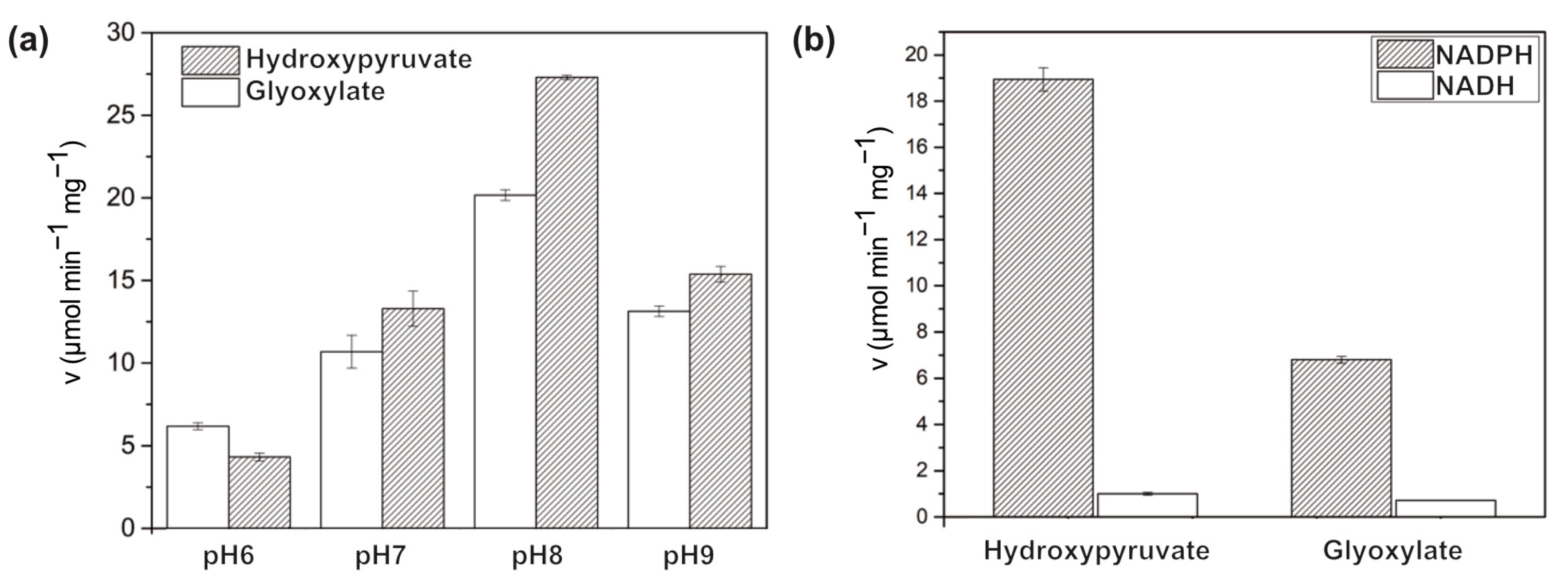
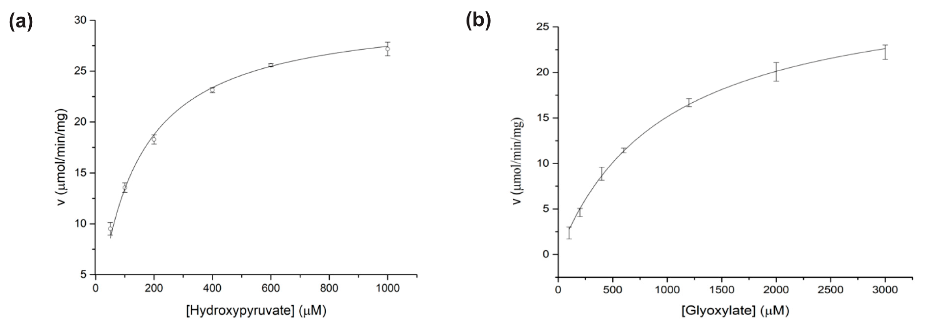
2.5. Activity Assay and Kinetic Analysis

Substrate reduction was monitored at 298 K. The absorbance at 340 nm was measured using an Ultrospec 8000 UV/Vis spectrophotometer (Cytiva, Wilmington, DE, USA), and the concentration of NAD(P)H was determined based on an extinction coefficient of 6.22 mM−1 cm−1. Triplicate assays were performed for each experiment, and all apparent kinetic parameters were calculated using GraphPad Prism (version 10.3.0). The Michaelis–Menten equation was used to calculate the apparent kinetic parameters. To determine the optimum pH range, two different buffer systems were employed to cover the pH range under investigation: Bis-Tris buffer at pH 6 and 7, and Tris-HCl buffer at pH 8 and 9. The reaction mixture consisted of 0.2 nM BsGRHPR, 2.0 mM glyoxylate or hydroxypyruvate, 0.2 mM NADPH, and 200 mM of the respective buffer solution. To assess the cofactor specificity between NADH and NADPH, 0.2 mM NADH or NADPH was added to the reaction mixture comprising 0.5 nM BsGRHPR, 2 mM glyoxylate, hydroxypyruvate, and 200 mM Tris-HCl pH 8. To determine the kinetic parameters for the two substrates, glyoxylate and hydroxypyruvate, the increase in substrate concentration was quantified spectrophotometrically by measuring the absorbance at 340 nm and 298 K, respectively. A total reaction volume of 500 µL was used, containing 0.2 nM BsGRHPR, 0.2 mM NADPH, and 200 mM Tris-HCl (pH 8.0). The initial reaction rate was measured for glyoxylate/hydroxypyruvate at six different concentrations ranging from 50 µM to 1 mM hydroxypyruvate and seven different concentrations ranging from 100 µM to 3 mM glyoxylate.

2.6. Circular Dichroism Spectroscopy and Thermal Denaturation

For CD measurements, the buffer was exchanged using a HiPrep 26/10 desalting column equilibrated with 20 mM Tris-HCl (pH 8.0) and 200 mM sodium chloride. The far-UV CD spectrum of BsGRHPR was measured at 293 K within a cell with a 0.1 cm path length using a JASCO J-1500 spectropolarimeter (JASCO, Tokyo, Japan). Three individual scans were recorded from 190 to 260 nm (0.1 nm step resolution, 1 nm bandwidth, and 1 s response time). The CD signal of the solvent was subtracted after averaging the three spectra. The CD intensity was normalized to the mean residue molar ellipticity. The thermal denaturation experiment was performed at 222 nm at a concentration of 0.5 mg/mL. The CD intensity was recorded every 30 s as the temperature was increased from 298 to 368 K at a rate of 2 K/min.

3. Results and Discussion

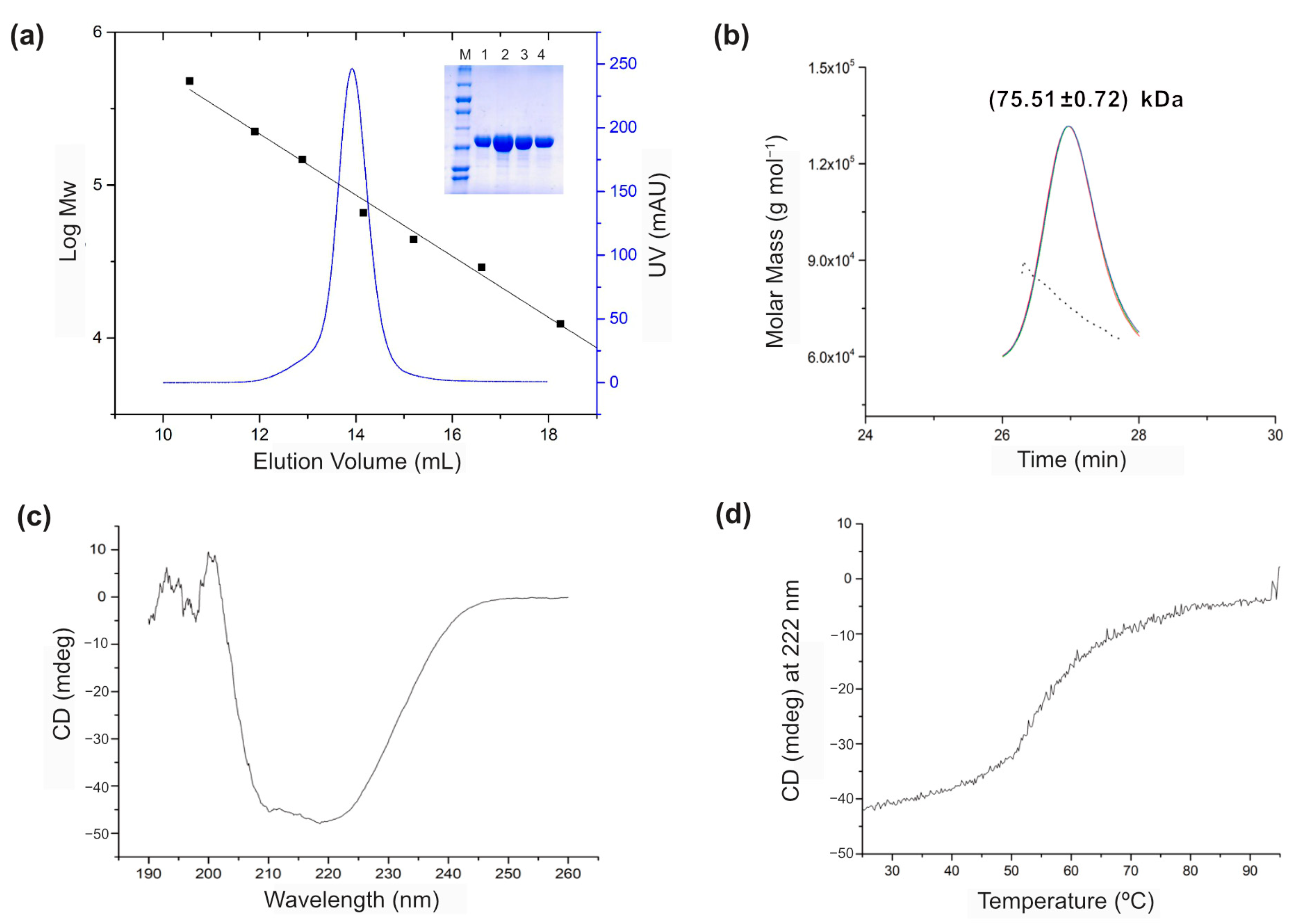
3.1. Biochemical Characterizations of BsGRHPR

BsGRHPR was expressed in Escherichia coli and purified using immobilized metal affinity and size-exclusion chromatography. The oligomeric status confirmed by size-exclusion chromatography combined with multi-angle light scattering was a dimer in solution (Figure 2a,b). The circular dichroism spectra indicated that the enzyme contained a high α-helix content (Figure 2c). The melting temperature (Tm) was determined to be 54.8 °C, which is relatively high, especially considering that Bacillus subtilis is a mesophile (Figure 2d).
In addition, we measured the specific activity of each substrate at four different pH values (Figure 3a). BsGRHPR exhibited maximum activity at pH 8.0 with both substrates. The specific activities of hydroxypyruvate and glyoxylate were 27.3 μmol min−1 mg−1 and 20.2 μmol min−1 mg−1, respectively. We also investigated the cofactor-dependent enzyme activity (Figure 3b). The specific activity of NADPH was higher than that of NADH in this study. These results are consistent with those of human GRHPR but differ from those of archaeal GRHPR [3,7,8,9].
The enzyme kinetics are shown in detail for the enzyme activity. Because of the low activity of NADH, only NADPH was used as a cofactor for enzyme kinetics, and pH 8.0 was found to be optimal for the reaction. Based on the initial velocity measured, the specific activity was calculated and is shown in μmol min−1 mg−1 (Figure 4). The kinetic parameters for glyoxylate were Vmax = 30.1 μmol min−1 mg−1, Km = 987.3 μM, kcat = 18.3 s−1, and kcat/Km = 0.02 s−1 mM−1 (Figure 4b). The values for hydroxypyruvate were Vmax = 31.0 μmol min−1 mg−1, Km = 130.9 μM, kcat = 18.8 s−1, and kcat/Km = 0.14 s−1 mM−1 (Figure 4a). Notably, the Michaelis constant implies that hydroxypyruvate binds more tightly to the active site than glyoxylate. The specificity constant for hydroxypyruvate was seven times higher than that for glyoxylate, although the kcat values for both substrates were similar. Taken together, these results indicate that hydroxypyruvate is the preferred substrate of BsGRHPR. To reveal the molecular details, we conducted structural analysis.

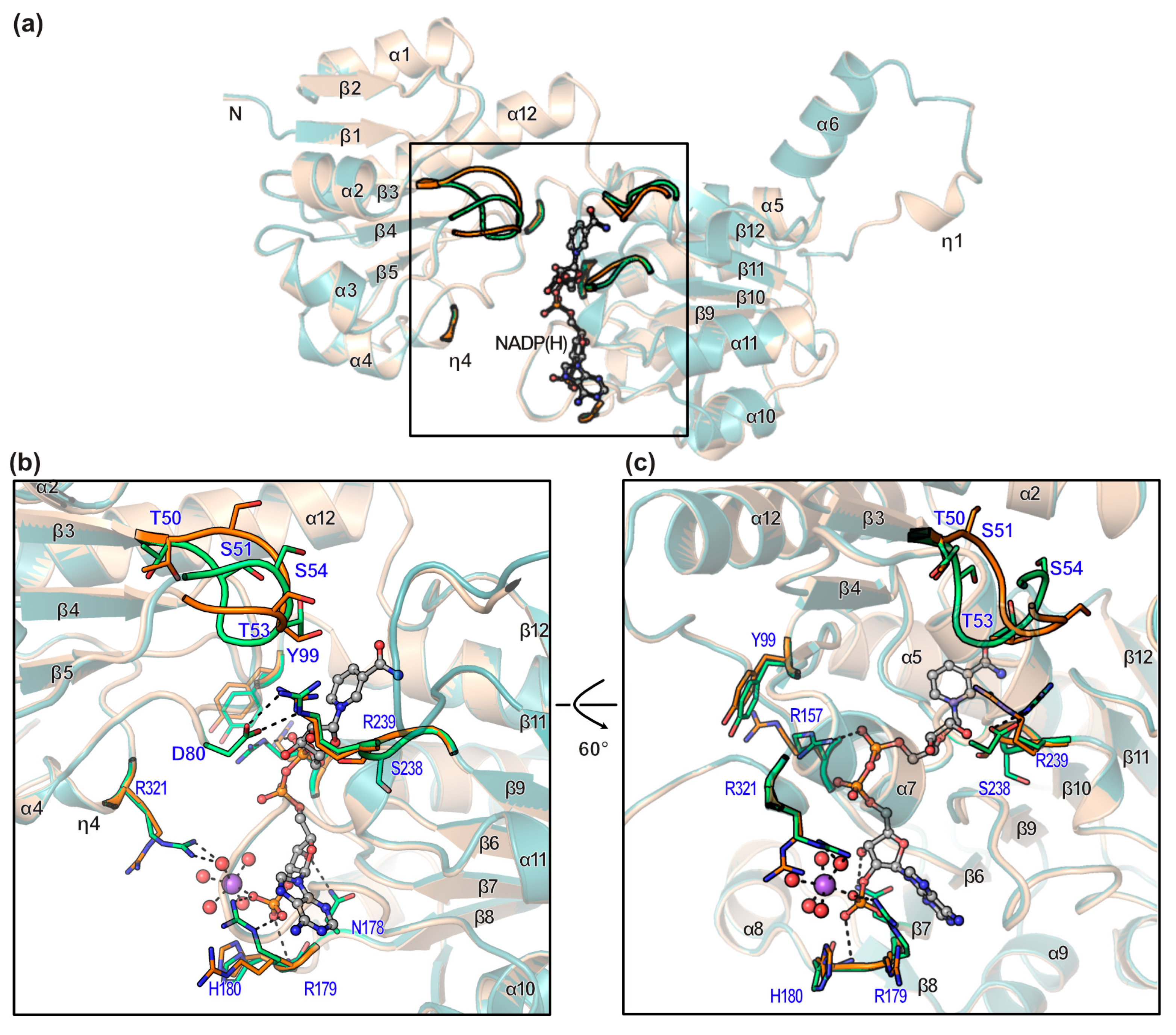
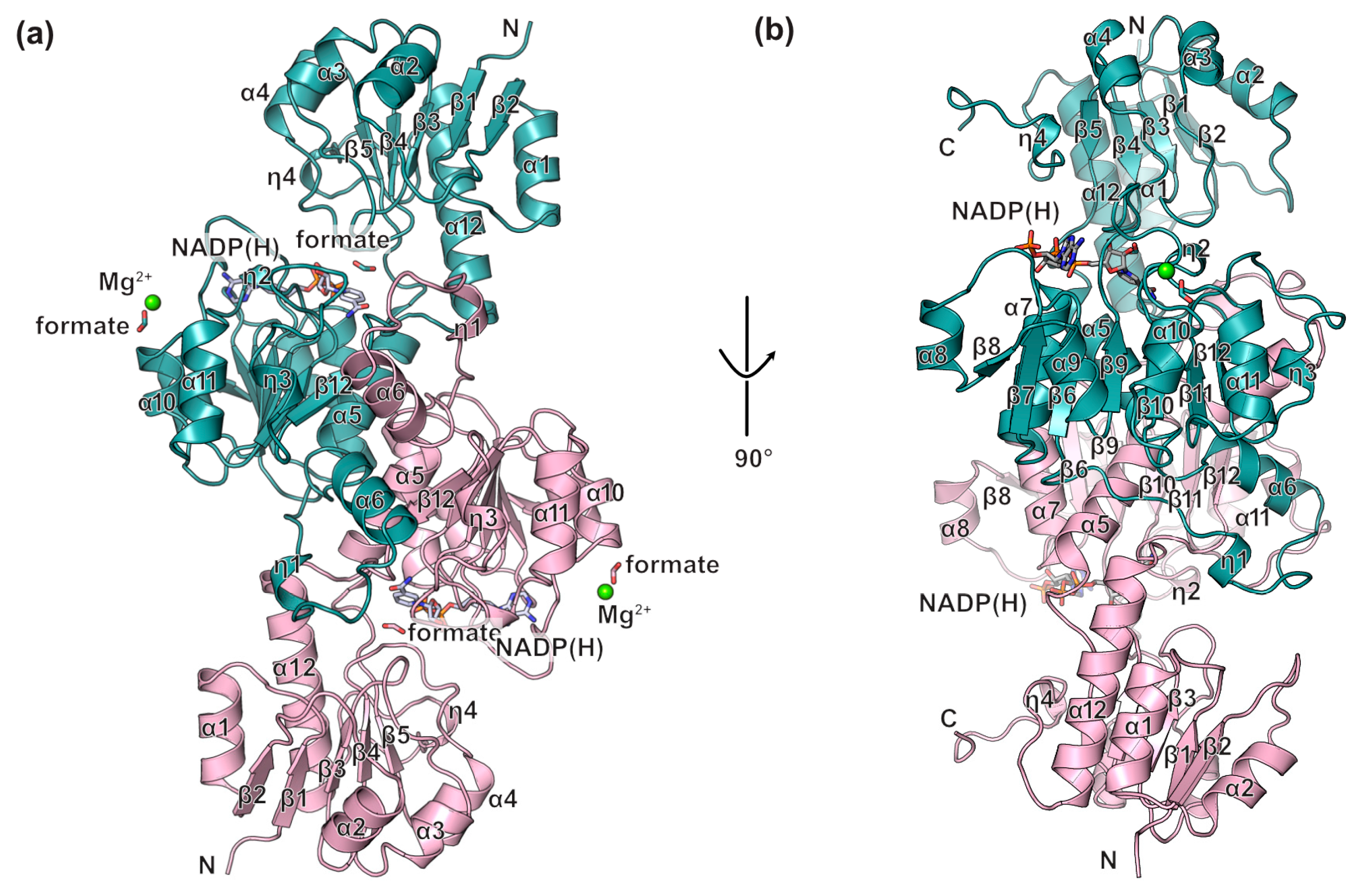
3.2. Overall Structure of BsGRHPR Complex with Both Formate and Cofactor

BsGRHPR crystallized in a monoclinic system, and the asymmetric unit contained a single monomer. The binary complex structure with formate was determined at a resolution of 1.2 Å, and the ternary complex structure with both the cofactor and formate was determined at a resolution of 1.5 Å (Table 1).
BsGRHPR comprises a coenzyme-binding domain (residues 101–292) consisting of seven α-helices (α5, α6, α7, α8, α9, α10, and α11) and three 310-helices (η1, η2, and η3) placed around seven stranded parallel β-sheet cores (β8–β7–β6–β9–β10–β11–β12) (Figure 5). This structure represents the classical NAD(P) Rossmann fold, with a dimerization loop (residues 118–146; between α6 and η1) (Figure 5). The substrate-binding domain (residues 1–100, 293–324) consists of five parallel β-sheet cores (β2–β1–β3–β4–β5) with five α-helices (α1, α2, α3, α4, and α12) and one 310-helix (η4) flanking on both sides (Figure 5). The magnesium ions and formate in the structure were derived from crystallization reagents (Figure 5). Formate is reminiscent of the substrate, which is why our attempts to co-crystallize or soak with glyoxylate or hydroxypyruvate were unsuccessful.
A dimer model of the crystal structure was generated using crystallographic operation. The PISA server analysis revealed an interface area of 2538 Å2. The α-helix (α6) and 310-helix (η1) primarily contribute to dimer formation (Figure 6a) [14]. Trp132 was inserted into the hydrophobic pocket formed by Pro269 and Pro286 in other subunits, whereas Glu137 formed a salt bridge with Arg296 in another subunit (Figure 6b,c). The distances between the carboxylate oxygens of Glu137 and the guanidinium group of Arg296 are 3.2 Å and 2.8 Å, respectively. W132, Pro269, Pro286, and Arg296 are highly conserved among homologs (Figure 7).

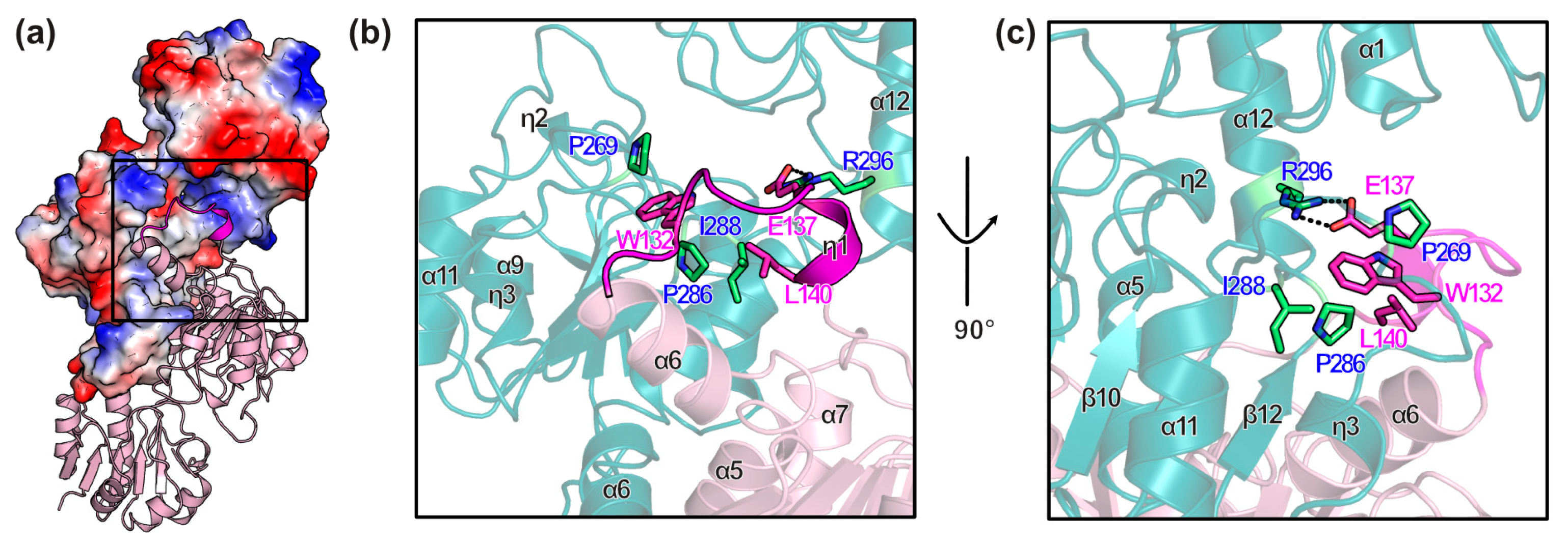
3.3. Conformational Changes on Binding of Cofactor

The superposition of the apo- and cofactor-bound forms explains the conformational changes in the enzyme. The overall fold did not change, as reflected in the rmsd value of 0.126, although the adjacent loops around the cofactor showed structural differences (Figure 8a). While the cofactor binds to the pocket, Arg239 is repelled and forms two salt bridges with Asp80 at a distance of 2.9 Å (Figure 8b). The most obvious change was observed for Arg157, which binds to the pyrophosphate of the cofactor (Figure 8c). Arg179 is also important for cofactor binding because it recognizes the phosphate of the cofactor (Figure 8c). The amide in the main chain of His180 interacts with the phosphate of the cofactor at a distance of 3.2 Å. In the substrate-binding domain, the loop between β3 and α3 (residues 50–55) shifts towards the cofactor. This shift allows for better accommodation of the cofactor within the binding pocket. The movement of the loop also creates additional space for substrate binding, potentially enhancing catalytic efficiency. Furthermore, the repositioning of these residues may contribute to the stabilization of the enzyme–substrate complex during the reaction. The distance between Gly52 in the two structures was 5.5 Å, and the distance between Ser51 in the two structures was 2.8 Å. This observation suggests that the active site moves as a rigid body to accommodate the cofactor (Figure 8c).

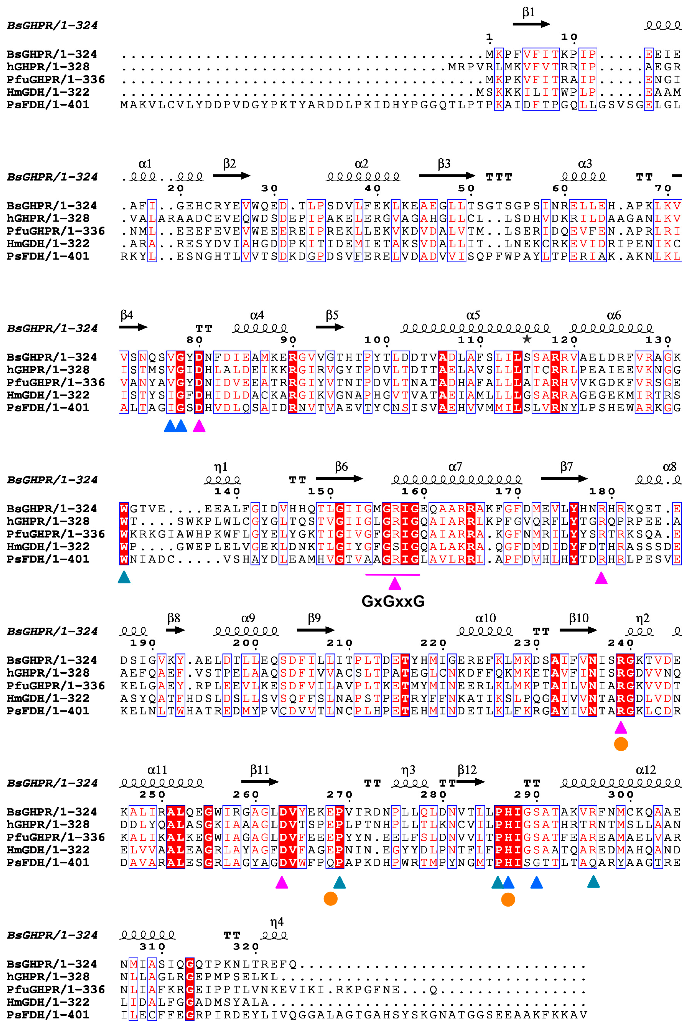
3.4. Cofactor Specificity

In our structure, the cofactor-binding site is exposed to the solvent channel, facilitating the exchange of both the cofactor and substrate (Figure 9a,b). The pyrophosphate of the cofactor is recognized by the glycine-rich loop (residues 154–159, GxGxxG, where x represents any amino acid), which is a conserved NAD(P)-binding motif (Figure 7). Furthermore, Asn178 interacts with the 3′-OH of adenosine ribose at a distance of 3.2 Å, and the 2′-phosphate of adenosine ribose interacts with Arg179 and His180 at distances of 2.7 Å and 3.2 Å, respectively (Figure 9c). If NADH replaces NADPH, Asn178 may exhibit weak interactions with the 2′- and 3′-OH of adenosine ribose. In contrast, FDH and HmGDH contain aspartate residues at this position (Figure 7). In addition, the nicotineamide moiety interacts with the carbonyl oxygen of Ile237 and the side chain of Asp263. The nicotinamide moiety also forms water-mediated hydrogen bonds with His287, Ser290, and Arg296. Interestingly, the position of glycerol in the apoGRHPR structure was well aligned with the nicotinamide moiety in the cofactor-bound structure, mimicking hydrogen-bond networking. Our observations indicated that the conserved Asn178 and Arg179 residues contributed to the selectivity of NADPH over NADH.

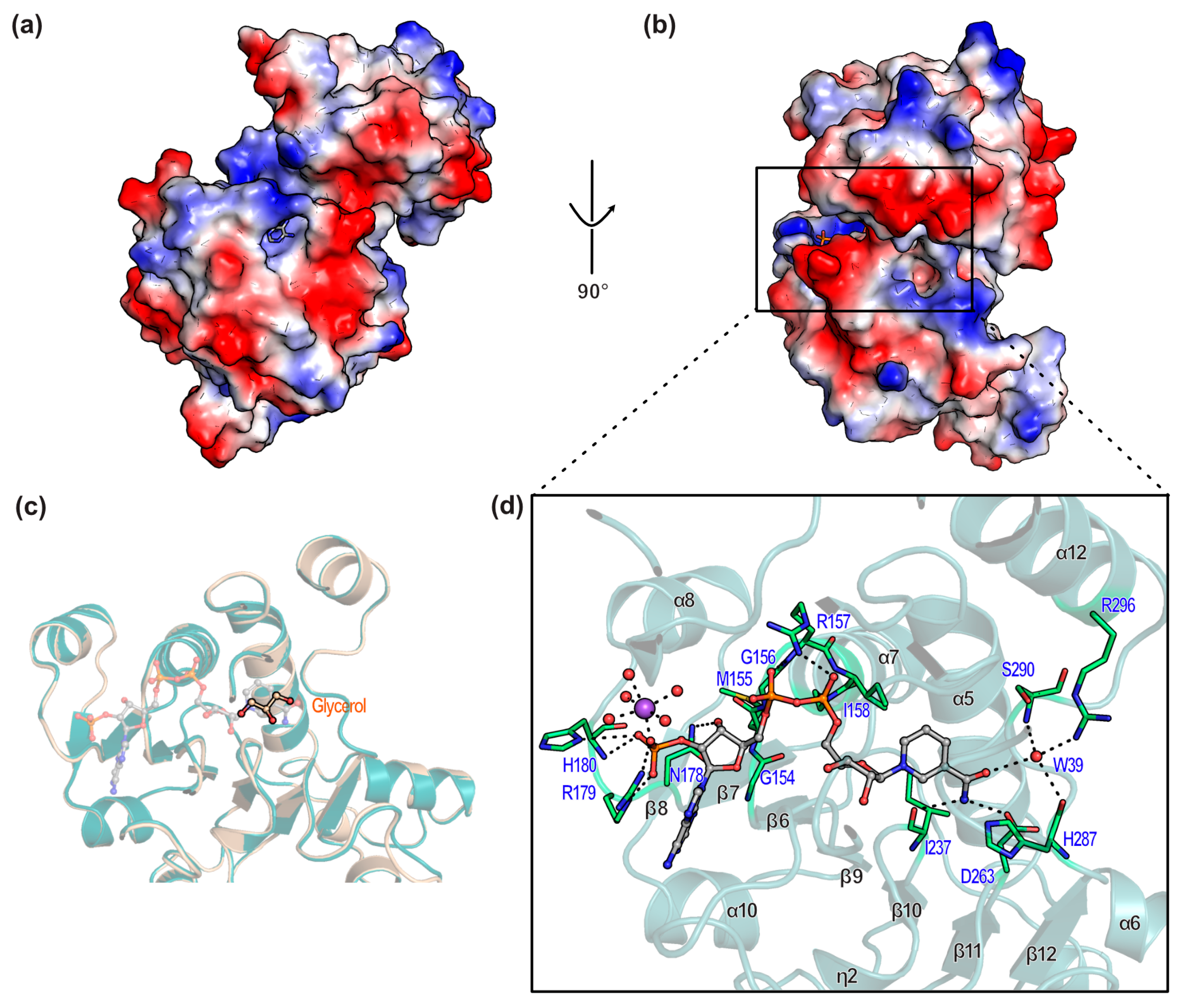
3.5. Substrate Binding Site

In our structure, the amide nitrogens of Val77 and Gly78 bind to the carboxyl groups of formate at distances of 2.9 Å and 2.7 Å, respectively (Figure 10). Val77 and Gly78 are highly conserved among homologs (Figure 7). Homology analyses revealed that S290, His287, and Arg239 were critical for substrate binding (Figure 7 and Figure 10c). The water molecules (W39, W55, W214, W257, and W273) were identified as representatives of the substrate in different binding modes. Their positions provide valuable insights into the molecular recognition process and potential binding mechanisms involved in the enzyme mechanism. The presence of formate establishes a stable network within the active site, elucidating the potential configuration of the active site when occupied by actual substrates or products rather than by formate. It was demonstrated that, besides valine and glycine, the conserved binding of the active site also included serine, histidine, and arginine, with the role of arginine being well-defined and described in previous studies. In detail, Glu238, Arg239, and His287 are known as the catalytic triad, which is supported by our structure [3,16]. In addition, BsGRHPR shows a clear preference for hydroxypyruvate in in vitro enzyme assays, which is consistent with hGRHPR and PfuGRHPR [7,8]. Our structure implies that Arg296 and S290 contribute to hydroxypyruvate binding, but not to glyoxylate binding.

4. Conclusions

In this study, we characterized the glyoxylate/hydroxypyruvate reductase (GRHPR) from Bacillus subtilis. We determined the crystal structures of both formate- and cofactor-bound GRHPR at near-atomic resolution (1.5 Å) and the crystal structure of formate-bound GRHPR at near-atomic resolution (1.2 Å). GRHPR from the mesophilic bacterium Bacillus subtilis prefers NADPH rather than NADH as a cofactor, which is consistent with human enzymes. Steady-state kinetics showed that the enzyme had maximum activity at pH 8.0 in the presence of both glyoxylate and hydroxypyruvate. BsGRHPR was remarkably thermostable, with a Tm of 54.82 °C. The crystal structure of BsGRHPR, compared to that of human, archaeal, and bacterial enzymes, revealed similarities and differences in the active site [7,8,9,17]. Additionally, the study revealed the conserved substrate- and cofactor-binding motifs of bacterial enzymes compared with those of human and archaeal enzymes. The hGRHPR study implies that the substrates are bound to the active site via Leu59, Val83, Gly84, W141* (from the adjacent subunit), Arg245, Glu274, Ser296, His293, and R302, which are described in terms of orientation to the substrate [7]. Similarly, in PfuGRHPR, the substrate is bound by Val76, Gly77, His288, and Arg241, which have the same function as Arg245 in humans [9]. In our structure, the water molecules observed near the active site were described and explained, providing insight into how substrates and products are linked to the enzyme active site by linking to a substrate analog (formate). The role of a conserved water molecule interacting with His287, Ser290, Arg296, and Glu137 residues as a gatekeeper at the entrance of the tunnel was also investigated. In addition, Trp132 is present at the dimer interface, where a distance of 2.9 Å is observed between Glu137 and Arg296. Overall, our findings provide a detailed biochemical and structural understanding of BsGRHPR, highlighting its substrate specificity and cofactor preference. These insights contribute to a broader understanding of GRHPR function across different species and may have implications for biotechnological applications involving redox metabolism.

Author Contributions

Conceptualization, J.K.Y. and W.K.; investigation, T.Q.N., J.K.Y. and W.K.; writing—original draft preparation, T.Q.N., J.K.Y. and W.K.; writing—review and editing, T.Q.N., T.H.D., J.K.Y. and W.K.; funding acquisition, W.K. All authors have read and agreed to the published version of the manuscript.

Funding

This research was supported by the National Research Foundation of Korea (NRF) grant funded by the Korean government (Ministry of Science and ICT, No. RS-2022-NR071729 and RS-2023-00217189; and Ministry of Education, No. RS-2021-NR060140).

Data Availability Statement

The structure factors and coordinates are deposited in the Protein Data Bank (www.rcsb.org, accessed on 17 March 2025) with PDB codes 9M0T and 9M0V.

Acknowledgments

We would like to thank the staff of the Korea Basic Science Institute for their assistance with SEC–MALS analyses and CD analyses. We would like to thank the staff at Beamline 11C of the Pohang Accelerator Laboratory (Pohang, Republic of Korea) for data collection.

Conflicts of Interest

The authors declare no conflicts of interest.

References

  1. Wanders, R.J.A.; Groothoff, J.W.; Deesker, L.J.; Salido, E.; Garrelfs, S.F. Human glyoxylate metabolism revisited: New insights pointing to multi-organ involvement with implications for siRNA-based therapies in primary hyperoxaluria. J. Inherit. Metab. Dis. 2025, 48, e12817. [Google Scholar] [CrossRef] [PubMed]
  2. Bhuyan, R.; Maggio, T.; Thomas, C.; Sambharia, M.; Gehrs, K.; Boyce, T. Late-onset retinal oxalosis in primary hyperoxaluria type 2. Am. J. Ophthalmol. Case Rep. 2024, 36, 102156. [Google Scholar] [CrossRef] [PubMed]
  3. Kutner, J.; Shabalin, I.G.; Matelska, D.; Handing, K.B.; Gasiorowska, O.; Sroka, P.; Gorna, M.W.; Ginalski, K.; Wozniak, K.; Minor, W. Structural, Biochemical, and Evolutionary Characterizations of Glyoxylate/Hydroxypyruvate Reductases Show Their Division into Two Distinct Subfamilies. Biochemistry 2018, 57, 963–977. [Google Scholar] [CrossRef] [PubMed]
  4. Goldberg, J.D.; Yoshida, T.; Brick, P. Crystal structure of a NAD-dependent D-glycerate dehydrogenase at 2.4 A resolution. J. Mol. Biol. 1994, 236, 1123–1140. [Google Scholar] [CrossRef] [PubMed]
  5. Rumsby, G.; Cregeen, D.P. Identification and expression of a cDNA for human hydroxypyruvate/glyoxylate reductase. Biochim. Biophys. Acta 1999, 1446, 383–388. [Google Scholar] [CrossRef] [PubMed]
  6. Allan, W.L.; Clark, S.M.; Hoover, G.J.; Shelp, B.J. Role of plant glyoxylate reductases during stress: A hypothesis. Biochem. J. 2009, 423, 15–22. [Google Scholar] [CrossRef] [PubMed]
  7. Booth, M.P.; Conners, R.; Rumsby, G.; Brady, R.L. Structural basis of substrate specificity in human glyoxylate reductase/hydroxypyruvate reductase. J. Mol. Biol. 2006, 360, 178–189. [Google Scholar] [CrossRef] [PubMed]
  8. Lassalle, L.; Engilberge, S.; Madern, D.; Vauclare, P.; Franzetti, B.; Girard, E. New insights into the mechanism of substrates trafficking in Glyoxylate/Hydroxypyruvate reductases. Sci. Rep. 2016, 6, 20629. [Google Scholar] [CrossRef]
  9. Yoshikawa, S.; Arai, R.; Kinoshita, Y.; Uchikubo-Kamo, T.; Wakamatsu, T.; Akasaka, R.; Masui, R.; Terada, T.; Kuramitsu, S.; Shirouzu, M.; et al. Structure of archaeal glyoxylate reductase from Pyrococcus horikoshii OT3 complexed with nicotinamide adenine dinucleotide phosphate. Acta Crystallogr. D Biol. Crystallogr. 2007, 63, 357–365. [Google Scholar] [CrossRef] [PubMed]
  10. Ohshima, T.; Nunoura-Kominato, N.; Kudome, T.; Sakuraba, H. A novel hyperthermophilic archaeal glyoxylate reductase from Thermococcus litoralis. Characterization, gene cloning, nucleotide sequence and expression in Escherichia coli. Eur. J. Biochem. 2001, 268, 4740–4747. [Google Scholar] [CrossRef] [PubMed]
  11. Otwinowski, Z.; Minor, W. Processing of X-ray diffraction data collected in oscillation mode. Methods Enzym. 1997, 276, 307–326. [Google Scholar]
  12. Emsley, P.; Lohkamp, B.; Scott, W.G.; Cowtan, K. Features and development of Coot. Acta Crystallogr. D Biol. Crystallogr. 2010, 66, 486–501. [Google Scholar] [CrossRef] [PubMed]
  13. Adams, P.D.; Afonine, P.V.; Bunkoczi, G.; Chen, V.B.; Davis, I.W.; Echols, N.; Headd, J.J.; Hung, L.W.; Kapral, G.J.; Grosse-Kunstleve, R.W.; et al. PHENIX: A comprehensive Python-based system for macromolecular structure solution. Acta Crystallogr. D Biol. Crystallogr. 2010, 66, 213–221. [Google Scholar] [CrossRef] [PubMed]
  14. Krissinel, E.; Henrick, K. Inference of macromolecular assemblies from crystalline state. J. Mol. Biol. 2007, 372, 774–797. [Google Scholar] [CrossRef] [PubMed]
  15. Robert, X.; Gouet, P. Deciphering key features in protein structures with the new ENDscript server. Nucleic Acids Res. 2014, 42, W320–W324. [Google Scholar] [CrossRef] [PubMed]
  16. Holton, S.J.; Anandhakrishnan, M.; Geerlof, A.; Wilmanns, M. Structural characterization of a D-isomer specific 2-hydroxyacid dehydrogenase from Lactobacillus delbrueckii ssp. bulgaricus. J. Struct. Biol. 2013, 181, 179–184. [Google Scholar] [CrossRef] [PubMed]
  17. Kumsab, J.; Tobe, R.; Kurihara, T.; Hirose, Y.; Omori, T.; Mihara, H. Characterization of a novel class of glyoxylate reductase belonging to the beta-hydroxyacid dehydrogenase family in Acetobacter aceti. Biosci. Biotechnol. Biochem. 2020, 84, 2303–2310. [Google Scholar] [CrossRef] [PubMed]
Figure 1. Reaction schemes of GRHPR.
Crystals 15 00298 g001
Figure 2. Biochemical characterization of BsGRHPR in solution. (a) Purified BsGRHPR was injected into a size-exclusion chromatography column. Molecular weights were calculated using the molecular weights of the seven standard proteins. SDS-PAGE revealed that the peak eluted from 13.5 to 15 mL. (b) Purified BsGRHPR was analyzed using size-exclusion chromatography combined with multi-angle light scattering. (c) CD spectra of the purified BsGRHPR. (d) Thermal denaturation was monitored using CD values at 222 nm.
Crystals 15 00298 g002
Figure 3. Optimum enzyme reaction conditions for BsGRHPR. (a) pH-dependence and (b) cofactor specificity of enzyme activity.
Crystals 15 00298 g003
Figure 4. Michaelis–Menten kinetic analysis. The data were fitted to the Michaelis–Menten equation to calculate Vmax and Km values. Triplicate assays were performed for each point. The bar represents the standard deviation and the circle represents the average. (a) Enzyme kinetics of hydroxypyruvate. (b) Enzyme kinetics of glyoxylate.
Crystals 15 00298 g004
Figure 8. Conformational changes upon cofactor binding. (a) Overall structural alignments of apoBsGRHPR and cofactor-bound BsGRHPR are shown in ribbon representations. The cofactor-bound BsGRHPR and apoBsGRHPR are represented as teal and orange ribbons, respectively. Except for the regions showing conformational changes, the ribbon diagrams are transparent. (b) A zoomed-in view of the boxed region in (a), showing the key residues involved in the interaction. The structural differences between the two conformations are highlighted. (c) Rotated view providing an alternative perspective of the binding site. Key residues are labelled and depicted in stick representation. Water is shown as red balls, and sodium ions are shown as purple balls.
Crystals 15 00298 g008
Figure 9. Structural details of the cofactor-binding site. (a,b) The surface of the monomer is colored according to the electrostatic potential from red (−73.2 kT/e, negatively charged) to blue (+73.2 kT/e, positively charged). (c) Overall structural alignment of apoBsGRHPR and cofactor-bound BsGRHPR is shown in ribbon representation, as in Figure 7. Glycerol was derived from apoGRHPR. (d) A detailed close-up of the cofactor-binding site showing the key residues involved in cofactor binding. Water is shown as red balls, and sodium ions are shown as purple balls.
Crystals 15 00298 g009
Figure 10. Structural details of active sites. (a,b) The surface of the monomer is colored according to the electrostatic potential from red (−73.2 kT/e, negatively charged) to blue (+73.2 kT/e, positively charged). (c) Structural alignment of BsGRHPR and hGRHPR (PDB ID: 2GCG). The structures were aligned using PyMOL, with a resulting root mean square deviation of 0.82. BsGRHPR is represented as teal-colored cartoon diagrams and gray stick models (cofactor and formate), and hGRHPR is represented as light-purple-colored cartoon diagrams and yellow stick models (cofactor and D-glycerate). (d) Zoomed-in view of the formate-binding site. Water molecules are represented by red balls.
Crystals 15 00298 g010
Figure 5. Overview of BsGRHPR in complex with both the cofactor and formate. The dimer is shown in ribbon diagrams with different colors for each monomer. The cofactor (NADP(H)) and formate are shown in stick model representations, and the magnesium ion is represented by a green ball. (a) Overview and (b) rotated view of BsGRHPR in complex with both the cofactor and formate.
Crystals 15 00298 g005
Figure 6. Structural details of the dimeric interface. (a) Overall structure of the dimer, showing the electrostatic potential from red (−73.2 kT/e, negatively charged) to blue (+73.2 kT/e, positively charged). The corresponding monomers are shown in the ribbon diagrams. A zoomed-in region (black box) highlights the interaction site of interest, where the dimerization loop (in magenta) is present. (b) Close-up view of the dimerization loop from (a), showing the key residues involved in the interaction. Hydrogen bonds are represented by black dotted lines. (c) Rotated perspective of the insertion loop shown in (b), providing an alternative view of the interface. Key residues are labelled and depicted in stick representation.
Crystals 15 00298 g006
Figure 7. Multiple sequence alignment of BsGRHPR. The sequences of homologs were used for multiple sequence alignment using the web interface of Clustal Omega (https://www.ebi.ac.uk/jdispatcher/msa/clustalo, accessed on 17 March 2025; v1.2.4). The diagrams for secondary structure generated using ESPrint/ENDscript (https://endscript.ibcp.fr, accessed on 17 March 2025) are represented based on the crystal structures in this study, were [15]. The synonyms of the homologs are shown as follows: BsGRHPR, Bacillus subtilis glyoxylate/hydroxypyruvate reductase, UniProt A0AAP1E3x5; hGRHPR, human glyoxylate/hydroxypyruvate reductase, UniProt Q9UBQ7; PfuGRHPR, Pyrococcus furiosus glyoxylate reductase, UniProt Q9U33Y2; HmGDH, Hyphomicrobium methylovorum glycerate dehydrogenase, UniProt P36234; PsFDH, Pseudomonas sp. Formate dehydrogenase, UniProt P33160. The marked triangles represent the characteristic motifs designated in this study as follows: magenta, cofactor-binding motif; teal, dimerization motif; blue, substrate-binding motif. The catalytic triad residues are denoted by orange circles.
Crystals 15 00298 g007
Table 1. Crystallographic statistics for data collection and refinement.
BsGRHPR + FormateBsGRHPR + Formate, NADPH
PDB ID9M0T9M0V
Data collection
 Wavelength (Å)0.9790.979
 Space groupC2C2
 Total reflections648,293 (26,705)336,953 (16,325)
 Unique reflections95,288 (4,200)50,733 (2502)
 Cell dimensions
  a, b, c (Å)93.1, 59.6, 59.993.2, 59.2, 59.7
  β (°)99.899.8
 Resolution (Å)29.8–1.2 (1.22–1.20)49.8–1.5 (1.53–1.50)
Rpim 10.022 (0.422)0.038 (0.478)
 CC1/20.999 (0.715)0.999 (0.648)
I/σ I16.2 (1.8)12.5 (1.7)
 Completeness (%)94.7 (85.0)98.8 (98.8)
 Multiplicity6.8 (6.4)6.6 (6.5)
Refinement
 Resolution (Å)29.5–1.2 (1.23–1.20)49.8–1.5 (1.54–1.50)
 No. reflections95,25550,703
Rwork/Rfree 20.165/0.1840.162/0.200
 No. atoms
  Protein26682658
  Ligand/ion1659
  Water421342
B-factors
  Protein17.9321.3
  Ligand17.6516.1
  Water28.73.1
 R.m.s. deviations
  Bond lengths (Å)0.0050.007
  Bond angles (°)0.870.91
 Ramachandran plot (%)
  Favored97.8696.94
  Allowed2.143.06
Values in parentheses correspond to the highest resolution shell. 1 Rpim = Σhkl|[1/(N − 1)]1/2 Σi|Ii(hkl) − I ( h k l ) |/Σhkl ΣiIi(hkl). 2 Rwork = Σ||Fobs| − |Fcalc||/Σ|Fobs|, where Rfree was calculated for a randomly chosen 2000 reflections that were not used for structure refinement, and Rwork was calculated for the remaining reflections.
Disclaimer/Publisher’s Note: The statements, opinions and data contained in all publications are solely those of the individual author(s) and contributor(s) and not of MDPI and/or the editor(s). MDPI and/or the editor(s) disclaim responsibility for any injury to people or property resulting from any ideas, methods, instructions or products referred to in the content.

