1. Introduction
The superior toughness and outstanding wear resistance of high-carbon high-manganese steel under high impact load mean that it is widely used in various industries [
1,
2,
3,
4]. And yet high-carbon high-manganese steel is generally processed by aging treatment in engineering on the basis of water toughening treatment in order to improve its wear resistance under non-high impact load service conditions. However, the transformation process of high-carbon high-manganese steel during aging treatment at different temperatures has been always a weak spot, and a lot of problems deserve further study such as its microstructure, morphology, crystallographic characteristics and precipitation location. Pearlite transformation can occur during aging treatment, and the mechanical properties of pearlite formed on austenite matrix are between that of ferrite and that of cementite [
5,
6,
7]. Therefore, studying pearlite transformation has great significance in enhancing the wear resistance of high-carbon high-manganese steel. While the eutectoid transformation of high-carbon high-manganese steel proceeds with an appropriate aging temperature, the formed pearlite is generally an integrated microstructure composed of ferrite and cementite (or carbide) [
8,
9,
10]. The decomposition equation of the Fe-C alloy is γ → α + Fe
3C. Moreover, the most classic pearlite morphology consists alternately of ferrite lamella and carbide lamella [
11]. When the size of the lamellae is larger than that of the critical nucleus, a pearlite colony can be formed, whose crystallographic characteristics can be confirmed by a TEM and electron back scattered diffraction (EBSD) [
12,
13,
14,
15]. Pearlite transformation is a process of nucleation and growth, and its transformation mechanism has been the research focus at home and abroad. In the 1930s, Carpenter and Robertson [
16] proposed the radial growth and parallel growth of pearlite by examining a series of eutectoid composition steels. Although the occasional formation of pearlite described as the “deposition of alternate layers of ferrite and cementite” was not fully discounted by Carpenter and Robertson, they drew the conclusion that pearlite is always formed by radial growth, and through edgewise growth, it continues to expand into austenite. And Jolivet [
17] confirmed Carpenter and Robertson’s research results. Two kinds of hypoeutectoid steels under isothermal transformation conditions were examined to verify that this common occurrence appears in the radial growth of pearlite. Despite the fact that the growth process of pearlite has been known via early studies, the Hull–Mehl mechanism was also reported by Hull and Mehl [
18,
19] after systematically examining hypoeutectoid steel, eutectoid steel and hypereutectoid steel, that is, the edgewise growth and sidewise nucleation/growth of pearlite. Conversely, after studying the pearlite formation process in hypoeutectoid steel and eutectoid steel, Hillert [
20] presented another pearlite growth mechanism, namely the branch mechanism, whereby ferrite nucleates at the grain boundary and continuously forms branches as it grows forward, while cementite continuously forms in coordination between the ferrite branches; finally, pearlite continuously develops during the repeated process. The branch mechanism of pearlite growth has been widely accepted, and this growth mechanism is also applicable to the growth process of lamellar pearlite morphology in high-carbon high-manganese steel [
21,
22,
23]. Nonetheless, Zhang and Kelly [
24] deemed that there is still a lack of direct evidence to prove the branch mechanism of pearlite, and whether this branch mechanism affects pearlite morphology remains unknown. Based on the above, the morphology and microstructure of 100Mn13 steel formed by hot forging, which was the experimental material aged at low temperature and high temperature, were observed and analyzed by a SEM and TEM in order to solve the issue of pearlite growth during eutectoid transformation in high-carbon high-manganese steel.
2. Experimental Materials and Methods
Moreover, 100Mn13 steel with a composition of Fe-0.92 wt.% C-10.24 wt.% Mn (
Table 1) was mainly hot-forged to use in this study.
Bars with a diameter of 32 mm of 100Mn13 steel forged following smelting and casting were, respectively, aged at 525 °C (low-temperature aging) and 650 °C (high-temperature aging) for 2 h after water toughening treatment at 1050 °C for 1.25 h. Then, they were quenched at room temperature with water. The heat treatment equipment was an SX-4-10 box-type resistance furnace (furnace temperature uniformity < 5 °C), equipped with a temperature controller and nickel–chrome–nickel–silicon electric couple, which could measure, indicate and automatically control the furnace temperature. The aging temperatures 525 °C and 650 °C were determined for this steel because pearlite was just formed from the grain boundary at 525 °C, when it was convenient to observe the initial chrysanthemum-like morphology, and 650 °C was the highest temperature in the series of experiments when the protrusions and branches at the growth frontier of pearlite lamella were fully grown, and this was when it was convenient to conduct the analysis.
The samples were sectioned, mechanically polished, and etched in a 4 pct nital solution for observation by a SUPRA 55 SEM. Thin foils were wire-cut into the thickness of 0.5 mm, mechanical grinded to approximately the thickness of 50 um, punched into disks with a diameter of 3 mm, perforated by a Gatan-691 ion milling machine to the edge thickness of less than 500 nm for observation by a JEM-2100F TEM operated at 200 kV. The morphology characteristics of the phase interface in pearlite were observed and analyzed by high-resolution transmission electron microscopy (HRTEM).
3. Experimental Results and Analysis
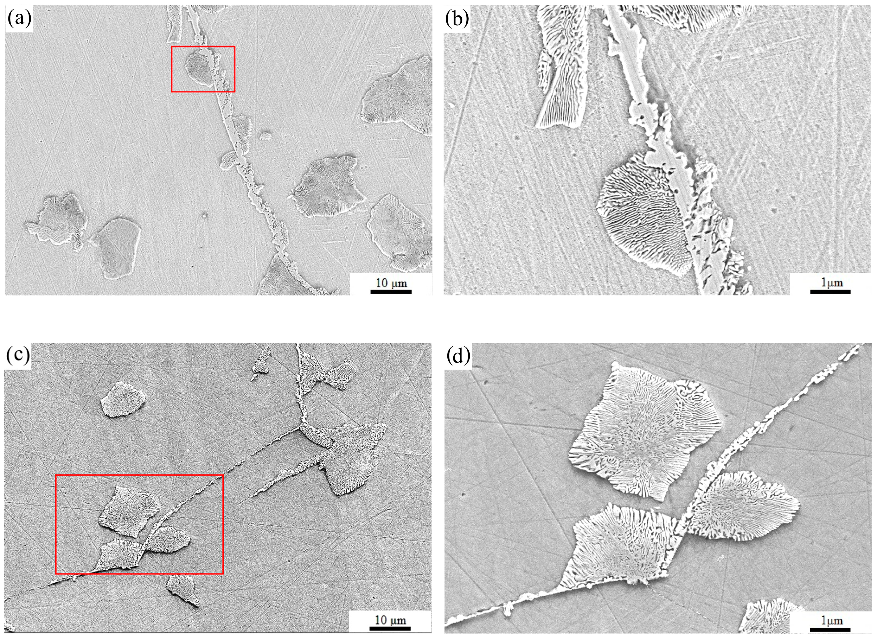
The SEM micrographs of chrysanthemum-like pearlite in 100Mn13 steel aged at 525 °C and 650 °C are shown in
Figure 1. And magnified images of the rectangular area in
Figure 1a,c are, respectively, shown in
Figure 1b,d. It can be seen that a large amount of the precipitated phase appears in 100Mn13 steel aged at 525 °C and 650 °C. Among them, some precipitates are distributed in a network along the austenite grain boundary, which are speculated to be carbides. Some precipitates are near the austenite grain boundary or preprecipitated phase or are located in the grain, showing a lamellar morphology, which should be pearlite.
Thereinto, some pearlite colonies are connected together, and the directions of pearlite lamellae change, with three or four directions. This is because the pearlite colonies simultaneously nucleate at different adjacent locations, then continuously develop, and finally have contact with each other and then connect together, indicating the dispersibility of pearlite nucleation or that the branching lamellae in pearlite colonies form chrysanthemum-like morphology. As in
Figure 1b, the growth direction of lamellae in pearlite colonies distributed along the austenite grain boundary is also slightly different. However, the different growth direction of lamellae in pearlite colonies did not affect the overall morphology of a very clear chrysanthemum flowering shape, which is called “chrysanthemum-like” in this paper. Simultaneously, there are also some pearlite colonies in the grains, as well as near the boundaries, that have chrysanthemum-like morphology, as shown in
Figure 1d. Although the “chrysanthemum-like” morphology of pearlite is relatively common, no in-depth study on the growth behavior of pearlite with such morphology has been reported so far. Furthermore, there are some protrusions, even a mass of branches on some lamellae of chrysanthemum-like pearlite, observed through a more careful observation, and whether the reason for the chrysanthemum-like morphology of pearlite is related to these structures needs further research.
The TEM micrographs including bright-field (BF) images, dark-field (DF) images and a selected area electron diffraction (SAED) pattern of chrysanthemum-like pearlite in 100Mn13 steel aged at 650 °C in the high-temperature aging treatment are shown in
Figure 2, in which chrysanthemum-like pearlite is composed alternatively of M
7C
3 (M represents Fe and Mn in this paper because of their approximate atomic radius dimensions) lamellae and ferrite lamellae, with the lamellar width between 60 nm and 350 nm, and the overall trend is basically the same, with only bending at the lamella growth frontier with a radial trend. The local magnified image of
Figure 2a is shown in
Figure 2b. By analyzing, the two sets of diffraction spots (
Figure 2c) are, respectively, calibrated as the M
7C
3 carbide and ferrite, with an OR of (
)
M7C3‖(
)
α, [
]
M7C3‖[
]
α. Thus, chrysanthemum-like pearlite consists of M
7C
3 lamellae and ferrite lamellae. It is worth noting that the carbide that makes up the pearlite is M
7C
3, not M
3C [
25,
26], M
13C
4, M
5C
2, or M
3C
2 [
27], as previously thought. From the Fe-C-Mn equilibrium phase diagram, it can be discerned that when Mn content is 5.1%, eutectoid transformation occurs at 570 °C and the carbide type is M
7C
3. The reason for this may be related to the effect of the Mn element on reducing the transformation temperature and stabilizing the M
7C
3 carbide [
28,
29].
The DF images of
Figure 2d,e display that the morphology of M
7C
3 lamellae and ferrite lamellae is different from that of M
3C lamellae and ferrite lamellae, which are usually straight, but there are some protrusions and branches at the lamella growth frontier in chrysanthemum-like pearlite. Meanwhile, both M
7C
3 lamellae and ferrite lamellae show a thickening tendency towards the growth frontier, and some protrusions are presented on the complementary phase interface at certain intervals, but M
7C
3 lamellae at the pearlite growth frontier are thicker than ferrite lamellae. It can be precisely seen in
Figure 2d that protrusions I, II and III increase successively along the growth direction of M
7C
3 lamellae, and protrusion III has a tendency to evolve into a branch of M
7C
3 lamellae, while a branching structure can be clearly observed on the fifth lamella of M
7C
3 (as shown in the arrow direction).
Similarly, a branch can be seen in the circled area of
Figure 2f, and the frontier interface of pearlite lamellae is almost at a 90° angle. This kind of branching structure is basically consistent with the branching structure presented in
Figure 1b and has been observed in Fe-12Mn-0.8C steel by Zhang and Kelly [
24] and Dippenaar and Honeycombe [
12]. The gradual thickening of M
7C
3 lamellae and successively increased protrusion structure demonstrate that not only the length of M
7C
3 lamellae elongates through edgewise growth but that the width of M
7C
3 lamellae also continuously thickens through sidewise growth, and the protrusion structure is even formed on the lamellae, and the growing protrusion then develops into the branching structure. Although the branching structure is common in pearlite colonies and is a crucial method to multiply the number of pearlite lamellae [
12], its detailed formation mechanism is rarely reported. Analogously, Hillert [
20] pointed out that cementite lamella forms the next cementite lamella through the bridging structure, which means that cementite grows into austenite through the “gap” on the adjacent ferrite lamella and forms the next cementite lamella. Nevertheless, the mode of the bridging structure is obviously not applicable to the branch mechanism. According to the morphology and location of protrusions and branches shown in
Figure 2d, the initial stage of the branching structure entails the formation of a lateral protrusion structure on the lamellae, which gradually evolves into the branching structure during their growth into austenite. In addition, protrusion I on ferrite lamella in
Figure 2e is low and flat, which can widen ferrite lamellae, while protrusion II is narrow and steep, which tends to develop into a branch. In consequence, the protrusions at the lateral interface of pearlite lamellae can not only increase the lamellae width but also evolve into branches. When the lamellae are narrow, the protrusions mainly grow along the lamella direction to increase their width, but when they increase to a certain extent, the protrusions formed again at lateral interfaces tend to develop into branches. Hence, the protrusion structure and branching structure make the lamellae of carbide and ferrite in the same pearlite colony bend and diverge along the growth direction, presenting chrysanthemum-like morphology macroscopically.
The frontier interface of pearlite lamella is almost at a 90° angle, as shown in the circled area of
Figure 2f, in which high-resolution analysis is performed (
Figure 3).
Figure 3b is an IFFT image of the interface in the rectangular area in
Figure 3a. The lattice plane of α (
) parallels that of M
7C
3 (
), and the overall arrangement of atoms is relatively regular in the direction of M
7C
3 [
], but the positions of some atoms are slightly offset. There is an obvious boundary between α lamella and M
7C
3 lamella with the step structure, which is different from the growth step at the interface between the austenite and pearlite matrix during edgewise growth. It is well known that the ledge mechanism of pearlite growth is an effective part of the interface diffusion control mechanism, and it includes not only growth steps at the growth frontier interface between carbide or ferrite and the matrix but also structural steps at the interface between carbide and ferrite. Since Bramfitt and Marder [
30] observed dislocation and growth steps in pearlite, the existence of structural steps on the habit plane of the eutectoid precipitated phase (i.e., the habit plane of ferrite/cementite) in various alloys has been confirmed by a TEM [
31,
32,
33,
34], and structural steps are caused by the movement of growth steps (i.e., the ferrite/austenite or cementite/austenite interface). In
Figure 3b, the number of atoms between adjacent lattice plane of (
) in M
7C
3 lamella tends to decrease with the decreasing distance from the frontier, which is the result of the upward and downward movement of atoms in space, indicating that the essence of the edgewise growth of M
7C
3 lamella is also a process of atom movement, through which the transition area of M
7C
3 to α is formed. The existence of this transition area also confirms the existence of structural steps between pearlite lamellae, that is, when the step structure moves from the adjacent α interface on one side of the M
7C
3 lamella to the adjacent α interface on the other side along the direction perpendicular to lamella growth, it shifts from the α interface of M
7C
3 to the other α side, forming structural steps along the direction of the ferrite lamella.
4. Discussion
The morphology and microstructure of the chrysanthemum-like pearlite colony formed in the austenite grain of 100Mn13 high-carbon high-manganese steel were researched after aging at 525 °C (low temperature) and 650 °C (high temperature). The morphology of this chrysanthemum-like pearlite has been reported in other studies, but the reason for its growth has not been determined. It is well known that the growth of pearlite includes the edgewise growth and sidewise growth of carbide lamellae and ferrite lamellae. At present, there is no lack of research on the edgewise growth of pearlite lamellae regarding aspects such as the interface movement [
35,
36], ledge mechanism [
37,
38], and so on. Yet the study on the sidewise growth of pearlite lamellae is still limited to the number of increasing lamellae; for instance, lateral nucleation, the thickening tendency of individual lamella and the branch mechanism are still not clear. In this paper, it is suggested that the chrysanthemum-like pearlite colony is related to the growth behavior of each lamella, which depends on the lateral protrusion structure thickening and the protrusion structure transforming into the branching structure.
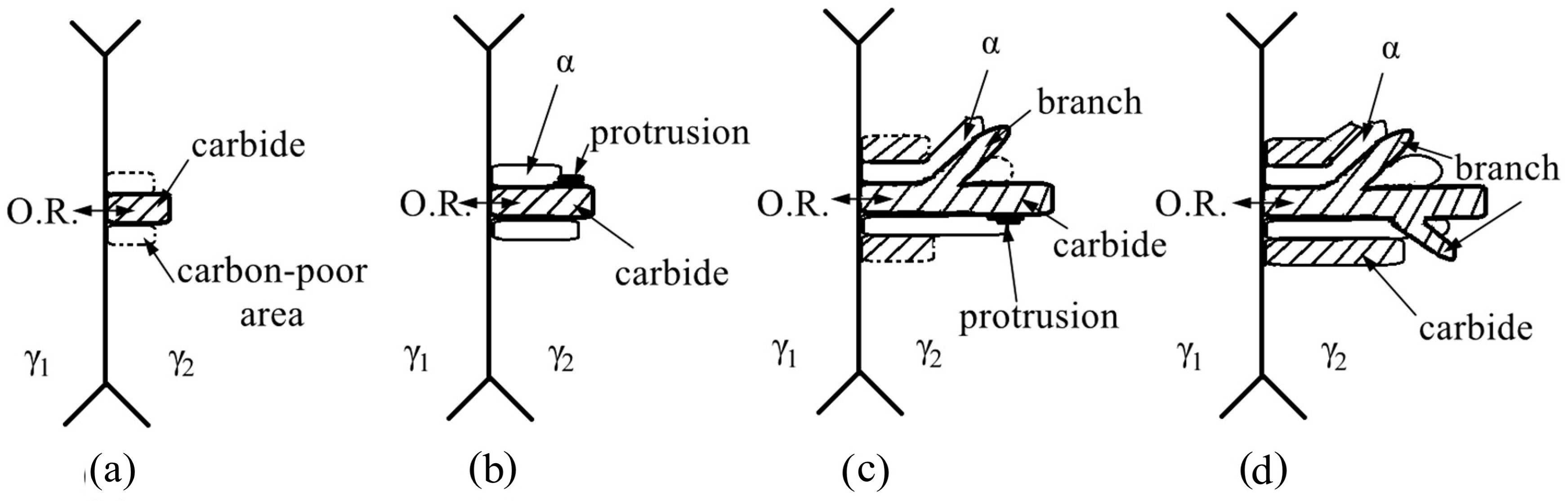
Figure 4 illustrates the growth process of chrysanthemum-like pearlite consisting of alternate lamellae of ferrite and carbide. Here, austenite decomposition directly forms the pearlite of M
7C
3+α, rather than that of M
3C+α. The reason for this is that the free energy of the M
7C
3+α system is lower than that of the M
3C+α system. In the formation process of the pearlite of M
7C
3+α, firstly, as the leading phase, the M
7C
3 carbide in pearlite preferentially nucleates perpendicular to the grain boundary at the interface, which results in a carbon-poor area formation on its side (
Figure 4a). Pearlite preferentially nucleates at grain boundaries because higher energy and more defects in these areas mean that it is easier to meet the structural fluctuations and composition fluctuations required for nucleation, and the stress concentration at the grain boundary may also promote nucleation and growth. When M
7C
3 lamella grows by edgewise growth, C atoms in austenite on the lamella’s lateral side are consumed, and atoms of the lattice plane stack column by column to induce M
7C
3 lamella thickening. However, if a certain factor causes problems in the process of stacking M
7C
3 lattice plane atoms, the stacking of atoms only occurs in the local part of the lamella, resulting in the formation of the protrusion structure at the local position, which leads to the easier formation of a carbon-poor area in austenite close to the protrusions, and then ferrite is formed, maintaining a certain OR with carbide precipitated preferentially (
Figure 4b). Although the formation of ferrite would hinder the accumulation process of the protrusion structure on M
7C
3 along the lamellae, the protrusion structure could still continue to accumulate and grow perpendicular to the lamellae, that is, the protrusion structure would continue to rise and get into the austenite approximately in the direction perpendicular to the lamella direction. When the protrusion grows to a certain height, it may begin to grow into the austenite along the lamella direction, and it eventually becomes a branch of pearlite lamellae (
Figure 4c). The process above is repeated, and the width of the growth frontier of M
7C
3 lamellae and ferrite lamellae becomes larger and even bends, and a pearlite colony appears chrysanthemum-like macroscopically due to the radial lamellae with the protrusion structure and branching structure (
Figure 4d).


