Review

Lithium Tetraborate as a Neutron Scintillation Detector: A Review

by
Elena Echeverria
1,*,
John McClory
2,
Lauren Samson
3,4,
Katherine Shene
3,
Juan A. Colón Santana
5,
Yaroslav Burak
6,
Volodymyr Adamiv
6,
Ihor Teslyuk
6,
Lu Wang
7,
Wai-Ning Mei
8,
Kyle A. Nelson
9,
Douglas S. McGregor
9,
Peter A. Dowben
10,*,
Carolina C. Ilie
3,*,
James Petrosky
2,* and
Archit Dhingra
11,*
1
Department of Physics, Oklahoma State University, 145 Physical Sciences Bldg., Rm 546, Stillwater, OK 74078, USA
2
Department of Engineering Physics, Air Force Institute of Technology, 2950 Hobson Way, Wright-Patterson AFB, OH 45433, USA
3
Physics Department, SUNY Oswego, 254 Shineman Center, Oswego, NY 13126, USA
4
Department of Physics, University at Buffalo, 239 Fronczak Hall, Buffalo, NY 14260-1500, USA
5
Department of Physics, Aurora University, 347 S. Gladstone Ave., Aurora, IL 60506-4892, USA
6
Institute of Physical Optics, 23 Dragomanov Str., 79-005 Lviv, Ukraine
7
CAS Key Lab of Materials for Energy Conversion, University of Science and Technology of China, Jinzhai Road 96, Hefei 230026, Anhui Province, China
8
Department of Physics, University of Nebraska–Omaha, Omaha, NE 68182, USA
9
S.M.A.R.T. Laboratory, Mechanical and Nuclear Engineering Department, Kansas State University, Manhattan, KS 66506, USA
10
Department of Physics and Astronomy, University of Nebraska-Lincoln, 855 North 16th Street, Lincoln, NE 68588, USA
11
Institut de Ciència dels Materials de la Universitat de València (ICMUV), University of Valencia, Carrer del Catedrátic José Beltrán Martinez, 2, 46980 Paterna, Valencia, Spain
*
Authors to whom correspondence should be addressed.
Crystals 2024, 14(1), 61; https://doi.org/10.3390/cryst14010061
Submission received: 24 November 2023 / Revised: 11 December 2023 / Accepted: 28 December 2023 / Published: 31 December 2023
(This article belongs to the Section Inorganic Crystalline Materials)

Abstract

:
The electronic structure and translucent nature of lithium tetraborate (Li2B4O7) render it promising as a scintillator medium for neutron detection applications. The inherently large neutron capture cross-section due to 10B and 6Li isotopes and the ease with which Li2B4O7 can be enriched with these isotopes, combined with the facile inclusion of rare earth dopants (occupying the Li+ sites), are expected to improve the luminescent properties, as well as the neutron detection efficiency, of Li2B4O7. The electronic structure of both doped and undoped Li2B4O7 were explored, using photoemission and inverse photoemission spectroscopies, optical measurements, and theoretical computational studies such as density functional theory. The scintillation properties are further enhanced because of the wide bandgap, making Li2B4O7 extremely translucent, so that capturing the neutron scintillation output is neither hindered nor diminished. Therefore, in this review, demonstrations of the possible amplification of neutron capture efficiencies, courtesy of rare-earth dopants, along with insights into a significantly large charge production (associated with neutron capture), are presented.

1. Introduction

Neutron detection is an inherent component of neutron radiation dosimetry, cross-border interdiction of fissile materials [1,2], nuclear reactor fuel and nuclear safety management [3,4], nonproliferation, nuclear stockpile monitoring, and nuclear medicine [5]. These particles are uncharged, which means that they do not provide a direct electronic signal, and they do not readily interact with most matter. In short, when compared to detection of other forms of radiation, neutron detection is nothing short of an ordeal. Therefore, when, in a Senate hearing, Dr. Robert J. Oppenheimer was asked what instrument he would use to detect an atomic bomb, his answer was “a screwdriver”, implying one would have to open every container to detect fissile materials because radiation emanations were extremely small [6].
Due to abovementioned reasons, practical neutron detection methods rely on indirect measurements based upon an initial neutron interaction producing a secondary species (conversion) that is readily measurable due to its effect on electronic and/or optical properties [7]. Neutron detectors are, therefore, divided into electronic (gas-filled or semiconductor devices, where ionization leads to an induced current or voltage pulse) or scintillation (absorption of radiation followed by luminescence in the material) detectors. However, the process of selecting just the right kind of materials to manufacture reliable neutron detectors faces a colossal challenge of circumventing the background radiation. To elaborate further, background γ-ray emissions, either from natural terrestrial sources or from the γ-ray emitters associated with the neutron source, can mask the secondary ionization or excitation signal from a neutron detector as well. Thus, many applications seek materials made of the lighter elements to remove or reduce the signals that might arise from associated X-ray and γ radiation, often referred to as being “γ-ray blind” [7], meaning that a very high neutron-to-gamma-ray detection ratio is sought [8]. Currently, there exist six kinds of materials used as scintillators: organic crystals, organic liquids, plastics, inorganic crystals, gases, and glasses. Among these materials, crystal, glass, and gas scintillators are often used for neutron detection; however, gases are less sensitive to β (beta) and γ (gamma) radiation, while the background from γ rays is generally higher for solids and liquids due to the higher atomic density. For thermal neutrons in particular, detectors with a high concentration of 6Li are employed because they enhance the scintillation sensitivity [9], which is why lithium tetraborate (Li2B4O7) has been touted to be a highly efficient material for applications in scintillation neutron detectors [10,11,12,13].
In this review article, the crystal and optical properties of the lithium tetraborate (Li2B4O7) are described. The optical properties and photoemission characteristics are discussed in detail to understand the advantage of using this material as a scintillator neutron detector. The most important results on the rare earth (RE) doping of this material and how this doping enhances the scintillation characteristics are also presented. Therefore, new research directions on scintillation efficiency and transparency can be identified through this review article. This information is critical to finally design and manufacture high-efficiency and low-cost Li2B4O7-based neutron scintillation detectors.

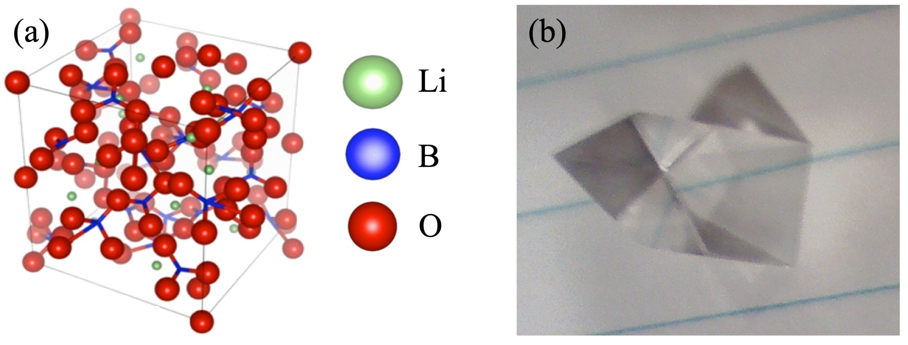
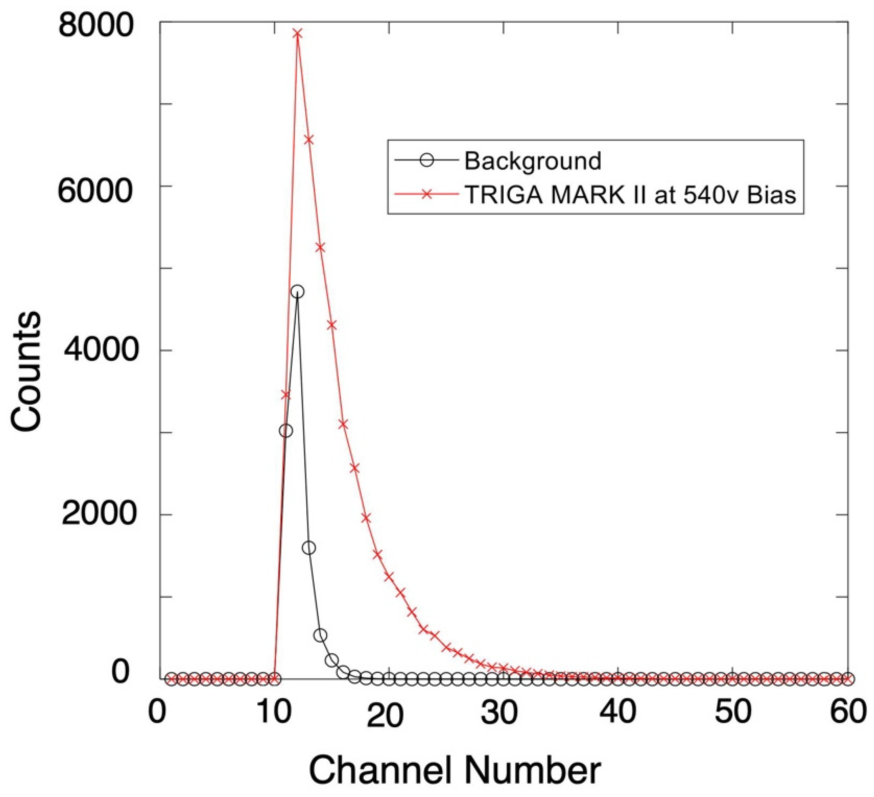
2. Lithium Tetraborate-Based Scintillation Detectors

Lithium tetraborate, usually known for its pyroelectric and piezoelectric properties [14,15,16], is a complex tetragonal crystal with 104 atoms per unit cell (see Figure 1a), with dimensions a = b = 9.470 Å and c = 10.290 Å and a space group of I41cd [17]. It has a characteristic wide electronic bandgap of ~8.9 to 10.1 eV [18], a large capability for thermal neutron capture, and high resistance to radiation damage. Li2B4O7 is also known to possess the best scintillation parameters among all the lithium borates [11,12,19,20], and multiple examples of experimental evidence advocating for the use of Li2B4O7 as a scintillator have existed for quite some time now [12].
Work by Zadneprovski et al. [11] confirmed that undoped Li2B4O7 is in fact γ blind, that is to say, largely insensitive to γ-ray radiation due to low γ-ray cross-sections, which is consistent with the fact that the primary elemental constituents of Li2B4O7 all have very low Z (i.e., atomic number) values. Since Li2B4O7 growth requires little post-material fabrication processing, scintillation detectors based on Li2B4O7 hold promise for an inexpensive and efficient detection system. Moreover, lightweight Li2B4O7 sheets can be combined with multiple scintillation–photomultiplier tubes into a single PIN diode (or photon sensor), so they can be scaled to large areas with little need for increased power or loss of detection area due to the need for pixelation and concomitant device connections, as would be the case in a solid-state device. Detectors based on Li2B4O7 can therefore be made thick enough to provide the necessary neutron moderation within the detector medium, leading to higher absolute efficiency. Lastly, Li2B4O7 is fairly immune to terrestrial-level temperature changes and unaffected by moisture and corrosion, making it well-suited for harsh environmental applications.
In terms of the physics of operation, the advantage of using Li2B4O7 as a neutron detection medium arises from the high thermal neutron capture cross-section inherent in the nuclear isotopes of B 5 10 B = 3935 barns) and L i 3 6 Li = 940 barns). Natural B consists of ~20% of 10B, and natural Li consists of ~6% of 6Li. Luminescence is generated by electron–hole pair creation and annihilation resulting from the energetic daughter products of 10B [9] and 6Li [10] capture reactions, as shown below:
  • 10B + n → 7Li (0.84 MeV) + 4He (1.47 MeV) + γ (0.48 MeV) (94%)
  • 10B + n → 7Li (1.015 MeV) + 4He (1.78 MeV) (6%)
  • 6Li + n → 3H + 4He + (4.8 MeV)
In order to improve the neutron interaction probability, Li2B4O7 can be formed using Li and B enriched with 6Li and 10B, respectively [10], thus increasing the thermal neutron capture cross-section. Both isotopes can be enriched using standard isotopic separation techniques. And even though standard isotopic separation techniques can be applied to enhance the enrichment of both the isotopes, usually Li is more widely used than B because its neutron capture reaction products have higher energy and lead to greater light output. Both the 6Li and 10B daughter fragments have significant kinetic energies, which, in turn, can lead to the creation of electron–hole pairs. The subsequent electron–hole pairs can then recombine to create light, but detection of the scintillation is more easily achieved if the light is in the visible range and the scintillation output is coupled with the photomultiplier or photodiode detector. Obviously, the light has to reach the detector, so a scintillator material that is close to transparent is ideal. This means that the optical properties and bandgap matter. But there are trade-offs, as discussed below. For example, a large bandgap may ensure a more translucent material but lead to the creation of fewer electron–hole pairs along the charge tracks left by the 6Li and 10B daughter fragments.
Single-crystal Li2B4O7, in its pure form, exhibits luminescence, but the scintillation efficiency is insufficient for practical neutron detection applications [21]. Another major drawback to using Li2B4O7 as a scintillation detector is that, like many glass-based materials, it is sensitive to electron (β), proton, and α radiation. Although it is possible to use a pulse height discrimination technique to separate 6Li or 10B neutron capture events from other events, the response time is on the order of 10 ns, and the light output is low, typically approximately 30% of that of anthracene [9]. In order to compensate for this disadvantage, the light output must be maximized to produce an adequate neutron capture scintillation response, obtained by select doping of the material. Fortunately, Li2B4O7 readily accepts the incorporation of dopants such as Cu and Ag, as well as rare earth elements, such as Yb [11,22], Ce [11], Nd [19,21], Sm [11], Eu [11,23], Gd [19], Tb [11], Er [19], and Tm [11,24], that enhance the luminescence by increasing recombination sites and adding luminescence lines [11,22,23,24,25,26,27,28], thus increasing the luminescent efficiency. Rare earth elements are especially useful, as they exhibit sharp luminescence originating from their intra-4f electronic transitions [22,23,24,25,26,29,30,31,32,33,34,35,36,37,38,39,40,41,42,43,44,45,46,47,48,49,50].
The partly filled 4f orbitals of the rare earth elements in conjunction with a filled 5s and 5p subshell provide enough shielding from the crystal field (electric field exerted by neighboring atoms) so that the energy levels of the rare earth ion closely resemble those of the free ions when incorporated into the Li2B4O7. This is evident from detailed electronic structure calculations of various rare earth dopants substitutionally placed in the Li2B4O7 lattice [25]. This property greatly increases luminescence efficiencies, i.e., signal above background, with the addition of small amounts of rare earths. Thus, the light output, generated as a consequence of neutron capture, is more readily detected in a photodetector and more easily distinguished from background. The doped Li2B4O7 results in a better linear dose response as compared to common thermoluminescent dosimeter materials (e.g., LiF), making it an attractive material for dosimetry applications [20,25,28,51]. Although many elements have been used for doping Li2B4O7, only cerium-activated lithium or borosilicate glass scintillators are well established and widely used as thermal (slow) neutron detectors [9,11,52,53,54,55]. And if the goal is to improve the sensitivity of Li2B4O7, it can be achieved by doping it with Ag [28]; in addition, combining the Ag-doped Li2B4O7 with solar-blind photomultiplier can also lead to a high signal-to-noise ratio [28].

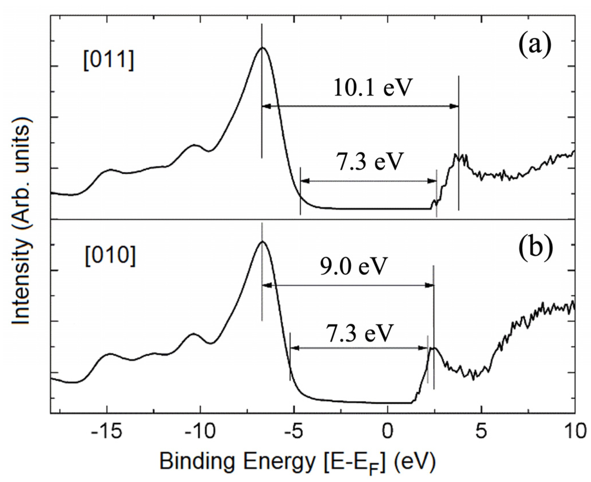
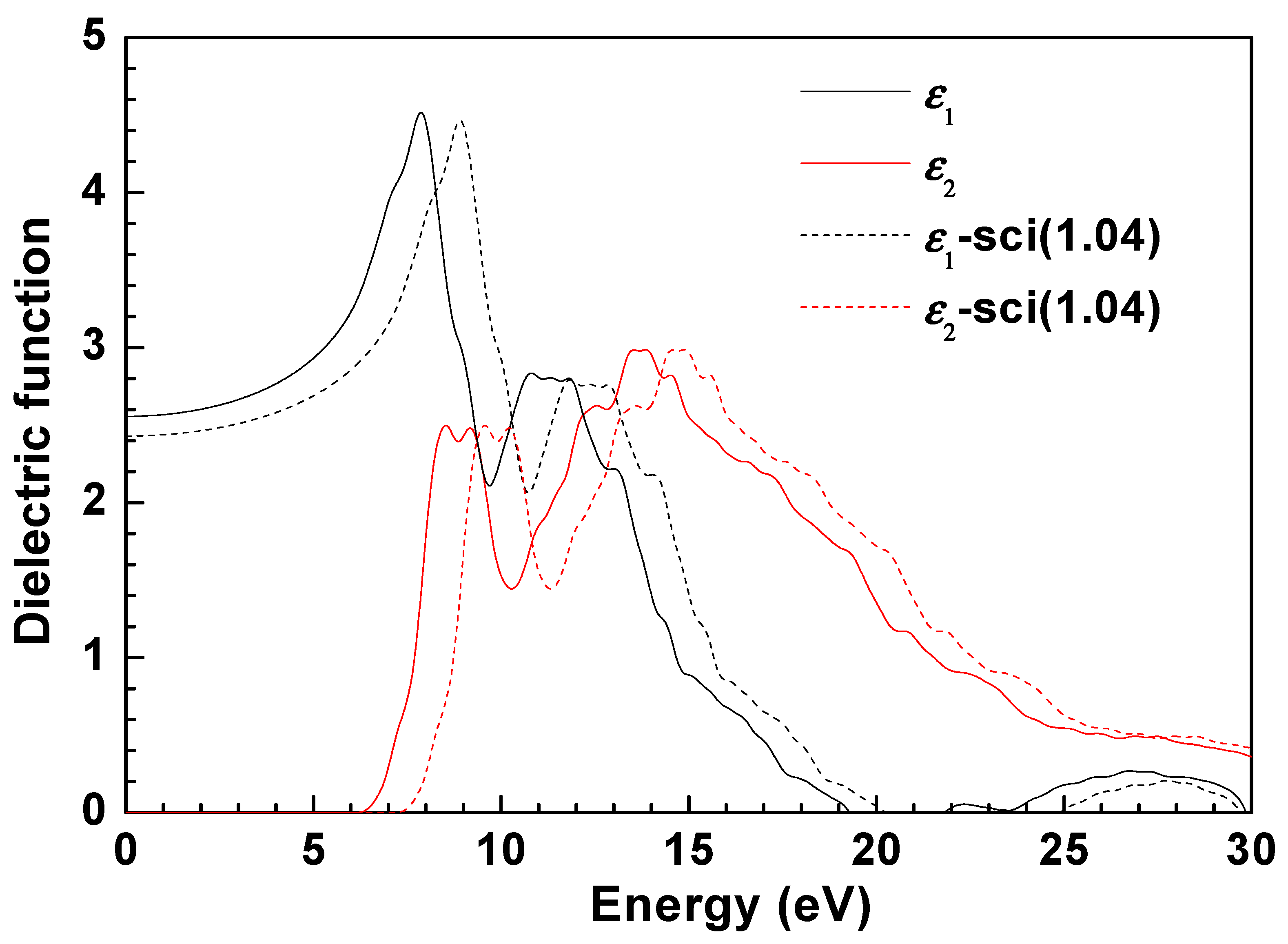
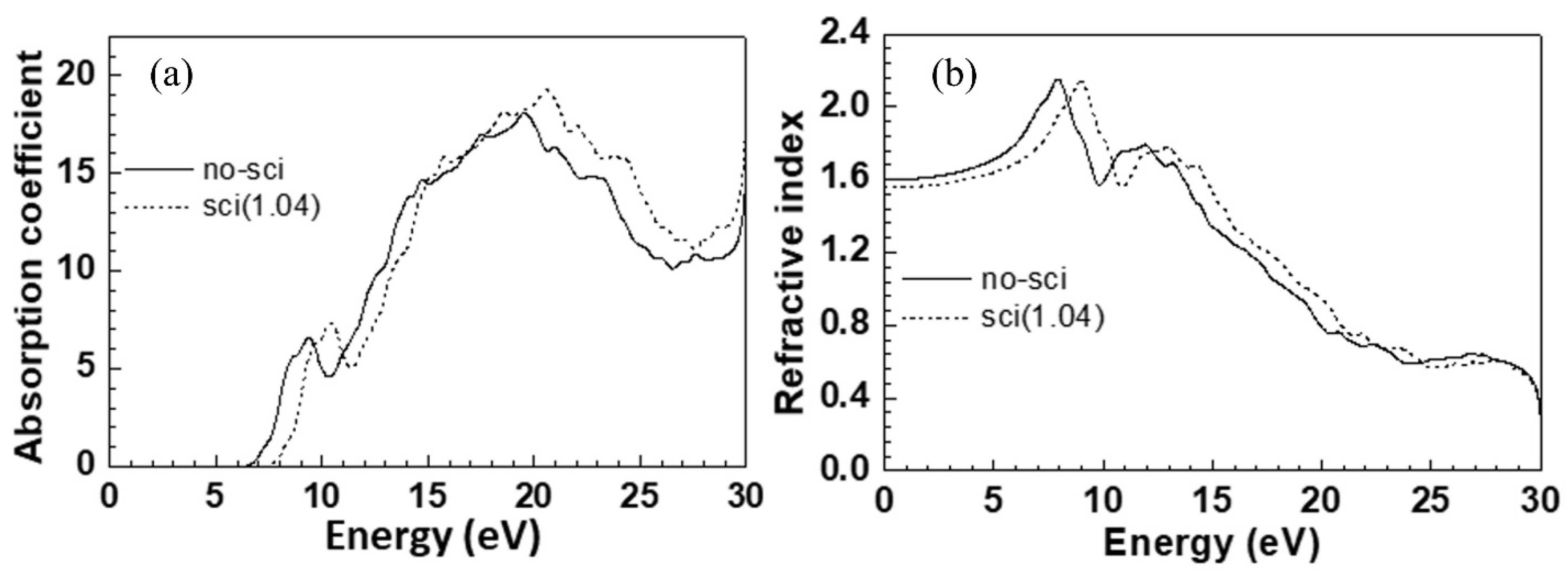
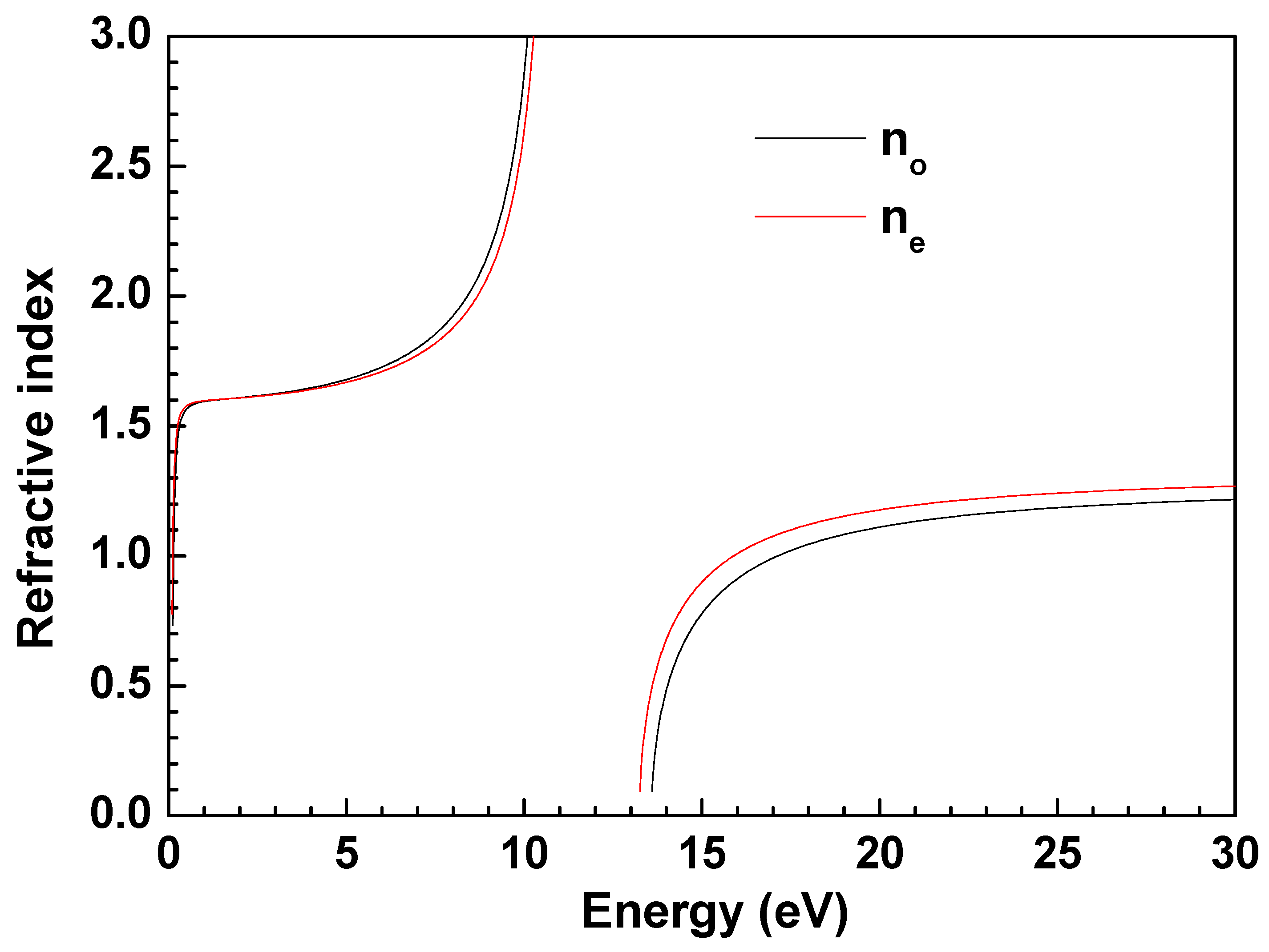
3. The Optical Characteristics of Li2B4O7

Owing to the wide electronic bandgap (~8.9 to 10.1 eV), as seen in the combined photoemission and inverse photoemission measurements [15,18], Li2B4O7 single crystals are transparent across a wide range of 165–6000 nm, and the fundamental absorption maximum is located at about 133 nm [15]. In nature, Li2B4O7 occurs as a clear, colorless mineral, as inclusions of diomignite in pegmatite, and it can be easily manufactured into crystals or glasses. As mentioned before, it can be fabricated into large sheets, using readily available manufacturing techniques, so the assembly of large area detector arrays is possible, and costs can be relatively low [51] because specialized materials processing is not required. Pure Li2B4O7 glasses, on the other hand, typically present high transparency in the range of 300–2600 nm, with three low-intensity emission bands centered at 402 nm, 520 nm, and 728 nm [11]. Regardless of whether it is a Li2B4O7 single crystal or a pure Li2B4O7 glass, the addition of dopants can be expected to alter their respective luminescence spectrum.
Figure 2 presents the transmission spectra of both undoped and doped single crystals of Li2B4O7. In this figure, signatures of an apparent trade-off between luminescence and transparency, as an increase in luminescence comes at the cost of transparency and efficiency of light collection, are observed. Doping Li2B4O7 single crystals with Ag gives birth to new absorption bands at 174 nm and 205 nm (indicated by the arrows in the figure), whereas both undoped and Cu-doped Li2B4O7 crystals present a broad low-intensity band [51].
Figure 3 presents the electronic band configuration of Li2B4O7 obtained from photoemission spectroscopy (PES) and inverse photoemission spectroscopy (IPES). The spectra reveal several sub-band transitions. The valence band has a high-intensity (or high electron density) primary peak in photoemission (below the Fermi level (EF)), and the strong feature above EF, observed in inverse photoemission, denotes the conduction band edge. A detailed analysis of the structures shown in Figure 3 unveils that the top of the valence band in a Li2B4O7 single crystal is mainly occupied by just boron–oxygen groups, while the bottom of its conduction band includes orbital contributions from lithium as well [15,57]. The energy interval between the two strong spectral features in Figure 3, one each in photoemission and inverse photoemission, represent the ground-state bandgap. From these measurements [15,18] on a Li2B4O7(100) crystal, the ground-state bandgap is found to be 9.8 ± 0.5 eV, falling right in the range between 8.9 ± 0.5 eV and 10.1 ± 0.5 eV [15,18,56,58,59], which is somewhat in line with theoretical expectations [57]. These measured values for the ground-state bandgap are higher than the previously measured values of the optical gap (Eg (opt) = 7.4 eV) extrapolated from the absorption plot [15,56], but they are closer to the theoretical ground-state bandgap. This means that incident photons possessing energy less than the ground-state bandgap, determined from photoemission and inverse photoemission, can still create electron–hole pairs by exciting electrons from the valence band to the conduction band. The creation of carriers will manifest as an increase in the conductivity of the crystal, especially if the carrier mobility and lifetimes are reasonable. Such an increase in the electron population in the conduction band due to optical excitations will, in turn, amplify the photoconductivity of Li2B4O7 crystal, while modifying its optical parameters. That is to say, once the Li2B4O7 crystal becomes conductive, the complex refractive index, n ~ = n 1 + i χ , becomes more relevant to the crystal structure model.
The overall dielectric function for all coordinate indices for Li2B4O7 can be determined via the crystallographic direction-dependent density functional theory (DFT). However, bandgaps estimated from DFT are subject to error and typically produce an incorrect bandgap that is smaller than the true ground-state bandgap [60,61,62,63]. Therefore, the scissor approximation method (SOA) was applied to the real ε1(E) and imaginary ε2(E) parts of the dielectric function (shown in Figure 4). In these calculations, the scissor correction error of 1.04 eV is towards the lower end of the range specified by Rasmussen [64] of 1-to-3 eV. This error is small, producing a near-identical approximation to the ground-state bandgap of 7.3 eV that is found using the generalized gradient approximation (GGA), shown in Figure 5. Here, it must be noted that this gap of 7.3 eV is close to the ~7.4 eV gap inferred from optical transmission (Figure 2) and the band edges seen in combined photoemission and inverse photoemission (Figure 3); therefore, the usage of these corrections to DFT appears somewhat reasonable. Figure 6 illustrates the calculated absorption coefficient and refractive index with and without using the scissor approximation averaged over three directions to account for the fact that all crystal faces are not identical in their symmetry.
In addition, with the use of Sellmeier equations, the refractive index of the single lithium tetraborate crystals can be easily verified. The Sellmeier equations applied to the Li2B4O7 crystals are as follows [65,66,67]:
n 0 2 = 2.56431 + 0.012337 λ 2 0.013013 0.019075 λ 2
n 0 2 = 2.38651 + 0.010664 λ 2 0.012878 0.012813 λ 2
The resulting refractive indices are plotted in Figure 7, where n 0 2 and n e 2 represent the ordinary and extraordinary part of the optical response to the incident light traversing a single lithium tetraborate crystal along the C4 axis. The curves center on a refractive index of 1.5, which matches the secondary peak seen in Figure 6. In addition, the small differences in the curves demonstrate nontrivial birefringence, with a bandgap of approximately 7.41 eV to 10.1 eV (with the calculated value being 6.37 eV) indicating an implicit correction of 1.04 eV to 3.73 eV. While 7.3 eV is close to the 7.4–7.5 eV gap determined from optical transmission (Figure 2), the bandgap value of 10.1 eV is close to the ground-state gap of 9–10 eV extrapolated from the combined photoemission and inverse photoemission spectra (Figure 3).
The fact that structural distortions, particularly at the interface of Li2B4O7 single crystals, affect the electron levels in atoms was clearly demonstrated by Wooten et al. [18,58] and the theory of [68]. The influences of imperfections and defects in the lattice of Li2B4O7 single crystals are highlighted in Figure 8, which illustrates that the absorption edge for the Li2B4O7 glass differs substantially from that of the Li2B4O7 single crystal [67]. From this figure, it is evident that the fundamental absorption maximum for borate glass occurs at much longer wavelengths [67], i.e., lower energies, in comparison with that of the Li2B4O7 single crystal [58,67,69]. For the Li2B4O7 glass, the absorption spectrum shows an indistinct absorption edge, which is common for glassy samples since the crystallographic direction-dependent anisotropic optical properties are expected to be suppressed [70]. The electronic structure of disordered media, which include Li2B4O7 glasses, can still be reconciled with the electronic states of Li2B4O7 single crystals [19], chiefly because of the similarities in the electron energy density distributions. With this in mind, the long wavelength shift of the absorption edge of the glass in comparison with single crystals can be explained by blurring the boundary of the electronic density of states. Moreover, the energy band model is still valid here, considering that the direct inter-band transitions are forbidden, with indirect transitions of phonons and excitons occurring through mediation. A detailed discussion regarding such indirect optical transitions is presented elsewhere [71].

4. Factors Affecting Charge Production

As noted above, lithium tetraborate has a ground-state bandgap of roughly 9.8 eV and a measured optical bandgap of 6.7 eV, and this significant bandgap, while improving light transmission, limits the number of electron–hole pairs created by the reaction products resulting from neutron capture. This optical bandgap corresponds to an approximate number of possible charge hole pairs of ~410,000 for the most probable reaction channel of 10B (94%), which has a much higher cross-section than 6Li. This calculation, of course, assumes that all the reaction energy is absorbed. Having said that, for a more accurate calculation, a correction factor to completely account for the production of an electron–hole pair due to the absorption of incoming radiation with an energy above the bandgap must be included. These correction factors have been estimated to be 3.17 [72] and 3.44 [73,74], and employing the correction factor calculated by Klein [72] indicates that a 10B reaction should produce roughly 200,000 charges. Using the same logic for 6Li, approximately 416,000 charges would be produced, but recall that this advantage is reduced since it is known that 6Li has a smaller cross-section and lower elemental concentration. These numbers are, of course, not completely realistic, as they are just the upper limits, as not all of the subsequently created electron–hole pairs will give rise to detectable scintillations. Instead, a neutron capture event near the surface (or an interface) can also result in either an incomplete electron–hole production or Auger-electron production or photoemission. Without defects or a dopant, excitonic decays are capable of producing photons well into the UV, given that the optical gap is 6.7 eV, while the ground-state bandgap is 9.8 eV.
Convincing evidence of electron–hole pair production from neutron irradiation can be collected by considering Li2B4O7 as a capacitive detector. The electrical response of a Li2B4O7 detector to a neutron fluence is expected to result in a distinctly different pulse count while being irradiated, as compared to the background measurement. A Li2B4O7 crystal was irradiated in the radial neutron beam of a TRIGA Mark II nuclear reactor, and the operating bias was increased (or decreased) until a signal was detected. Once the operating biases were fixed upon the detection of a signal, the pulse height spectroscopy data were recorded, as shown in Figure 9. The shutter to the beam was opened and then closed cyclically between 10-min irradiation measurements and 10-min background measurements, using a multichannel analyzer. These results demonstrate an increase in conductance with neutron capture, consistent with the electron–hole pair creation from the Li and He or 3H and 4He ion tracks. Although counts were observed above the background during irradiation, there are no distinct spectral differences. This outcome indicates that the background electrical noise is likely due to dielectric breakdown or an increase in conductivity due to electron–hole pair creation, much like the expected increase in conductivity due to photocarrier creation discussed above.

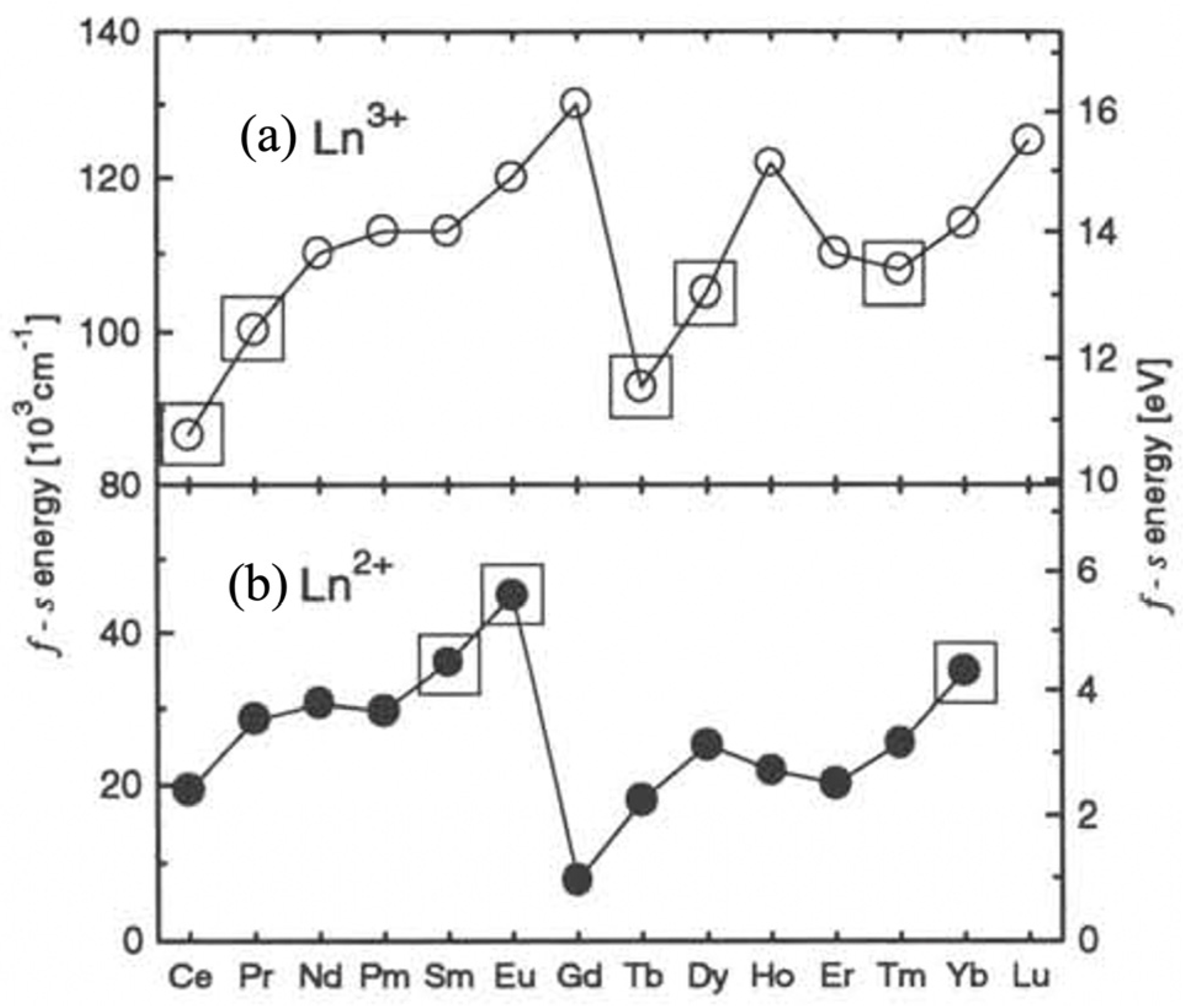
5. Factors Affecting Light Production

Based upon the bandgap of Li2B4O7, scintillation is expected to produce a photon in the UV energy range. Figure 10 provides the scintillation response of undoped Li2B4O7 to α particle radiation from 241Am (Figure 10a), and neutrons plus α particles from a 239Pu source (Figure 10b). In these cases, assessing the interactions with incoming α radiation is particularly important, as α particles are also among the main reaction products of the 10B or 6Li neutron capture reactions. The results shown in Figure 10 confirm that the majority of the light response falls below ~450 nm, which is largely in the UV spectrum (10–400 nm). However, it is desirable to produce visible light in order to exploit the high efficiencies of PIN diodes and photomultiplier tubes. Therefore, if highly efficient scintillator neutron detection systems are to be realized using Li2B4O7, then the neutron capture must be maximized, as the bandgap is engineered to produce more transitions to a longer wavelength (i.e., in the visible range) while being unaffected by environmental factors such as temperature. One bandgap-engineering option for increasing the wavelength in the light emission spectrum is through the inclusion of defects into the Li2B4O7 structure. It has been shown that surface states [68,75] produce photovoltaic charging effects on the material, pinning the surface potential 3.5 eV away from the conduction band minimum (see Figure 3) [76]. Therefore, surfaces states and defects can lead to scintillation in the near-visible [77,78,79], as shown in Figure 10b.
Most of the rare earths exhibit emission in the visible region and into the near-infrared region [37,44]. Depending on the host, these transitions can be modified; however, all the electronic levels of the rare earth will remain inside of the bandgap of the host. When the rare earth takes the place of one of the atoms in the host, such sites become a trap center. The new transitions or trap energies can be observed via thermoluminescence, radioluminescence, or light output measurements. In 1996, Wojtowicz [80] used a simple band structure model to study the scintillation mechanism of a compound in the form of AB3 doped with a rare earth ion and found that, depending on the f-s energy promotion [80] a rare earth ion will act as an electron or hole trap. Energy calculations, based upon the f-s transition energy, propose lanthanide ions as the prime candidates to be used as activators for electron or hole traps. These ions can be used in Li2B4O7-based compounds (as shown in Figure 11) to act as outstanding activators for modification of the luminescence spectrum [80].
Not only are the luminescence spectra of rare earth-doped Li2B4O7 affected by the kind of rare earth dopant [11,22,24,25,26,30,32,34,40,41,47,81]; they are also affected by the rare earth dopant’s concentration [30,81] and the growth atmosphere [34]. The preliminary work of Zadneprovski et al. [11] suggests that co-doping Li2B4O7 with Cu along with many rare earth dopant additions leads to a very efficient neutron scintillation (Figure 12), and this may limit the available concentrations of activators when managing the overall luminescence spectrum.
Rare earth elements tend to occupy the Li+ sites of Li2B4O7 [25,40,47], and the structural geometry of rare earth-doped Li2B4O7, as shown in Figure 13, does not change significantly with dopants. The occupation of B sites by a rare earth element is quite unlikely owing to the large difference in the ionic radii of B ions and that of the rare earth elements (and the oxygen coordination number of the rare earth). Li+ substitution is not the only consequence of rare earth doping; a few site distortions and site disorders are present as well, due to the change in the lengths of the bonds between the rare earth and surrounding atoms. X-ray absorption near-edge structure (EXAFS) data have shown that bond lengths decrease with the increase in atomic number [25]. It is also known that rare earth impurities on Li2B4O7 are present in the form of trivalent (RE3+) ions [32,40,47]. Kelly et al. [25], using density functional theory (DFT), show indications of strong hybridization between rare earth states and the Li2B4O7 host. In their study [25], they used five rare earth elements: Nd, Gd, Dy, Er, and Yb; however, only the first four demonstrate overlapping of the unoccupied 4f levels of the rare earth with the conduction band of Li2B4O7 [25]. This finding is another indication that rare earth elements tend to occupy Li+ instead of B, because Li+ is bonded to the B4O7 by ionic bonds, while boron and oxygen are strongly tied via covalent bonds. The importance of understanding hybridization in scintillators cannot be emphasized enough because significant hybridization between the rare earth states and the Li2B4O7 host can increase luminescence and decrease excited-state lifetimes. The rare earths will add states within the undoped Li2B4O7, reducing the bandgap at high concentrations without affecting the transparency [24].
A final challenge when adding activators to Li2B4O7 is in avoiding degradation to transparency. This is still a ripe area for research and material advancement. In experiments involving the doping of Li2B4O7 with 3% Er via concentration, photo-optical characterization demonstrated no reduction in transparency; however, little effort has been made to characterize its total scintillation efficiency [25]. Er, like Ce, Eu, Tm, and Yb, is a rare earth element with similar chemical behavior, and, thus, other rare earth elements also appear quite promising.
The preliminary results of Kelly et al. [25] are not too surprising given the prior successes with the rare earth doping of Li2B4O7. What is still not known is the range of possible elemental concentrations or combinatory mixtures of co-doping Li2B4O7 with Cu along with other rare earth elements, such as Ce, Tb, and Er, and their effects on its optical and mechanical properties. In some cases, the optical transparency was improved, while it stayed unaltered in other cases [11]. Additionally, about 80–85% of the transmission in doped Li2B4O7 is revealed to be in the range of 350 nm to 800 nm [11], demonstrating the high optical quality of the pure and doped glasses, which is quite an encouraging result (to say the least).

6. Conclusions

In conclusion, Li2B4O7 has several inherent physical, atomic, and nuclear properties that establish it as a promising candidate for a high-efficiency, low-power, robust, low false-positive neutron detection medium. Although the elemental content and structure of Li2B4O7, along with the possibilities for 6Li and 10B isotopic enrichment, render it a great candidate for neutron scintillation detector material, there is still substantial room for improvement in order to attain higher quantum efficiencies. Such a goal can be achieved through a better understanding of the parameters affecting detection efficiencies, improved signal-to-noise ratio, background rejections, and self-moderation of MeV neutrons so as to determine the best moderator combination. The interplay of these factors complicates finding the ideal dopants and their respective doping concentration for producing higher scintillation following interactions with neutrons. As it stands, doping Li2B4O7 with either europium, ytterbium, samarium, or copper is especially promising, as existing investigations indicate that they can be readily introduced into its crystal structure, barring a few minor detrimental effects to its luminescence and mechanical qualities. All things considered, it is fair to say that further experimental studies on both the scintillation efficiency and transparency of Li2B4O7 are needed.

Author Contributions

Conceptualization, E.E., Y.B., J.A.C.S., P.A.D., C.C.I. and A.D.; methodology, J.M., J.P., K.A.N. and D.S.M.; software, L.W. and W.-N.M.; validation, E.E., Y.B., J.A.C.S., I.T., V.A., J.M., J.P., K.A.N., D.S.M.; formal analysis, E.E., Y.B., J.A.C.S., I.T., V.A., J.M., J.P., K.A.N., P.A.D., L.S., K.S., D.S.M. and A.D.; investigation, E.E., Y.B., J.A.C.S., I.T., V.A., J.M., J.P., K.A.N. and D.S.M.; resources, J.M., J.P., P.A.D., C.C.I., D.S.M., Y.B., V.A., A.D. and W.-N.M.; data curation, L.W., W.-N.M., J.M., Y.B., V.A., I.T., K.A.N., D.S.M., J.P.; writing—original draft preparation, E.E., L.S., K.S., C.C.I., J.A.C.S. and A.D.; writing—review and editing, E.E., J.P., D.S.M., P.A.D., C.C.I. and A.D.; visualization, J.P., W.-N.M., Y.B., J.M. and A.D.; supervision, J.M., J.P., A.D., Y.B., D.S.M. and C.C.I.; project administration, C.C.I., E.E., J.M., A.D. and J.P.; funding acquisition, J.P., J.M., W.-N.M. and P.A.D. All authors have read and agreed to the published version of the manuscript.

Funding

This work was supported by Project PID2020-112507GB-I00, funded by MCIN/AEI/10.13039/501100011033; the National Aeronautics and Space Administration, through grants NNX13AN16A, NNX14AL11A, and NNX15AI09H; the Defense Threat Reduction Agency (Grant No. HDTRA1-14-1-0041); Nebraska Public Power District through the Nebraska Center for Energy Sciences Research; and the National Science Foundation through EPSCoR RII Track-1: Emergent Quantum Materials and Technologies (EQUATE), Award OIA-2044049.

Data Availability Statement

The datasets generated during and/or analyzed during the current study are available from the corresponding authors upon reasonable request.

Acknowledgments

The authors would like to thank Benjamin W. Montag, Brant E. Kananen, and T. D. Kelly for their technical support.

Conflicts of Interest

The authors declare that they have no known competing financial interests or personal relationships that could have appeared to influence the work reported in this article.

References

  1. Kouzes, R.T. Detecting Illicit Nuclear Materials: The Installation of Radiological Monitoring Equipment in the United States and Overseas Is Helping Thwart Nuclear Terrorism. Am. Sci. 2005, 93, 422–427. [Google Scholar] [CrossRef]
  2. Kouzes, R.T.; Ely, J.H.; Erikson, L.E.; Kernan, W.J.; Lintereur, A.T.; Siciliano, E.R.; Stephens, D.L.; Stromswold, D.C.; Van Ginhoven, R.M.; Woodring, M.L. Neutron Detection Alternatives to 3He for National Security Applications. Nucl. Instrum. Methods Phys. Res. A 2010, 623, 1035–1045. [Google Scholar] [CrossRef]
  3. Seymour, R.S.; Richardson, B.; Morichi, M.; Bliss, M.; Craig, R.A.; Sunberg, D.S. Scintillating-Glass-Fiber Neutron Sensors, Their Application and Performance for Plutonium Detection and Monitoring. J. Radioanal. Nucl. Chem. 2000, 243, 387–388. [Google Scholar] [CrossRef]
  4. Van der Ende, B.M.; Li, L.; Godin, D.; Sur, B. Stand-off Nuclear Reactor Monitoring with Neutron Detectors for Safeguards and Non-Proliferation Applications. Nat. Commun. 2019, 10, 1959. [Google Scholar] [CrossRef] [PubMed]
  5. Eijk, C.W.E. van Inorganic Scintillators in Medical Imaging. Phys. Med. Biol. 2002, 47, R85–R106. [Google Scholar] [CrossRef] [PubMed]
  6. Bird, K.; Sherwin, M.J. American Prometheus: The Triumph and Tragedy of J. Robert Oppenheimer; Alfred A. Knopf: New York, NY, USA, 2005. [Google Scholar]
  7. Caruso, A.N. The Physics of Solid-State Neutron Detector Materials and Geometries. J. Phys. Condens. Matter 2010, 22, 443201. [Google Scholar] [CrossRef]
  8. Frangville, C.; Grabowski, A.; Dumazert, J.; Montbarbon, E.; Lynde, C.; Coulon, R.; Venerosy, A.; Bertrand, G.H.V.; Hamel, M. Nanoparticles-Loaded Plastic Scintillators for Fast/Thermal Neutrons/Gamma Discrimination: Simulation and Results. Nucl. Instrum. Methods Phys. Res. A 2019, 942, 162370. [Google Scholar] [CrossRef]
  9. Leo, W.R. Techniques for Nuclear and Particle Physics Experiments; Springer: Berlin/Heidelberg, Germany, 1994; ISBN 978-3-540-57280-0. [Google Scholar]
  10. Burak, Y.V.; Adamiv, V.T.; Teslyuk, I.M.; Shevel, V.M. Optical Absorption of Isotopically Enriched Li2B4O7 Single Crystals Irradiated by Thermal Neutrons. In Radiation Measurements; Pergamon: Bergama, Turkey, 2004; Volume 38, pp. 681–684. [Google Scholar]
  11. Zadneprovski, B.I.; Eremin, N.V.; Paskhalov, A.A. New Inorganic Scintillators on the Basis of LBO Glass for Neutron Detection. Funct. Mater. 2005, 12, 261–268. [Google Scholar]
  12. Seidl, E.; Schwertführer, W. Lithiumborat-Einkristalle Als Neutronendetektoren. Atomkernenergie 1966, 11, 155–162. [Google Scholar]
  13. Vinograd, E.L.; Vydai, Y.T.; Zagariy, L.B.; Kosmyna, M.B.; Kudin, A.M.; Levin, A.B.; Nazarenko, B.P.; Tarasov, V.A.; Chernikov, V.V. Scintillation Parameters of Single Crystals Containing Lithium: Li2B4O7, LiTaO3, LiNbO3. Funct. Mater. 1994, 1, 152–154. [Google Scholar]
  14. Bhalla, A.S.; Cross, L.E.; Whatmore, R.W. Pyroelectric and Piezoelectric Properties of Lithium Tetraborate Single Crystal. Jpn. J. Appl. Phys. 1985, 24, 727. [Google Scholar] [CrossRef]
  15. Adamiv, V.T.; Burak, Y.V.; Wooten, D.J.; McClory, J.; Petrosky, J.; Ketsman, I.; Xiao, J.; Losovyj, Y.B.; Dowben, P.A. The Electronic Structure and Secondary Pyroelectric Properties of Lithium Tetraborate. Materials 2010, 3, 4550–4579. [Google Scholar] [CrossRef]
  16. Ketsman, I.; Wooten, D.; Xiao, J.; Losovyj, Y.B.; Burak, Y.V.; Adamiv, V.T.; Sokolov, A.; Petrosky, J.; McClory, J.; Dowben, P.A. The Off-Axis Pyroelectric Effect Observed for Lithium Tetraborate. Phys. Lett. A 2010, 374, 891–895. [Google Scholar] [CrossRef]
  17. Marzouk, Z.G.; Dhingra, A.; Burak, Y.; Adamiv, V.; Teslyuk, I.; Dowben, P.A. Long Carrier Lifetimes in Crystalline Lithium Tetraborate. Mater. Lett. 2021, 297, 129978. [Google Scholar] [CrossRef]
  18. Wooten, D.; Ketsman, I.; Xiao, J.; Losovyj, Y.B.; Petrosky, J.; McClory, J.; Burak, Y.V.; Adamiv, V.T.; Brown, J.M.; Dowben, P.A. The Electronic Structure of Li2B4O7(110) and Li2B4O7(100). EPJ Appl. Phys. 2010, 52, 31601. [Google Scholar] [CrossRef]
  19. Baumer, V.N.; Chernikov, V.V.; Dubovik, M.F.; Gavrylyuk, V.P.; Grinyov, B.V.; Grin, L.A.; Korshikova, T.I.; Shekhovtsov, A.N.; Sysoeva, E.P.; Tolmachev, A.V.; et al. Comparative Analysis of Scintillation Parameters Peculiarities of Li2B4O7, LaB3O6, Li6Gd(BO3)3 Single Crystals. Funct. Mater. 2001, 8, 736–741. [Google Scholar]
  20. Özdemir, Z.; Özbayoğlu, G.; Yilmaz, A. Investigation of Thermoluminescence Properties of Metal Oxide Doped Lithium Triborate. J. Mater. Sci. 2007, 42, 8501–8508. [Google Scholar] [CrossRef]
  21. Ogorodnikov, I.N.; Pustovarov, V.A.; Kruzhalov, A.V.; Isaenko, L.I.; Kirm, M.; Zimmerer, G. Self-Trapped Excitons in LiB3O5 and Li2B4O7 Lithium Borates: Time-Resolved Low-Temperature Luminescence VUV Spectroscopy. Phys. Solid State 2000, 42, 464–472. [Google Scholar] [CrossRef]
  22. Podgórska, D.; Kaczmarek, S.M.; Drozdowski, W.; Berkowski, M.; Worsztynowicz, A. Growth and Optical Properties of Li2B4O7 Single Crystals Pure and Doped with Yb, Co and Mn Ions for Nonlinear Applications. Acta Phys. Pol. A 2005, 107, 507–518. [Google Scholar] [CrossRef]
  23. Dubovik, M.F.; Tolmachev, A.V.; Grinyov, B.V.; Grin, L.A.; Dolzhenkova, E.F.; Dobrotvorskaya, M.V. Luminescence and Radiation-Induced Defects in Li₂B₄O₇:Eu Single Crystals. Quantum Electron. Optoelectron. 2000, 3, 420–422. [Google Scholar] [CrossRef]
  24. Kindrat, I.I.; Padlyak, B.V.; Lisiecki, R.; Adamiv, V.T. Spectroscopic and Luminescent Properties of the Lithium Tetraborate Glass Co-Doped with Tm and Ag. J. Lumin. 2020, 225, 117357. [Google Scholar] [CrossRef]
  25. Kelly, T.D.; Petrosky, J.C.; McClory, J.W.; Adamiv, V.T.; Burak, Y.V.; Padlyak, B.V.; Teslyuk, I.M.; Lu, N.; Wang, L.; Mei, W.-N.; et al. Rare Earth Dopant (Nd, Gd, Dy, and Er) Hybridization in Lithium Tetraborate. Front. Phys. 2014, 2, 31. [Google Scholar] [CrossRef]
  26. Kindrat, I.I.; Padlyak, B.V.; Lisiecki, R.; Adamiv, V.T. Spectroscopic and Luminescent Properties of the Lithium Tetraborate Glass Co-Doped with Nd and Ag. J. Alloys Compd. 2021, 853, 157321. [Google Scholar] [CrossRef]
  27. Nagirnyi, V.; Kotlov, A.; Corradi, G.; Watterich, A.; Kirm, M. Electronic Transitions in Li2B4O7:Cu Single Crystals. Phys. Status Solidi C 2007, 4, 885–888. [Google Scholar] [CrossRef]
  28. Patra, G.; Singh, A.; Tiwari, B.; Singh, S.; BARe, D.D. Undefined Development of Thermal Neutron Detector Based on Lithium Tetraborate (LTB) Single Crystal. 2015. Available online: barc.gov.in (accessed on 24 November 2023).
  29. Chen, Y.; Huang, Y.; Luo, Z. Spectroscopic Properties of Yb3+ in Bismuth Borate Glasses. Chem. Phys. Lett. 2003, 382, 481–488. [Google Scholar] [CrossRef]
  30. Danilyuk, P.S.; Popovich, K.P.; Puga, P.P.; Gomonai, A.I.; Primak, N.V.; Krasilinets, V.N.; Turok, I.I.; Puga, G.D.; Rizak, V.M. Optical Absorption Spectra and Energy Levels of Er3+ Ions in Glassy Lithium Tetraborate Matrix. Opt. Spectrosc. 2014, 117, 759–763. [Google Scholar] [CrossRef]
  31. Ignatovych, M.; Holovey, V.; Watterich, A.; Vidóczy, T.; Baranyai, P.; Kelemen, A.; Chuiko, O. Luminescence Characteristics of Cu- and Eu-Doped Li2B4O7. Radiat. Meas. 2004, 38, 567–570. [Google Scholar] [CrossRef]
  32. Ishii, M.; Kuwano, Y.; Asaba, S.; Asai, T.; Kawamura, M.; Senguttuvan, N.; Hayashi, T.; Koboyashi, M.; Nikl, M.; Hosoya, S.; et al. Luminescence of Doped Lithium Tetraborate Single Crystals and Glass. Radiat. Meas. 2004, 38, 571–574. [Google Scholar] [CrossRef]
  33. Jayasankar, C.K.; Babu, P. Optical Properties of Sm3+ Ions in Lithium Borate and Lithium Fluoroborate Glasses. J. Alloys Compd. 2000, 307, 82–95. [Google Scholar] [CrossRef]
  34. Kaczmarek, S.M. Li2B4O7 Glasses Doped with Cr, Co, Eu and Dy. Opt. Mater. 2002, 19, 189–194. [Google Scholar] [CrossRef]
  35. Kassab, L.; Tatumi, S.; Morais, A.; Courrol, L.; Wetter, N.; Salvador, V. Spectroscopic Properties of Lead Fluoroborate Glasses Doped with Ytterbium. Opt. Express 2001, 8, 585. [Google Scholar] [CrossRef] [PubMed]
  36. Kelly, T.D.; Kong, L.; Buchanan, D.A.; Brant, A.T.; Petrosky, J.C.; McClory, J.W.; Adamiv, V.T.; Burak, Y.V.; Dowben, P.A. EXAFS and EPR Analysis of the Local Structure of Mn-Doped Li2B4O7. Phys. Status Solidi B 2013, 250, 1376–1383. [Google Scholar] [CrossRef]
  37. Kenyon, A. Recent Developments in Rare-Earth Doped Materials for Optoelectronics. Prog. Quantum Electron. 2002, 26, 225–284. [Google Scholar] [CrossRef]
  38. Kobayashi, M.; Ishii, M.; Senguttuvan, N. Scintillation Characteristics of Undoped and Cu+-Doped Li2B4O7 Single Crystals. arXiv 2015, arXiv:1503.03759. [Google Scholar]
  39. Lin, H.; Yang, D.; Liu, G.; Ma, T.; Zhai, B.; An, Q.; Yu, J.; Wang, X.; Liu, X.; Yue-Bun Pun, E. Optical Absorption and Photoluminescence in Sm3+- and Eu3+-Doped Rare-Earth Borate Glasses. J. Lumin. 2005, 113, 121–128. [Google Scholar] [CrossRef]
  40. Padlyak, B.; Ryba-Romanowski, W.; Lisiecki, R.; Adamiv, V.; Burak, Y.; Teslyuk, I.; Banaszak-Piechowska, A. Optical Spectra and Luminescence Kinetics of the Sm3+ and Yb3+ Centres in the Lithium Tetraborate Glasses. Opt. Appl. 2010, 40, 427–438. [Google Scholar]
  41. Padlyak, B.; Ryba-romanowski, W.; Lisiecki, R.; Pieprzyk, B.; Drzewiecki, A.; Adamiv, V.; Burak, Y.; Teslyuk, I. Synthesis and Optical Spectroscopy of the Lithium Tetraborate Glasses, Doped with Terbium and Dysprosium. Opt. Appl. 2012, XLII. [Google Scholar] [CrossRef]
  42. Vivien, D.; Georges, P. Crystal Growth, Optical Spectroscopy and Laser Experiments on New Yb3+-Doped Borates and Silicates. Opt. Mater. 2003, 22, 81–83. [Google Scholar] [CrossRef]
  43. Polisadova, E.F.; Valiev, D.T.; Belikov, K.N.; Egorova, N.L. Scintillation Lithium-Phosphate-Borate Glasses Doped by REI. Glass Phys. Chem. 2015, 41, 98–103. [Google Scholar] [CrossRef]
  44. Rivera, V.A.G.; Ferri, F.A.; Marega, E. Localized Surface Plasmon Resonances: Noble Metal Nanoparticle Interaction with Rare-Earth Ions. In Plasmonics—Principles and Applications; InTech: Houston, TX, USA, 2012. [Google Scholar]
  45. Shimizugawa, Y.; Umesaki, N.; Qiu, J.; Hirao, K. Local Structure around Europium Ions Doped in Borate Glasses. J. Synchrotron Radiat. 1999, 6, 624–626. [Google Scholar] [CrossRef]
  46. Pisarski, W.A.; Pisarska, J.; Dominiak-Dzik, G.; Ryba-Romanowski, W. Visible and Infrared Spectroscopy of Pr3+ and Tm3+ Ions in Lead Borate Glasses. J. Phys. Condens. Matter 2004, 16, 6171–6184. [Google Scholar] [CrossRef]
  47. Senguttuvan, N.; Ishii, M.; Shimoyama, M.; Kobayashi, M.; Tsutsui, N.; Nikl, M.; Dusek, M.; Shimizu, H.M.; Oku, T.; Adachi, T.; et al. Crystal Growth and Luminescence Properties of Li2B4O7 Single Crystals Doped with Ce, In, Ni, Cu and Ti Ions. Nucl. Instrum. Methods Phys. Res. A 2002, 486, 264–267. [Google Scholar] [CrossRef]
  48. Santos, C.; Lima, A.F.; Lalic, M.V. First-Principles Study of Structural, Electronic, Energetic and Optical Properties of Substitutional Cu Defect in Li2B4O7 Scintillator. J. Alloys Compd. 2018, 735, 756–764. [Google Scholar] [CrossRef]
  49. Rzyski, B.M.; Morato, S.P. Luminescence Studies of Rare Earth Doped Lithium Tetraborate. Nucl. Instrum. Methods 1980, 175, 62–64. [Google Scholar] [CrossRef]
  50. Saisudha, M.B.; Ramakrishna, J. Effect of Host Glass on the Optical Absorption Properties of Nd3+, Sm3+, and Dy3+ in Lead Borate Glasses. Phys. Rev. B 1996, 53, 6186–6196. [Google Scholar] [CrossRef]
  51. Pekpak, E.; Yilmaz, A.; Ozbayoglu, G. An Overview on Preparation and TL Characterization of Lithium Borates for Dosimetric Use. Open Miner. Process. J. 2010, 3, 14–24. [Google Scholar] [CrossRef]
  52. Knoll, G.F. Radiation Detection and Measurement, 3rd ed.; John Wiley & Sons Inc: New York, NY, USA, 1999. [Google Scholar]
  53. Chaminade, J.P.; Viraphong, O.; Guillen, F.; Fouassier, C.; Czirr, B. Crystal Growth and Optical Properties of New Neutron Detectors Ce3+:Li6R(BO3)3 (R = Gd,Y). IEEE Trans. Nucl. Sci. 2001, 48, 1158–1161. [Google Scholar] [CrossRef]
  54. Chernikov, V.V.; Dubovik, M.F.; Gavrylyuk, V.P.; Grinyov, B.V.; Griǹ, L.A.; Korshikova, T.I.; Shekhovtsov, A.N.; Sysoeva, E.P.; Tolmachev, A.V.; Zelenskaya, O.V. Peculiarities of Scintillation Parameters of Some Complex Composition Borate Single Crystals. Nucl. Instrum. Methods Phys. Res. A 2003, 498, 424–429. [Google Scholar] [CrossRef]
  55. Ishii, M.; Kuwano, Y.; Asai, T.; Asaba, S.; Kawamura, M.; Senguttuvan, N.; Hayashi, T.; Kobayashi, M.; Nikl, M.; Hosoya, S.; et al. Boron Based Oxide Scintillation Glass for Neutron Detection. Nucl. Instrum. Methods Phys. Res. A 2005, 537, 282–285. [Google Scholar] [CrossRef]
  56. Burak, Y.V.; Adamiv, V.T.; Teslyuk, I.M.; Antonyak, O.T.; Malynych, S.Z.; Pidzyrailo, M.S. Thermoluminescence in Doped Single Crystals Li2B4O7:A (A = Cu, Ag). Ukrayins’kij Fyizichnij Zhurnal Kiev 2005, 50, 1153–1158. [Google Scholar]
  57. Islam, M.M.; Maslyuk, V.V.; Bredow, T.; Minot, C. Structural and Electronic Properties of Li2B4O7. J. Phys. Chem. B 2005, 109, 13597–13604. [Google Scholar] [CrossRef] [PubMed]
  58. Wooten, D.; Ketsman, I.; Xiao, J.; Losovyj, Y.B.; Petrosky, J.; McClory, J.; Burak, Y.V.; Adamiv, V.T.; Dowben, P.A. The Surface Core Level Shift for Lithium at the Surface of Lithium Borate. Phys. B Condens. Matter 2010, 405, 461–464. [Google Scholar] [CrossRef]
  59. Sugawara, T.; Komatsu, R.; Uda, S. Linear and Nonlinear Optical Properties of Lithium Tetraborate. Solid State Commun. 1998, 107, 233–237. [Google Scholar] [CrossRef]
  60. Borlido, P.; Schmidt, J.; Huran, A.W.; Tran, F.; Marques, M.A.L.; Botti, S. Exchange-Correlation Functionals for Band Gaps of Solids: Benchmark, Reparametrization and Machine Learning. NPJ Comput. Mater. 2020, 6, 96. [Google Scholar] [CrossRef]
  61. Lentz, L.C.; Kolpak, A.M. Predicting HSE Band Gaps from PBE Charge Densities via Neural Network Functionals. J. Phys. Condens. Matter 2020, 32, 155901. [Google Scholar] [CrossRef]
  62. Perdew, J.P. Density Functional Theory and the Band Gap Problem. Int. J. Quantum Chem. 1985, 28, 497–523. [Google Scholar] [CrossRef]
  63. Perdew, J.P.; Yang, W.; Burke, K.; Yang, Z.; Gross, E.K.U.; Scheffler, M.; Scuseria, G.E.; Henderson, T.M.; Zhang, I.Y.; Ruzsinszky, A.; et al. Understanding Band Gaps of Solids in Generalized Kohn–Sham Theory. Proc. Natl. Acad. Sci. USA 2017, 114, 2801–2806. [Google Scholar] [CrossRef]
  64. Rasmussen, A.; Deilmann, T.; Thygesen, K.S. Towards Fully Automated GW Band Structure Calculations: What We Can Learn from 60.000 Self-Energy Evaluations. NPJ Comput. Mater. 2021, 7, 22. [Google Scholar] [CrossRef]
  65. Kwon, T.Y.; Ju, J.J.; Kim, H.K.; Cha, J.W.; Kim, J.N.; Cha, M.; Yun, S.I. Linear Optical Properties and Characteristics of Critically Phase-Matched Second Harmonic Generation of a Li2B4O7 Crystal Grown by the Czochralski Method. Mater. Lett. 1996, 27, 317–321. [Google Scholar] [CrossRef]
  66. Petrov, V.; Rotermund, F.; Noack, F.; Komatsu, R.; Sugawara, T.; Uda, S. Vacuum Ultraviolet Application of Li2B4O7 Crystals: Generation of 100 Fs Pulses down to 170 Nm. J. Appl. Phys. 1998, 84, 5887–5892. [Google Scholar] [CrossRef]
  67. Burak, Y.V.; Adamiv, V.T.; Teslyuk, I.M.; Moroz, I.Y.; Malynych, S.Z. What Is the True Value of Bulk Band Gap of Lithium Tetraborate Single Crystal? Phys. Chem. Solid State 2022, 23, 113–119. [Google Scholar] [CrossRef]
  68. Wang, L.; Mei, W.N.; Dowben, P.A. The Surface States of Lithium Tetraborate. J. Phys. Condens. Matter 2013, 25, 045014. [Google Scholar] [CrossRef] [PubMed]
  69. Antonyak, O.T.; Burak, Y.V.; Lyseiko, I.T.; Pidzyrailo, N.S.; Khapko, Z.A. Luminescence of Li2B4O7 Crystals. Opt. Spectrosc. 1986, 61, 550–553. [Google Scholar]
  70. Wettlaufer, J.S.; Jackson, M.; Elbaum, M. A Geometric Model for Anisotropic Crystal Growth. J. Phys. A Math. Gen. 1994, 27, 5957–5967. [Google Scholar] [CrossRef]
  71. Moustafa, Y.M.; Hassan, A.K.; El-Damrawi, G.; Yevtushenko, N.G. Structural Properties of V2O5:Li2O:B2O3 Glasses Doped with Copper Oxide. J. Non-Cryst. Solids 1996, 194, 34–40. [Google Scholar] [CrossRef]
  72. Klein, C.A. Bandgap Dependence and Related Features of Radiation Ionization Energies in Semiconductors. J. Appl. Phys. 1968, 39, 2029–2038. [Google Scholar] [CrossRef]
  73. Bussolati, C.; Fiorentini, A.; Fabri, G. Energy for Electron-Hole Pair Generation in Silicon by Electrons and α Particles. Phys. Rev. 1964, 136, A1756–A1758. [Google Scholar] [CrossRef]
  74. Emery, F.E.; Rabson, T.A. Average Energy Expended Per Ionized Electron-Hole Pair in Silicon and Germanium as a Function of Temperature. Phys. Rev. 1965, 140, A2089–A2093. [Google Scholar] [CrossRef]
  75. Wooten, D.; Ketsman, I.; Xiao, J.; Losovyj, Y.B.; Petrosky, J.C.; McClory, J.; Burak, Y.; Adamiv, V.; Dowben, P.A. Differences in the Surface Charging at the (100) and (110) Surfaces of Li2B4O7. MRS Proc. 2009, 1164, 1164-L04-04. [Google Scholar] [CrossRef]
  76. Xiao, J.; Lozova, N.; Losovyj, Y.B.; Wooten, D.; Ketsman, I.; Swinney, M.W.; Petrosky, J.; McClory, J.; Burak, Y.V.; Adamiv, V.T.; et al. Surface Charging at the (100) Surface of Cu Doped and Undoped Li2B4O7. Appl. Surf. Sci. 2011, 257, 3399–3403. [Google Scholar] [CrossRef]
  77. Brant, A.T.; Kananan, B.E.; Murari, M.K.; McClory, J.W.; Petrosky, J.C.; Adamiv, V.T.; Burak, Y.V.; Dowben, P.A.; Halliburton, L.E. Electron and Hole Traps in Ag-Doped Lithium Tetraborate (Li2B4O7) Crystals. J. Appl. Phys. 2011, 110, 093719. [Google Scholar] [CrossRef]
  78. Brant, A.T.; Buchanan, D.A.; McClory, J.W.; Dowben, P.A.; Adamiv, V.T.; Burak, Y.V.; Halliburton, L.E. EPR Identification of Defects Responsible for Thermoluminescence in Cu-Doped Lithium Tetraborate (Li2B4O7) Crystals. J. Lumin. 2013, 139, 125–131. [Google Scholar] [CrossRef]
  79. Swinney, M.W.; McClory, J.W.; Petrosky, J.C.; Yang, S.; Brant, A.T.; Adamiv, V.T.; Burak, Y.V.; Dowben, P.A.; Halliburton, L.E. Identification of Electron and Hole Traps in Lithium Tetraborate (Li2B4O7) Crystals: Oxygen Vacancies and Lithium Vacancies. J. Appl. Phys. 2010, 107, 113715. [Google Scholar] [CrossRef]
  80. Wojtowicz, A.J. Scintillation Mechanism: The Significance of Variable Valence and Electron-Lattice Coupling in RE-Activated Scintillators. In Proceedings of the SCINT-95, Delft, The Netherlands, 28 August–1 September 1995; University Press: Delft, The Netherlands, 1996; pp. 95–103. [Google Scholar]
  81. Puga, P.P.; Puga, G.D.; Popovich, K.P.; Kel’man, V.A.; Krasylynec, V.N.; Turok, I.I.; Prymak, M.V.; Danyliuk, P.S. Optical Absorption and X-Ray Luminescence of Glassy Lithium Tetraborate Activated by Terbium Oxide. Glass Phys. Chem. 2012, 38, 190–195. [Google Scholar] [CrossRef]
Figure 1. (a) The structure of lithium tetraborate (Li2B4O7) and (b) a lithium tetraborate single crystal showing excellent translucence.
Crystals 14 00061 g001
Figure 2. Room-temperature transmission spectra of Li2B4O7 single crystals: 1—undoped; 2—Cu doped; 3—Ag doped. Adapted from [56].
Crystals 14 00061 g002
Figure 3. The intensity of the combined experimental photoemission (left) and inverse photoemission (right) data for a Li2B4O7(100) crystal along (a) [011] and (b) [010] as a function of the binding energy E–EF, where EF is the Fermi level. Adapted from [18].
Crystals 14 00061 g003
Figure 4. The real ε1(E) and imaginary ε2(E) parts of the dielectric constant of a Li2B4O7 crystal for the average of the three index directions (solid lines), and then calculated after application of the scissor operator (dashed lines) to correct for the underestimated bandgap that is typical of DFT.
Crystals 14 00061 g004
Figure 5. Calculated spectra of real ε1(E) and imaginary ε2(E) parts of the dielectric constant of Li2B4O7 crystal from DFT with the generalize gradient approximation (GGA). Eg represents the calculated bandgap (6.37 eV). (a) Data for incident light E perpendicular to the z-axis. (b) E parallel to the z-axis.
Crystals 14 00061 g005
Figure 6. Both (a) absorption coefficient and (b) refractive index are calculated before (solid line) and after the application of the scissor operator (dashed line) to correct for the underestimated bandgap that is typical for DFT calculations.
Crystals 14 00061 g006
Figure 7. The refractive index as a function of energy of the incident photon.
Crystals 14 00061 g007
Figure 8. Intrinsic absorption edge of Li2B4O7: (1) glass sample and (2) single crystal [67].
Crystals 14 00061 g008
Figure 9. The differential pulse height spectrum obtained for a 10-min count of background- and neutron-irradiated biased Li2B4O7 crystal.
Crystals 14 00061 g009
Figure 10. Luminescent response in Li2B4O7 to α radiation from 241Am without (a) and with (b) Ag doping. Luminescent response in Li2B4O7 peaks at around 371 nm (a) but peaks at 533 nm with (b) Ag doping.
Crystals 14 00061 g010
Figure 11. Energies of f-s transition for rare earth (RE = Ln in the figure) ions in their 3+ and 2+ states. The ions most likely to act as an electron (b) (bottom curve) or hole (a) (top curve) traps are indicated by squares. Adapted from [80].
Crystals 14 00061 g011
Figure 12. The neutron pulse height spectra (1) taken from a Pu(Be) source (●) compared to γ radiation (2) from a 60Co source (o) and background (3,▼) from various doped and undoped lithium tetraborates. Adapted from [11].
Crystals 14 00061 g012
Figure 13. Structural model of the rare earth-doped Li15B32O56. Oxygen (red), boron (light pink), lithium (purple), and RE (green). Both the theory and experiment evince that the RE occupies the Li+ site. Modified from [25].
Crystals 14 00061 g013
Disclaimer/Publisher’s Note: The statements, opinions and data contained in all publications are solely those of the individual author(s) and contributor(s) and not of MDPI and/or the editor(s). MDPI and/or the editor(s) disclaim responsibility for any injury to people or property resulting from any ideas, methods, instructions or products referred to in the content.

