


Synthesis, Characterization of Dy2NdSbO7/Bi2WO6 Heterojunction Photocatalyst and the Application for the Photocatalytic Degradation of Chlorpyrifos under Visible Light Irradiation


Abstract
:1. Introduction
2. Experimental Section
2.1. Materials and Reagents
2.2. Preparation Method of Bi2WO6
2.3. Preparation Method of Dy2NdSbO7
2.4. Synthesis of N-Doped TiO2
2.5. Synthesis of Dy2NdSbO7/Bi2WO6 Heterojunction Photocatalyst
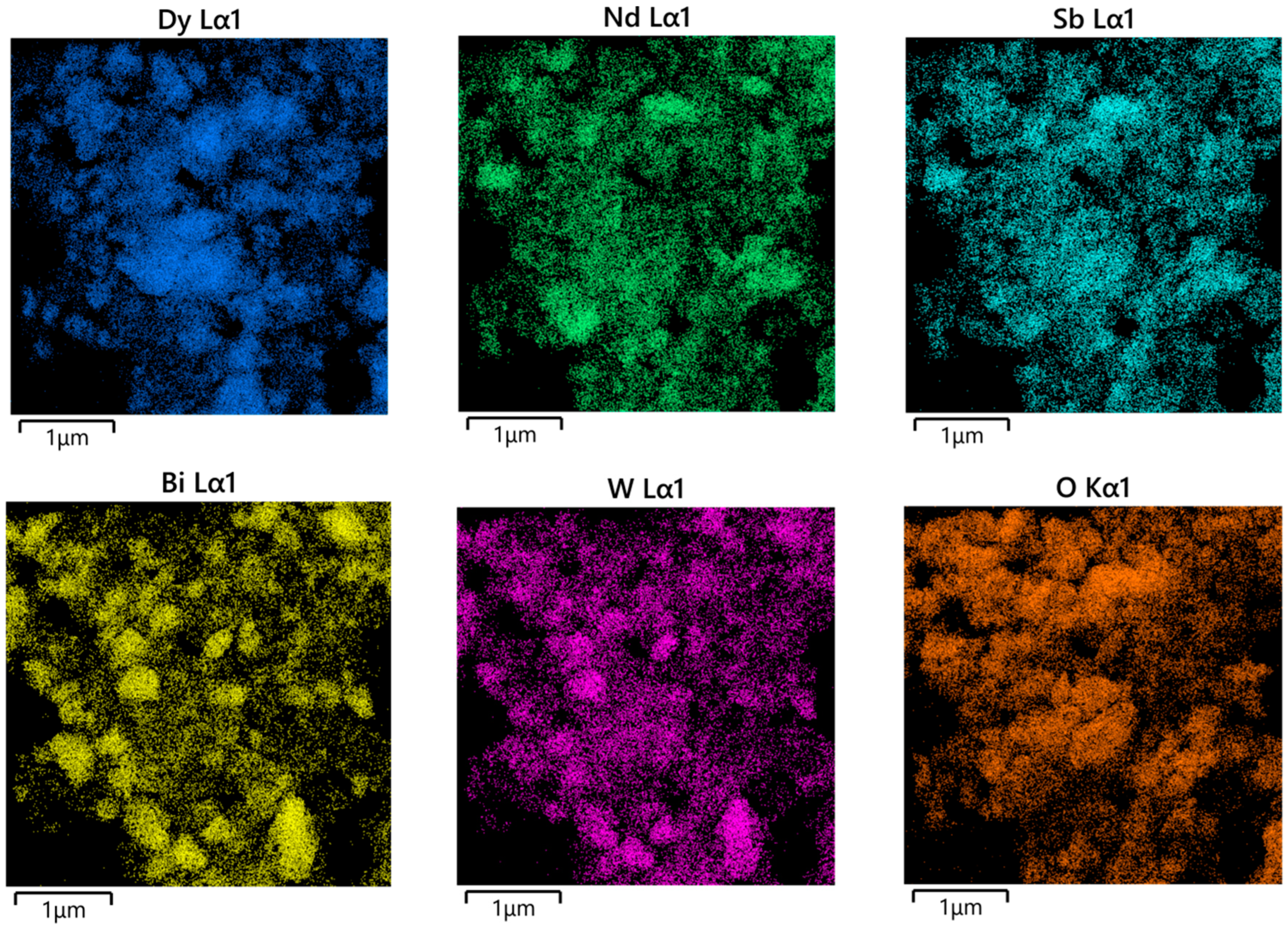
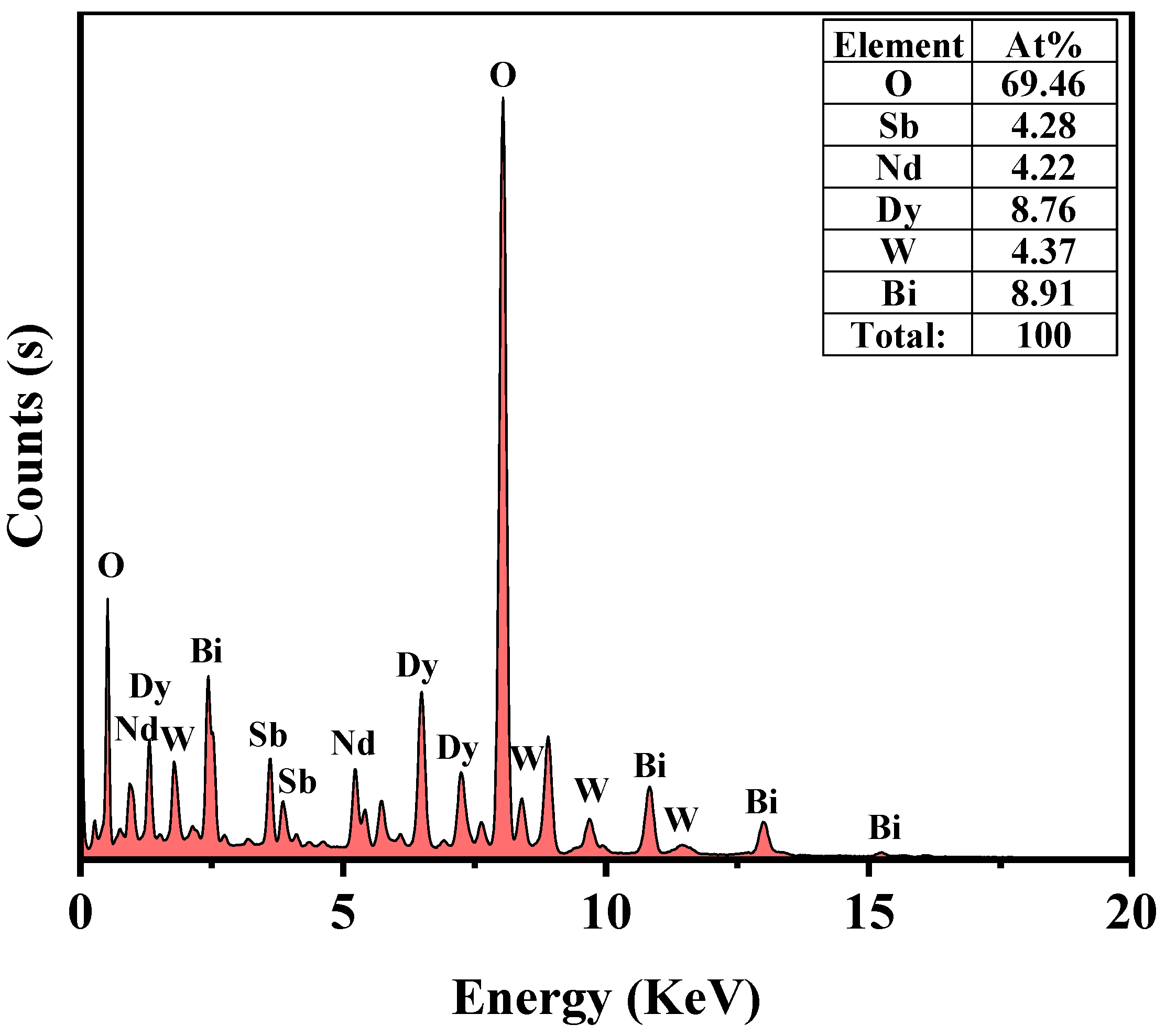
2.6. Characterization
2.7. Photoelectrochemical Experiments
2.8. Experimental Setup and Procedure
3. Result and Discussion
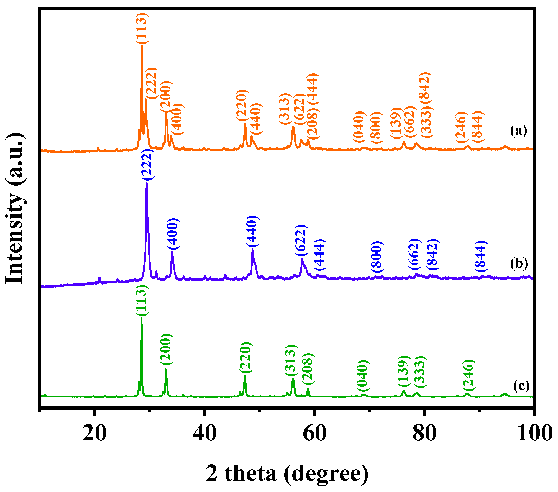
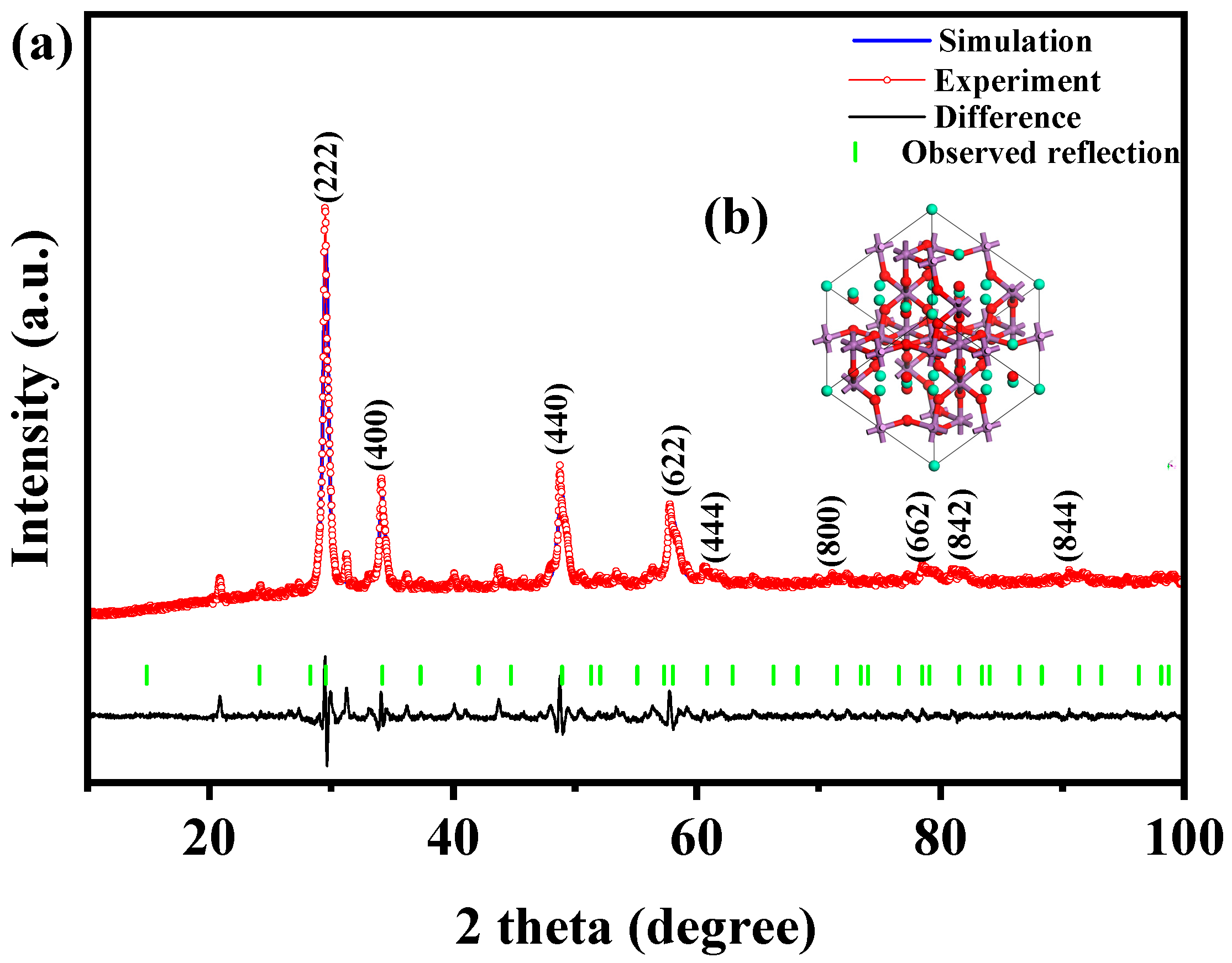
3.1. XRD Analysis
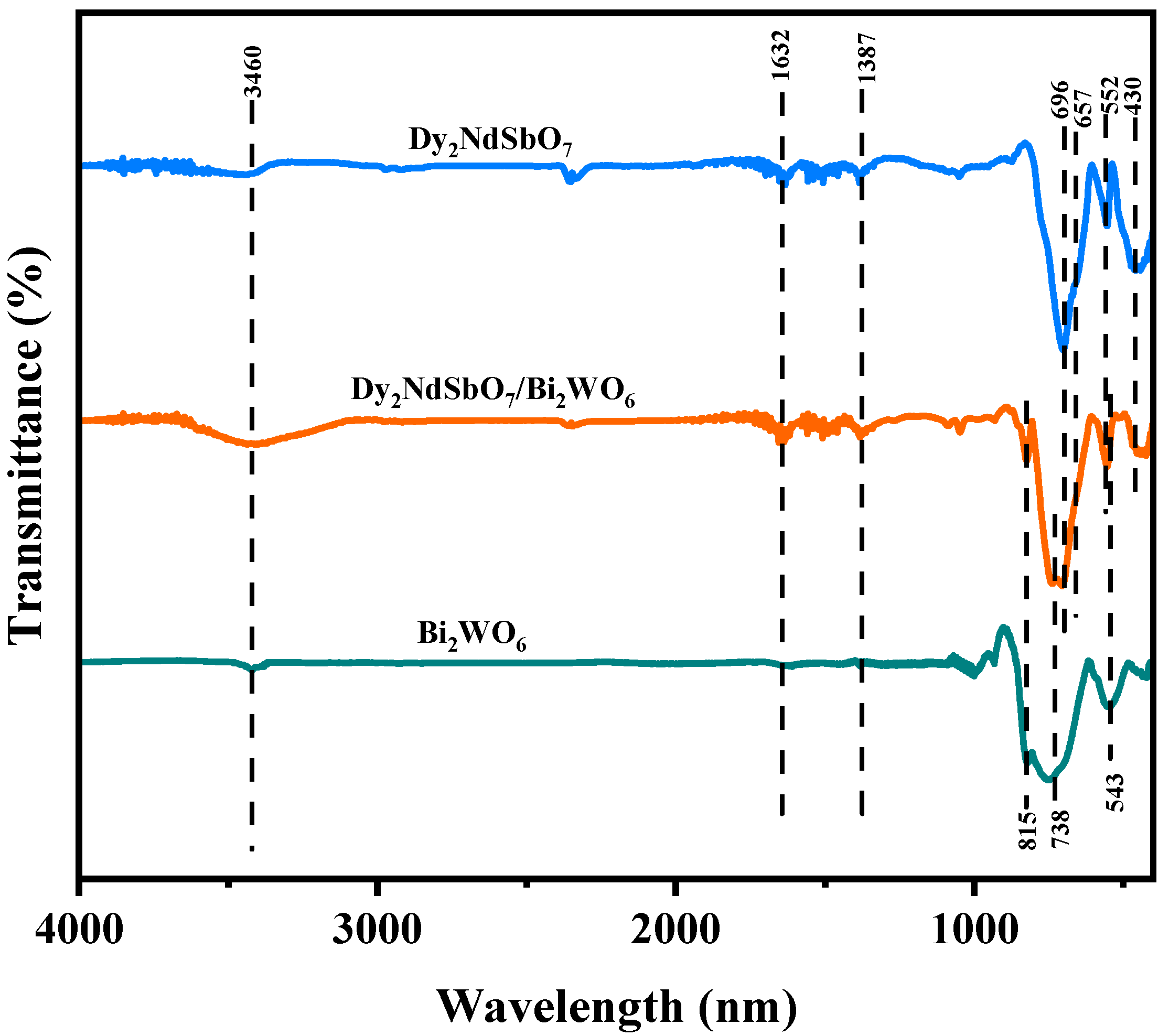
3.2. FTIR Analysis
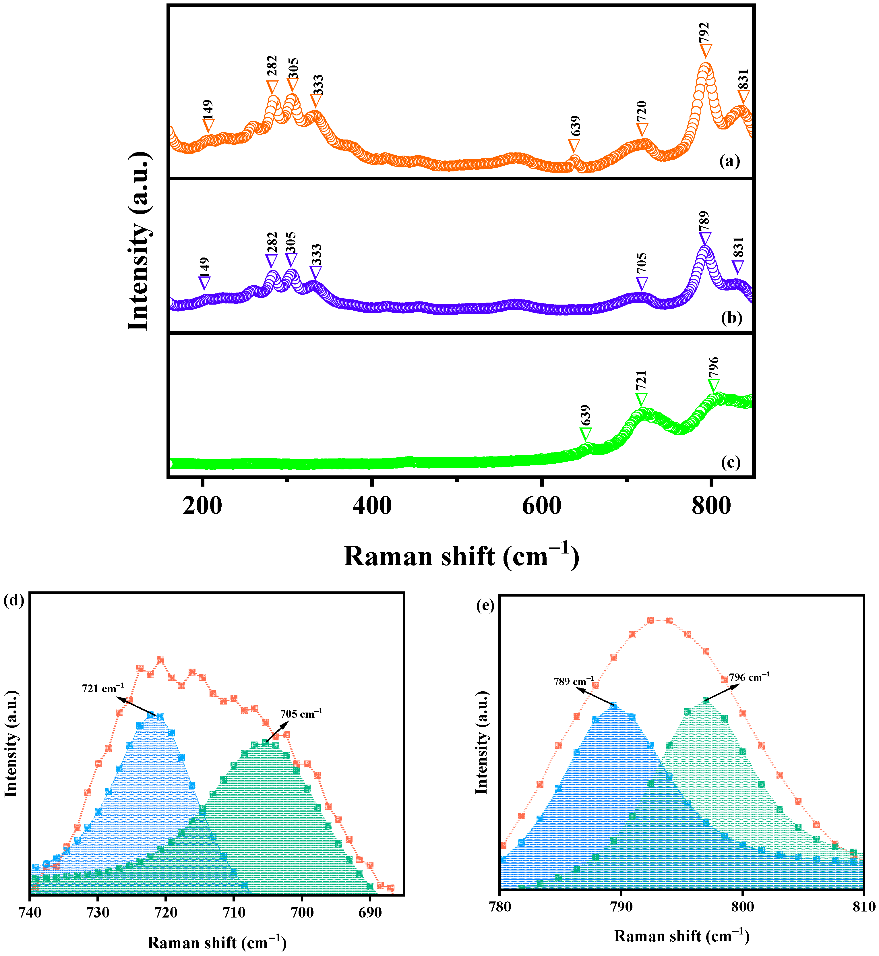
3.3. Raman Analysis
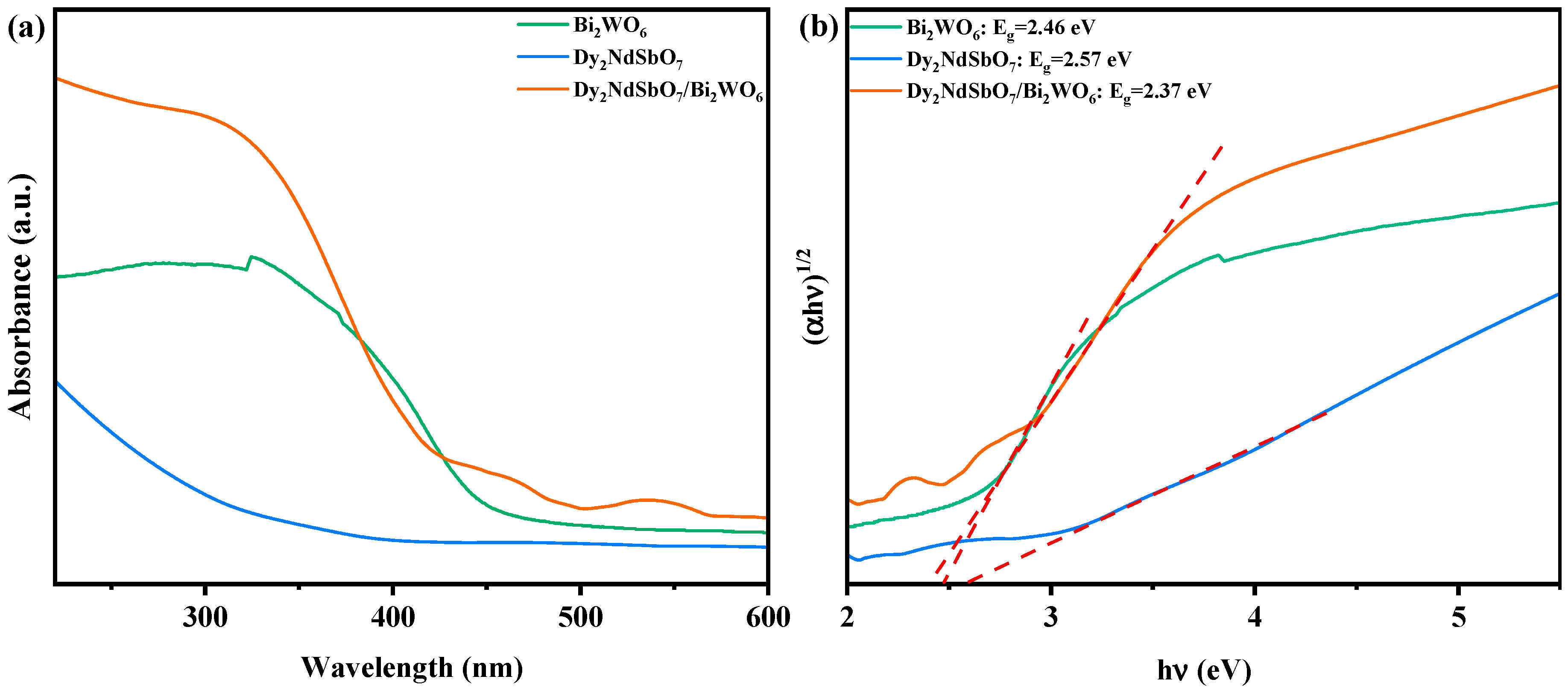
3.4. UV-Vis Diffuse Reflectance Spectra
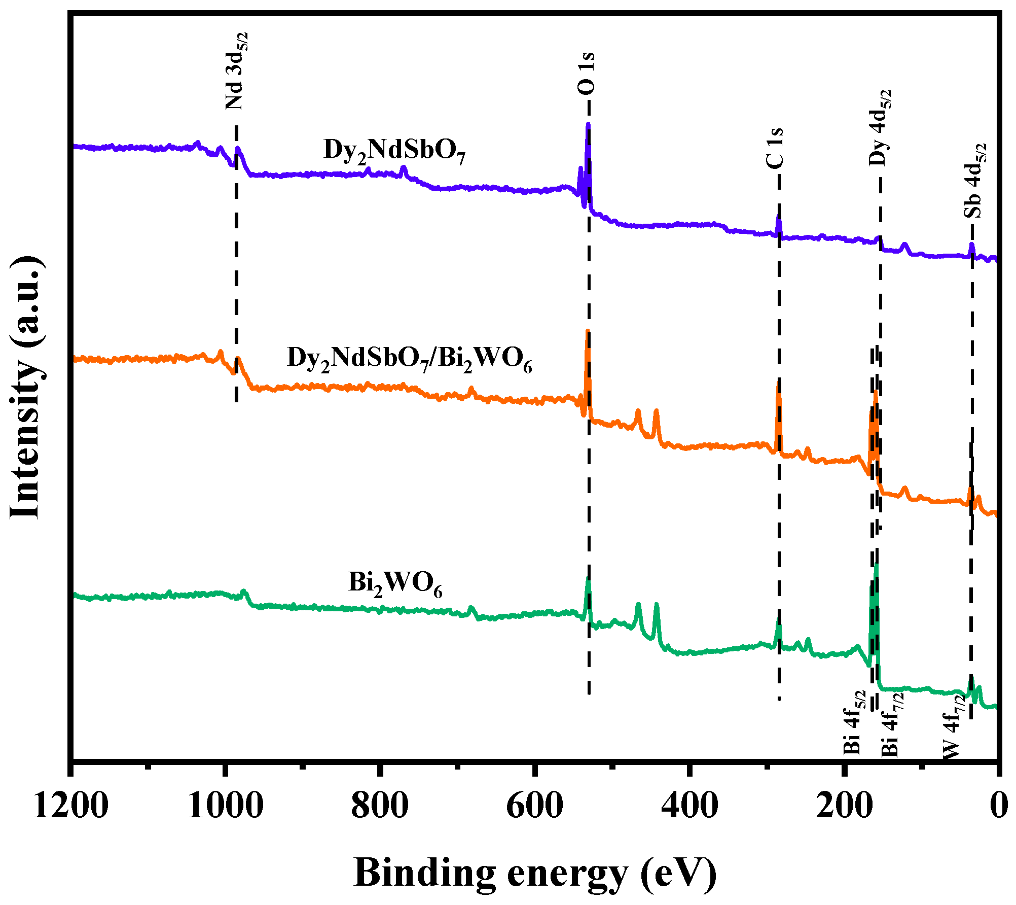
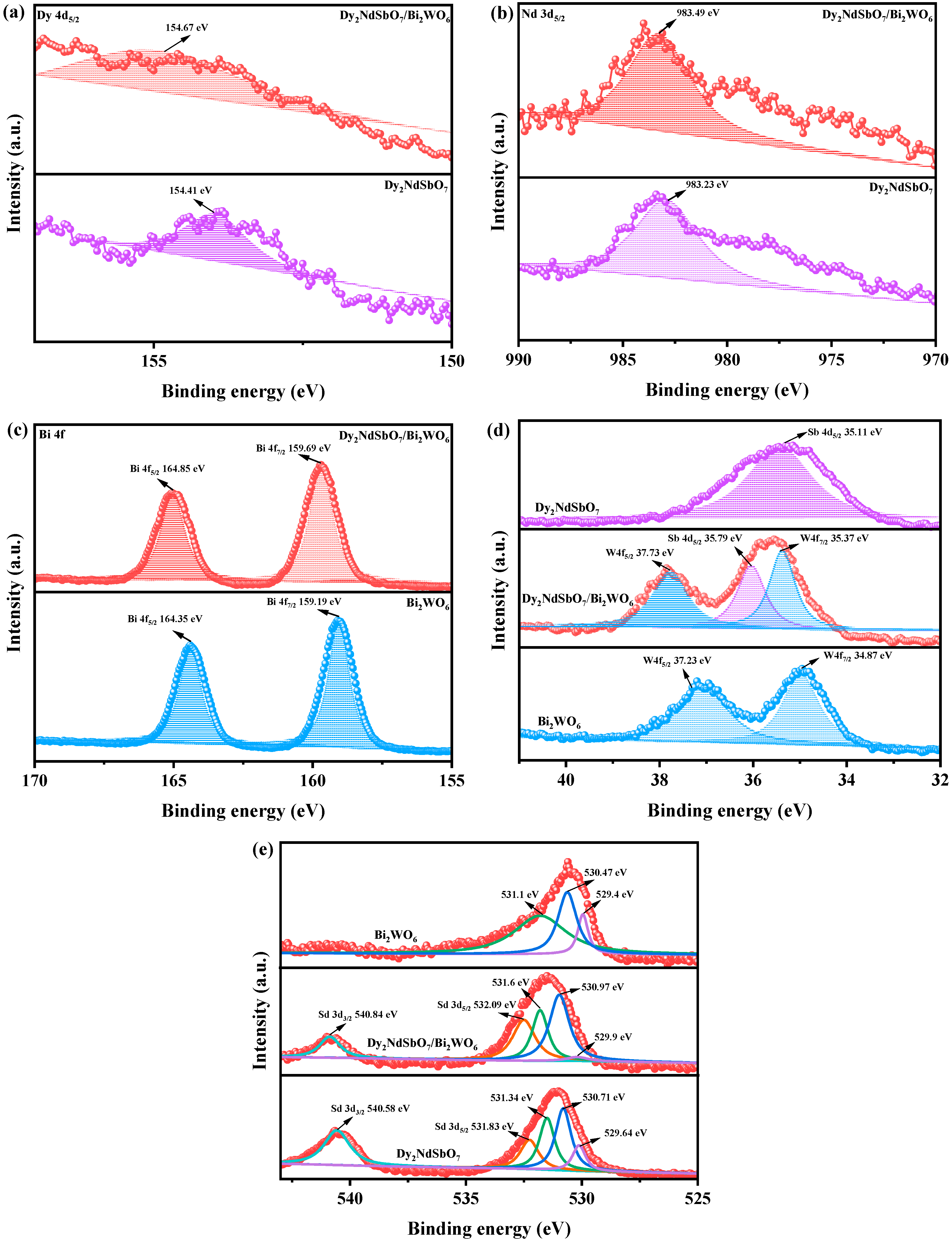
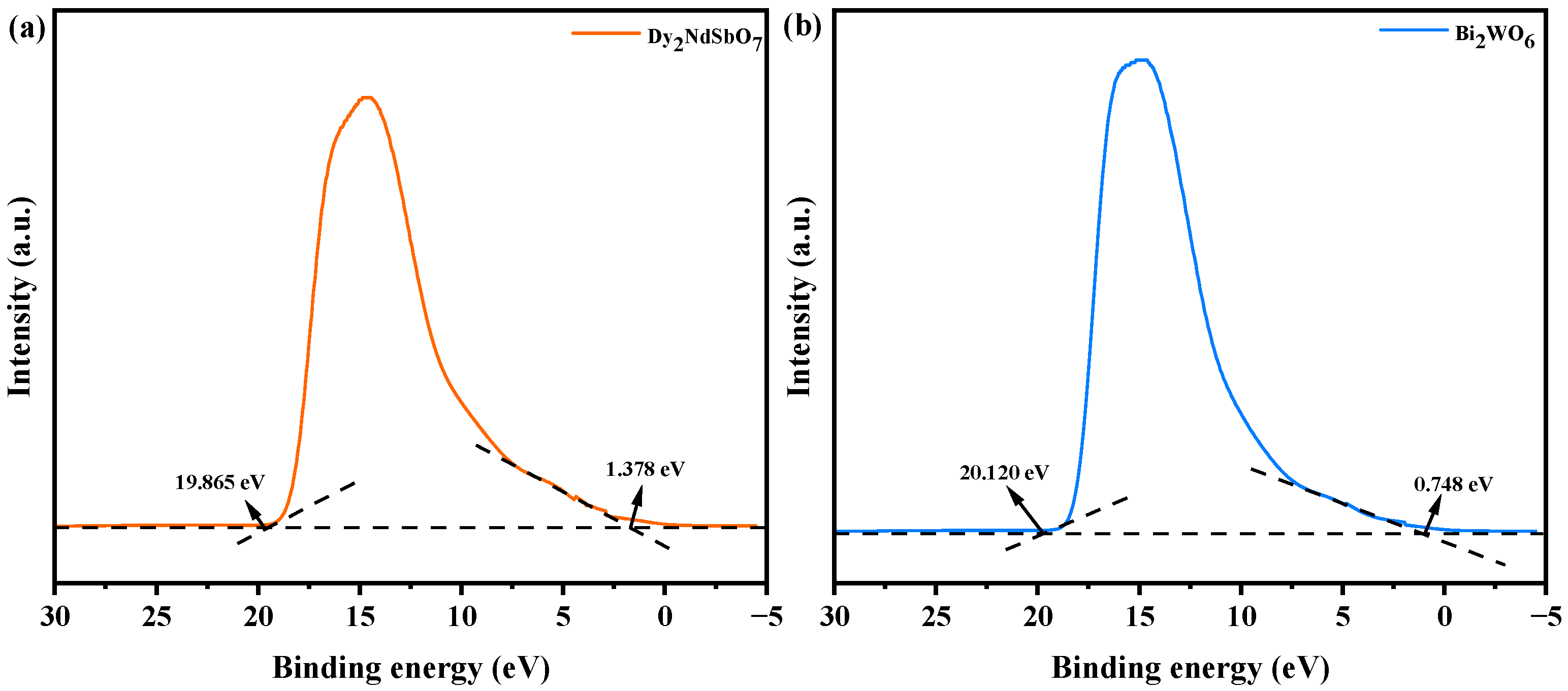
3.5. X-ray Photoelectron Spectroscopy Analysis
3.6. Photocatalytic Activity
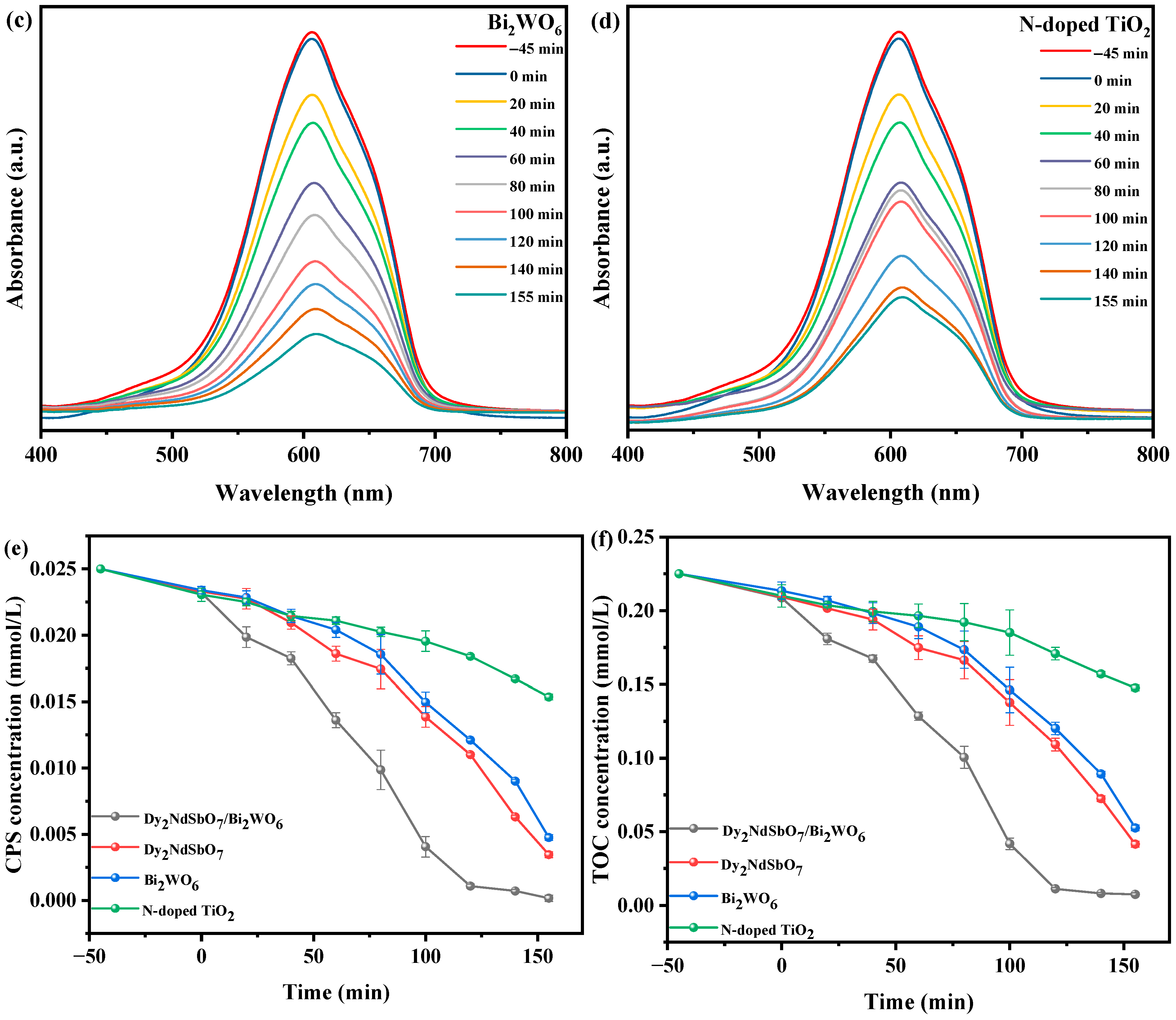
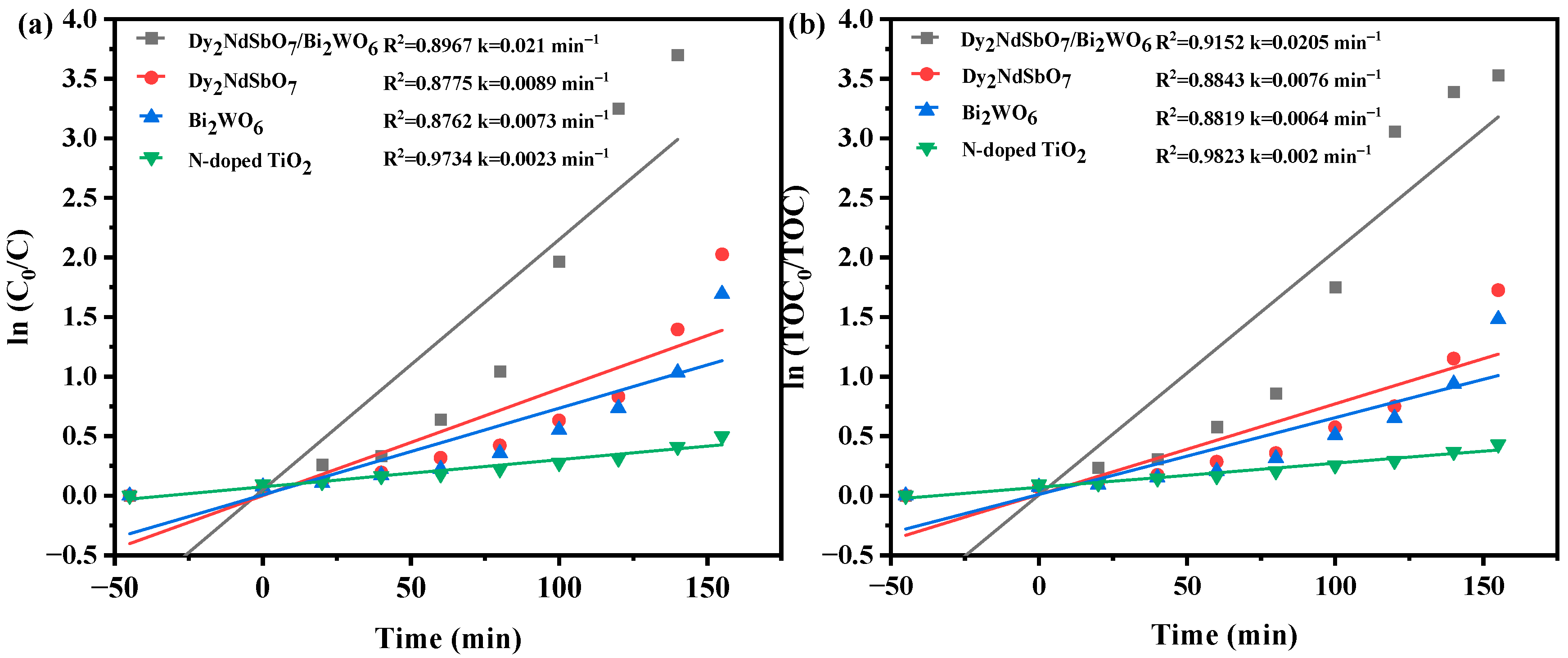
3.6.1. Photocatalytic Activity in Photodegradation Experiments
3.6.2. Comparison of Photocatalytic Activity
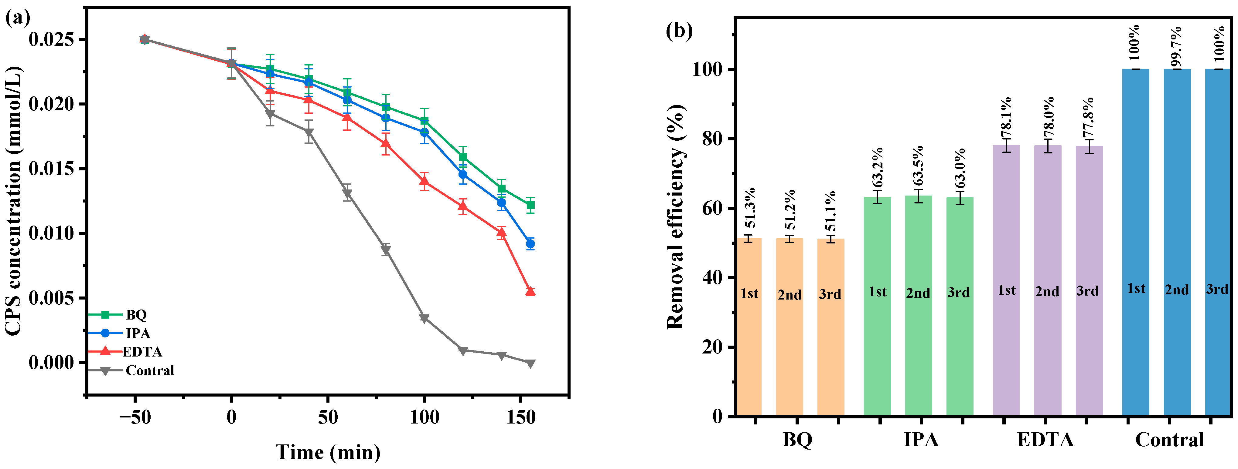
3.6.3. Impact of Photocatalytic Activity Influenced by Different Factors
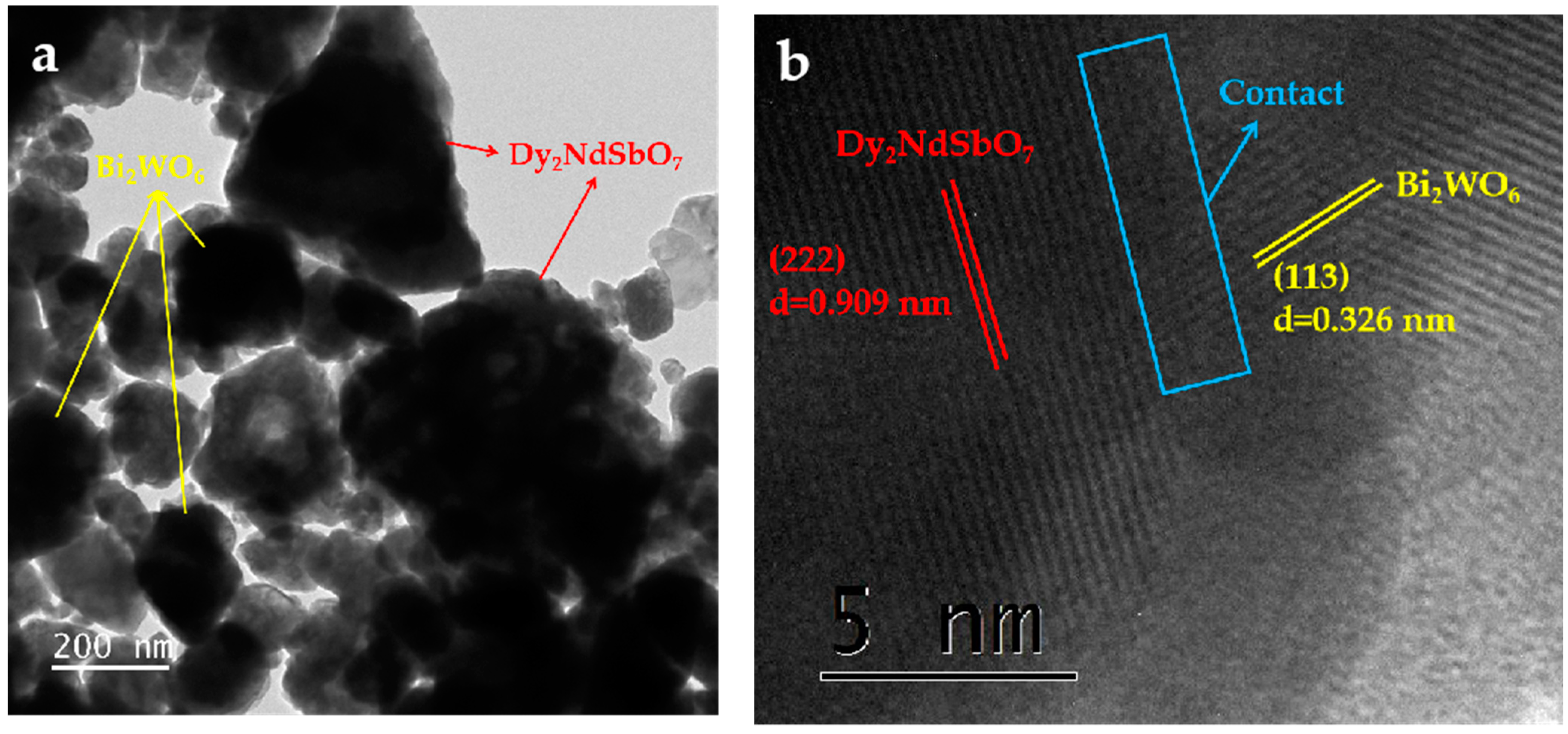
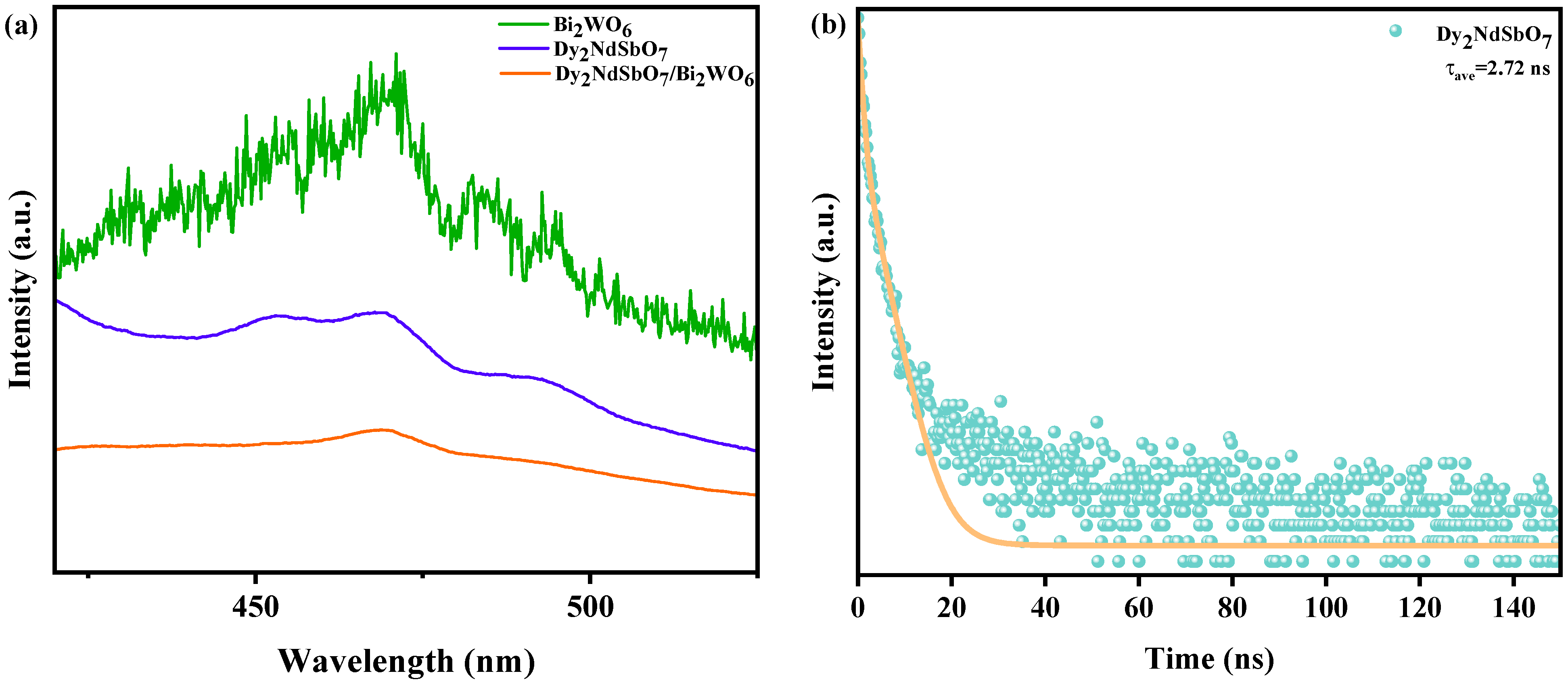
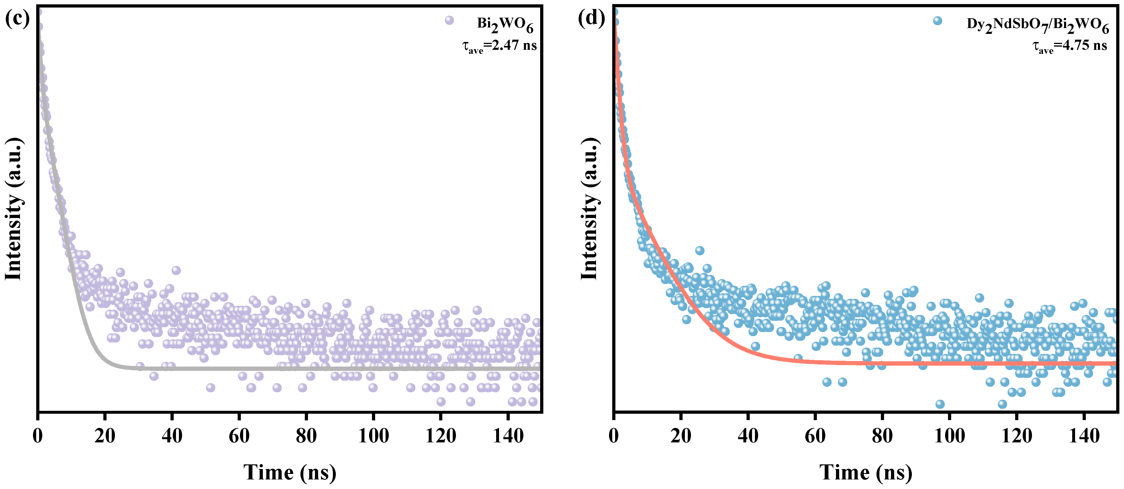
3.7. Property Characterization
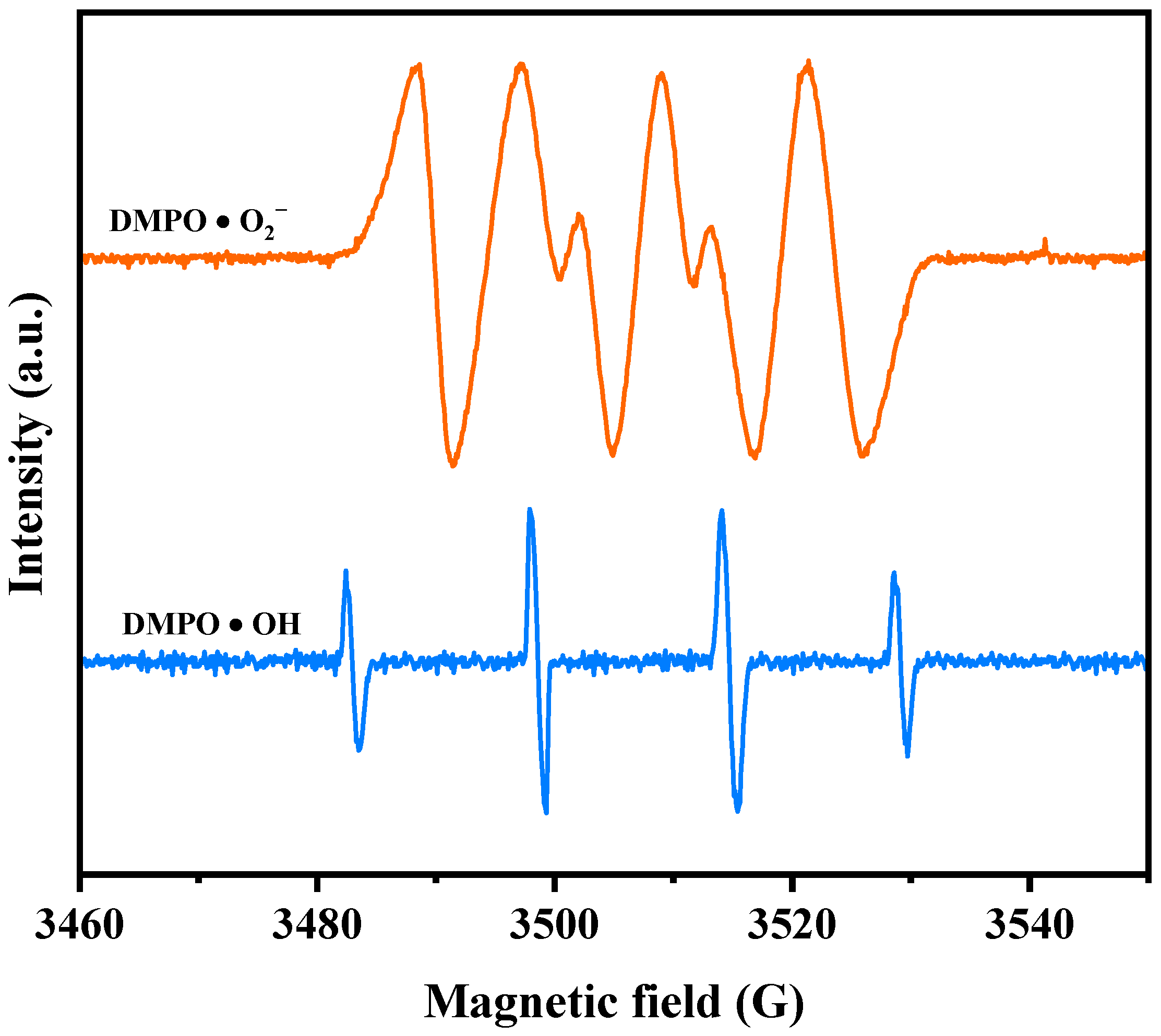
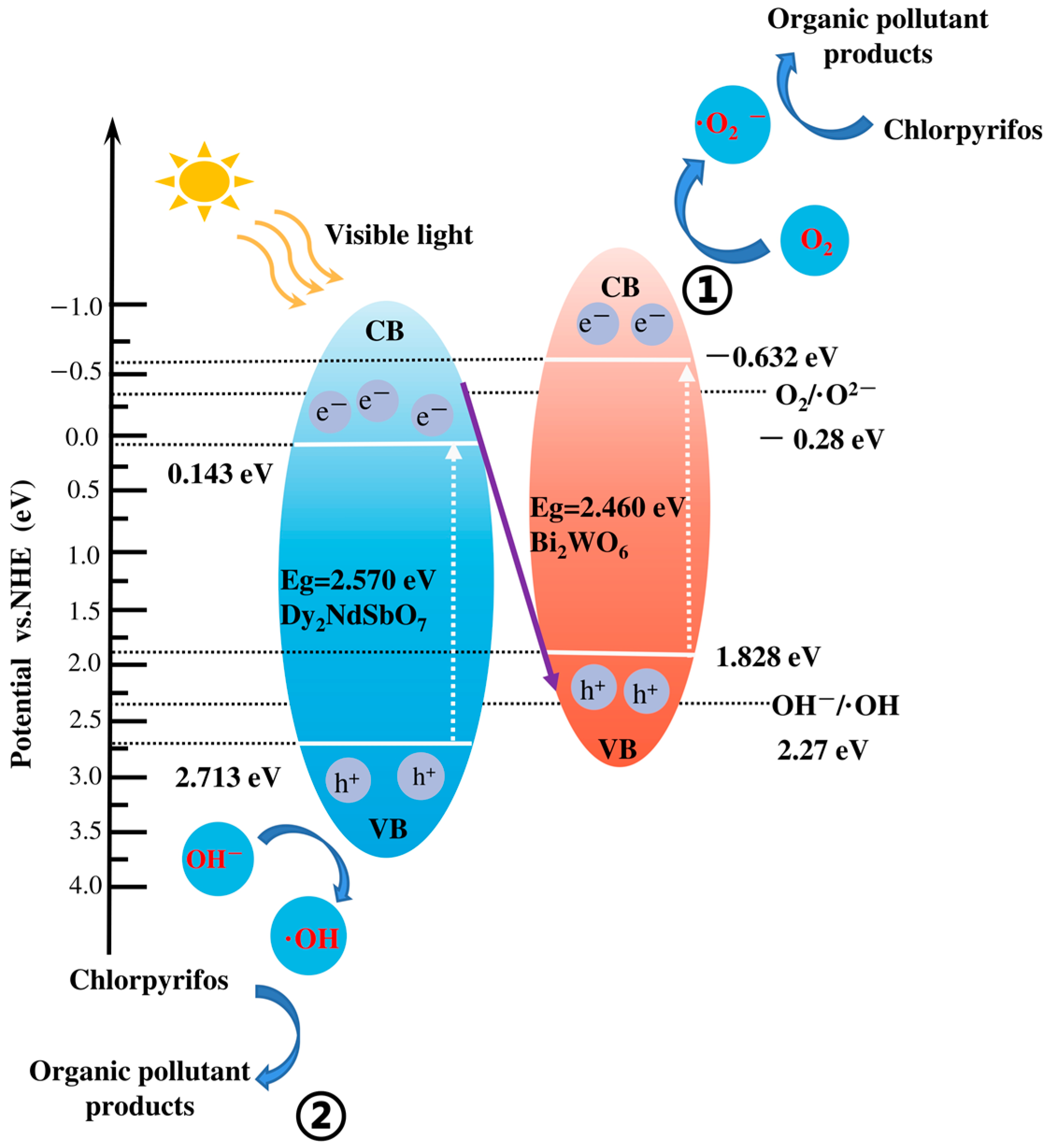
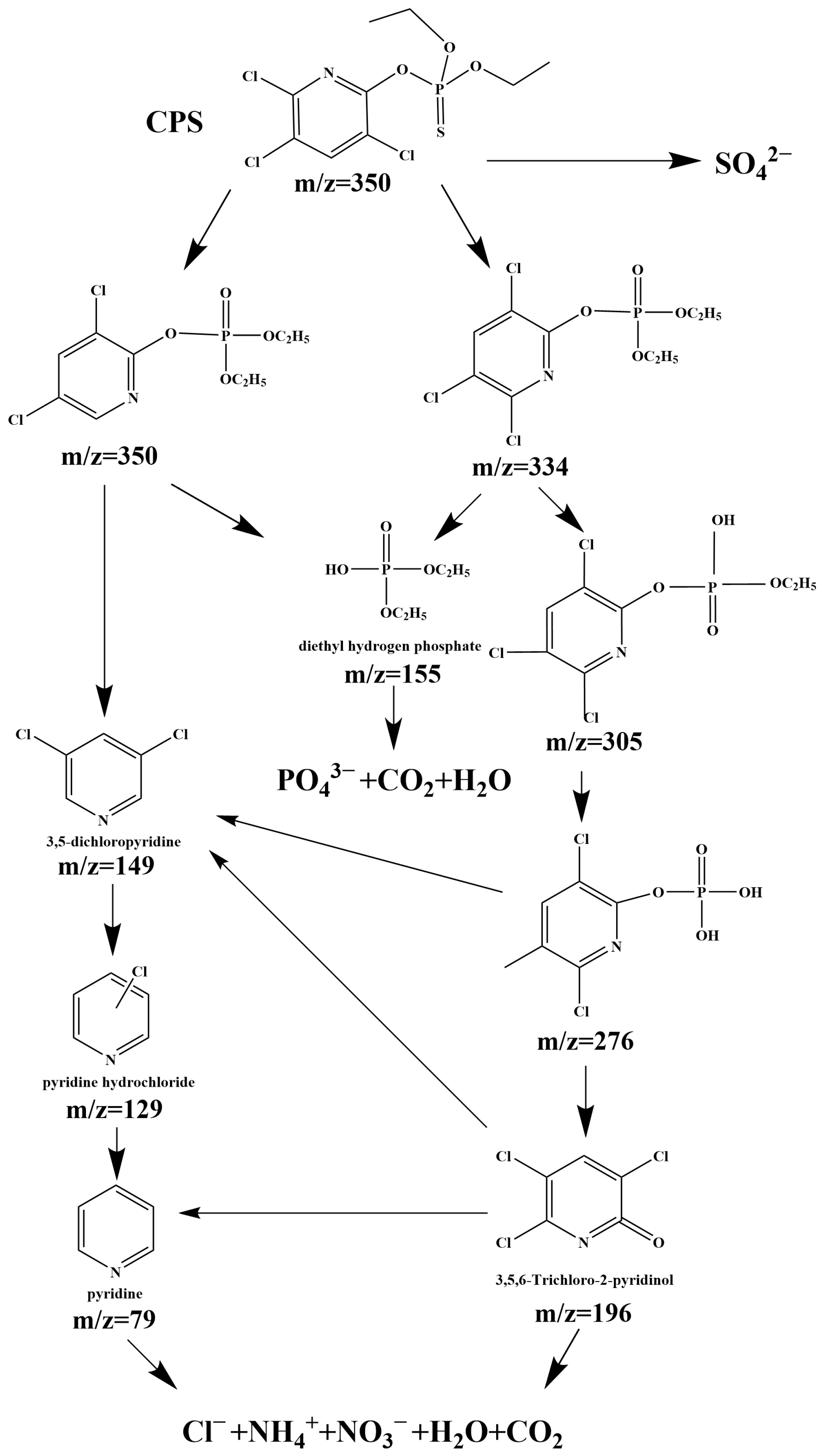
3.8. Analysis of Possible Degradation Mechanisms
4. Conclusions
Supplementary Materials
Author Contributions
Funding
Institutional Review Board Statement
Informed Consent Statement
Data Availability Statement
Conflicts of Interest
References
- Zhang, Y.J.; Zhou, B.H.; Chen, H.L.; Yuan, R.F. Heterogeneous photocatalytic oxidation for the removal of organophosphorus pollutants from aqueous solutions: A review. Sci. Total Environ. 2023, 856, 159048. [Google Scholar] [CrossRef] [PubMed]
- Gordi, Z.; Bayat, A. Development and synthesis of a metal-organic framework on metal-organic framework composite as a novel catalyst for photodegradation of two organophosphorus pesticides under visible light. React. Kinet. Mech. Catal. 2023, 136, 1673–1688. [Google Scholar] [CrossRef]
- Sibmah, S.; Kirupavasam, E.K. An efficient photocatalytic degradation of Quinalphos pesticide under visible light using zinc oxide/magnesium oxide nanocomposites as a novel photocatalyst. Indian J. Chem. 2022, 61, 901–908. [Google Scholar] [CrossRef]
- Mumtaz, N.; Javaid, A.; Imran, M.; Latif, S.; Hussain, N.; Nawaz, S.; Bilal, M. Nanoengineered metal-organic framework for adsorptive and photocatalytic mitigation of pharmaceuticals and pesticide from wastewater. Environ. Pollut. 2022, 308, 119690. [Google Scholar] [CrossRef] [PubMed]
- Fu, H.Y.; Tan, P.; Wang, R.J.; Li, S.N.; Liu, H.Z.; Yang, Y.; Wu, Z.L. Advances in organophosphorus pesticides pollution: Current status and challenges in ecotoxicological, sustainable agriculture, and degradation strategies. J. Hazard. Mater. 2022, 424, 127494. [Google Scholar] [CrossRef] [PubMed]
- Li, G.L.; Wen, A.; Liu, J.H.; Wu, D.; Wu, Y.N. Facile extraction and determination of organophosphorus pesticides in vegetables via magnetic functionalized covalent organic framework nanocomposites. Food Chem. 2021, 337, 127974. [Google Scholar] [CrossRef]
- Sharma, A.; Shukla, A.; Attri, K.; Kumar, M.; Kumar, P.; Suttee, A.; Singh, G.; Barnwal, R.P.; Singla, N. Global trends in pesticides: A looming threat and viable alternatives. Ecotoxicol. Environ. Saf. 2020, 201, 110812. [Google Scholar] [CrossRef] [PubMed]
- Zhu, X.Y.; Li, B.; Yang, J.; Li, Y.S.; Zhao, W.R.; Shi, J.L.; Gu, J.L. Effective Adsorption and Enhanced Removal of Organophosphorus Pesticides from Aqueous Solution by Zr-Based MOFs of UiO-67. ACS Appl. Mater. Interfaces 2015, 7, 223–231. [Google Scholar] [CrossRef]
- Gilani, R.A.; Rafique, M.; Rehman, A.; Munis, M.F.H.; Rehman, S.U.; Chaudhary, H.J. Biodegradation of chlorpyrifos by bacterial genus Pseudomonas. J. Basic Microbiol. 2016, 56, 105–119. [Google Scholar] [CrossRef]
- Yadav, M.; Srivastva, N.; Singh, R.S.; Upadhyay, S.N.; Dubey, S.K. Biodegradation of chlorpyrifos by Pseudomonas sp. in a continuous packed bed bioreactor. Bioresour. Technol. 2014, 165, 265–269. [Google Scholar] [CrossRef]
- Sasikala, C.; Jiwal, S.; Rout, P.; Ramya, M. Biodegradation of chlorpyrifos by bacterial consortium isolated from agriculture soil. World J. Microbiol. Biotechnol. 2012, 28, 1301–1308. [Google Scholar] [CrossRef] [PubMed]
- Gearhart, D.A.; Sickles, D.W.; Buccafusco, J.J.; Prendergast, M.A.; Terry, A.V. Chlorpyrifos, chlorpyrifos-oxon, and diisopropylfluorophosphate inhibit kinesin-dependent microtubule motility. Toxicol. Appl. Pharmacol. 2007, 218, 20–29. [Google Scholar] [CrossRef] [PubMed]
- Steevens, J.A.; Benson, W.H. Toxicokinetic interactions and survival of Hyalella azteca exposed to binary mixtures of chlorpyrifos, dieldrin, and methyl mercury. Aquat. Toxicol. 2001, 51, 377–388. [Google Scholar] [CrossRef] [PubMed]
- Steevens, J.A.; Benson, W.H. Interactions of chlorpyrifos and methyl mercury: A mechanistic approach to assess chemical mixtures. Mar. Environ. Res. 2000, 50, 113–117. [Google Scholar] [CrossRef] [PubMed]
- Thind, P.S.; Kumari, D.; John, S. TiO2/H2O2 mediated UV photocatalysis of Chlorpyrifos: Optimization of process parameters using response surface methodology. J. Environ. Chem. Eng. 2018, 6, 3602–3609. [Google Scholar] [CrossRef]
- Sarangapani, C.; Misra, N.N.; Milosavljevic, V.; Bourke, P.; O’Regan, F.; Cullen, P.J. Pesticide degradation in water using atmospheric air cold plasma. J. Water Process Eng. 2016, 9, 225–232. [Google Scholar] [CrossRef]
- Chakma, S.; Moholkar, V.S. Mechanistic analysis of hybrid sono-photo-ferrioxalate system for decolorization of azo dye. J. Taiwan Inst. Chem. E 2016, 60, 469–478. [Google Scholar] [CrossRef]
- Ahmad, T.; Rafatullah, M.; Ghazali, A.; Sulaiman, O.; Hashim, R.; Ahmad, A. Removal of Pesticides from Water and Wastewater by Different Adsorbents: A Review. J. Environ. Sci. Health Part C Environ. Carcinog. Ecotoxicol. Rev. 2010, 28, 231–271. [Google Scholar] [CrossRef]
- Agarwal, S.; Tyagi, I.; Gupta, V.K.; Dehghani, M.H.; Bagheri, A.; Yetilmezsoy, K.; Amrane, A.; Heibati, B.; Rodriguez-Couto, S. Degradation of azinphos-methyl and chlorpyrifos from aqueous solutions by ultrasound treatment. J. Mol. Liq. 2016, 221, 1237–1242. [Google Scholar] [CrossRef]
- Soltani-nezhad, F.; Saljooqi, A.; Mostafavi, A.; Shamspur, T. Synthesis of Fe3O4/CdS-ZnS nanostructure and its application for photocatalytic degradation of chlorpyrifos pesticide and brilliant green dye from aqueous solutions. Ecotoxicol. Environ. Saf. 2020, 189, 109886. [Google Scholar] [CrossRef]
- Merci, S.; Saljooqi, A.; Shamspur, T.; Mostafavi, A. Investigation of photocatalytic chlorpyrifos degradation by a new silica mesoporous material immobilized by WS2 and Fe3O4 nanoparticles: Application of response surface methodology. Appl. Organomet. Chem. 2020, 34, e5343. [Google Scholar] [CrossRef]
- Zangiabadi, M.; Shamspur, T.; Saljooqi, A.; Mostafavi, A. Evaluating the efficiency of the GO-Fe3O4/TiO2 mesoporous photocatalyst for degradation of chlorpyrifos pesticide under visible light irradiation. Appl. Organomet. Chem. 2019, 33, e4813. [Google Scholar] [CrossRef]
- Mansourian, R.; Mousavi, S.M.; Alizadeh, S.; Sabbaghi, S. CeO2/TiO2/SiO2 nanocatalyst for the photocatalytic and sonophotocatalytic degradation of chlorpyrifos. Can. J. Chem. Eng. 2022, 100, 451–464. [Google Scholar] [CrossRef]
- Nekooie, R.; Shamspur, T.; Mostafavi, A. Novel CuO/TiO2/PANI nanocomposite: Preparation and photocatalytic investigation for chlorpyrifos degradation in water under visible light irradiation. J. Photochem. Photobiol. A 2021, 407, 113038. [Google Scholar] [CrossRef]
- Anirudhan, T.S.; Manjusha, V.; Shainy, F. Magnetically retrievable cysteine modified graphene oxide@nickelferrite@titanium dioxide photocatalyst for the effective degradation of chlorpyrifos from aqueous solutions. Environ. Technol. Innov. 2021, 23, 101633. [Google Scholar] [CrossRef]
- Ikrari, K.I.; Hasbullah, H.; Salleh, W.N.W.; Nakagawa, K.; Yoshioka, T. Photocatalytic membrane technologies for removal of recalcitrant pollutants. Mater. Today Proc. 2022, 65, 3101–3108. [Google Scholar] [CrossRef]
- Zuraimi, M.S.; Magee, R.J.; Won, D.Y.; Nong, G.; Arsenault, C.D.; Yang, W.; So, S.; Nilsson, G.; Abebe, L.; Alliston, C. Performance of sorption- and photocatalytic oxidation-based indoor passive panel technologies. Build. Environ. 2018, 135, 85–93. [Google Scholar] [CrossRef]
- Chong, M.N.; Jin, B.; Chow, C.W.K.; Saint, C. Recent developments in photocatalytic water treatment technology: A review. Water Res. 2010, 44, 2997–3027. [Google Scholar] [CrossRef]
- Sun, Y.; Zhang, W.; Li, Q.; Liu, H.; Wang, X. Preparations and applications of zinc oxide based photocatalytic materials. Adv. Sens. Energy Mater. 2023, 2, 100069. [Google Scholar] [CrossRef]
- Lee, S.F.; Jimenez-Relinque, E.; Martinez, I.; Castellote, M. Effects of Mott-Schottky Frequency Selection and Other Controlling Factors on Flat-Band Potential and Band-Edge Position Determination of TiO2. Catalysts 2023, 13, 1000. [Google Scholar] [CrossRef]
- Lee, S.F.; Jimenez-Relinque, E.; Martinez, I.; Castellote, M. Photoelectrochemical global approach to the behaviour of nanostructured anatase under different irradiation conditions. Catal. Today 2022, 397, 286–295. [Google Scholar] [CrossRef]
- Supin, K.K.; Namboothiri, P.P.M.; Vasundhara, M. Enhanced photocatalytic activity in ZnO nanoparticles developed using novel Lepidagathis ananthapuramensis leaf extract. RSC Adv. 2023, 13, 1497–1515. [Google Scholar] [CrossRef]
- Ziental, D.; Czarczynska-Goslinska, B.; Mlynarczyk, D.T.; Glowacka-Sobotta, A.; Stanisz, B.; Goslinski, T.; Sobotta, L. Titanium Dioxide Nanoparticles: Prospects and Applications in Medicine. Nanomaterials 2020, 10, 387. [Google Scholar] [CrossRef] [PubMed]
- Ghosal, K.; Agatemor, C.; Spitalsky, Z.; Thomas, S.; Kny, E. Electrospinning tissue engineering and wound dressing scaffolds from polymer-titanium dioxide nanocomposites. Chem. Eng. J. 2019, 358, 1262–1278. [Google Scholar] [CrossRef]
- Lu, P.J.; Huang, S.C.; Chen, Y.P.; Chiueh, L.C.; Shih, D.Y.C. Analysis of titanium dioxide and zinc oxide nanoparticles in cosmetics. J. Food Drug Anal. 2015, 23, 587–594. [Google Scholar] [CrossRef] [PubMed]
- Dimapilis, E.A.S.; Hsu, C.S.; Mendoza, R.M.O.; Lu, M.C. Zinc oxide nanoparticles for water disinfection. Sustain. Environ. Res. 2018, 28, 47–56. [Google Scholar] [CrossRef]
- Kolodziejczak-Radzimska, A.; Jesionowski, T. Zinc Oxide-From Synthesis to Application: A Review. Materials 2014, 7, 2833–2881. [Google Scholar] [CrossRef] [PubMed]
- Roohandeh, L.; Hakimyfard, A.; Samimifar, M. High efficient solar light photocatalytic degradation of malachite green by solid state synthesized Bi2Sn2O7 and Bi2MxSn2O7 (M = Y3+, Eu3+, Gd3+ and Yb3+) nanomaterials. J. Nanoanal. 2020, 7, 240–247. [Google Scholar]
- Abe, R.; Higashi, M.; Zou, Z.G.; Sayama, K.; Abe, Y. Photocatalytic water splitting into H2 and O2 over R2Ti2O7 (R = Y, rare earth) with pyrochlore structure. Chem. Lett. 2004, 33, 954–955. [Google Scholar] [CrossRef]
- Luan, J.F.; Hu, Z.T. Synthesis, Property Characterization, and Photocatalytic Activity of Novel Visible Light-Responsive Photocatalyst Fe2BiSbO7. Int. J. Photoenergy 2012, 2012, 301954. [Google Scholar] [CrossRef]
- Devi, V.R.; Ravi, G.; Velchuri, R.; Muniratnam, N.R.; Prasad, G.; Vithal, M. Preparation, Characterization, Photocatalytic Activity and Conductivity Studies of YLnTi2O7(Ln = Nd, Sm, Eu and Gd). Trans. Indian Ceram. Soc. 2013, 72, 241–251. [Google Scholar] [CrossRef]
- Zhang, S.; Wen, Z.; Zhao, Y.; Li, G.; Li, Z. Preparation, Characterization of A2Ce2O7 (A = La and Gd) and Their Photo-Catalytic Properties. Energy Environ. Focus 2015, 4, 324–329. [Google Scholar] [CrossRef]
- Li, J.; Zhang, X.; Ai, Z.; Jia, F.; Zhang, L.; Lin, J. Efficient Visible Light Degradation of Rhodamine B by a Photo-Electrochemical Process Based on a Bi2WO6 Nanoplate Film Electrode. J. Phys. Chem. C 2007, 111, 6832–6836. [Google Scholar] [CrossRef]
- Zhao, C.R.; Cai, L.Y.; Wang, K.Q.; Li, B.X.; Yuan, S.D.; Zeng, Z.H.; Zhao, L.H.; Wu, Y.; He, Y.M. Novel Bi2WO6/ZnSnO3 heterojunction for the ultrasonic-vibration-driven piezocatalytic degradation of RhB. Environ. Pollut. 2023, 319, 120982. [Google Scholar] [CrossRef] [PubMed]
- Zhu, Y.Y.; Wang, Y.J.; Ling, Q.; Zhu, Y.F. Enhancement of full-spectrum photocatalytic activity over BiPO4/Bi2WO6 clomposites. Appl. Catal. B Environ. 2017, 200, 222–229. [Google Scholar] [CrossRef]
- Tian, Y.L.; Chang, B.B.; Lu, J.L.; Fu, J.; Xi, F.N.; Dong, X.P. Hydrothermal Synthesis of Graphitic Carbon Nitride-Bi2WO6 Heterojunctions with Enhanced Visible Light Photocatalytic Activities. ACS Appl. Mater. Interfaces 2013, 5, 7079–7085. [Google Scholar] [CrossRef]
- Zhao, Y.; Wang, Y.; Liu, E.; Fan, J.; Hu, X. Bi2WO6 nanoflowers: An efficient visible light photocatalytic activity for ceftriaxone sodium degradation. Appl. Surf. Sci. 2018, 436, 854–864. [Google Scholar] [CrossRef]
- Lai, M.T.L.; Lai, C.W.; Lee, K.M.; Chook, S.W.; Yang, T.C.K.; Chong, S.H.; Juan, J.C. Facile one-pot solvothermal method to synthesize solar active Bi2WO6 for photocatalytic degradation of organic dye. J. Alloys Compd. 2019, 801, 502–510. [Google Scholar] [CrossRef]
- Zou, Z.G.; Ye, J.H.; Abe, R.; Arakawa, H. Photocatalytic decomposition of water with Bi2InNbO7. Catal. Lett. 2000, 68, 235–239. [Google Scholar] [CrossRef]
- Zuo, G.; Ma, S.; Yin, Z.; Chen, W.; Wang, Y.; Ji, Q.; Xian, Q.; Yang, S.; He, H. Z-Scheme Modulated Charge Transfer on InVO4@ZnIn2S4 for Durable Overall Water Splitting. Small 2023, 19, 2207031. [Google Scholar] [CrossRef]
- Wang, Q.; Li, Y.; Huang, F.; Song, S.; Ai, G.; Xin, X.; Zhao, B.; Zheng, Y.; Zhang, Z. Recent Advances in g-C3N4-Based Materials and Their Application in Energy and Environmental Sustainability. Molecules 2023, 28, 432. [Google Scholar] [CrossRef] [PubMed]
- Yu, Y.; Cao, C.Y.; Liu, H.; Li, P.; Wei, F.F.; Jiang, Y.; Song, W.G. A Bi/BiOCl heterojunction photocatalyst with enhanced electron-hole separation and excellent visible light photodegrading activity. J. Mater. Chem. A 2014, 2, 1677–1681. [Google Scholar] [CrossRef]
- Wang, W.J.; Yu, J.C.; Xia, D.H.; Wong, P.K.; Li, Y.C. Graphene and g-C3N4 Nanosheets Cowrapped Elemental α-Sulfur As a Novel Metal-Free Heterojunction Photocatalyst for Bacterial Inactivation under Visible-Light. Environ. Sci. Technol. 2013, 47, 8724–8732. [Google Scholar] [CrossRef] [PubMed]
- Shen, T.; Shi, X.K.; Guo, J.X.; Li, J.; Yuan, S.D. Photocatalytic removal of NO by light-driven Mn3O4/BiOCl heterojunction photocatalyst: Optimization and mechanism. Chem. Eng. J. 2021, 408, 128014. [Google Scholar] [CrossRef]
- Jiang, T.G.; Wang, K.; Guo, T.; Wu, X.Y.; Zhang, G.K. Fabrication of Z-scheme MoO3/Bi2O4 heterojunction photocatalyst with enhanced photocatalytic performance under visible light irradiation. Chin. J. Catal. 2020, 41, 161–169. [Google Scholar] [CrossRef]
- Liu, Y.; Kong, J.J.; Yuan, J.L.; Zhao, W.; Zhu, X.; Sun, C.; Xie, J.M. Enhanced photocatalytic activity over flower-like sphere Ag/Ag2CO3/BiVO4 plasmonic heterojunction photocatalyst for tetracycline degradation. Chem. Eng. J. 2018, 331, 242–254. [Google Scholar] [CrossRef]
- Sohrabian, M.; Mahdikhah, V.; Alimohammadi, E.; Sheibani, S. Improved photocatalytic performance of SrTiO3 through a Z-scheme polymeric-perovskite heterojunction with g-C3N4 and plasmonic resonance of Ag mediator. Appl. Surf. Sci. 2023, 618, 156682. [Google Scholar] [CrossRef]
- Zhu, B.C.; Cheng, B.; Fan, J.J.; Ho, W.K.; Yu, J.G. g-C3N4-Based 2D/2D Composite Heterojunction Photocatalyst. Small Struct. 2021, 2, 2100086. [Google Scholar] [CrossRef]
- Xu, H.; Xu, Y.G.; Li, H.M.; Xia, J.X.; Xiong, J.; Yin, S.; Huang, C.J.; Wan, H.L. Synthesis, characterization and photocatalytic property of AgBr/BiPO4 heterojunction photocatalyst. Dalton Trans. 2012, 41, 3387–3394. [Google Scholar] [CrossRef]
- Wen, X.J.; Qian, L.; Lv, X.X.; Sun, J.; Guo, J.; Fei, Z.H.; Niu, C.G. Photocatalytic degradation of sulfamethazine using a direct Z-Scheme AgI/Bi4V2O11 photocatalyst: Mineralization activity, degradation pathways and promoted charge separation mechanism. J. Hazard. Mater. 2020, 385, 121508. [Google Scholar] [CrossRef]
- Pathania, D.; Sharma, A.; Kumar, S.; Srivastava, A.K.; Kumar, A.; Singh, L. Bio-synthesized Cu-ZnO hetro-nanostructure for catalytic degradation of organophosphate chlorpyrifos under solar illumination. Chemosphere 2021, 277, 130315. [Google Scholar] [CrossRef] [PubMed]
- Farner Budarz, J.; Cooper, E.M.; Gardner, C.; Hodzic, E.; Ferguson, P.L.; Gunsch, C.K.; Wiesner, M.R. Chlorpyrifos degradation via photoreactive TiO2 nanoparticles: Assessing the impact of a multi-component degradation scenario. J. Hazard. Mater. 2019, 372, 61–68. [Google Scholar] [CrossRef] [PubMed]
- Wang, J.H.; Zou, Z.G.; Ye, J.H. Synthesis, structure and photocatalytic property of a new hydrogen evolving photocatalyst Bi2InTaO7. In Functionally Graded Materials Vii; Pan, W., Gong, J., Zhang, L., Chen, L., Eds.; Trans Tech Publications Ltd.: Zurich-Uetikon, Switzerland, 2003; Volume 423-4, pp. 485–490. [Google Scholar]
- Muniz, F.T.L.; Miranda, M.A.R.; Morilla dos Santos, C.; Sasaki, J.M. The Scherrer equation and the dynamical theory of X-ray diffraction. Acta Crystallogr. Sect. A Found. Adv. 2016, 72, 385–390. [Google Scholar] [CrossRef] [PubMed]
- Hargreaves, J.S.J. Some considerations related to the use of the Scherrer equation in powder X-ray diffraction as applied to heterogeneous catalysts. Catal. Struct. React. 2016, 2, 33–37. [Google Scholar] [CrossRef]
- Mustapha, S.; Ndamitso, M.M.; Abdulkareem, A.S.; Tijani, J.O.; Shuaib, D.T.; Mohammed, A.K.; Sumaila, A. Comparative study of crystallite size using Williamson-Hall and Debye-Scherrer plots for ZnO nanoparticles. Adv. Nat. Sci. Nanosci. Nanotechnol. 2019, 10, 045013. [Google Scholar] [CrossRef]
- Bibi, H.; Iqbal, M.; Wahab, H.; Öztürk, M.; Ke, F.; Iqbal, Z.; Khan, M.I.; Alghanem, S.M. Green synthesis of multifunctional carbon coated copper oxide nanosheets and their photocatalytic and antibacterial activities. Sci. Rep. 2021, 11, 10781. [Google Scholar] [CrossRef] [PubMed]
- Lok, R.; Budak, E.; Yilmaz, E. Structural characterization and electrical properties of Nd2O3 by sol–gel method. J. Mater. Sci.-Mater. Electron. 2020, 31, 3111–3118. [Google Scholar] [CrossRef]
- Yuvakkumar, R.; Hong, S.I. Nd2O3: Novel synthesis and characterization. J. Sol-Gel Sci. Technol. 2015, 73, 511–517. [Google Scholar] [CrossRef]
- Garbout, A.; Ben Taazayet-Belgacem, I.; Férid, M. Structural, FT-IR, XRD and Raman scattering of new rare-earth-titanate pyrochlore-type oxides LnEuTi2O7 (Ln = Gd, Dy). J. Alloys Compd. 2013, 573, 43–52. [Google Scholar] [CrossRef]
- Bosca, M.; Pop, L.; Borodi, G.; Pascuta, P.; Culea, E. XRD and FTIR structural investigations of erbium-doped bismuth–lead–silver glasses and glass ceramics. J. Alloys Compd. 2009, 479, 579–582. [Google Scholar] [CrossRef]
- Pascuta, P.; Culea, E. FTIR spectroscopic study of some bismuth germanate glasses containing gadolinium ions. Mater. Lett. 2008, 62, 4127–4129. [Google Scholar] [CrossRef]
- Kaviyarasu, K.; Sajan, D.; Devarajan, P.A. A rapid and versatile method for solvothermal synthesis of Sb2O3 nanocrystals under mild conditions. Appl. Nanosci. 2012, 3, 529–533. [Google Scholar] [CrossRef]
- Rada, S.; Rus, L.; Rada, M.; Zagrai, M.; Culea, E.; Rusu, T. Compositional dependence of structure, optical and electrochemical properties of antimony(III) oxide doped lead glasses and vitroceramics. Ceram. Int. 2014, 40, 15711–15716. [Google Scholar] [CrossRef]
- Duan, F.; Zhang, Q.; Shi, D.; Chen, M. Enhanced visible light photocatalytic activity of Bi2WO6 via modification with polypyrrole. Appl. Surf. Sci. 2013, 268, 129–135. [Google Scholar] [CrossRef]
- Janani, B.; Okla, M.K.; Abdel-Maksoud, M.A.; AbdElgawad, H.; Thomas, A.M.; Raju, L.L.; Al-Qahtani, W.H.; Khan, S.S. CuO loaded ZnS nanoflower entrapped on PVA-chitosan matrix for boosted visible light photocatalysis for tetracycline degradation and anti-bacterial application. J. Environ. Manag. 2022, 306, 114396. [Google Scholar] [CrossRef] [PubMed]
- Li, R.; Cai, M.; Xie, Z.; Zhang, Q.; Zeng, Y.; Liu, H.; Liu, G.; Lv, W. Construction of heterostructured CuFe2O4/g-C3N4 nanocomposite as an efficient visible light photocatalyst with peroxydisulfate for the organic oxidation. Appl. Catal. B Environ. 2019, 244, 974–982. [Google Scholar] [CrossRef]
- Cheng, T.; Gao, H.; Liu, G.; Pu, Z.; Wang, S.; Yi, Z.; Wu, X.; Yang, H. Preparation of core-shell heterojunction photocatalysts by coating CdS nanoparticles onto Bi4Ti3O12 hierarchical microspheres and their photocatalytic removal of organic pollutants and Cr(VI) ions. Colloids Surf. A Physicochem. Eng. Asp. 2022, 633, 127918. [Google Scholar] [CrossRef]
- Al-Oweini, R.; El-Rassy, H. Synthesis and characterization by FTIR spectroscopy of silica aerogels prepared using several Si(OR)4 and R″Si(OR′)3 precursors. J. Mol. Struct. 2009, 919, 140–145. [Google Scholar] [CrossRef]
- Isari, A.A.; Hayati, F.; Kakavandi, B.; Rostami, M.; Motevassel, M.; Dehghanifard, E. N, Cu co-doped TiO2@functionalized SWCNT photocatalyst coupled with ultrasound and visible-light: An effective sono-photocatalysis process for pharmaceutical wastewaters treatment. Chem. Eng. J. 2020, 392, 123685. [Google Scholar] [CrossRef]
- Shao, B.; Liu, X.; Liu, Z.; Zeng, G.; Liang, Q.; Liang, C.; Cheng, Y.; Zhang, W.; Liu, Y.; Gong, S. A novel double Z-scheme photocatalyst Ag3PO4/Bi2S3/Bi2O3 with enhanced visible-light photocatalytic performance for antibiotic degradation. Chem. Eng. J. 2019, 368, 730–745. [Google Scholar] [CrossRef]
- Zhang, Y.; Bhadbhade, M.; Scales, N.; Karatchevtseva, I.; Price, J.R.; Lu, K.; Lumpkin, G.R. Dysprosium complexes with mono-/di-carboxylate ligands—From simple dimers to 2D and 3D frameworks. J. Solid State Chem. 2014, 219, 1–8. [Google Scholar] [CrossRef]
- Khadraoui, Z.; Bouzidi, C.; Horchani-Naifer, K.; Ferid, M. Crystal structure, energy band and optical properties of dysprosium monophosphate DyPO4. J. Alloys Compd. 2014, 617, 281–286. [Google Scholar] [CrossRef]
- Refat, M.S.; Elsabawy, K.M. Infrared spectra, Raman laser, XRD, DSC/TGA and SEM investigations on the preparations of selenium metal, (Sb2O3, Ga2O3, SnO and HgO) oxides and lead carbonate with pure grade using acetamide precursors. Bull. Mater. Sci. 2011, 34, 873–881. [Google Scholar] [CrossRef]
- Gilliam, S.J.; Jensen, J.O.; Banerjee, A.; Zeroka, D.; Kirkby, S.J.; Merrow, C.N. A theoretical and experimental study of Sb4O6: Vibrational analysis, infrared, and Raman spectra. Spectrochim. Acta A Mol. Biomol. Spectrosc. 2004, 60, 425–434. [Google Scholar] [CrossRef] [PubMed]
- Serqueira, E.O.; Dantas, N.O.; Anjos, V.; Bell, M.J.V. Raman Spectroscopy of SiO2–Na2O–Al2O3–B2O3 glass doped with Nd3+ and CdS nanocrystals. J. Alloys Compd. 2014, 582, 730–733. [Google Scholar] [CrossRef]
- Xiang, Y.; Ju, P.; Wang, Y.; Sun, Y.; Zhang, D.; Yu, J. Chemical etching preparation of the Bi2WO6/BiOI p–n heterojunction with enhanced photocatalytic antifouling activity under visible light irradiation. Chem. Eng. J. 2016, 288, 264–275. [Google Scholar] [CrossRef]
- Chahine, A.; Et-tabirou, M.; Pascal, J.L. FTIR and Raman spectra of the Na2O–CuO–Bi2O3–P2O5 glasses. Mater. Lett. 2004, 58, 2776–2780. [Google Scholar] [CrossRef]
- Nowak, M.; Kauch, B.; Szperlich, P. Determination of energy band gap of nanocrystalline SbSI using diffuse reflectance spectroscopy. Rev. Sci. Instrum. 2009, 80, 046107. [Google Scholar] [CrossRef]
- Zhou, F.; Kang, K.; Maxisch, T.; Ceder, G.; Morgan, D. The electronic structure and band gap of LiFePO4 and LiMnPO4. Solid State Commun. 2004, 132, 181–186. [Google Scholar] [CrossRef]
- Butler, M.A.; Ginley, D.S.; Eibschutz, M. Photoelectrolysis with YFeO3 electrodes. J. Appl. Phys. 1977, 48, 3070–3072. [Google Scholar] [CrossRef]
- Tauc, J.; Grigorovici, R.; Vancu, A. Optical properties and electronic structure of amorphous germanium. Phys. Status Solidi 1966, 15, 627–637. [Google Scholar] [CrossRef]
- Makula, P.; Pacia, M.; Macyk, W. How To Correctly Determine the Band Gap Energy of Modified Semiconductor Photocatalysts Based on UV-Vis Spectra. J. Phys. Chem. Lett. 2018, 9, 6814–6817. [Google Scholar] [CrossRef] [PubMed]
- Liu, X.; Fan, H.Q. Theoretical studies on electronic structure and optical properties of Bi2WO6. Optik 2018, 158, 962–969. [Google Scholar] [CrossRef]
- Saeed, M.; ul Haq, A.; Muneer, M.; Ahmad, A.; Bokhari, T.H.; Sadiq, Q. Synthesis and characterization of Bi2O3 and Ag-Bi2O3 and evaluation of their photocatalytic activities towards photodegradation of crystal violet dye. Phys. Scr. 2021, 96, 125707. [Google Scholar] [CrossRef]
- Liu, B.Y.; Du, J.Y.; Ke, G.L.; Jia, B.; Huang, Y.J.; He, H.C.; Zhou, Y.; Zou, Z.G. Boosting O2 Reduction and H2O Dehydrogenation Kinetics: Surface N-Hydroxymethylation ofg-C3N4 Photocatalysts for the Efficient Production of H2O2. Adv. Funct. Mater. 2022, 32, 2111125. [Google Scholar] [CrossRef]
- Liu, C.; Feng, Y.; Han, Z.T.; Sun, Y.; Wang, X.Q.; Zhang, Q.F.; Zou, Z.G. Z-scheme N-doped K4Nb6O17/g-C3N4 heterojunction with superior visible-light-driven photocatalytic activity for organic pollutant removal and hydrogen production. Chin. J. Catal. 2021, 42, 164–174. [Google Scholar] [CrossRef]
- Wang, L.; Wang, J.; Fei, Y.; Cheng, H.; Pan, H.; Wu, C. Ag3PO4/Bi2WO6 Heterojunction Photocatalyst with Remarkable Visible-Light-Driven Catalytic Activity. Crystals 2023, 13, 1531. [Google Scholar] [CrossRef]
- Zhang, H.; Fan, Z.; Chai, Q.; Li, J. Facile Synthesis of a Bi2WO6/BiO2−x Heterojunction for Efficient Photocatalytic Degradation of Ciprofloxacin under Visible Light Irradiation. Catalysts 2023, 13, 469. [Google Scholar] [CrossRef]
- Liu, A.; Hu, J.; He, J.; Huang, X.; Hu, N.; Li, Y.; Huang, Q.; Guo, S.; Liu, X.; Yang, Z.; et al. Direct Z-scheme hierarchical heterostructures of oxygen-doped g-C3N4/In2S3 with efficient photocatalytic Cr(vi) reduction activity. Catal. Sci. Technol. 2021, 11, 7963–7972. [Google Scholar] [CrossRef]
- Ashfaq, M.; Ali, A.; Abbood, N.K.; Panchal, S.; Akram, N.; Saeed, M.; Doshi, O.P.; Ali, F.; Muhammad, S.; Sameeh, M.Y.; et al. Enhanced Photocatalytic Activity of the Bi2O3-NiO Heterojunction for the Degradation of Methyl Orange under Irradiation of Sunlight. Water 2023, 15, 3182. [Google Scholar] [CrossRef]
- Al Bawab, A.; Abu-Dalo, M.; Khalaf, A.; Abu-Dalo, D. Olive Mill Wastewater (OMW) Treatment Using Photocatalyst Media. Catalysts 2022, 12, 539. [Google Scholar] [CrossRef]
- Cabir, B.; Yurderi, M.; Caner, N.; Agirtas, M.S.; Zahmakiran, M.; Kaya, M. Methylene blue photocatalytic degradation under visible light irradiation on copper phthalocyanine-sensitized TiO2 nanopowders. Mater. Sci. Eng. B 2017, 224, 9–17. [Google Scholar] [CrossRef]
- Morrison, C.; Sun, H.; Yao, Y.; Loomis, R.A.; Buhro, W.E. Methods for the ICP-OES Analysis of Semiconductor Materials. Chem. Mater. 2020, 32, 1760–1768. [Google Scholar] [CrossRef]
- Taleghani, M.S.; Tabrizi, N.S.; Sangpour, P. Enhanced visible-light photocatalytic activity of titanium dioxide doped CNT-C aerogel. Chem. Eng. Res. Des. 2022, 179, 162–174. [Google Scholar] [CrossRef]
- Senasu, T.; Lorwanishpaisarn, N.; Hemavibool, K.; Nijpanich, S.; Chanlek, N.; Nanan, S. Construction of g-C3N4/BiOCl/CdS heterostructure photocatalyst for complete removal of oxytetracycline antibiotic in wastewater. Sep. Purif. Technol. 2023, 306, 122735. [Google Scholar] [CrossRef]
- Sabzehparvar, M.; Kiani, F.; Tabrizi, N.S. Mesoporous-assembled TiO2-NiO-Ag nanocomposites with p-n/Schottky heterojunctions for enhanced photocatalytic performance. J. Alloys Compd. 2021, 876, 160133. [Google Scholar] [CrossRef]
- Esfandian, H.; Rostamnejad Cherati, M.; Khatirian, M. Electrochemical behavior and photocatalytic performance of chlorpyrifos pesticide decontamination using Ni-doped ZnO-TiO2 nanocomposite. Inorg. Chem. Commun. 2024, 159, 111750. [Google Scholar] [CrossRef]
- Khan, S.H.; Pathak, B.; Fulekar, M.H. Synthesis, characterization and photocatalytic degradation of chlorpyrifos by novel Fe: ZnO nanocomposite material. Nanotechnol. Environ. Eng. 2018, 3, 13. [Google Scholar] [CrossRef]
- Luan, J.; Wei, Z.; Niu, B.; Yang, G.; Huang, C.; Ma, B.; Liu, W. Synthesis, Property Characterization and Photocatalytic Activity of the Ag3PO4/Gd2BiTaO7 Heterojunction Catalyst under Visible Light Irradiation. Catalysts 2021, 12, 22. [Google Scholar] [CrossRef]
- Zafar, Z.; Fatima, R.; Kim, J.-O. Experimental studies on water matrix and influence of textile effluents on photocatalytic degradation of organic wastewater using Fe–TiO2 nanotubes: Towards commercial application. Environ. Res. 2021, 197, 111120. [Google Scholar] [CrossRef]
- Cruz, M.; Gomez, C.; Duran-Valle, C.J.; Pastrana-Martínez, L.M.; Faria, J.L.; Silva, A.M.T.; Faraldos, M.; Bahamonde, A. Bare TiO2 and graphene oxide TiO2 photocatalysts on the degradationof selected pesticides and influence of the water matrix. Appl. Surf. Sci. 2017, 416, 1013–1021. [Google Scholar] [CrossRef]
- Zhuang, Y.; Luan, J. Improved photocatalytic property of peony-like InOOH for degrading norfloxacin. Chem. Eng. J. 2020, 382, 122770. [Google Scholar] [CrossRef]
- Balakrishnan, G.; Velavan, R.; Mujasam Batoo, K.; Raslan, E.H. Microstructure, optical and photocatalytic properties of MgO nanoparticles. Results Phys. 2020, 16, 103013. [Google Scholar] [CrossRef]
- Liqiang, J.; Yichun, Q.; Baiqi, W.; Shudan, L.; Baojiang, J.; Libin, Y.; Wei, F.; Honggang, F.; Jiazhong, S. Review of photoluminescence performance of nano-sized semiconductor materials and its relationships with photocatalytic activity. Sol. Energy Mater. Sol. Cells 2006, 90, 1773–1787. [Google Scholar] [CrossRef]
- Chen, J.; Zhao, X.; Kim, S.G.; Park, N.G. Multifunctional Chemical Linker Imidazoleacetic Acid Hydrochloride for 21% Efficient and Stable Planar Perovskite Solar Cells. Adv. Mater. 2019, 31, 1902902. [Google Scholar] [CrossRef] [PubMed]
- Gao, Z.W.; Wang, Y.; Ouyang, D.; Liu, H.; Huang, Z.F.; Kim, J.; Choy, W.C.H. Triple Interface Passivation Strategy-Enabled Efficient and Stable Inverted Perovskite Solar Cells. Small Methods 2020, 4, 2000478. [Google Scholar] [CrossRef]
- Chen, J.; Kim, S.-G.; Ren, X.; Jung, H.S.; Park, N.-G. Effect of bidentate and tridentate additives on the photovoltaic performance and stability of perovskite solar cells. J. Mater. Chem. A 2019, 7, 4977–4987. [Google Scholar] [CrossRef]
- Ma, Q.; Kumar, R.K.; Xu, S.Y.; Koppens, F.H.L.; Song, J.C.W. Photocurrent as a multiphysics diagnostic of quantum materials. Nat. Rev. Phys. 2023, 5, 170–184. [Google Scholar] [CrossRef]
- Cheng, Y.X.; Ye, J.H.; Lai, L.; Fang, S.; Guo, D.Y. Ambipolarity Regulation of Deep-UV Photocurrent by Controlling Crystalline Phases in Ga2O3 Nanostructure for Switchable Logic Applications. Adv. Electron. Mater. 2023, 9, 2201216. [Google Scholar] [CrossRef]
- Bredar, A.R.C.; Chown, A.L.; Burton, A.R.; Farnum, B.H. Electrochemical Impedance Spectroscopy of Metal Oxide Electrodes for Energy Applications. ACS Appl. Energy Mater. 2020, 3, 66–98. [Google Scholar] [CrossRef]
- Behera, A.; Mansingh, S.; Das, K.K.; Parida, K. Synergistic ZnFe2O4-carbon allotropes nanocomposite photocatalyst for norfloxacin degradation and Cr (VI) reduction. J. Colloid Interface Sci. 2019, 544, 96–111. [Google Scholar] [CrossRef] [PubMed]
- Annadi, A.; Gong, H. Success in both p-type and n-type of a novel transparent AgCuI alloy semiconductor system for homojunction devices. Appl. Mater. Today 2020, 20, 100703. [Google Scholar] [CrossRef]
- Xu, S.; Gong, S.; Jiang, H.; Shi, P.; Fan, J.; Xu, Q.; Min, Y. Z-scheme heterojunction through interface engineering for broad spectrum photocatalytic water splitting. Appl. Catal. B 2020, 267, 118661. [Google Scholar] [CrossRef]
- Cheng, T.; Gao, H.; Sun, X.; Xian, T.; Wang, S.; Yi, Z.; Liu, G.; Wang, X.; Yang, H. An excellent Z-scheme Ag2MoO4/Bi4Ti3O12 heterojunction photocatalyst: Construction strategy and application in environmental purification. Adv. Powder Technol. 2021, 32, 951–962. [Google Scholar] [CrossRef]
- Huang, W.; Li, Y.F.; Fu, Q.M.; Chen, M. Fabrication of a novel biochar decorated nano-flower-like MoS2 nanomaterial for the enhanced photodegradation activity of ciprofloxacin: Performance and mechanism. Mater. Res. Bull. 2022, 147, 111650. [Google Scholar] [CrossRef]
- Yan, S.W.; Yang, J.; Li, Y.; Jia, X.H.; Song, H.J. One-step synthesis of ZnS/BiOBr photocatalyst to enhance photodegradation of tetracycline under full spectral irradiation. Mater. Lett. 2020, 276, 128232. [Google Scholar] [CrossRef]
- Zhang, W.; Zhou, L.; Shi, J.; Deng, H.P. Synthesis of Ag3PO4/G-C3N4 Composite with Enhanced Photocatalytic Performance for the Photodegradation of Diclofenac under Visible Light Irradiation. Catalysts 2018, 8, 45. [Google Scholar] [CrossRef]
- Zhang, J.L.; Ma, Z. Ag3VO4/AgI composites for photocatalytic degradation of dyes and tetracycline hydrochloride under visible light. Mater. Lett. 2018, 216, 216–219. [Google Scholar] [CrossRef]
- Zhang, J.F.; Hu, Y.F.; Jiang, X.L.; Chen, S.F.; Meng, S.G.; Fu, X.L. Design of a direct Z-scheme photocatalyst: Preparation and characterization of Bi2O3/g-C3N4 with high visible light activity. J. Hazard. Mater. 2014, 280, 713–722. [Google Scholar] [CrossRef]
- Zuo, C.; Tai, X.S.; Jiang, Z.Y.; Liu, M.F.; Jiang, J.H.; Su, Q.; Yan, X.Y. S-Scheme 2D/2D Heterojunction of ZnTiO3 Nanosheets/Bi2WO6 Nanosheets with Enhanced Photoelectrocatalytic Activity for Phenol Wastewater under Visible Light. Molecules 2023, 28, 3495. [Google Scholar] [CrossRef]
- Tamtam, M.R.; Koutavarapu, R.; Choi, G.S.; Shim, J. Z-Scheme Photocatalytic Degradation of Potassium Butyl Xanthate by a 2D/2D Heterojunction of Bi2WO6 Using MoS2 as a Co-Catalyst. Catalysts 2023, 13, 1238. [Google Scholar] [CrossRef]
- Majhi, D.; Bhoi, Y.P.; Samal, P.K.; Mishra, B.G. Morphology controlled synthesis and photocatalytic study of novel CuS-Bi2O2CO3 heterojunction system for chlorpyrifos degradation under visible light illumination. Appl. Surf. Sci. 2018, 455, 891–902. [Google Scholar] [CrossRef]
- Samy, M.; Ibrahim, M.G.; Gar Alalm, M.; Fujii, M.; Diab, K.E.; ElKady, M. Innovative photocatalytic reactor for the degradation of chlorpyrifos using a coated composite of ZrV2O7 and graphene nano-platelets. Chem. Eng. J. 2020, 395, 124974. [Google Scholar] [CrossRef]


| Atom | x | y | z | Occupation Factor |
|---|---|---|---|---|
| Dy | 0 | 0 | 0 | 1 |
| Nd | 0.5 | 0.5 | 0.5 | 0.5 |
| Sb | 0.5 | 0.5 | 0.5 | 0.5 |
| O(1) | −0.168 | 0.125 | 0.125 | 1 |
| O(2) | 0.125 | 0.125 | 0.125 | 1 |
| Atom | x | y | z | Occupation Factor |
|---|---|---|---|---|
| Bi(1) | 0.0000 | 0.1386 | 0.5665 | 0.64 |
| W(1) | 0.0000 | 0.1386 | 0.5665 | 0.36 |
| Bi(2) | 0.0000 | 0.1842 | 0.2500 | 0.78 |
| W(2) | 0.0000 | 0.1842 | 0.2500 | 0.22 |
| O(1) | 0.0000 | 0.7554 | 0.25000 | 1.00 |
| O(2) | 0.0000 | 0.0429 | 0.1172 | 1.00 |
| O(3) | 0.0000 | 0.3368 | 0.0845 | 1.00 |
| Photocatalyst | ICP-OES Results (mg/Kg) | mol/Kg | Theoretical Mole Ratio (Dy:Nd:Sb:Bi:W) | Experimental Mole Ratio (Dy:Nd:Sb:Bi:W) |
|---|---|---|---|---|
| Dy2NdSbO7/Bi2WO6 | Dy = 142,378.60 Nd = 64,890.11 Sb = 53,160.15 Bi = 185,742.25 W = 80,642.25 | 0.89 0.45 0.44 0.89 0.44 | 2:1:1:2:1 | 2.03:1.02:1.00:2.03:1.00 |
| Photocatalyst | Radiation | Irradiation Time (min) | Pesticide | Removal Rate (%) | Ref. |
|---|---|---|---|---|---|
| Ni-doped ZnO-TiO2 | UV | 140 | Chlorpyrifos | 92.66 | [108] |
| Fe-ZnO nanocomposite | UV | 60 | Chlorpyrifos | 81.4 | [109] |
| Ni-doped ZnO-TiO2 | Simulated solar light | 140 | Chlorpyrifos | 75.22 | [108] |
| CuO/TiO2 | Visible light | 90 | Chlorpyrifos | 60 | [24] |
| TiO2/PANI | Visible light | 90 | Chlorpyrifos | 82 | [24] |
| TiO2 nanoparticles | Visible light | 1440 | Chlorpyrifos | 80 | [62] |
| CuO-ZnO | Visible light | 240 | Chlorpyrifos | 81 | [61] |
| Dy2NdSbO7 | Visible light | 155 | Chlorpyrifos | 86.8 | This study |
| DBHP | Visible light | 155 | Chlorpyrifos | 100 | This study |
Disclaimer/Publisher’s Note: The statements, opinions and data contained in all publications are solely those of the individual author(s) and contributor(s) and not of MDPI and/or the editor(s). MDPI and/or the editor(s) disclaim responsibility for any injury to people or property resulting from any ideas, methods, instructions or products referred to in the content. |
© 2023 by the authors. Licensee MDPI, Basel, Switzerland. This article is an open access article distributed under the terms and conditions of the Creative Commons Attribution (CC BY) license (https://creativecommons.org/licenses/by/4.0/).
Share and Cite
Luan, J.; Hao, L.; Yao, Y.; Wang, Y.; Yang, G.; Li, J. Synthesis, Characterization of Dy2NdSbO7/Bi2WO6 Heterojunction Photocatalyst and the Application for the Photocatalytic Degradation of Chlorpyrifos under Visible Light Irradiation. Crystals 2024, 14, 55. https://doi.org/10.3390/cryst14010055
Luan J, Hao L, Yao Y, Wang Y, Yang G, Li J. Synthesis, Characterization of Dy2NdSbO7/Bi2WO6 Heterojunction Photocatalyst and the Application for the Photocatalytic Degradation of Chlorpyrifos under Visible Light Irradiation. Crystals. 2024; 14(1):55. https://doi.org/10.3390/cryst14010055
Chicago/Turabian StyleLuan, Jingfei, Liang Hao, Ye Yao, Yichun Wang, Guangmin Yang, and Jun Li. 2024. "Synthesis, Characterization of Dy2NdSbO7/Bi2WO6 Heterojunction Photocatalyst and the Application for the Photocatalytic Degradation of Chlorpyrifos under Visible Light Irradiation" Crystals 14, no. 1: 55. https://doi.org/10.3390/cryst14010055
APA StyleLuan, J., Hao, L., Yao, Y., Wang, Y., Yang, G., & Li, J. (2024). Synthesis, Characterization of Dy2NdSbO7/Bi2WO6 Heterojunction Photocatalyst and the Application for the Photocatalytic Degradation of Chlorpyrifos under Visible Light Irradiation. Crystals, 14(1), 55. https://doi.org/10.3390/cryst14010055