Article

Optical Photon Propagation Characteristics and Thickness Optimization of LaCl3:Ce and LaBr3:Ce Crystal Scintillators for Nuclear Medicine Imaging

1
Radiation Physics, Materials Technology and Biomedical Imaging Laboratory, Department of Biomedical Engineering, University of West Attica, Ag. Spyridonos, 12210 Athens, Greece
2
Department of Biomedical Sciences, University of West Attica, Ag. Spyridonos, 12210 Athens, Greece
*
Author to whom correspondence should be addressed.
Crystals 2024, 14(1), 24; https://doi.org/10.3390/cryst14010024
Submission received: 4 December 2023 / Revised: 18 December 2023 / Accepted: 25 December 2023 / Published: 26 December 2023
(This article belongs to the Special Issue Feature Papers in Crystals 2023)

Abstract

:
The present study focuses on the determination of the optimal crystal thickness of LaCl3:Ce and LaBr3:Ce crystal scintillators for Nuclear Medicine Imaging applications. A theoretical model was applied for the estimation of the optical efficiency of the two single-crystal scintillators in terms of Detector Optical Gain (DOG). The theoretical model was validated against the experimental values of the Absolute Efficiency (AE) of the two crystals, obtained in the energy range 110 kVp–140 kVp. By fitting the theoretical model to these experimental data, the propagation probability per elementary thickness k was determined and DOG was theoretically calculated for crystal thicknesses from 0.005 cm to 2 cm, in the energy range of Nuclear Medicine Imaging. k values for LaCl3:Ce and LaBr3:Ce crystals were significantly higher compared to other single-crystal scintillators. The DOG values of the two crystals may serve as evidence that the LaBr3:Ce crystal exhibits significantly better performance compared to the LaCl3:Ce crystal. With an increase in energy, the optimum thickness increases for both crystals. Additionally, crystal efficiency generally demonstrates a decrease beyond a certain thickness. The aforementioned insights may provide valuable guidance for the design and optimization of crystal scintillators in Nuclear Medicine Imaging systems.

1. Introduction

Nuclear Medicine Imaging provides functional or metabolic information by imaging the distribution and concentration of a radiolabeled compound within the body. There are two main modalities in Nuclear Medicine Imaging, single photon imaging, which includes conventional (planar) scintigraphy along with single photon emission computed tomography (SPECT) and positron emission tomography (PET) [1]. The radionuclides used in single photon imaging are gamma emitters in the energy range from about 60 keV to 400 keV [1,2,3]. Such radioisotopes, as shown in Table 1, are 123I, 201Th, 67Ga, 111In, 131I, and 99mTc, the latter being the most frequently utilized due to its numerous advantages [1,2,3,4].
The radioisotopes used in PET emit β+ particles which interact with electrons inside the patient’s body and then annihilate, producing two photons, each with an energy of 511 keV. [1]. Nowadays, SPECT and PET are combined with X-ray Computed Tomography (CT) in hybrid systems SPECT/CT and PET/CT. These systems, through the combination of anatomical and functional information, obtained in the same imaging session very close together in time, and the improvement in attenuation correction, provide the opportunity for a more accurate diagnosis [1,5,6,7,8,9,10,11,12]. The efficiency of these hybrid systems could be further improved if a common detector was utilized to operate both modalities [13,14].
Two scintillators that are potential candidates for that common detector unit, owing to their appealing characteristics, are LaCl3:Ce and LaBr3:Ce crystals. These crystals have a large light yield, 49,000 photons/MeV and 63,000 photons/MeV, fast response with decay times 28 ns and 25 ns, excellent energy resolution, 3.8% and 2.6%, and max wavelength emission at 350 and 380 nm, respectively [15,16,17,18,19,20]. In Table 2, some of the properties of the two crystals are presented. Their suitability for hybrid imaging systems has already been investigated under X-ray excitation in previous studies [21,22].
Due to their exceptional performance in brightness and response time, these crystals continue to be subjects of investigation and comparison with other crystals already in use [23,24,25,26]. Apart from medical imaging, their use has been explored in the detection of protons in nuclear reactions [27], simultaneous detection of high-energy protons and gamma rays in nuclear reactions, as well as in the precise determination of the radioactive dose in carbon ion therapy, and in proton tomography [28]. Furthermore, their use has been examined in innovative gamma-ray detection systems with position sensitivity [29], in a common detector for neutrons and gamma rays [30], in the detection of gamma rays in a nuclear power reactor [31], and in the application for a Compton camera used in proton beam monitoring in hadron therapy [32].
The optical performance of the detector is greatly influenced by the thickness of the scintillation crystal when considering a specific X-ray or gamma-ray energy spectrum. In particular, the thickness of the crystal affects the detection efficiency and the intrinsic spatial resolution [1]. In addition, an optimal thickness can lead to the production of a high-intensity signal, thus reducing the amount of radiopharmaceutical required to be injected into the patient, leading to faster imaging and a lower patient dose. The present study focuses on the determination of the optimal crystal thickness of the aforementioned crystals for Nuclear Medicine Imaging applications. For this purpose, a theoretical model [33,34,35,36] is applied for the estimation of the optical efficiency of the two single-crystal scintillators in terms of Detector Optical Gain (DOG), i.e., the number of emitted optical photons per incident X- or gamma-ray photons [33,35]. The model describes the light collection efficiency of a single crystal as a function of thickness and incident X-ray or gamma-ray energy.
The theoretical model was validated against the experimental values of the Absolute Efficiency (AE) of the two crystals, i.e., the ratio of the energy flux of light emitted by the crystal to the rate of incident X-ray exposure [21,22]. The propagation probability per elementary thickness k was used to theoretically calculate DOG for crystal thicknesses from 0.005 cm to 2 cm, in the energies corresponding to Nuclear Medicine Imaging utilized radiopharmaceuticals. The optimum scintillator thickness determined for both generic (i.e. bone imaging, kidney imaging) or more specific (i.e., thyroid, heart) examinations, as well as for PET applications, may lead to a design of Nuclear Medicine Imaging instrumentation based on LaCl3:Ce and LaBr3:Ce scintillators, with increased examination speed and radiation dose savings to the patient.

2. Materials and Methods
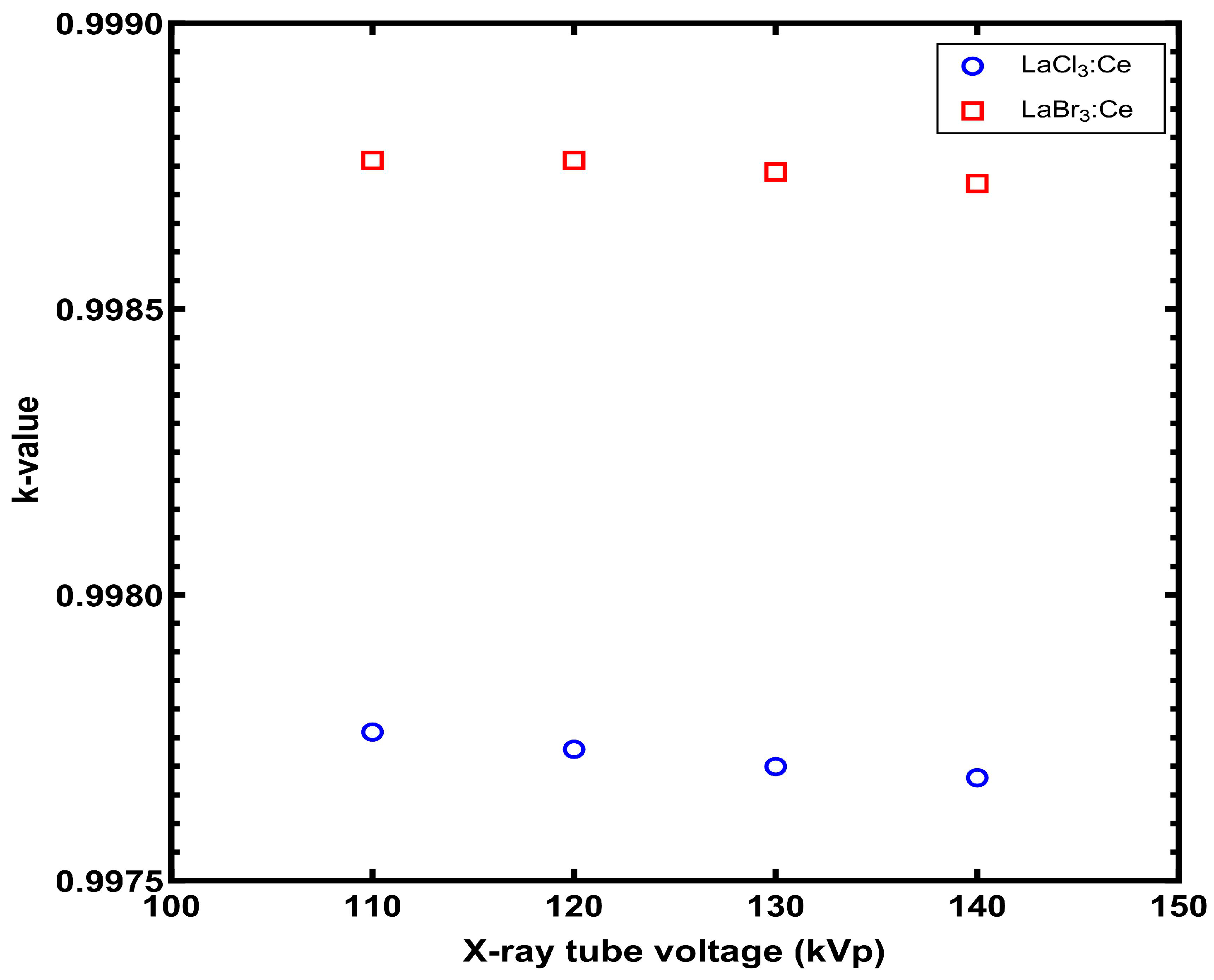
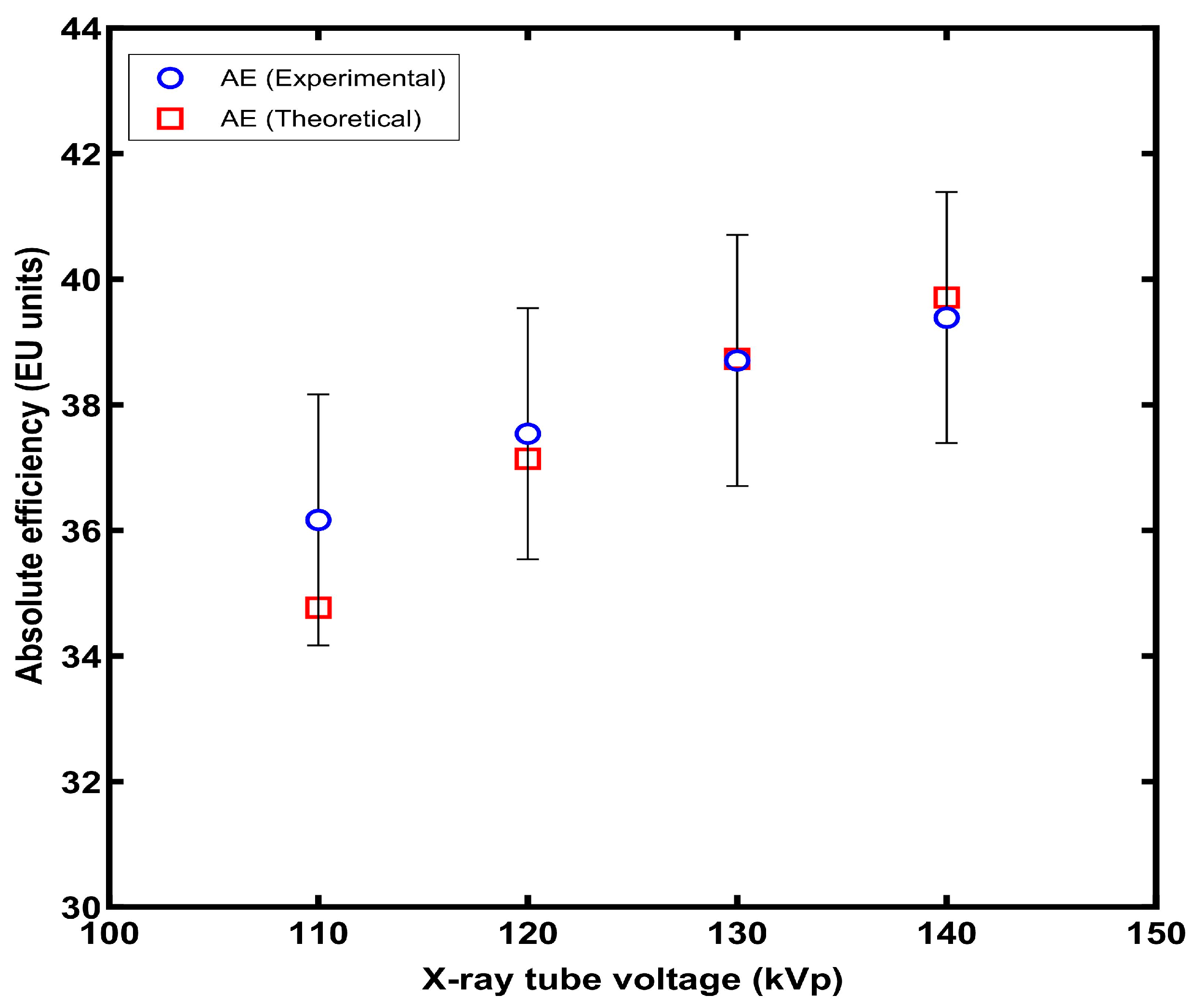
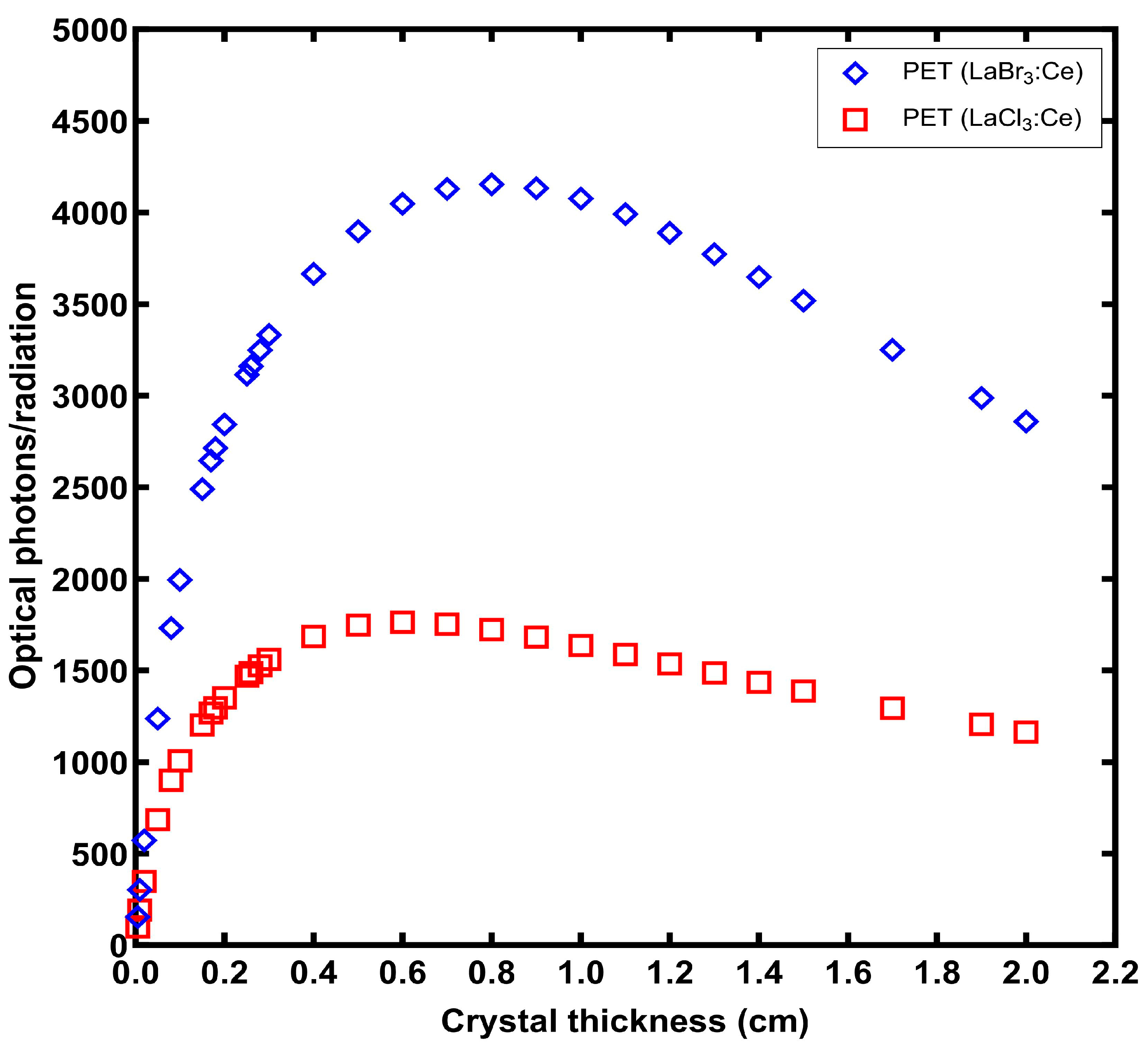
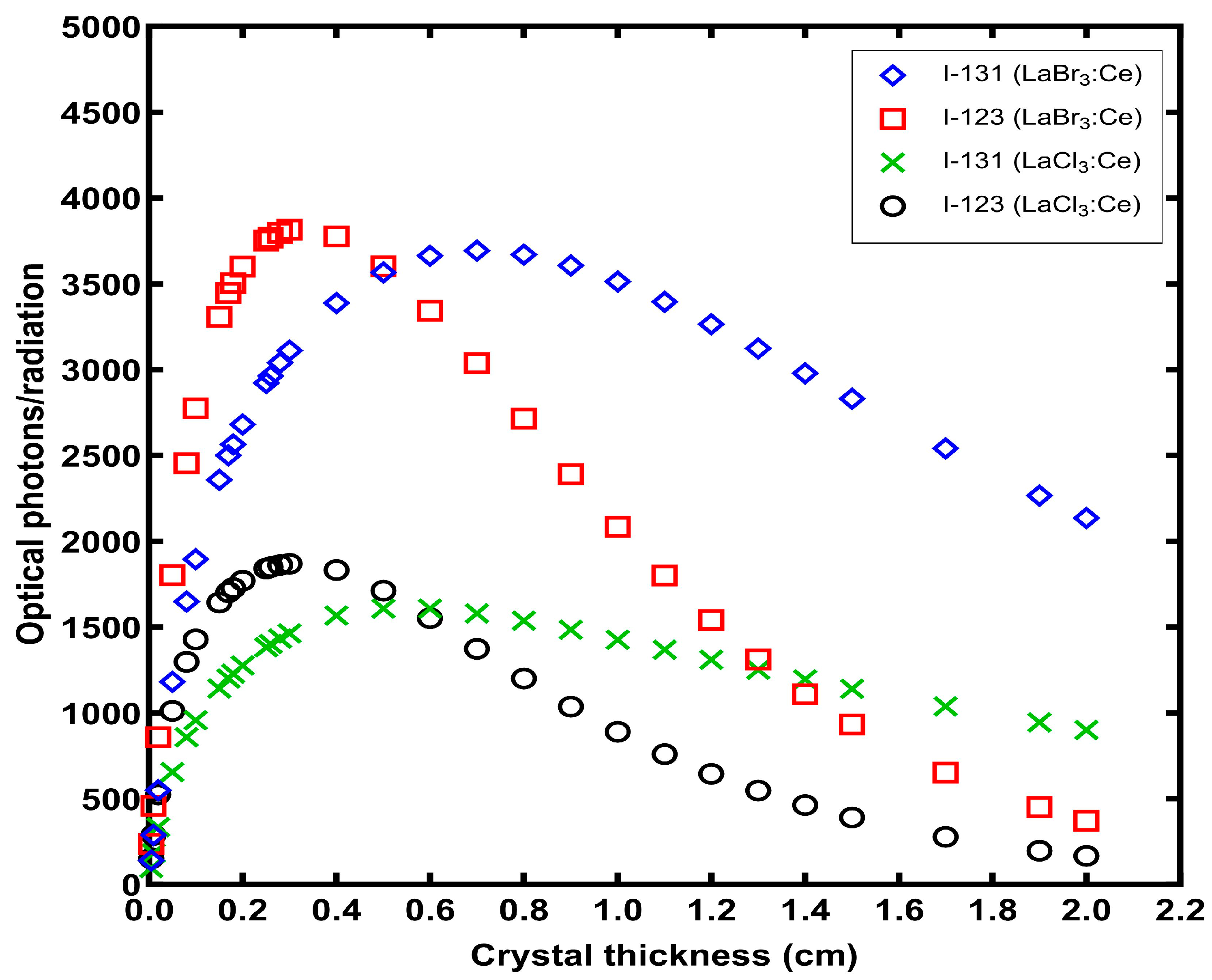
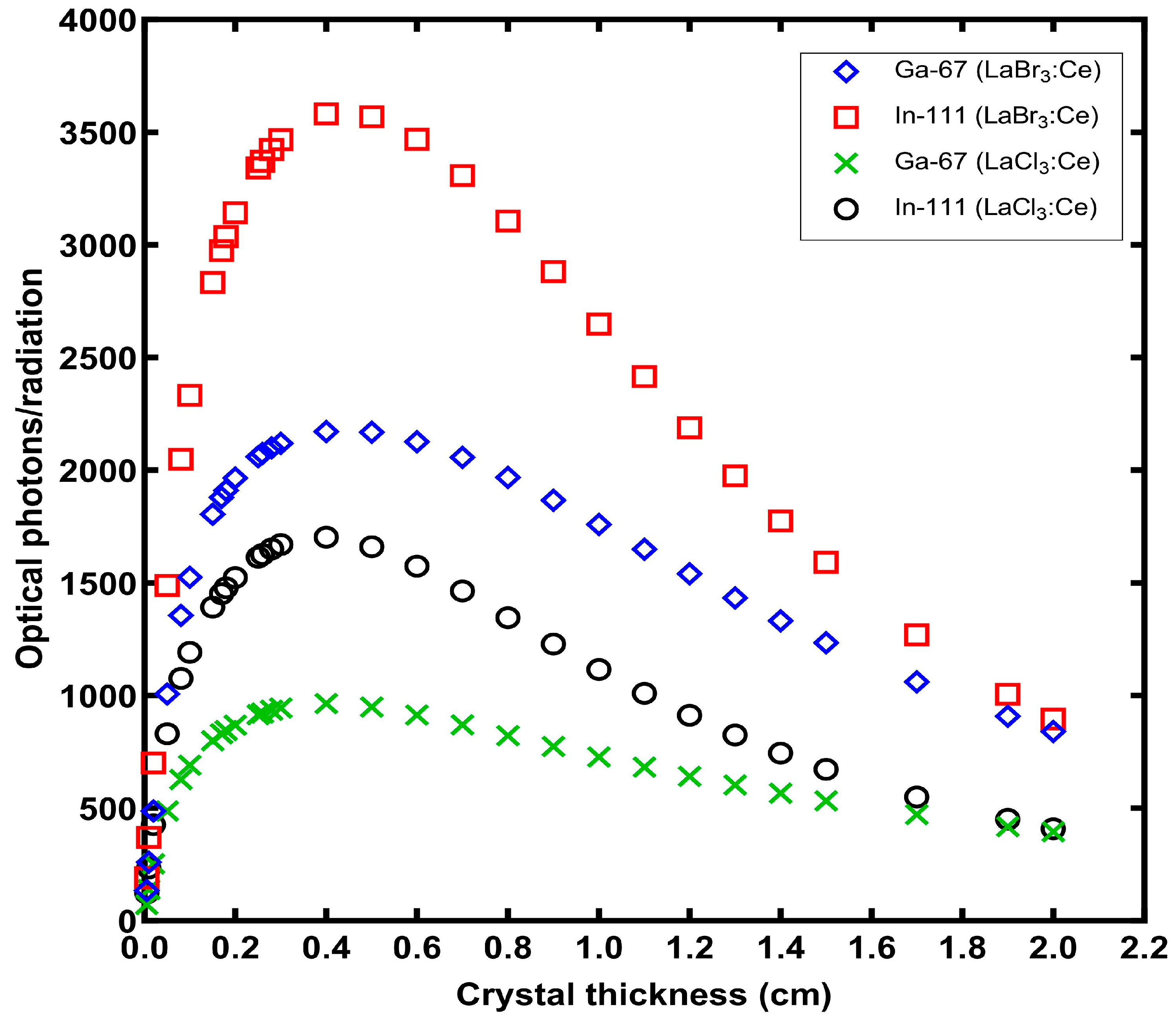
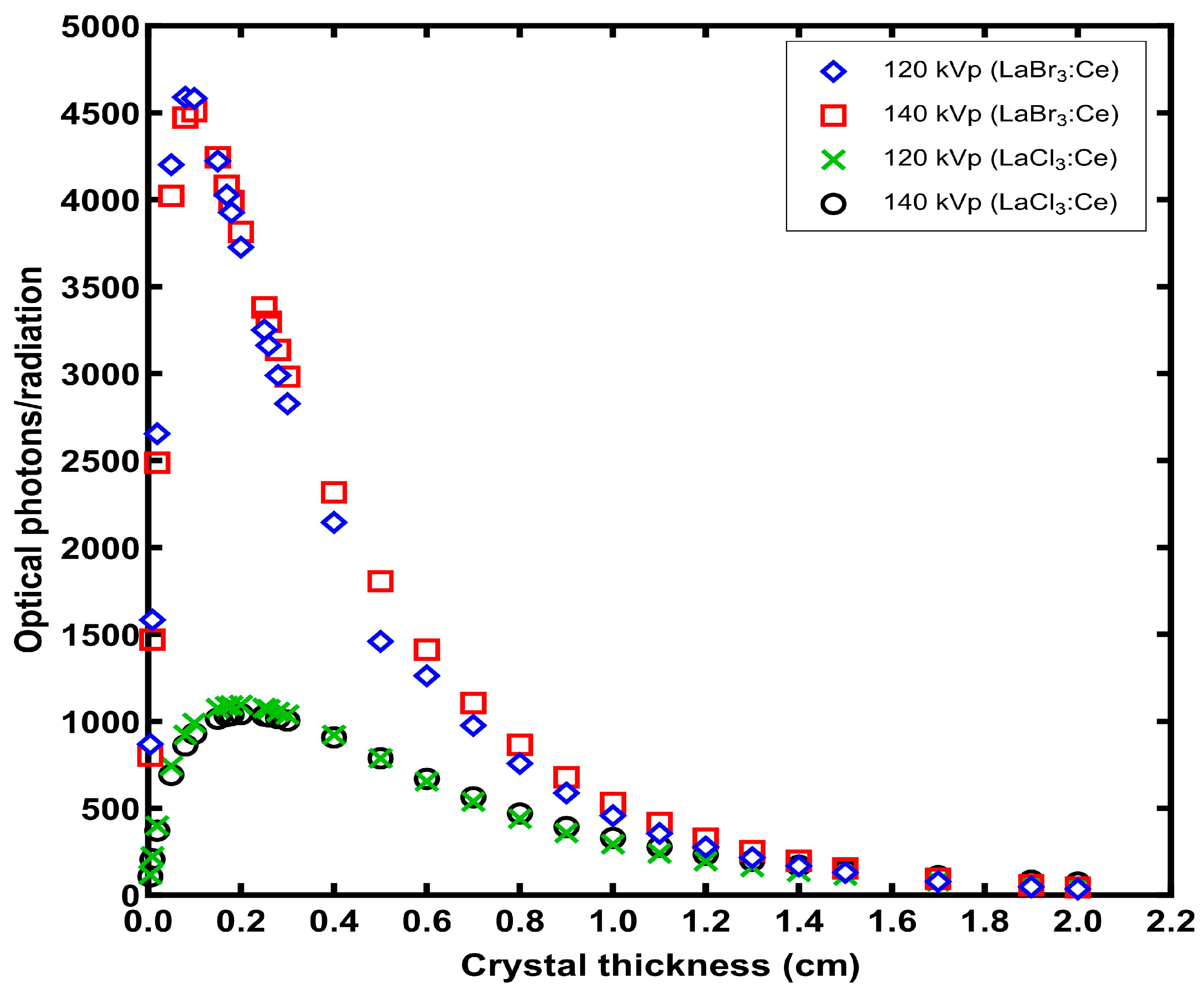
According to the applied theoretical model, the crystal can be divided into N elementary layers. Only a percentage, hereafter called k, of the photons generated in the nth layer passes to the n + 1 layer and from there to the next, eventually reaching the output surface of the crystal. The model assumes that this percentage k remains constant for each layer. The loss of optical photon energy in the crystal is assumed to occur either by internal optical photon absorption in the crystal mass or by optical photon escape when the photons reach the crystal surfaces. The model also considers the X-ray attenuation properties in the crystal, the intrinsic conversion efficiency, showing the fraction of the absorbed X-ray or gamma-ray energy converted into optical photon energy [36], and the reflection of the optical photons at the input and output crystal surfaces. A more in-depth explanation of the model has been described in previous studies [33,35].
It is hypothesized that an equal number of optical photons, produced within the crystal’s mass, are traveling in both forward and backward directions [33,35]. By considering all the reflections that occur between the entrance and the exit surfaces of the scintillator, we can determine the proportion of optical photons, produced in the nth layer, that manage to escape the crystal by the relation [35]:
M n = 1 R 0 k N n 1 k 2 N R 0   R 1 + R 1 k n + N k 2 N R 0 R 1 1 k 2 N R 0 R 1
where R0 is the output reflectivity, denoting the fraction of photons reflected back into the scintillator when incident at its exit surface, and R1 is the input reflectivity, referring to the proportion of optical photons that are reflected back into the scintillator upon encountering its entrance surface. Both cubic crystals are wrapped with Teflon tape on their five surfaces, resulting in a high reflectivity close to 100% (R1 = 1) for these surfaces [37]. The exit surface of the crystals in contact with the photodetector is protected with silicon glass. Consequently, when determining the R0, the reflection coefficients of crystal–silicon glass interface (Rcr,Si) and silicon glass–air interface (RSi,air) were taken into consideration. A fraction (1 − Rcr,Si) of the incident light on the exit surface of the crystal passes through the interface between the crystal and silicon glass. Subsequently, this fraction reaches the interface between glass and air, where a fraction RSi,air of it, is reflected and directed back towards the crystal. In this approach, an error of approximately 1.5% arises due to reflections on the crystal–glass interface. Considering the above, R0 is given by the following relation:
R 0 = 1 R c r , S i   R S i , a i r
Rcr,Si and RSi,air are defined as [33]:
R c r , S i = n 1 n 2 2 n 1 + n 2 2
and
R S i , a i r = n 2 1 2 n 2 + 1 2
The refractive indexes n1 = 1.9 of the crystals and n2 = 1.47 of the silicon glass used in relations (3) and (4) were obtained from the literature [38,39,40].
Considering all of the layers, the total amount of optical photons generated by X-rays with energy E and manage to exit through the output is equal to [35,36]:
M E = n = 1 N f E e μ A l t e μ n Δ t μ Δ t n c E E λ 1 R 0 k N n 1 k 2 N R 0   R 1 + R 1 k n + N k 2 N R 0   R 1 1 k 2 N R 0   R 1
where f(E) is the incident X-ray fluence obtained from the TASMIP Spectra Calculator [41], μ is the attenuation coefficient, Δt is the thickness of each layer n of the scintillator, Eλ is the energy of the optical photons, and μAl and t are the attenuation coefficient and the thickness of the aluminum encapsulation of the crystal, respectively. nc is the intrinsic conversion efficiency and it was calculated for a light yield of LY = 49.000 photons/MeV and LY = 63.000 photons/MeV for LaCl3:Ce and LaBr3:Ce crystals, respectively, by the relation [34,36]:
n C = L Y E λ 10 6
The theoretical value of AE for a photon of specific energy is given by the following relation [33,34]:
A E E = n = 1 N γ E e μ A l t e μ n Δ t μ Δ t n c 1 R 0 k N n 1 k 2 N R 0   R 1 + R 1 k n + N k 2 N R 0   R 1 1 k 2 N R 0   R 1
where γ(E) is a conversion factor converting energy fluence (W/m2) into exposure rate (mR/s). Considering the whole energy spectrum, AE equals to [34]:
A E = E = E m i n E m a x f E A E E E = E m i n E m a x f E
The propagation probability per elementary thickness k was determined from relation (8) by fitting the theoretical equation to experimental AE(E) data. During the fitting procedure, different values of k were tested in Equation (8) and compared with the corresponding experimental AE results calculated. Different k values were tested arbitrarily (trial and error method). The final selected k value was the one where the results of Equation (8) were equal to the corresponding experimental AE data. These experimental data were obtained from crystals samples with 10 mm thicknesses, that are commonly used in CT applications (110 kVp–140 kVp) [21,22]. X-ray irradiation of the crystal samples was performed using an X-ray tube equipped with a 1.5 mm Al filter, connected to a CPI series CMP 200DR 50kW generator while maintaining a constant current-time product of 63 mAs and an irradiation duration of 1 s. Additionally, a 20mm Al filtration was introduced at the exit of the X-ray tube. The exposure rate was measured with RTI Piranha P100B dosimeter. The experimental setup for the energy flux measurement comprised a light integration sphere (Oriel 70451) coupled to a photomultiplier tube (PMT) (EMI 9798B) which was connected to an electrometer (Cary 400) [21,22].
Similarly, the DOG can be determined across the entire range of X-ray energy by the relation [35,36]:
D O G = E = E m i n E m a x M E E = E m i n E m a x f E e μ A l t
For the calculation of DOG for radioisotopes, assuming that fi is the probability of energy emission Ei, which has an attenuation coefficient μi, relation (5) transforms into the following form:
M i E i = n = 1   N e μ A l t e μ i Δ t μ i Δ t n c 1 E λ 1 R 0 k N n 1 k 2 N R 0   R 1 + R 1 k n + N k 2 N R 0   R 1 1 k 2 N R 0   R 1
Finally, the DOG was calculated as:
D O G = i f i E i M i i f i

3. Results

Figure 1 displays the values of parameter k for LaCl3:Ce and LaBr3:Ce crystals within the tube voltage range of 110 kVp to 140 kVp, demonstrating the propagation of optical photons through each layer of 5 μm thickness. Both crystals present a stable performance, with the k values remaining almost constant (from 0.99776 to 0.99768 for LaCl3:Ce and from 0.99876 to 0.99872 for LaBr3:Ce). The results obtained for both crystals can be considered adequate, as the values of k remain above 0.99 over the entire range of voltages investigated.
The data presented in Figure 2 and Figure 3 allow for a comparison between the experimental [21,22] and theoretical values of AE for the two crystals. As shown in these figures, a strong agreement exists between the experimental observations and the theoretical predictions of AE, for both crystals. For the LaCl3:Ce crystal, the relative difference between the experimental and the theoretical AE is between 0.05% and 3.86%. On the contrary, for the LaBr3:Ce crystal, the relative difference between the experimental and the theoretical values ranges from 0.03% to 2.57%.
The theoretical AE values for LaCl3:Ce and LaBr3:Ce crystals were determined using an average k value of 0.9977 and 0.9987, respectively. For other scintillators, the values of the k coefficient, which were calculated using the same model and for an elementary layer thickness of 1 μm, range between 0.74 and 0.994. Specifically, for the single-crystal scintillators GSO:Ce, LSO:Ce, LYSO:Ce, and YAP:Ce, the corresponding k values were 0.923, 0.845, 0.860, and 0.74, respectively [33,36]. In the case of CsI:Tl columnar phosphor, the k value ranged from 0.980 to 0.994 for different column thicknesses [35].
Figure 4, Figure 5, Figure 6, Figure 7 and Figure 8 present the DOG values that have been predicted for crystal thicknesses ranging from 0.005 cm to 2 cm, corresponding to various radioisotopes and X-ray energies. The results indicate that DOG values attributed to the LaBr3:Ce crystal are generally higher than those of the LaCl3:Ce crystal for all radioisotopes and X-ray energies examined. Another deduction derived from the findings is that the crystal efficiency demonstrates a decrease beyond a certain thickness. This is because although a higher crystal thickness enhances energy absorption, it also leads to increased loss of optical photons during propagation towards the output.
For various medical applications, different optimal crystal thicknesses have been identified for LaCl3:Ce and LaBr3:Ce crystals. Specifically, for PET applications, the LaCl3:Ce crystal demonstrates optimal performance with a thickness of 0.6 cm, whereas the LaBr3:Ce crystal performs best at 0.8 cm. Both crystals, however, show ideal thicknesses of 0.3 cm and 0.1 cm for 99mTc and 201Tl applications, respectively. For 131I applications, the optimal thickness is 0.5 cm for LaCl3:Ce and 0.7 cm for LaBr3:Ce, whereas for 123I applications, both crystals exhibit an ideal thickness of 0.3 cm. Additionally, both crystals demonstrate ideal performance for 111In and 67Ga applications with a thickness of 0.4 cm. Moreover, for X-ray energies of 120 kVp and 140 kVp, the LaCl3:Ce crystal shows optimal thickness at 0.2 cm, whereas the LaBr3:Ce crystal exhibits the best performance at 0.08 cm and 0.1 cm, respectively.

4. Discussion

The presented results are based on a theoretical analytical model which employs basic physical principles in ionizing radiation transfer and absorption. As such, it can provide a fast insight into the steps of radiation transfer, radiation absorption, optical photon production, and optical photon escape as a function of crystal thickness. The ionizing radiation interactions are treated assuming exponential attenuation of the radiation in the crystal while adopting the total attenuation coefficient as the radiation extinction parameter. The use of the total attenuation coefficient in low energies where the photoelectric absorption dominates provides accurate radiation absorption results. In the higher energies considered in this study, especially above 300 keV, the effect of scatter is of importance. The presented analytical method uses the attenuation coefficient as an extinction factor and may overestimate the radiation absorption since it assumes that all scatter photons and the characteristic radiation will be reabsorbed in the crystal. This may be valid for scatter photons of lower energy that might be subjected to photoelectric absorption, or for interactions in crystals with large dimensions. However, if small crystal thicknesses or secondary radiation generated close to the crystal surface are considered, then these photons possess a higher probability of escaping the crystal. Thus, analytical modeling overestimates the absorbed radiation energy. The extent of the overestimation depends upon the material, the interaction site, and the radiation energy. Subsequently, the optical photon propagation in the presented work is treated via an already published semi-empirical approach where the percentage of the optical photon transmission per layer is obtained by fitting experimental data, and the variation in the optical photon escape angles is not explicitly considered.
A more accurate prediction of the radiation and optical photons transfer and escape in the material, without a priori knowledge of the experimental AE results would require simulation tools, where an accurate geometry is necessary and the radiation and the optical transfer properties as well as the photon absorption lengths can be more accurately considered [42,43,44,45,46,47,48]. Despite a degree of overestimation in higher energies by the utilized analytical methodology and the corresponding assumption in the optical photon exit layer, the presented method can still provide useful qualitative results in terms of comparing the efficiency of different crystalline materials at different thicknesses when excited by ionizing radiation.
As observed in Figure 1, the average values of k for the LaCl3:Ce and LaBr3:Ce crystals are significantly higher compared to other single-crystal scintillators and comparable to those for CsI:Tl columnar phosphor [33,35,36]. Based on the assumptions of the model for the propagation of light within crystals, this difference is mainly attributed to the fact that all surfaces of the examined crystals, except the output surface, were covered with Teflon, thus minimizing losses by optical photon escape when the photons were incident on the crystal surfaces. As previously mentioned, according to the model, light losses, in addition to lateral light escape, can also occur due to internal optical photon absorption in the crystal mass. However, the absorption of the generated photons depends on the band gap width of the crystal in relation to the energy of the photons. In the case of LaCl3:Ce, the energy of the majority of generated photons is 3.54 eV, whereas the estimated band gap of the crystal lies between 4.34 and 8.25 eV, with an intermediate value being more probable [49]. For LaBr3:Ce, the photons have an energy of 3.26 eV, and the band gap is estimated to be between 3.42 and 7.43 eV [49]. Consequently, the reabsorption of the generated photons occurs in a very small percentage. In other words, both crystals are highly transparent to the visible radiation they produce.
The DOG values of the two crystals may be evidence that the LaBr3:Ce crystal exhibits significantly better performance compared to the LaCl3:Ce crystal, primarily due to its higher light yield [21,22]. The thickness of the crystal affects its optical performance in two ways. As the thickness increases, so does the absorption of incident X or gamma radiation and thus the production of optical photons. However, with increasing thickness, the losses of generated optical photons also exhibit a corresponding rise. Therefore, the optimal thickness is determined by the optimal combination of these two phenomena. For low energies of incident radiation, where their absorption occurs at a shallow depth within the crystal, increasing the thickness of the crystal would decrease its optical performance, as it would increase the losses of optical photons without significantly contributing to their production. For higher energies of incident radiation, which are absorbed at greater depths, a thicker crystal is required, up to the point where the increased production of optical photons is not outweighed by their increased losses. Therefore, the optical performance of the crystal depends on the energy of the incident radiation, the thickness of the crystal, and the type of crystal.
It is worth commenting that the increased efficiency of LaBr3:Ce in every case, compared to LaCl3:Ce, is not only affected by the notable difference in the corresponding light yields (i.e., 63,000 photons/MeV for LaBr3:Ce vs. 49,000 photons/MeV for LaCl3:Ce). The total light output is also affected by the radiation energy absorbed and the probability of optical photon escape. For the same crystal thickness, the higher LaBr3:Ce density (5.2 g/cm3) with regard to that of LaCl3:Ce (3.86 g/cm3) enhances the probability of radiation interaction and subsequent energy absorption in LaBr3:Ce with respect to LaCl3:Ce. Consequently, a greater number of optical photons are produced in the LaBr3:Ce scintillator when all the absorbed radiation is considered. Finally, the optical photon propagation probability per layer of LaBr3:Ce is slightly higher than the corresponding LaCl3:Ce as shown in Figure 1. Therefore, the higher probability of radiation absorption, the higher optical photon yield, and the more efficient optical photon propagation characteristics of LaBr3:Ce combined result in the higher efficiency of LaBr3:Ce with respect to LaCl3:Ce.
Generally, it is evident that with the increase in energy, the optimum thickness increases for both crystals and reaches its maximum at the higher energy value of 511 keV in PET. As mentioned earlier, this was expected since the absorption of higher energy X or gamma photons occurs at a greater depth within the crystal. Thus, for PET applications, the optimal thickness for LaCl3:Ce and LaBr3:Ce crystals is 0.6 and 0.8 cm, respectively. For the energy range at which the radionuclides used in SPECT emit, the optimal thickness of LaCl3:Ce and LaBr3:Ce ranges from 0.1–0.5 cm and 0.1–0.7 cm, respectively, whereas for 120 and 140 KVp, the optimal thickness is 0.2 cm and 0.08–0.1 cm, respectively.
Approximately 80% of all nuclear medicine scans performed globally consist of SPECT and planar scintigraphy [50]. The most common clinical use of SPECT is in the assessment of myocardial perfusion and the evaluation of myocardial and coronary artery status following a cardiac ischemic episode [2,5]. The cardiological application of SPECT surpasses all other diagnostic applications of SPECT to such an extent that manufacturers offer SPECT systems that have been optimized for this type of imaging [5]. The most widely used isotope worldwide in this imaging technique, but not limited to it, is 99mTc [50,51]. For this isotope, it has been observed that the optimal thickness is 0.3 cm for both crystals. Another isotope that was initially used in cardiology nuclear imaging but continues to be utilized for certain advantages it offers, as well as in cases of 99mTc shortage, is 201Tl [52], for which the optimal thickness of the two crystals is 0.1 cm.
123I and 131I are extensively used in nuclear imaging as well as in the radiation therapy of neuroendocrine tumors, such as thyroid cancer, comprising a theranostic pair [4,51,53,54]. 123I, due to its shorter half-life and absence of beta particle emission, imposes a lower radiation burden on the subject compared to 131I; therefore, it is more commonly used in nuclear imaging [4]. It is also particularly effective in visualizing serotonin and dopamine neurotransmitters, as well as in diagnosing neurodegenerative diseases, cerebrovascular disorders, and brain injuries [51]. From the results, for applications involving 123I, the ideal crystal thickness for both crystals stands at 0.3 cm, whereas for 131I, it stands at 0.5 cm and 0.7 cm for LaCl3:Ce and LaBr3:Ce, respectively.
67Ga has been utilized for over fifty years in nuclear imaging, particularly for the diagnosis and localization of inflammatory areas within the body, such as in the case of pyrexia of unknown origin [53]. Furthermore, the scintigraphy with this isotope is used adjunctively for distinguishing between malignant and benign tumors, as well as in the evaluation of head and neck squamous cell carcinoma [55]. The optimal thickness for 67Ga was calculated as equal to 0.4 cm for both crystals. 111In exhibits exceptional characteristics for nuclear imaging and is used for similar purposes as 67Ga. 111In is typically bound to antibodies, polypeptides, or white blood cells for the diagnosis and localization of inflammations within the body, for the diagnosis of cases with fever of unknown origin, or in cases where other diagnostic and imaging techniques yield ambiguous results [56,57]. Regarding uses related to 111In, the ideal crystal thickness was found equal to 0.4 cm for both crystals.
In the field of nuclear medicine applications, the choice of the most suitable crystal scintillator thickness plays a pivotal role in optimizing patient care. An optimal crystal scintillator thickness allows for improved sensitivity in detecting gamma rays emitted by radiopharmaceuticals, which in turn translates to reduced required radioactivity administered to patients to achieve the desired diagnostic outcomes. This reduction translates to a substantial decrease in patient radiation exposure. Consequently, patients undergoing nuclear medicine procedures experience lower radiation exposure, which not only enhances their safety but also contributes to a more sustainable and patient-centric approach to nuclear medicine practice.
The presented theoretical DOG results demonstrate the applicability of different LaBr3:Ce and LaCl3:Ce crystal thicknesses with the radioisotopes currently in use in nuclear medicine applications and can be used as an initial reference for the development of crystals with different thicknesses. A more precise quantitative approach will require experimental AE results for each case.

5. Conclusions

In the current study, a theoretical model was developed to describe the light propagation within a crystal scintillator and to estimate the optimal thickness of LaCl3:Ce and LaBr3:Ce single-crystal scintillators in terms of Detector Optical Gain (DOG). Notably, the strong alignment between experimental observations and theoretical predictions of AE in both crystals underscores the reliability of the model. The values of k for LaCl3:Ce and LaBr3:Ce crystals are markedly superior in comparison to other single-crystal scintillators. Moreover, the discernibly superior DOG values of LaBr3:Ce imply its enhanced performance compared to LaCl3:Ce. Our observations also reveal that the optimal thickness depends on both the material of the scintillator and the energy of the gamma-rays and X-rays. These findings offer valuable insights into the design and optimization of crystal scintillators in the realm of Nuclear Medicine Imaging systems, providing a robust foundation for future advancements in this critical field.

Author Contributions

Conceptualization, S.T., C.M. and N.K.; methodology, S.T., C.M., I.V., G.F. and N.K.; software, S.T., N.K. and I.V.; validation, K.N., G.F. and I.K.; formal analysis, S.T., C.M., I.V. and A.B.; investigation, S.T., I.K., A.B. and K.N.; resources, C.M., I.V. and G.F.; data curation, I.V. and N.K.; writing—original draft preparation, S.T., C.M. and N.K.; writing—review and editing, S.T., C.M., I.K. and N.K.; visualization, C.M. and N.K.; supervision, N.K.; project administration, C.M. and N.K. All authors have read and agreed to the published version of the manuscript.

Funding

This research received no external funding.

Data Availability Statement

The data associated with our study are included or referenced in the article and can be made available on reasonable request.

Conflicts of Interest

The authors declare no conflicts of interest.

References

  1. Cherry, S.R.; Sorenson, J.A.; Phelps, M.E. Physics in Nuclear Medicine, 4th ed.; Elsevier/Saunders: Philadelphia, PA, USA, 2012. [Google Scholar]
  2. Lecomte, R.; Granja, C.; Leroy, C.; Stekl, I. Biomedical Imaging: SPECT and PET. AIP Conf. Proc. 2007, 958, 115–122. [Google Scholar] [CrossRef]
  3. Eijk, C.W.E.V. Inorganic scintillators in medical imaging. Phys. Med. Biol. 2002, 47, 85–106. [Google Scholar] [CrossRef] [PubMed]
  4. Morphis, M.; Van Staden, J.A.; du Raan, H.; Ljungberg, M. Evaluation of Iodine-123 and Iodine-131 SPECT activity quantification: A Monte Carlo study. EJNMMI Phys. 2021, 8, 61. [Google Scholar] [CrossRef] [PubMed]
  5. Madsen, M.T. Recent Advances in SPECT Imaging. J. Nucl. Med. 2007, 48, 661–673. [Google Scholar] [CrossRef]
  6. Zanzonico, P. Positron emission tomography: A review of basic principles, scanner design and performance, and current systems. Semin. Nucl. Med. 2004, 34, 87–111. [Google Scholar] [CrossRef]
  7. Chowdhury, F.U.; Scarsbrook, A.F. The role of hybrid SPECT-CT in oncology: Current and emerging clinical applications. Clin. Radiol. 2008, 63, 241–251. [Google Scholar] [CrossRef] [PubMed]
  8. Bailey, D.L. Transmission scanning in emission tomography. Eur. J. Nucl. Med. Mol. Imaging 1998, 25, 774–787. [Google Scholar] [CrossRef] [PubMed]
  9. Cal-Gonzalez, J.; Rausch, I.; Shiyam Sundar, L.K.; Lassen, M.L.; Muzik, O.; Moser, E.; Papp, L.; Beyer, T. Hybrid Imaging: Instrumentation and Data Processing. Front. Phys. 2018, 6, 47. [Google Scholar] [CrossRef]
  10. Beyer, T.; Freudenberg, L.S.; Townsend, D.W.; Czernin, J. The Future of Hybrid Imaging—Part 1: Hybrid Imaging Technologies and SPECT/CT. Insights Imaging 2011, 2, 161–169. [Google Scholar] [CrossRef]
  11. Beyer, T.; Townsend, D.W.; Czernin, J.; Freudenberg, L.S. The Future of Hybrid Imaging—Part 2: PET/CT. Insights Imaging 2011, 2, 225–234. [Google Scholar] [CrossRef]
  12. Guo, Z. The Principle and State-of-Art Facilities for PET. J. Phys. Conf. Ser. 2022, 2386, 012062. [Google Scholar] [CrossRef]
  13. Marsden, P.K. Detector technology challenges for nuclear medicine and PET. Nucl. Instrum. Methods Phys. Res. Sect. A Accel. Spectrometers Detect. Assoc. Equip. 2003, 513, 1–7. [Google Scholar] [CrossRef]
  14. Berard, P.; Pepin, C.M.; Rouleau, D.; Cadorette, J.; Lecomte, R. CT acquisition using PET detectors and electronics. IEEE Trans. Nucl. Sci. 2005, 52, 634–637. [Google Scholar] [CrossRef]
  15. Van Loef, E.V.D.; Dorenbos, P.; Van Eijk, C.W.E.V.; Kramer, K.; Gudel, H.U. Scintillation properties of LaCl3:Ce3+ crystals: Fast, efficient and high-energy-resolution scintillators. IEEE Nucl. Sci. Symp. Conf. Rec. 2000, 6, 31–34. [Google Scholar] [CrossRef]
  16. Iltis, A.; Mayhugh, M.R.; Menge, P.; Rozsa, C.M.; Selles, O.; Solovyev, V. Lanthanum halide scintillators: Properties and applications. Nucl. Instrum. Methods Phys. Res. Sect. A Accel. Spectrometers Detect. Assoc. Equip. 2006, 563, 359–363. [Google Scholar] [CrossRef]
  17. Van Loef, E.V.D.; Dorenbos, P.; Van Eijk, C.W.E.V.; Krämer, K.; Güdel, H.U. High-energy-resolution scintillator: Ce3+ activated LaBr3. Appl. Phys. Lett. 2001, 79, 1573–1575. [Google Scholar] [CrossRef]
  18. Van Loef, E.V.D.; Dorenbos, P.; Van Eijk, C.W.E.V.; Krämer, K.; Güdel, H.U. Scintillation properties of LaBr3:Ce3+ crystals: Fast, efficient and high-energy-resolution scintillators. Nucl. Instrum. Methods Phys. Res. Sect. A Accel. Spectrometers Detect. Assoc. Equip. 2002, 486, 254–258. [Google Scholar] [CrossRef]
  19. Advatech, U.K. LaCl3: Ce-Lanthanum Chloride (Ce). Available online: https://www.advatech-uk.co.uk/lacl3_ce.html (accessed on 12 May 2023).
  20. Advatech, U.K. LaBr3: Ce-Lanthanum Bromide (Ce). Available online: https://www.advatech-uk.co.uk/labr3_ce.html (accessed on 12 May 2023).
  21. Tseremoglou, S.; Michail, C.; Valais, I.; Ninos, K.; Bakas, A.; Kandarakis, I.; Fountos, G.; Kalyvas, N. Evaluation of Cerium-Doped Lanthanum Bromide (LaBr3:Ce) Single-Crystal Scintillator’s Luminescence Properties under X-ray Radiographic Conditions. Appl. Sci. 2022, 13, 419. [Google Scholar] [CrossRef]
  22. Tseremoglou, S.; Michail, C.; Valais, I.; Ninos, K.; Bakas, A.; Kandarakis, I.; Fountos, G.; Kalyvas, N. Efficiency Properties of Cerium-Doped Lanthanum Chloride (LaCl3:Ce) Single Crystal Scintillator under Radiographic X-ray Excitation. Crystals 2022, 12, 655. [Google Scholar] [CrossRef]
  23. Bagheri, R.; Shirmardi, S.P.; Heidari, B.; Ahmadi, B.; Tuzemen, S. Comparison of LaBr3(CE) and NaI(TL) Scintillation Crystals Responses to Various Gamma Energies Using Monte Carlo Simulations. Int. J. Adv. Res. 2016, 4, 494–500. [Google Scholar] [CrossRef]
  24. Sharma, S.; Ranga, V.; Dhibar, M.; Rawat, S.; Kumar, G.A. Intrinsic Resolution of Compton Electrons in LaBr3: Ce and LaCl3: Ce Detectors Using Compton Coincidence Technique. Nucl. Phys. 2016, 61, 1020–1021. [Google Scholar]
  25. Sariyal, R.; Mazumdar, I.; Patel, S.M. Measurement of Absolute Light Yield and Quantum Efficiency of LaBr3: Ce Scintillator Detector Using SiPM. J. Inst. 2021, 16, P12024. [Google Scholar] [CrossRef]
  26. Dey Chaudhuri, S.; Banerjee, D.; Bhattacharjee, T.; Wasim Raja, S.; Acharya, R.; Pujari, P.K. Performance Study of LaBr3: Ce Detectors Coupled to R2083 PM Tube for Energy and Timing Characteristics. J. Radioanal. Nucl. Chem. 2020, 324, 829–835. [Google Scholar] [CrossRef]
  27. Cazzaniga, C.; Nocente, M.; Tardocchi, M.; Fazzi, A.; Hjalmarsson, A.; Rigamonti, D.; Ericsson, G.; Gorini, G. Thin YAP:Ce and LaBr3:Ce Scintillators as Proton Detectors of a Thin-Film Proton Recoil Neutron Spectrometer for Fusion and Spallation Sources Applications. Nucl. Instrum. Methods Phys. Res. Sect. A Accel. Spectrometers Detect. Assoc. Equip. 2014, 751, 19–22. [Google Scholar] [CrossRef]
  28. Nácher, E.; Mårtensson, M.; Tengblad, O.; Álvarez-Pol, H.; Bendel, M.; Cortina-Gil, D.; Gernhäuser, R.; Le Bleis, T.; Maj, A.; Nilsson, T.; et al. Proton Response of CEPA4: A Novel LaBr3(Ce)–LaCl3(Ce) Phoswich Array for High-Energy Gamma and Proton Spectroscopy. Nucl. Instrum. Methods Phys. Res. Sect. A Accel. Spectrometers Detect. Assoc. Equip. 2015, 769, 105–111. [Google Scholar] [CrossRef]
  29. Babiano, V.; Caballero, L.; Calvo, D.; Ladarescu, I.; Olleros, P.; Domingo-Pardo, C. γ -Ray Position Reconstruction in Large Monolithic LaCl3(Ce) Crystals with SiPM Readout. Nucl. Instrum. Methods Phys. Res. Sect. A Accel. Spectrometers Detect. Assoc. Equip. 2019, 931, 1–22. [Google Scholar] [CrossRef]
  30. Taggart, M.P.; Henderson, J. Fast-Neutron Response of LaBr3(Ce) and LaCl3(Ce) Scintillators. Nucl. Instrum. Methods Phys. Res. Sect. A Accel. Spectrometers Detect. Assoc. Equip. 2020, 975, 164201. [Google Scholar] [CrossRef]
  31. Garnett, R.; Prestwich, W.V.; Atanackovic, J.; Wong, M.; Byun, S.H. Characterization of a LaBr3(Ce) Detector for Gamma-Ray Spectrometry for CANDU Power Reactors. Radiat. Meas. 2017, 106, 628–631. [Google Scholar] [CrossRef]
  32. Aldawood, S.; Castelhano, I.; Gernhäuser, R.; Van Der Kolff, H.; Lang, C.; Liprandi, S.; Lutter, R.; Maier, L.; Marinšek, T.; Schaart, D.R.; et al. Comparative Characterization Study of a LaBr3(Ce) Scintillation Crystal in Two Surface Wrapping Scenarios: Absorptive and Reflective. Front. Oncol. 2015, 5, 270. [Google Scholar] [CrossRef]
  33. Nikolopoulos, D.; Kalyvas, N.; Valais, I.; Argyriou, X.; Vlamakis, E.; Sevvos, T.; Kandarakis, I. A semi-empirical Monte Carlo based model of the Detector Optical Gain of Nuclear Imaging scintillators. J. Inst. 2012, 7, P11021. [Google Scholar] [CrossRef]
  34. David, S.; Michail, C.; Seferis, I.; Valais, I.; Fountos, G.; Liaparinos, P.; Kandarakis, I.; Kalyvas, N. Evaluation of Gd2O2S: Pr granular phosphor properties for X-ray mammography imaging. J. Lumin. 2016, 169, 706–710. [Google Scholar] [CrossRef]
  35. Kalyvas, N.; Valais, I.; Michail, C.; Fountos, G.; Kandarakis, I.; Cavouras, D. A theoretical study of CsI:Tl columnar scintillator image quality parameters by analytical modeling. Nucl. Instrum. Methods Phys. Res. Sect. A Accel. Spectrometers Detect. Assoc. Equip. 2015, 779, 18–24. [Google Scholar] [CrossRef]
  36. Kalyvas, N.; Valais, I.; David, S.; Michail, C.; Fountos, G.; Liaparinos, P.; Kandarakis, I. Studying the energy dependence of intrinsic conversion efficiency of single crystal scintillators under X-ray excitation. Opt. Spectrosc. 2014, 116, 743–747. [Google Scholar] [CrossRef]
  37. Roncali, E.; Stockhoff, M.; Cherry, S.R. An integrated model of scintillator-reflector properties for advanced simulations of optical transport. Phys. Med. Biol. 2017, 62, 4811–4830. [Google Scholar] [CrossRef] [PubMed]
  38. Bernabei, R.; Belli, P.; Montecchia, F.; Nozzoli, F.; d’Angelo, A.; Cappella, F.; Incicchitti, A.; Prosperi, D.; Castellano, S.; Cerulli, R.; et al. Performances and potentialities of a LaCl3:Ce scintillator. Nucl. Instrum. Methods Phys. Res. Sect. A Accel. Spectrometers Detect. Assoc. Equip. 2005, 555, 270–281. [Google Scholar] [CrossRef]
  39. Giaz, A.; Pellegri, L.; Riboldi, S.; Camera, F.; Blasi, N.; Boiano, C.; Bracco, A.; Brambilla, S.; Ceruti, S.; Coelli, S.; et al. Characterization of large volume 3.5″×8″ LaBr3:Ce detectors. Nucl. Instrum. Methods Phys. Res. Sect. A Accel. Spectrometers Detect. Assoc. Equip. 2013, 729, 910–921. [Google Scholar] [CrossRef]
  40. RefractiveIndex. INFO. Available online: https://refractiveindex.info/ (accessed on 12 May 2023).
  41. TASMIP Spectra Calculator. Available online: https://www.solutioinsilico.com/medical-physics/applications/tasmip-app.php?ans=0 (accessed on 14 March 2023).
  42. Averyanov, D.; Blau, D. New Inogranic Scintillators’ Application in the Electromagnetic Calorimetry in High-Energy Physics. Appl. Sci. 2023, 13, 6189. [Google Scholar] [CrossRef]
  43. Nicolaidis, R.; Nozzoli, F.; Pepponi, G.; on behalf of the NUSES Collaboration. A Compact Particle Detector for Space-Based Applications: Development of a Low-Energy Module (LEM) for the NUSES Space Mission. Instruments 2023, 7, 40. [Google Scholar] [CrossRef]
  44. Lakshminarayana, G.; Elmahroug, Y.; Kumar, A.; Tekin, H.O.; Rekik, N.; Dong, M.; Lee, D.-E.; Yoon, J.; Park, T. Detailed Inspection of γ-Ray, Fast and Thermal Neutrons Shielding Competence of Calcium Oxide or Strontium Oxide Comprising Bismuth Borate Glasses. Materials 2021, 14, 2265. [Google Scholar] [CrossRef]
  45. Sarrut, D.; Bała, M.; Bardiès, M.; Bert, J.; Chauvin, M.; Chatzipapas, K.; Dupont, M.; Etxebeste, A.; M Fanchon, L.; Jan, S.; et al. Advanced Monte Carlo Simulations of Emission Tomography Imaging Systems with GATE. Phys. Med. Biol. 2021, 66, 10TR03. [Google Scholar] [CrossRef]
  46. Sarrut, D.; Arbor, N.; Baudier, T.; Borys, D.; Etxebeste, A.; Fuchs, H.; Gajewski, J.; Grevillot, L.; Jan, S.; Kagadis, G.C.; et al. The OpenGATE Ecosystem for Monte Carlo Simulation in Medical Physics. Phys. Med. Biol. 2022, 67, 184001. [Google Scholar] [CrossRef]
  47. Zhang, J.; Xiao, X.; Chen, Y.; Zhang, B.; Ma, X.; Ai, X.; Li, J. A Portable Three-Layer Compton Camera for Wide-Energy-Range Gamma-Ray Imaging: Design, Simulation and Preliminary Testing. Sensors 2023, 23, 8951. [Google Scholar] [CrossRef]
  48. Zarrini-Monfared, Z.; Karbasi, S.; Zamani, A.; Mosleh-Shirazi, M.A. Full modulation transfer functions of thick parallel- and focused-element scintillator arrays obtained by a Monte Carlo optical transport model. Med. Phys. 2023, 50, 3651–3660. [Google Scholar] [CrossRef]
  49. McIlwain, M.E.; Gao, D.; Thompson, N. First principle quantum description of the energetics associated with LaBr3, LaCl3, and Ce doped scintillators. In Proceedings of the 2007 IEEE Nuclear Science Symposium Conference Record, Honolulu, HI, USA, 26 October–3 November 2007; pp. 2460–2465. [Google Scholar] [CrossRef]
  50. Crișan, G.; Moldovean-Cioroianu, N.S.; Timaru, D.G.; Andrieș, G.; Căinap, C.; Chiș, V. Radiopharmaceuticals for PET and SPECT Imaging: A Literature Review over the Last Decade. Int. J. Mol. Sci. 2022, 23, 5023. [Google Scholar] [CrossRef]
  51. Pimlott, S.L.; Sutherland, A. Molecular tracers for the PET and SPECT imaging of disease. Chem. Soc. Rev. 2011, 40, 149–162. [Google Scholar] [CrossRef]
  52. Pagnanelli, R.A.; Basso, D.A. Myocardial Perfusion Imaging with 201Tl. J. Nucl. Med. Technol. 2010, 38, 1–3. [Google Scholar] [CrossRef]
  53. Bailey, D.; Sabanathan, D.; Aslani, A.; Campbell, D.; Walsh, B.; Lengkeek, N. RetroSPECT: Gallium-67 as a Long-Lived Imaging Agent for Theranostics. Asia Ocean. J. Nucl. Med. Biol. 2021, 9, 1–8. [Google Scholar] [CrossRef]
  54. Israel, O.; Pellet, O.; Biassoni, L.; De Palma, D.; Estrada-Lobato, E.; Gnanasegaran, G.; Kuwert, T.; la Fougere, C.; Mariani, G.; Massalha, S.; et al. Two decades of SPECT/CT—The coming of age of a technology: An updated review of literature evidence. Eur. J. Nucl. Med. Mol. Imaging 2019, 46, 1990–2012. [Google Scholar] [CrossRef]
  55. Oohashi, M.; Toshima, H.; Hayama, K.; Ogura, I. Gallium-67 SPECT-CT for the evaluation of head and neck: Preliminary study on maximum standardised uptake value in lesions, and in the parotid and submandibular glands. Pol. J. Radiol. 2020, 85, 224–229. [Google Scholar] [CrossRef]
  56. Lewis, S.S.; Cox, G.M.; Stout, J.E. Clinical Utility of Indium 111–Labeled White Blood Cell Scintigraphy for Evaluation of Suspected Infection. Open Forum Infect. Dis. 2014, 1, ofu089. [Google Scholar] [CrossRef]
  57. Roca, M.; De Vries, E.F.J.; Jamar, F.; Israel, O.; Signore, A. Guidelines for the labelling of leucocytes with 111In-oxine. Eur. J. Nucl. Med. Mol. Imaging 2010, 37, 835–841. [Google Scholar] [CrossRef] [PubMed]
Figure 1. k values of LaCl3:Ce and LaBr3:Ce single crystals in X-ray tube voltage range of 110 to 140 kVp.
Crystals 14 00024 g001
Figure 2. Theoretical and experimental AE (5.3% error bar) results for LaCl3:Ce crystal.
Crystals 14 00024 g002
Figure 3. Theoretical and experimental AE (4.25% error bar) results for LaBr3:Ce crystal.
Crystals 14 00024 g003
Figure 4. The predicted DOG values of LaCl3:Ce and LaBr3:Ce crystals for various thicknesses for 511 keV.
Crystals 14 00024 g004
Figure 5. The predicted DOG values of LaCl3:Ce and LaBr3:Ce crystals for various thicknesses.
Crystals 14 00024 g005
Figure 6. The predicted DOG values of LaCl3:Ce and LaBr3:Ce crystals for various thicknesses.
Crystals 14 00024 g006
Figure 7. The predicted DOG values of LaCl3:Ce and LaBr3:Ce crystals for various thicknesses.
Crystals 14 00024 g007
Figure 8. The predicted DOG values of LaCl3:Ce and LaBr3:Ce crystals for various thicknesses.
Crystals 14 00024 g008
Table 1. Nuclear Medicine radionuclides and their corresponding energies [2,4].
Radionuclidesγ Energy (keV) and % Ratio
123I159 (83%)
201Tl71 (47%), 80 (20%), 167 (10%)
67Ga93 (40%), 185 (20%), 300 (17%), 393 (20%)
111In171 (90%), 245 (94%)
131I365 (82%)
99mTc140 (89%)
Table 2. LaCl3:Ce and LaBr3:Ce crystal properties [19,20].
PropertiesLaCl3:CeLaBr3:Ce
Wavelength, max emission (nm)350380
Decay Time (ns)2825
Light Yield (photons/MeV)49,00063,000
Radiation Length (cm)2.8131.881
Density (g/cm3)3.865.2
Hardness (Mho)33
Reflection Loss/Surface (%)6.86.8
Lattice constant (nm)0.61960.6196
Disclaimer/Publisher’s Note: The statements, opinions and data contained in all publications are solely those of the individual author(s) and contributor(s) and not of MDPI and/or the editor(s). MDPI and/or the editor(s) disclaim responsibility for any injury to people or property resulting from any ideas, methods, instructions or products referred to in the content.

