Abstract
CsCu2I3 crystal is a promising Cu-based halide material for scintillation detection. In this paper, Na+ ion-doped CsCu2I3 crystals with a size of ϕ12 mm × 50 mm were grown successfully using the vertical Bridgman method, and the properties were systematically investigated. CsCu2I3:Na crystals exhibit yellow light emission peaking at 575 nm and a large Stokes shift of 1.55 eV. Based on the results of the XRD and XPS, the Na+ was introduced successfully. The optical absorption spectra show that the band gap of CsCu2I3 crystals was narrowed when the Na+ was doped. The photoluminescence quantum efficiency (PLQY) is improved from 16.4% to 19.6%. Finally, the X-ray-induced afterglow, and scintillation (energy resolution, light yield and decay time) under a 137Cs source were measured and discussed. These results illustrate that CsCu2I3:Na crystals have potential applications in the radiation detection field.
1. Introduction
Halide or oxide scintillators are capable of detecting high-energy rays, such as X-rays and γ rays. They have been widely used in nuclear medicine imaging, high-energy physics and high-tech industry [1,2,3]. In particular, halide scintillation crystals present a high light output and excellent energy resolution, including LaBr3:Ce, SrI2:Eu, CLYC (Cs2LiYCl6:Ce), CLLB (Cs2LiLaBr6:Ce), etc. [4]. However, those crystals have a strong self-absorption effect and highly hygroscopic nature. It is important that they are externally activated compounds. In comparison to externally activated scintillators, self-activated scintillators have the advantage of luminescent homogeneity, preventing them from performance degradation when scaling up the crystal size.
Fortunately, due to their confinement of exciton emission, large Stokes shift, great air stability and high PLQY (photoluminescence quantum yield) [5,6,7,8], novel Cu-based intrinsic luminescent materials were developed and draw increasing attention. Cu-based halide scintillators such as CsCu2I3 can possess both excellent performance and nontoxicity perfectly, which is different from traditional lead halide perovskites [9]. CsCu2I3 is a one-dimensional perovskite structure. This structure is responsible for its high radiation recombination rate, which is the key to achieving a high PLQY [10]. Furthermore, it exhibits congruent melting with a low melting point (371 °C), high density of 5.01 g/cm3, and high Zeff of 50.6. Additionally, it can detect a wide energy range from soft X-ray to hard gamma radiation as a single-crystal scintillators [5,11,12]. It is very worth noting that CsCu2I3 crystals have an ultralow afterglow level of 0.008% at 10 ms, which is three orders of magnitude lower than that of commercial CsI:Tl scintillators.
However, compared to the cutting-edge halide scintillators, CsCu2I3 crystals have a fast decay time but a low light yield (16,000 photons/MeV) under 137Cs γ-ray irradiation [3]. The quantum efficiency of STE emission itself at room temperature is less than 10%. Therefore, the low light yield is related with the low quantum efficiency of exciton emission, which is caused by thermal quenching or disintegration. In principle, scintillation efficiency can be improved by up to one order of magnitude by adjusting the thermal stability, and the light yield will be up to 100,000 photons/MeV [3]. In previous work, Li-doped CsCu2I3 crystals were grown using the Bridgman method and the PLQY (photoluminescence quantum yield) was improved to 18.7% [11], which is a higher PLQY than that of the pure CsCu2I3 [13]. Additionally, the photoluminescence intensity of Cs3Cu2I5 crystals was enhanced while the oxidation of Cu+ to Cu2+ was prevented via Na+ doping [14]. Therefore, this paper aims to obtain high-quality bulk CsCu2I3 single crystals with high PLQY and excellent properties through doping Na ions.
In this paper, the CsCu2I3:n% Na (n = 1, 3, and 5) crystals were grown using the Bridgman method. The effect of the Na+ doping concentration on the light yield, energy resolution, decay time and PLQY was studied and discussed systematically. The X-ray-excited luminescence (XEL) spectra and afterglow were also measured. Finally, the Na-doped CsCu2I3 single crystals showed an energy resolution of 9.3% under a 137Cs source at 662 keV, and the PLQY was also improved.
2. Materials and Methods
CsCu2I3:Na single crystals were grown using the vertical Bridgman method; the schematic diagram is shown in Figure 1a. The crystal growth furnace used was self-made with a single temperature provided by MoSi2 rods. The temperature gradient is about 20 °C/cm. CuI (99.999%, ALDRICH, St. Louis, MI, USA), CsI and NaI (99.99%, APL Engineered Materials, Inc., Urbana, IL, USA) were used as starting materials. According to the phase diagram presented in [5], the CsCu2I3 crystal is a congruent melting compound. The formula CsCu2I3:n% Na (n = 1, 3, and 5) was used to designate the composition. These starting materials were weighed according to the stoichiometric ratio. The ϕ12 mm quartz crucible used was cylindrical in shape, with a capillary tube measuring 3 mm in diameter at the bottom. All materials were fully ground and filled in a quartz crucible within a dry nitrogen glove box. In order to remove residual water and oxygen impurities, the crucibles were evacuated to 5 × 10⁻⁵ mbar and sealed using a oxy-hydrogen flame before the progression of growth. The furnace was heated first from 30 °C to 300 °C manually in 2 h, and then the temperature was raised to 550 °C in 3 h and maintained for 15 h. A slow translation rate of 0.5 mm∙h−1 was adopted. At last, the furnace was cooled with a rate of 5 °C∙h−1. The transparent samples were obtained when the as-grown crystals were cut perpendicularly to the growth direction, as shown in Figure 1b. The crystals were cut and polished for optical and spectral measurements.
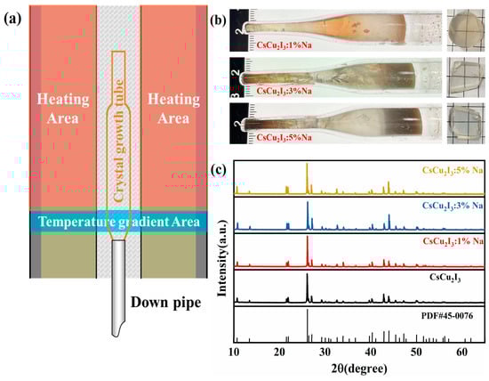
Figure 1.
(a) Schematic diagram of crystal growth using the Bridgman method. (b) As-grown crystals and corresponding samples. (c) XRD patterns of CsCu2I3:Na crystals.
The XRD of samples was completed on a Bruker D8 Advance diffractometer with Cu Ka radiation (λ = 0.1541 nm). The scanning step was 5 °C/min with a 5% error. The XPS spectra were measured using a Thermo ESCALAB 250XI spectrometer with a 0.1 ev error. The optical absorption spectra were obtained using UV2600 UV-Visible Spectrophotometer with a 1 nm error. The photoluminescence (PL) spectra, photoluminescence excitation (PLE) spectra and decay time measurements under UV light irradiation were recorded on an FLS1000 spectrometer with a pulsed nano-LED; the error was 1 nm and 0.1 ns, respectively. The slit widths for both the excitation and emission measurements were set at 2.5 nm, and the scan speed was fixed at 240 nm/min. The PLQY determination was performed with a Hamamatsu quantum yield measurement system C9920-02G with a 0.1 error. The XEL and afterglow were measuredusing HORIBA FluoroMax-4/Plus with a Hamamastu R928P PMT, a Moxtek MAGPRO X-ray tube (50 kV; 100 μA) with a W target used as the excitation source. The energy resolution, light yield, and decay time were acquired using a Hamamatsu R6233-100 photomultiplier tube (PMT) under 137Cs@662 keV gamma-ray irradiation with a shaping time of 5 µs. The samples were directly coupled to the PMT using mineral oil. A CsI:Tl crystal was also measured as a reference.
3. Results
The powder XRD patterns of CsCu2I3:Na crystals are shown in Figure 1c; the pure CsCu2I3 crystal was chosen as a reference. The patterns are well-matched with PDF card #45-0076 [15]. The second phase cannot be identified, indicating that Na+ doping had no obvious effect on the crystal structure. In order to prove the presence of Na+, the XPS spectra were measured and the results are shown in Figure 2. The Na+-related peaks were observed and located at 1071.6 eV, as shown in Figure 2d, showing that the Na+ was introduced successfully. Furthermore, the peaks of different elements exhibited a slight shift after Na+ doping. The peaks of Cs, Cu moved toward to the higher binding energy as Na+ concentration increased, which resulted from the lattice shrinking when the smaller-radius Na+ ions were doped, as shown in Figure 2b,c. Meantime, the binding energies of 952.3 and 932.0 eV were attributed to Cu 2p1/2 and Cu 2p3/2, respectively, which demonstrated the monovalent state of copper in the crystal host. Therefore, Na+ doping had no effect on the chemical valence of Cu+ for CsCu2I3:Na crystals. However, the Na-related peaks showed an obvious shift to low energy with the increase in the Na+ doping concentration. According to the radius of Na+(0.59 Å), Cs+(1.74 Å), and Cu+(0.60 Å) [11], Na+ ions are small enough to occupy both positions of Cu+ and Cs+. Considering the electronegativity of Cs+, Cu+, and Na+ ions (Na+ > Cs+ > Cu+), the electronegativity of Na+ is closer to that that of Cs+. Na+ ions are more likely to occupy the position of Cs+, and CsCu2I3 is composed of a series of [Cu2I6]4− tetrahedral units which are combined by two spatially oriented [CuI4]3− tetrahedral units connecting alternately. The [Cu2I6]4− chains were separated by Cs and formed the 1D electronic structure. This unique localized structure resulted in the formation of self-trapping excitons [11]. When the Na+ replaced the position of Cs+, the distance between Na+ and tetrahedral unit was reduced. The strengthened binding between the ions and groups led to the movement of XPS peaks with the increased binding energy. According to the reference [8], the isolated A cation also has a minimal impact on the electronic properties in elpasolite structures. The XPS results are consistent with the conclusion.
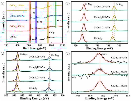
Figure 2.
(a) XPS spectra of pure and Na-doped samples; partially enlarged XPS spectra of certain elements: (b) Cs, (c) Cu and (d) Na.
Figure 3 shows the optical absorption spectra of the pure CsCu2I3 and Na+-doped samples. In accordance with the appearance of a light absorption edge, the band gaps of pure CsCu2I3, CsCu2I3:1%Na, CsCu2I3:3%Na and CsCu2I3:5%Na are calculated to be 3.31 eV, 3.28 eV, 3.21 eV and 3.16 eV via the Tauc plot method, respectively, as shown in the insets. Apparently, the band gaps of the CsCu2I3 crystals became smaller as the concentration of Na+ increased. A similar phenomenon was also observed in a Li+-doped CsCu2I3 crystal [11]. The valence band and conduction band consist of the 3d and 4s orbitals of Cu. Hence, Na+ doping has an effect on the local structure of Cu+, and as reported in ref. [16], the relationship of LY (light yield) and Eg (forbidden energy gap) can be described using the formula below:
where β is a parameter range from 2 to 7, S is the efficiency of transfer of the excitation to the luminescent center, and Q is the quantum efficiency of the luminescent center. Based on this inverse ratio of LY and Eg, a conclusion can be given that the LY is related to the PLQY and the Eg, and the LY will be increased when the Eg becomes smaller, which means the Na+ doping can theoretically improve the light yield of CsCu2I3 crystals.
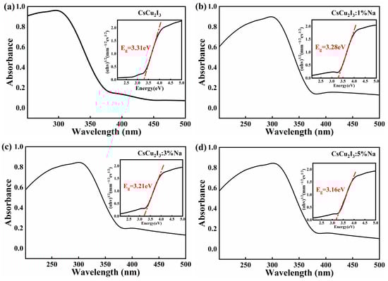
Figure 3.
The optical absorption spectra of (a) pure CsCu2I3, (b) CsCu2I3:1%Na, (c) CsCu2I3:3%Na and (d) CsCu2I3:5%Na.
In order to investigate the effect of Na+ doping on the luminescence properties, the emission spectra, excitation spectra and normalized spectra of pure CsCu2I3 and Na+ doped crystals (λex = 334 nm, λem = 575 nm) were measured and are shown in Figure 4a,b. The excitation and emission bands of Na+-doped samples were all located at 334 nm and 575 nm, which is well in agreement with those of pure CsCu2I3 crystals. According to reference [17], the emission is caused by STE instead of the defects, and the single-peak emission suggests that basic STE progress is independent of Na+ doping. Obviously, the CsCu2I3:Na crystals still have the advantage of a big Stokes shift of 1.55 eV (241 nm) and no self-absorption. The photoluminescence intensity was improved with the increase in the Na+ doping concentration. A similar result was reported in ref. [14]. A normalized partially enlarged graph of emission spectra is shown in Figure 4b, which proves further that no luminescent center was observed when the Na+ was introduced.
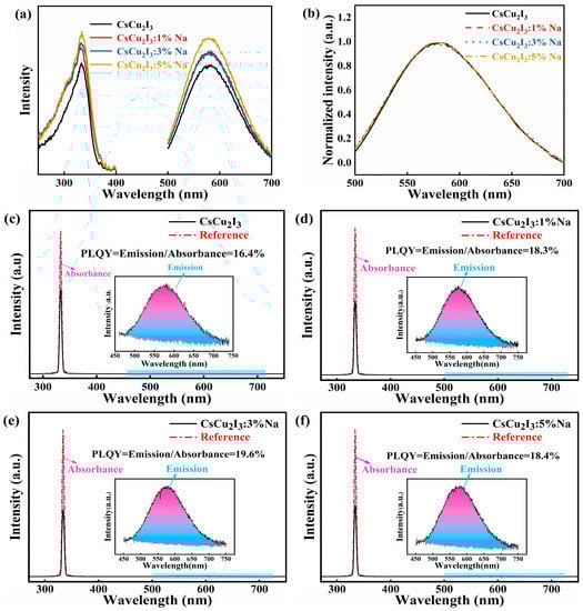
Figure 4.
(a) The PLE and PL spectra of pure and Na+-doped CsCu2I3 samples (λex = 334 nm; λem = 575 nm); (b) normalized PL spectra; the PLQY spectra of (c) pure CsCu2I3, (d) CsCu2I3:1%Na, (e) CsCu2I3:3%Na, (f) and CsCu2I3:5%Na. The reference curve was measured by placing a blank quartz plate in the integrating sphere.
The photoluminescence quantum efficiency of pure and Na+-doped CsCu2I3 crystals was measured at room temperature, as shown in Figure 4c–f, using an absolute photoluminescence measurement system with an integrating sphere, and a blank quartz plate as a reference in the integrating sphere under excitation. According to ref. [6,9,18,19], the highest PLQY of pure CsCu2I3 and CsCu2I3:Li reaches 15.7% and 18.7%, respectively. As shown in Figure 4c,e, the PLQY first increased and then decreased when the Na+ doping concentration increased, and the PLQY of the CsCu2I3:Na crystal was improved from 16.4% to 19.6% when the doped concentration of Na+ was up to 3%.
The XEL spectra of Na+-doped CsCu2I3 crystals are shown in Figure 5a. The commercial CsI:Tl crystal was chosen as a reference; the inset is the normalized integrated intensity. According to the integration value of the STE emission intensity in the XEL spectra, the yield of CsCu2I3:Na samples increased first then decreased as the Na+ content increased under X-ray. The light yield of CsCu2I3:1%, CsCu2I3:3%Na, and CsCu2I3:5%Na samples was determined to be 9.3%, 16.3% and 10.1% of that of the CsI:Tl crystal, respectively. The CsCu2I3:3%Na sample presents the highest yield, which is consistent with that of the PLQY. Combined with Equation (1), the light output is strongly dependent on the PLQY and Eg while the PLQY dominates. Hence, the light yield was increased first then decreased when the Na+ concentration was varied from 1% to 5%.
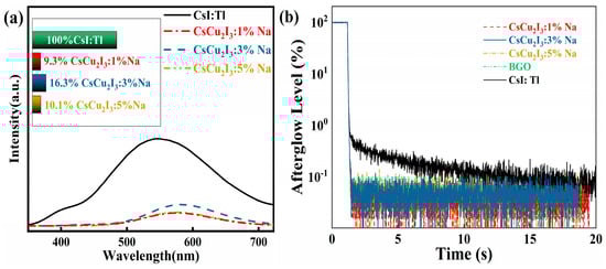
Figure 5.
(a) XEL spectra of Na−doped CsCu2I3 samples and CsI:Tl; the inset is the normalized integrated intensity. (b) Afterglow profile of CsCu2I3:1%, CsCu2I3:3%Na, CsCu2I3:5%Na, commercial BGO and CsI:Tl.
As previously reported, the CsCu2I3 single crystal presents an extremely low afterglow level. Here, the X-ray excited afterglow profiles of CsCu2I3:Na were also measured; the commercial BGO and CsI:Tl crystal were chosen as a reference, as shown in Figure 5b. CsCu2I3:Na crystals have a similar afterglow level with that of a BGO crystal, which is lower than that of a commercial CsI:Tl crystal. Therefore, Na+ doping has no significant effect on the afterglow characteristics. It still has an excellent afterglow property, indicating that the CsCu2I3:Na crystal has a promising prospect of application in X-ray imaging.
The energy resolution and light yield of the CsCu2I3:Na crystals were evaluated using Hamamatsu R6233-100 PMT under a 137Cs gamma ray source at 662 keV. Additionally, a commercial CsI:Tl crystal was measured as a reference to determine the absolute light yield of the samples, as shown in Figure 6. The energy resolution of varied-Na+-doped CsCu2I3 crystal was determined to be about 12.1%,10.1% and 9.3%, respectively, while that of the commercial CsI:Tl crystal was 5.8%. The energy resolution was worse than that of the CsI:Tl crystal, but the energy resolution was improved as the Na+ doping concentration increased. The CsCu2I3:3%Na crystal presented the best energy resolution. Based on the commercial CsI:Tl crystal (about 56,000 photons/MeV) [11], the light yield of CsCu2I3:Na crystals was determined to be 4465 photons/MeV, 9376 photons/MeV and 8929 photons/MeV, respectively. It had a similar variation tendency with that of XEL and PLQY. The CsCu2I3:3%Na crystal had the highest light yield.
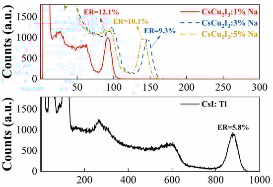
Figure 6.
Pulse height spectra of Na-doped CsCu2I3 and CsI:Tl under 137Cs γ-ray irradiation.
The decay curves provided in Figure 7 were measured under a 137Cs gamma ray source at 662 keV. The double exponential fitting formula used was as follows:
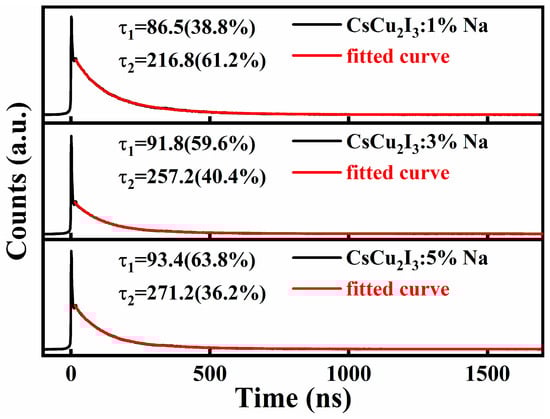
Figure 7.
Scintillation decay curves of CsCu2I3:1%Na, CsCu2I3:3%Na and CsCu2I3:5%Na.
Based on the fitting results, two decay times can be obtained, as discussed in refs. [11,15]. Here, the fast component and slow component was about 86.5 ns and 216.8 ns for the CsCu2I3:1%Na crystal. As the Na+ concentration increased, the decay time was slightly slower from 86.5 ns to 93.4 ns while the proportion of the fast-decay component gradually enhanced from 38.8% to 63.8%. In other words, the Na+-doped crystals still kept a fast decay time, presenting a potential method to improve the scintillation properties of CsCu2I3 crystals. Further improvements in crystal quality and scintillation properties need to be achieved in the future.
4. Discussion
The XRD patterns suggest that CsCu2I3:Na single crystals were successfully grown using raw materials with the stoichiometric ratio of CsCu2I3:n%Na. The XPS spectra show that Na+ was successfully introduced. As the Na+ concentration increased, the peaks of Cs and Cu showed a slight shift toward a higher binding energy while the peaks of Na+ showed a slight shift toward a lower binding energy. Because Na+ has a smaller effective ionic radius, the space between [Cu2I6]4− tetrahedral units was narrowed when the Na+ replaced the position of Cs+. This could have made it more difficult to excite the outermost electrons of Cs and Cu ions, perhaps resulting in an increase in the binding energy of Cs and Cu peaks, as demonstrated in our experimental results.
The results of the absorption spectra of CsCu2I3:Na samples indicate that the band gap has a trend of decreasing with the increasing concentration of Na+. From the perspective of band structure, the valence band and conduction band originate primarily from the Cu 3d and 4s orbitals in the CsCu2I3 crystal, and the incorporation of Na+ may reduce the distance between the adjacent Cu+, which leads to enhanced hybridization between Cu 4s orbitals and a smaller band gap [20]. Additionally, the variation of ions in the Cs position can still influence the electronic structure [8]. As mentioned above, the distance between [Cu2I6]4− tetrahedral units was narrowed via Na+ doping, which suggests that the Cu ions from different [Cu2I6]4− units became closer. Therefore, a conclusion can be given that a smaller bandgap results from the proximity of [Cu2I6]4− units.
The results of PL and PLE of pure and Na+-doped CsCu2I3 crystal show that the crystal still had a large Stokes shift of 1.55 eV, which is precisely one of the properties of a 1D perovskite structure. It is fortunate that there was no new luminescent center introduced after Na+ doping. Moreover, the PLQY of CsCu2I3 was improved. As an alkali, the metal halide crystal exhibits STE emission, while strong electroacoustic interaction causes distortion surrounding electrons and holes. Because the excitons are localized, non-radiative energy transfer is reduced during the recombination of electrons and holes, which is the reason for the high PLQY of CsCu2I3 crystals. Therefore, it is possible that Na+ doping enhances electroacoustic interaction, which results in stronger lattice distortions and a more localized position of excitons, and finally improves the PLQY. The highest PLQY reaches 19.6% when the doping concentration is 3%. The electronic structure dimension of the crystals can be reduced by doping appropriate elements to obtain a high PLQY [10]. The results indicate that Na doping does have a positive effect on the PLQY of CsCu2I3 crystals.
The results of the XEL spectra of the CsCu2I3:Na samples show that the sample with 3% of Na doping exhibited the best performance, which is well in agreement with the PLQY result. The emission peak located at 575 nm, ascribed to the STE, proves that no new luminescent center was introduced. The afterglow results of the CsCu2I3:Na samples show that the afterglow level of the samples was significantly better than that of CsI:Tl. Interestingly, the curves of the CsCu2I3:Na samples almost overlapped, which illustrates that the Na doping concentration did not have a significant impact on the afterglow performance of CsCu2I3 crystals.
The relative light yield and energy resolution of the Na-doped samples were calculated based on the pulse height spectra. The results show that the varied trends of energy resolution and light yield were consistent with that of the XEL and PLQY. This improvement results from the enhancement of the PLQY. Meanwhile, it was found that there were two decay components via double exponential fitting. The fast decay component (about 90 ns) primarily arises from the trap-assisted recombination of photo-excited electrons and holes at the surface of CsCu2I3, whereas the slow decay component (250 ns) is associated with recombination processes occurring within CsCu2I3 crystals. The proportion of fast decay was enhanced after Na+ doping. It is possible that Na+ doping improves the trap-assisted recombination of the surface and hinders the internal recombination of electrons and holes. Hence, Na+ doping improves scintillation properties, which is advantageous in practical applications.
5. Conclusions
In summary, Na+-doped CsCu2I3 single crystals were grown successfully using the Bridgman method. Intense STE emission peaked at 575 nm, presenting a large Stokes shift of 241 nm (1.55 eV). Na+ dopIng results a smaller bandgap and higher PLQY. The PLQY was improved from 16.4% to 19.6%. Additionally, the CsCu2I3:Na crystal still had a low afterglow level under X-ray. Similarly, the best energy resolution of 9.3% and light yield (9376 photons/MeV) were obtained with a decay time of 91.8 ns for the CsCu2I3:3%Na crystal under 137Cs radiation. The strategy of Na+ doping obviously has an effect on improving PLQY and scintillation properties. In general, CsCu2I3:Na crystals are promising single-crystal scintillators with high density, a high effective atomic number, non-hygroscopicity, and negligible self-absorption, which means its has promising applications in the field of X-ray and γ-ray detection.
Author Contributions
Formal analysis, H.Y.; investigation, W.L. and H.L.; data curation, C.S. and D.L.; writing—original draft preparation, C.S.; writing—review and editing, Q.W.; supervision, Q.W. and L.Q.; project administration, Q.W.; funding acquisition, Q.W., G.T. and L.Q. All authors have read and agreed to the published version of the manuscript.
Funding
This research was funded by National Key R&D Program of China [no. 2022YFB3503600], National Natural Science Foundation of China [NSFC] [nos. 12275262 and 51972291], Natural Science Foundation of Zhejiang [nos. LGG22E020001 and Z23E020002], “Pioneer” and “Leading Goose” R&D Program of Zhejiang [no. 2022C01046].
Data Availability Statement
Data are available at reasonable request from the authors.
Conflicts of Interest
The authors declare no conflict of interest.
References
- Nikl, M.; Yoshikawa, A. Recent R&D trends in inorganic single-crystal scintillator materials for radiation detection. Adv. Opt. Mater. 2015, 3, 463–481. [Google Scholar]
- Van Eijk, C.W.E. Inorganic-scintillator development. Nucl. Instrum. Methods Phys. Res. Sect. A Accel. Spectrometers Detect. Assoc. Equip. 2001, 460, 1–14. [Google Scholar] [CrossRef]
- Cheng, S.; Beitlerova, A.; Kucerkova, R.; Mihokova, E.; Nikl, M.; Zhou, Z.; Ren, G.; Wu, Y. Non-hygroscopic, self-absorption free, and efficient 1D CsCu2I3 perovskite single crystal for radiation detection. ACS Appl. Mater. Interfaces 2021, 13, 12198–12202. [Google Scholar] [CrossRef]
- Jin, T.; Hao, S.; Shang, Y.; Lei, Z.; Yang, C. Recent Trends in Elpasolite Single Crystal Scintillators for Radiation Detection. Crystals 2022, 12, 887. [Google Scholar] [CrossRef]
- Wojakowska, A.; Górniak, A.; Kuznetsov, A.Y.; Wojakowski, A.; Josiak, J. Phase diagram of the system copper (I) iodide+ cesium iodide. J. Chem. Eng. Data 2003, 48, 468–471. [Google Scholar] [CrossRef]
- Lin, R.; Zhu, Q.; Guo, Q.; Zhu, Y.; Zheng, W.; Huang, F. Dual self-trapped exciton emission with ultrahigh photoluminescence quantum yield in CsCu2I3 and Cs3Cu2I5 perovskite single crystals. J. Phys. Chem. C 2020, 124, 20469–20476. [Google Scholar] [CrossRef]
- Maddalena, F.; Tjahjana, L.; Xie, A.; Arramel; Zeng, S.; Wang, H.; Coquet, P.; Drozdowski, W.; Dujardin, C.; Dang, C.; et al. Inorganic, organic, and perovskite halides with nanotechnology for high–light yield X-and γ-ray scintillators. Crystals 2019, 9, 88. [Google Scholar] [CrossRef]
- Garcia de Mendoza Creus, S. Optical Characterization of CsCu2I3 and Cs3Cu2I5 Metal Halide Perovskites. Bachelor’s Thesis, Universitat de Barcelona, Barcelona, Spain, 2022. [Google Scholar]
- Zhu, M.; Wen, H.; Wang, B.; Wang, Q.; Li, J.; Wang, J. Research Progress of Perovskite-Type Scintillation Crystals. J. Synth. Cryst. 2021, 50, 1844–1857. [Google Scholar]
- Lin, R.; Guo, Q.; Zhu, Q.; Zhu, Y.; Zheng, W.; Huang, F. All-inorganic CsCu2I3 single crystal with high-PLQY (≈15.7%) intrinsic white-light emission via strongly localized 1D excitonic recombination. Adv. Mater. 2019, 31, 1905079. [Google Scholar] [CrossRef] [PubMed]
- Liu, D.; Wei, Q.; Tong, Y.; Xiang, P.; Cai, P.; Tang, G.; Shi, H.; Qin, L. A novel Li+-doped CsCu2I3 single crystal for dual gamma–neutron detection. CrystEngComm 2023, 25, 58–63. [Google Scholar] [CrossRef]
- Li, Z.; Li, Z.; Shi, Z.; Fang, X. Facet-dependent, fast response, and broadband photodetector based on highly stable all-inorganic CsCu2I3 single crystal with 1D electronic structure. Adv. Funct. Mater. 2020, 30, 2002634. [Google Scholar] [CrossRef]
- Xiang, P.; Wei, Q.; Wang, C.; Cai, P.; Tong, Y.; Tang, G.; Sun, X.; Yang, F.; Shi, H.; Liu, Z.; et al. An all-inorganic Li-doped Cs3Cu2I5 single crystal for dual gamma ray and neutron detection applications. J. Mater. Chem. C 2022, 10, 15400–15407. [Google Scholar] [CrossRef]
- Xie, L.; Chen, B.; Zhang, F.; Zhao, Z.; Jiang, T.; Wang, M.; Wu, Y.; Huang, L.; Song, W.; Liu, Y.; et al. Stability enhancement of Cs3Cu2I5 powder with high blue emission realized by Na+ doping strategy. J. Lumin. 2021, 239, 118333. [Google Scholar] [CrossRef]
- Mo, X.; Li, T.; Huang, F.; Li, Z.; Zhou, Y.; Lin, T.; Ouyang, Y.; Tao, X.; Pan, C. Highly-efficient all-inorganic lead-free 1D CsCu2I3 single crystal for white-light emitting diodes and UV photodetection. Nano Energy 2021, 81, 105570. [Google Scholar] [CrossRef]
- Weber, M.J. Scintillation: Mechanisms and new crystals. Nucl. Instrum. Methods Phys. Res. Sect. A Accel. Spectrometers Detect. Assoc. Equip. 2004, 527, 9–14. [Google Scholar] [CrossRef]
- Xing, X.; Tong, T.; Mohebinia, M.; Wang, D.; Ren, Z.; Hadjiev, V.G.; Wang, Z.; Bao, J. Photoluminescence and Raman Spectra of One-Dimensional Lead-free Perovskite CsCu2I3 Single-Crystal Wires. J. Phys. Chem. Lett. 2022, 13, 6447–6454. [Google Scholar] [CrossRef] [PubMed]
- Ma, Z.; Shi, Z.; Qin, C.C.; Cui, M.; Yang, D.; Wang, X.; Wang, L.; Ji, X.; Chen, X.; Sun, J.; et al. Stable yellow light-emitting devices based on ternary copper halides with broadband emissive self-trapped excitons. ACS Nano 2020, 14, 4475–4486. [Google Scholar] [CrossRef] [PubMed]
- Roccanova, R.; Yangui, A.; Seo, G.; Creason, T.D.; Wu, Y.; Kim, D.Y.; Du, M.-H.; Saparov, B. Bright luminescence from nontoxic CsCu2X3 (X = Cl, Br, I). ACS Mater. Lett. 2019, 1, 459–465. [Google Scholar] [CrossRef]
- Du, M.H. Emission trend of multiple self-trapped excitons in luminescent 1D copper halides. ACS Energy Lett. 2020, 5, 464–469. [Google Scholar] [CrossRef]
Disclaimer/Publisher’s Note: The statements, opinions and data contained in all publications are solely those of the individual author(s) and contributor(s) and not of MDPI and/or the editor(s). MDPI and/or the editor(s) disclaim responsibility for any injury to people or property resulting from any ideas, methods, instructions or products referred to in the content. |
© 2023 by the authors. Licensee MDPI, Basel, Switzerland. This article is an open access article distributed under the terms and conditions of the Creative Commons Attribution (CC BY) license (https://creativecommons.org/licenses/by/4.0/).