Article

High pO2 Flux Growth and Characterization of NdNiO3 Crystals

1
State Key Laboratory of Crystal Materials, Shandong University, Jinan 250100, China
2
NSF’s ChemMatCARS, The University of Chicago, Argonne, IL 60439, USA
*
Authors to whom correspondence should be addressed.
Crystals 2023, 13(2), 180; https://doi.org/10.3390/cryst13020180
Submission received: 27 December 2022 / Revised: 16 January 2023 / Accepted: 16 January 2023 / Published: 19 January 2023

Abstract

:
Single crystals of the perovskite nickelate NdNiO3 with dimensions of up to 50 μm on edge have been successfully grown using the flux method at a temperature of 400 °C and oxygen pressure of 200 bar. The crystals were investigated by a combination of techniques, including high-resolution synchrotron X-ray single-crystal and powder diffraction and physical property measurements such as magnetic susceptibility and resistivity. Resistivity measurements revealed a metal-insulator transition (MIT) at TMIT~180 K with apparent thermal hysteresis; however, no superlattice peaks or peak splitting below TMIT, which corresponds to a structural transition from Pbnm to P21/n, was observed. The successful growth of NdNiO3 crystals at relatively low temperatures and oxygen pressure provides an alternative approach for preparing single crystals of interesting perovskites such as RNiO3 (R = Sm-Lu) and parent phases of superconducting square planar nickelates.

1. Introduction

The discovery of superconductivity in the thin film of Nd0.8Sr0.2NiO2 by Li et al. [1] has attracted intense attention in the fields of condensed matter physics and materials science and launched the Nickel Age of Superconductivity [2,3,4,5,6,7,8,9,10,11,12,13,14]. Until today, superconductivity with a critical temperature (Tc) in the range of 9–15 K has been observed in the infinite-layer R1-xMxNiO2 (R = La, M = Ca, Sr; R = Pr, Nd, M = Sr) [15,16,17,18,19] and the quintuple-layer Nd6Ni5O12 [20]. Under high static pressure, Tc has been increased to 30 K in Pr0.8Sr0.2NiO2 films [21]. After 3 years of intense research, several fundamental issues still exist [2,3,4,5,6,7,8,9,10,11,12,13,14], including the mechanism of superconductivity, the similarity and difference between nickelate and cuprate superconductivity, whether the ground state is magnetic, and whether competing phases such as pseudogaps and strange metals exist. Single crystals are the ideal platform to solve the abovementioned open questions. However, no direct method has been found to synthesize these nickelates; instead, they were prepared by topotactical reduction through their parent compounds, i.e., Ruddlesden–Popper (RP) phases Rn+1-xMxNinO3n+1 (R = La-Nd, M = Ca, Sr, n = 5, and ∞) [4,13].
The n = ∞ RP nickelates is the well-known perovskite RNiO3 (R = La-Lu, Y), which has attracted extensive attention in the past few decades in two areas: (1) as a model system for studying metal-insulator transition (MIT, R ≠ La) and the lattice-charge-spin-orbital interaction [22] and (2) potential applications such as new morphological computing, bioelectronic interfaces, and electrocatalysis [23]. For RNiO3 (R = Pr, Nd), antiferromagnetic ordering occurs simultaneously at TMIT while for R3+ < Nd3+, TN < TMIT. Regarding the underlying physics behind MIT, there have been various proposals, including charge disproportionation, bipolaron condensation, and negative charge transfer, but it remains under debate [22]. In addition, type-II multiferroics have been theoretically predicted [24,25].
Single crystals of RNiO3 are in high demand in preparing infinite-layer nickelate crystals via topotactical reduction and for addressing the underlying mechanism of MIT and magnetism. Nickel ions usually exist in the divalent form, and the preparation of RNiO3 requires high oxygen pressure to stabilize the trivalent nickel ion. With the decrease of R3+ ions, the required oxygen pressure increases sharply. LaNiO3 and PrNiO3 single crystals have been grown using the high-pressure floating zone method under pO2 of 40 bar [26] and 290 bar [27], respectively. However, for RNiO3 whose R3+ is smaller than Pr3+, the required oxygen pressure exceeds 300 bar, which is the highest pressure that can be achieved using a commercial floating zone furnace [28]. RNiO3 crystals can also be grown using a high-pressure flux method. Among the literature [29,30,31,32], chloride or perchlorate (NaCl/NaClO4/KCl/KClO4) are mostly used as the flux, and growth was conducted at a temperature of 900–1500 °C and isostatic pressure of 4–4.5 GPa. In 2021, Klein et al. [33] first successfully grew crystals of RNiO3 (R= Nd, Sm, Gd, Dy, Y, Ho, Er, Lu) under pO2 = 2000 bar and T = 850 °C using used LiCl-KCl flux. The dimensions of NdNiO3 crystals are up to 10 μm on edge [33].
In this contribution, we report the successful growth of NdNiO3 crystals with dimensions up to 50 μm under pO2 = 200 and T = 400 °C using NaOH-KOH as flux. The crystals show a MIT as evidenced by transport measurements. High-resolution synchrotron X-ray single-crystal diffraction and high-resolution X-ray powder diffraction have been used to investigate the structural response across MIT. Our successful growth of NdNiO3 crystals using hydroxide flux at low temperature and reduced pO2 opens the door to a new opportunity for preparing single crystals of other interesting nickelates, including RNiO3 (R = Sm-Lu), parent phases of superconducting square planar nickelates, and potential multiferroic nickelates.

2. Materials and Synthesis

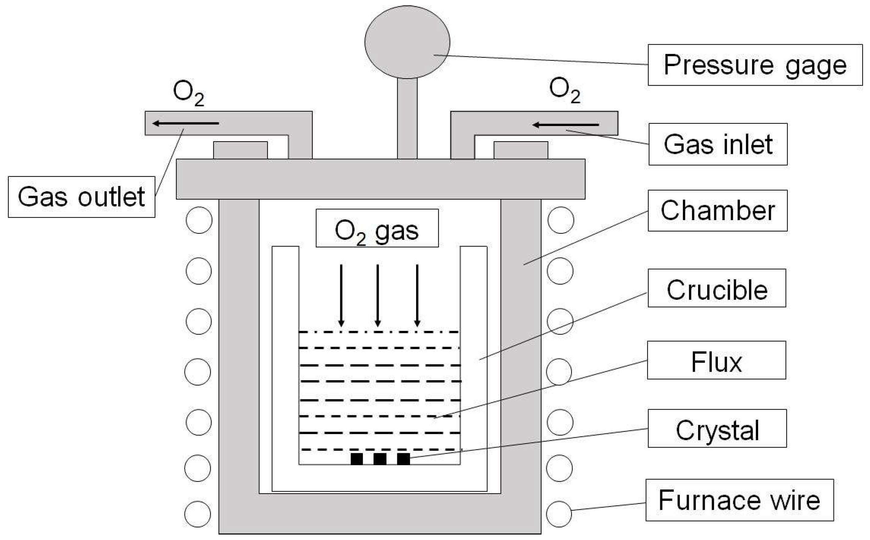
2.1. High-Pressure Flux Growth of NdNiO3 Crystals

NdNiO3 crystals were grown using a high-pressure flux furnace designed for operation at Tmax = 650 °C and Pmax = 300 bar (Model HP45, TOP Industrie). A scheme of the reaction chamber within the furnace is shown in Figure 1. Nd2O3 (Alfa Aesar, Haverhill, MA, USA, 99.99%) and NiO (Sigma-Aldrich, St. Louis, MO, USA, 99.99%) were used as starting materials. Nd2O3 was baked at 900 °C for 24 h before use. A stoichiometric ratio of Nd2O3 and NiO powder was weighted, mixed, and thoroughly ground, and then it was mixed with NaOH/KOH (molar ratio 1:1) flux in a glove box at a weight ratio of 1:10. The mixture was loaded into a Pt crucible, which was then put into the high-pressure furnace HP45. The furnace was filled with oxygen gas at room temperature so as to reach the targeted pO2 (5, 20, 40, 100, 200, or 300 bar) when the furnace was heated to 400 or 500 °C. During this process, oxygen is consumed due to the reaction of raw materials to form NdNiO3. Therefore, when the pressure is lower than the expected value, gas was manually filled in: (1) in the range of 150–300 bar, we used a booster pump to increase the pressure (error bar 5%), and (2) below 150 bar, gas was added by opening the valve of gas cylinder. The furnace was maintained at targeted temperature and pO2 for 48 h in order to dissolve raw materials. After dwelling, the furnace was cooled down to 360 °C at the rate of 5 °C/h and then to 260 °C at 10 or 1 °C/h, followed by furnace cooling to room temperature. No gas was filled during cooling. Finally, flux was dissolved using distilled water and black polycrystals were obtained.

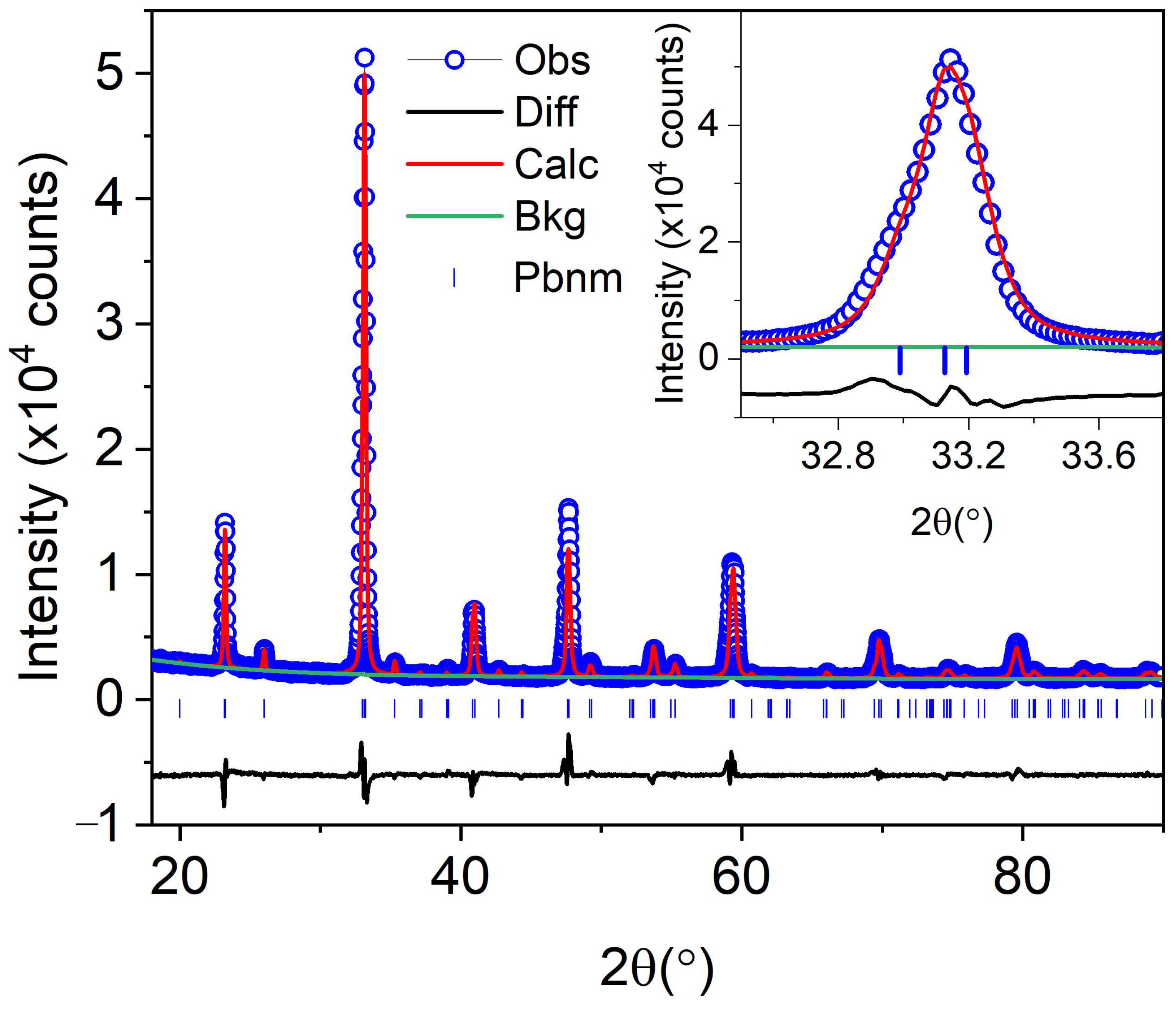
2.2. In-House X-ray Powder Diffraction (PXRD)

Bruker AXS D2 Phaser X-ray powder diffractometer was used to check phase purity. Data were collected at room temperature using Cu-Kα radiation (λ = 1.5418 Å) in the 2θ range of 10–90° with a scan step of 0.02° and 0.2 s per step. Rietveld refinement was performed using TOPAS 6. Refined parameters include background (Chebychev function, 5 order), sample displacement, lattice parameters, crystallite size (L and G), strain L, and atomic positions. Thermal parameters were not refined.

2.3. Synchrotron X-ray Single-Crystal Diffraction (SXRD)

X-ray single-crystal diffraction data were collected with a Pilatus3 X 1M (CdTe) area detector using synchrotron radiation (λ = 0.41328 Å) at 250 K and 120 K at Beamline 15-ID-D (NSF’s ChemMatCARS) at the Advanced Photon Source, Argonne National Laboratory. A piece of a single crystal with dimensions of 0.015 × 0.015 × 0.015 mm3 was mounted to the tip of glass fiber and measured using a Huber 3-circle diffractometer. Indexing, data reduction, and image processing were performed using Bruker APEX3 software [34].

2.4. High-Resolution Synchrotron X-ray Powder Diffraction (HRPXRD)

HRPXRD data were collected at Beamline 11-BM in the 2θ range of 0.5–50° with a step size of 0.001°, counting time of 0.1 s per step, and a wavelength of λ = 0.458961 Å at 295 K and λ = 0.458956 Å at 100 K. Fine-ground polycrystalline powders were loaded into a ϕ0.8-mm Kapton capillary for installation on a magnetic sample base used by the beamline sample changer. The sample was spun continuously at 5600 rpm during data collection. An Oxford Cryostream 700 Plus N2 gas blower was used to control the temperature at 100 K. Rietveld refinement was performed using TOPAS 6. Refined parameters include background (Chebychev function, 5 order), zero error, lattice parameters, crystallite size (L and G), strain (L and G), atomic positions, and thermal parameters (Beq). Simple axial Model was used for axial convolutions.

2.5. Scanning Electron Microscopy (SEM)

SEM was used to investigate the microstructure of the crystals. SEM images were obtained with a Hitachi S-4800 microscope at 5.0 kV incident electrons.

2.6. Energy Dispersive Spectrometer (EDS)

The X-ray spectrometer EDAX GENESIS XM2 SYSTEM 60x on S-4800 was used for qualitative and quantitative analysis of the as-grown crystals. The experiment was carried out on a 0.040 × 0.030 × 0.030 mm3 crystal.

2.7. Electrical Transport

Resistivity was measured using the standard four-probe method. The single crystals were too small to make four contacts, so instead, crystals were ground into powders, pressed into thin pellets, and then annealed under different conditions. The pellets were cut into thin bars for making four contacts with silver paint. The temperature was controlled using the Quantum Design Physical Properties Measurement System (Dynacool-9) in the range of 2–300 K.

2.8. Magnetic Susceptibility

The DC magnetization of the samples was measured using a PPMS Dynacool-9. The polycrystalline sample was packed in a clear plastic capsule. The sample was cooled in zero fields to 2 K, and DC magnetization data were recorded on warming (3 K/min). We also measured the magnetization of the empty capsule under the same conditions, which was negligible compared with the sample.

3. Results and Discussion

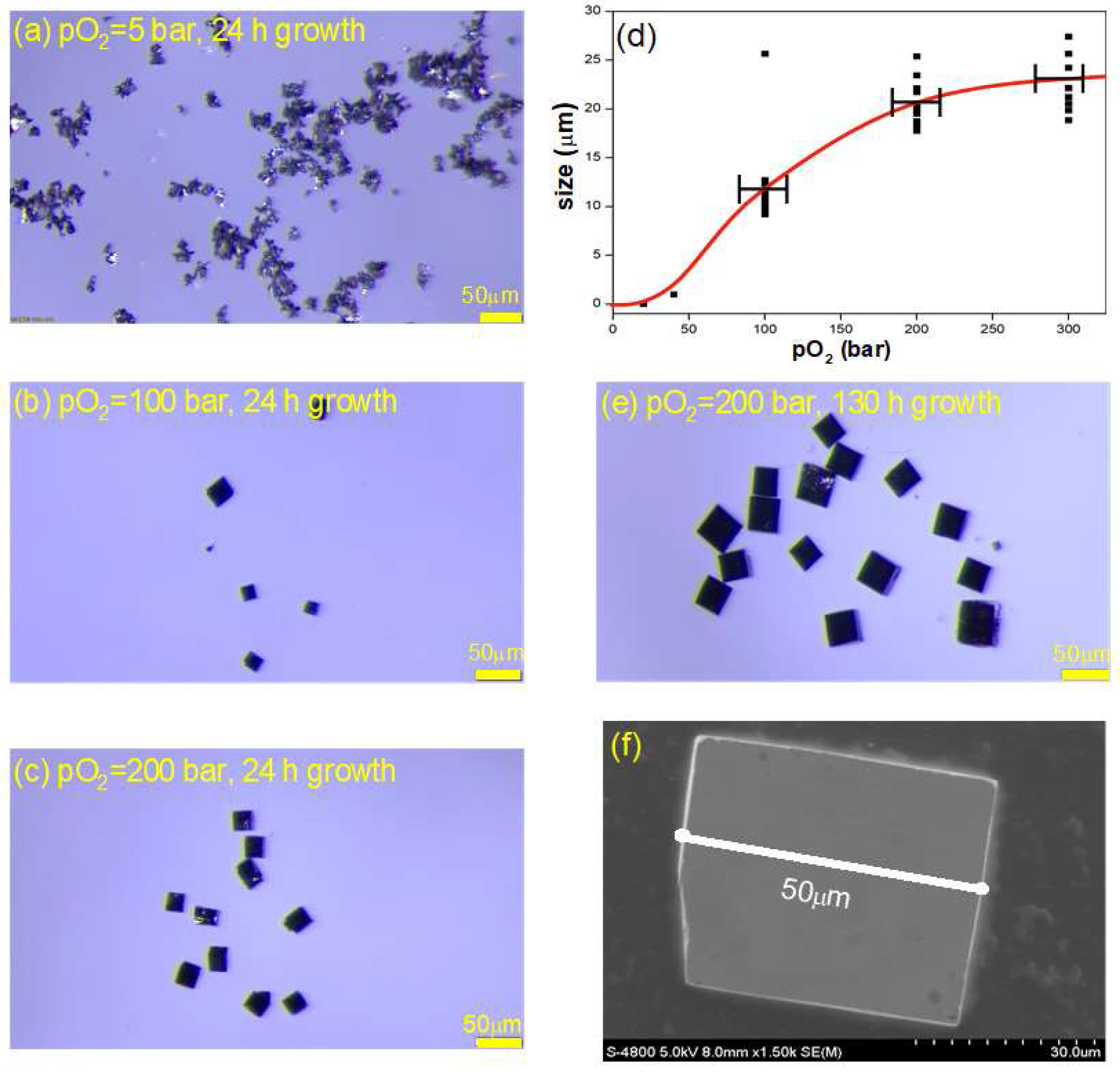
3.1. High pO2 Single Crystal Growth

The flux method was used to grow NdNiO3 crystals. We first consider the LiCl-KCl flux system, as Klein et al. [33] grew NdNiO3 crystals of 10 µm under 2000 bar at 850 °C, and Prakash et al. [35] successfully synthesized polycrystalline powders of EuNiO3 and GdNiO3 at 180 bar O2. Higher pO2 is needed for RNiO3 with smaller R ions [26,27], thus we expect to grow NdNiO3 crystals below pO2 = 180 bar. Unfortunately, we failed to grow crystals but found erosion in our furnace due to the reaction between LiCl-KCl and the chamber (Ni-superalloy).
We noticed that the mixture of NaOH/KOH (molar ratio 1:1) has a low melting point of ~170 °C, which could possibly lower the growth temperature of NdNiO3 significantly and reduce defects in the as-grown crystals. In addition, we found that single crystals of YBa2Cu4O8 and NaNiO2 were successfully grown by Song et al. [36] and Brion et al. [37], respectively, using such a hydroxide flux system. Initially, we attempted to grow NdNiO3 at pO2 = 20 bar at 400 °C. Black powders with tiny shiny crystals of ~10 µm were obtained. Figure 2 shows the Rietveld refinement of in-house powder X-ray diffraction data with Rwp = 6.52% (see Table S1 for Rietveld details). All peaks can be indexed in the space group Pbnm with lattice parameters a = 5.4019 (3) Å, b = 5.3697 (3) Å, c = 7.6009 (5) Å, which are comparable to the results reported by Wang et al. [38]. Therefore, we obtained the same size of NdNiO3 crystals at less than a half in temperature and two orders lower in oxygen pressure of Klein et al [33].
We then optimized the growth conditions of NdNiO3, including pO2, dwelling time, dwelling temperature, and cooling rate (see Table 1) in order to improve the dimensions and quality of crystals. We first optimized the dwelling time under pO2 = 20 bar, and the content of the NiO impurity phase in the product decreased significantly after the dwelling time was extended from 24 to 48 h; there was no obvious improvement after 48 h. Therefore, the dwelling time was selected as 48 h in the subsequent experiments. We then turned to optimizing pO2. Unexpectedly, the pure phase of NdNiO3 could be obtained at the lowest oxygen pressure of 5 bar, but the overall particles were small and no crystals could be observed under the microscope using 185 times magnification (Figure 3a). When the oxygen pressure was increased to 100 bar, prismatic crystals appeared (see Figure 3b). With the increase of pO2 to 200 bar, the crystal size and morphology are remarkably improved (Figure 3c). Further increasing the pO2 to 300 bar, the crystal size increased to a small extent (not shown). Figure 3d presents the crystal size as a function of pO2 under the same conditions: NdNiO3/flux = 1:10 (weight), dwelling time 48 h at 400 °C, and cooling rate 10 °C/h. As can be seen, the size of crystals increases significantly as pO2 increases and improves limitedly beyond 200 bar. For dwelling temperature, we went to 500 °C under pO2 of 300 bar, and the dimensions of the obtained NdNiO3 were the same level as 400 °C, while the corrosion of the flux to crucible was much worse. Furthermore, NaOH in the flux reacted with NiO to form Na0.6Ni3O5.4 impurities. Thus, we determined 200 bar as the optimized pO2. We further slowed down the cooling rate to 1 °C/h, and finally obtained shiny NdNiO3 crystals with a maximum size of about 50 μm (Figure 3e). SEM showed that the crystal had a regular appearance without obvious defects (Figure 3f). The composition of the crystals was estimated using EDS Figure S1 (See Supplementary Materials). The EDS data showed that the crystals contained Nd, Ni, and O elements, and the molar ratio of Nd: Ni is close to 1.

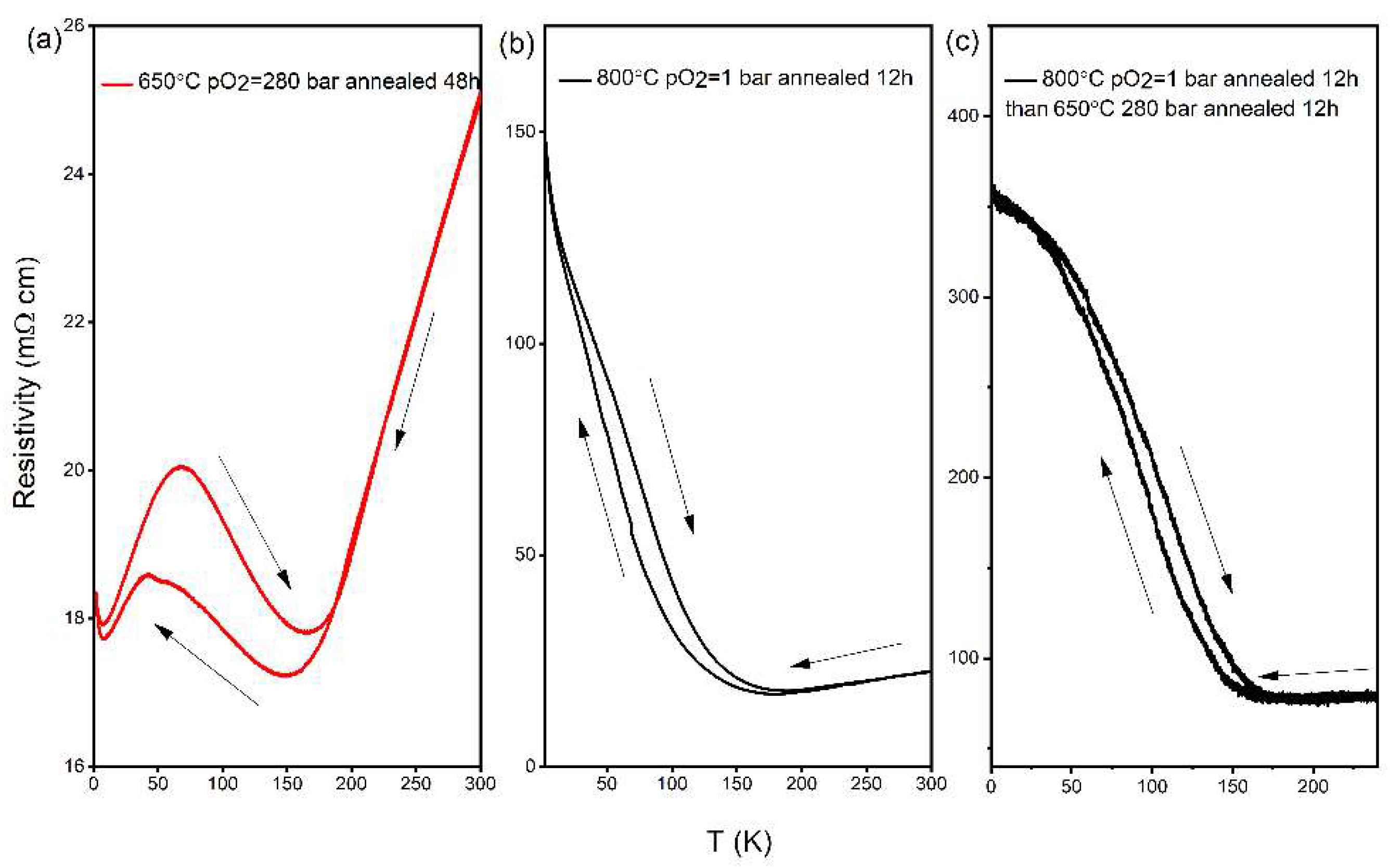
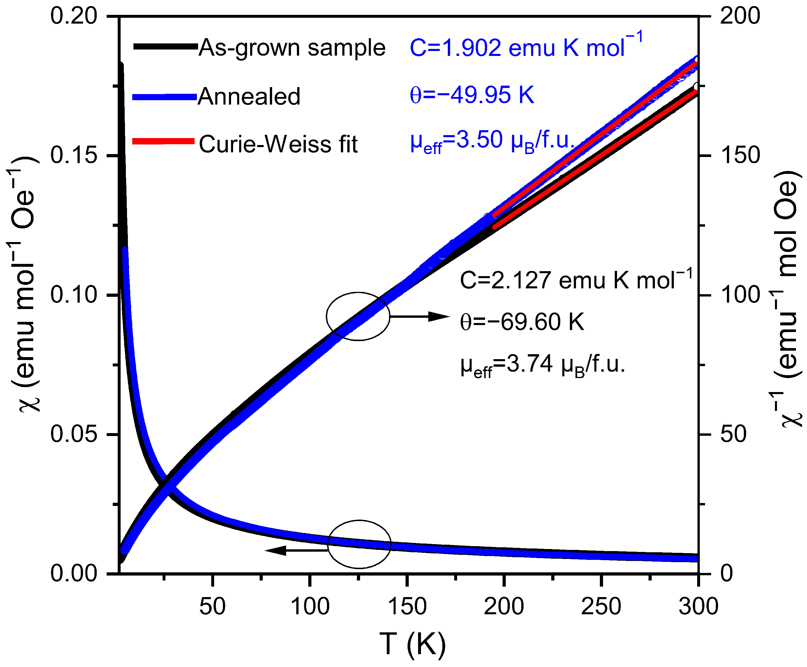
3.2. Physical Properties

Resistivity. To check if our crystals show metal-insulator transition, we performed resistivity measurements. As the crystals are not large enough, we measured resistivity on polycrystalline thin bars using the four-probe method. Figure 4 shows the resistivity of NdNiO3 as a function of temperature on heating and cooling from three samples: (a) annealed at 650 °C for 48 h under pO2 = 280 bar, (b) annealed at 800 °C for 12 h under pO2 = 1 bar (see Figure S2 for PXRD), and (c) initially annealed at 800 °C for 12 h under pO2 = 1 bar and then at 650 °C for 12 h under pO2 = 280 bar. All three samples exhibit metal-insulator transition, although the transition temperature is a bit lower compared with 196 K reported in Ref. [33]. This TMIT-reduced behavior is consistent with previously reported polycrystalline samples that contain oxygen vacancies [39]. Sample (a) shows a large hysteresis and re-enters a metallic state at a lower temperature. This re-entry behavior has also been observed in PrNiO3 single crystals, which was attributed to the presence of oxygen vacancies [27]. The existence of oxygen deficiency in our samples is not unexpected as this is a common phenomenon in perovskite oxides and the pO2 we used for crystal growth is one order smaller than Klein et al. [33] The above three samples are expected to have different oxygen contents, and the oxygen content of as-grown crystals should fall into their range; thus, one expects to see metal-insulator transition in the as-grown crystals. To achieve stoichiometric oxygen contents, annealing at high pO2 and longer time are needed.
Magnetic susceptibility. Complementary to transport properties, magnetic measurements offer an additional way to assess the properties of our sample. It was reported that antiferromagnetic order occurs concomitantly with MIT for NdNiO3 [22]. The temperature dependent DC magnetic susceptibility (χ) and the inverse magnetic susceptibility (1/χ) of as-grown NdNiO3 and annealed at 650 °C for 48 h under pO2 = 280 bar are shown in Figure 5. No anomaly, discontinuity, or change in slope, which would mark the onset of magnetic ordering, was observed down to 2 K. This is likely because the magnetic susceptibility is dominated by the paramagnetic contribution of Nd3+, which mask the weak signal of the Ni sublattice. A Curie–Weiss fit to the as-grown data in the range of 200–300 K, χ = C/(T-θ), where C and θ are the Curie and Weiss constants, respectively, yields C = 2.127 emu K mol−1 and θ = −69.6 K. The resulting effective moment is µeff = 3.74 µB/f.u., which is close to the expected value of 3.89 µB/f.u. of the free ion Nd3+. The large negative value of the Weiss constant indicates strong antiferromagnetic interactions.

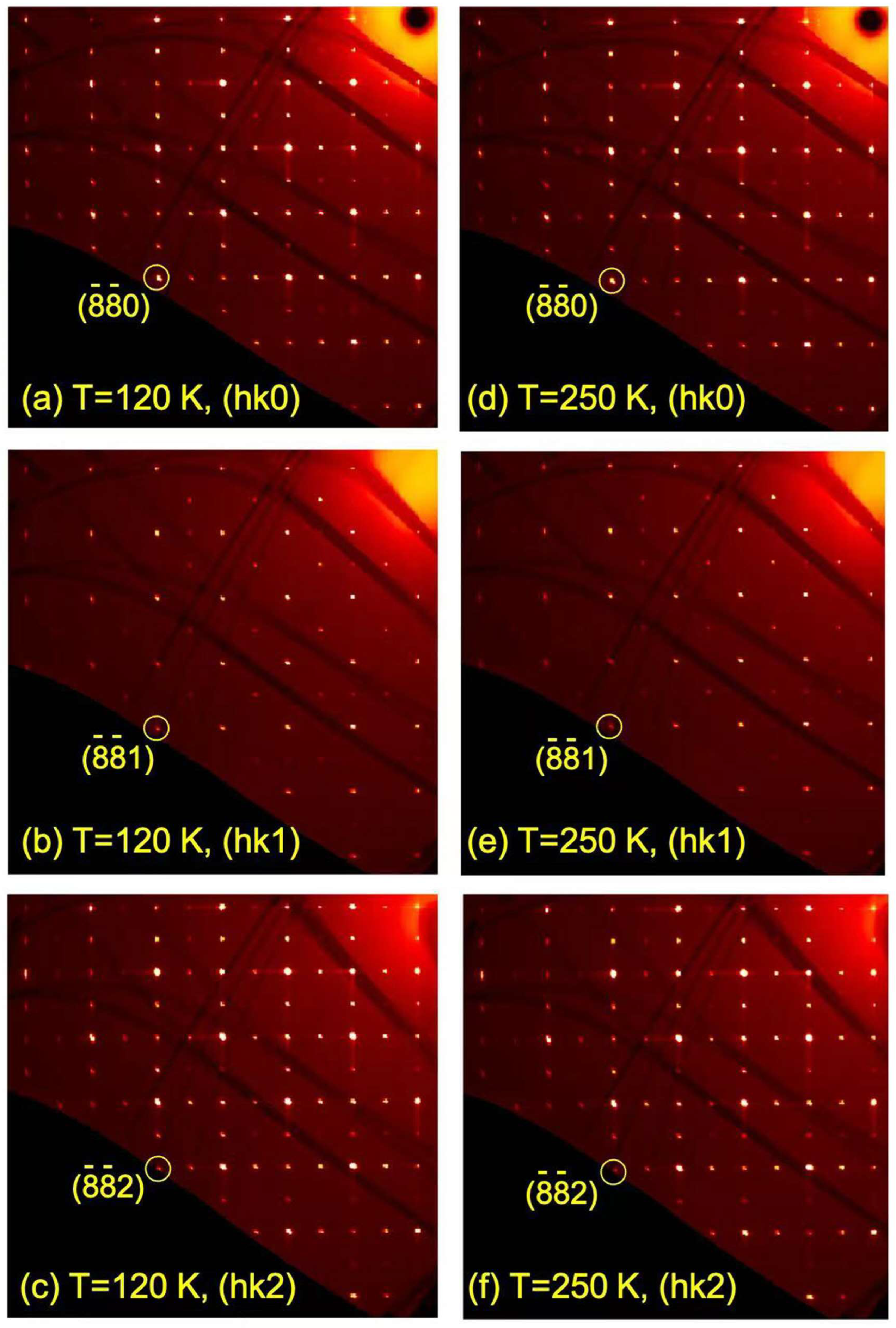
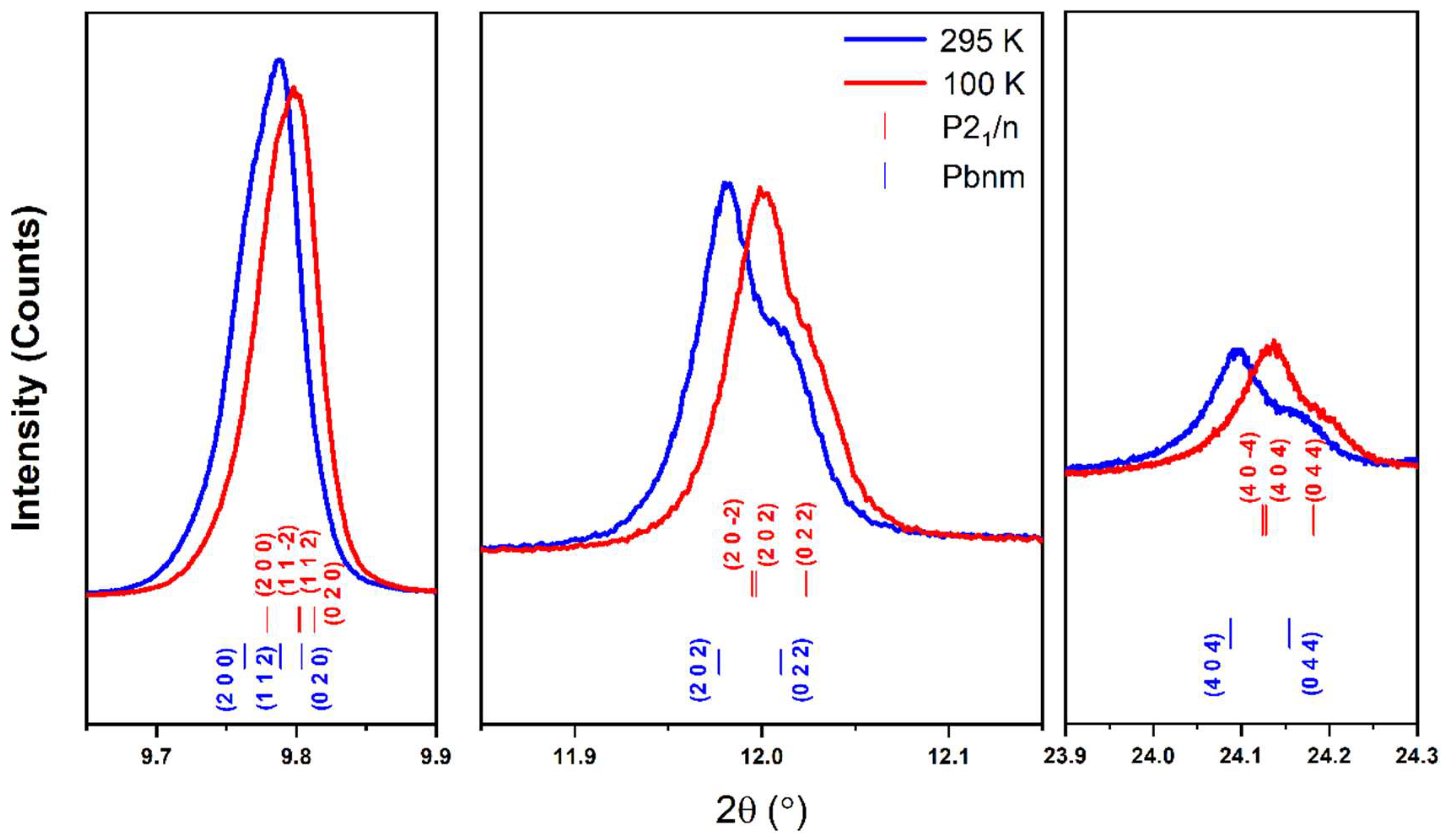
3.3. Structural Study at Various Temperatures

Perovskite nickelates (R = Pr-Lu) exhibit an MIT from a high-temperature metallic state to a low-temperature antiferromagnetic insulator. It has been reported that NdNiO3 has subtle PbnmP21/n symmetry breaking and associated Ni site splitting below TMIT [22]. To check the monoclinic distortion and possible orbital ordering [40], we have performed high-resolution X-ray single-crystal and powder diffraction using synchrotron radiation.
We found our NdNiO3 single crystals are twinned even at room temperature, resulting in a pseudocubic lattice with an average lattice parameter of a~7.6 Å in our synchrotron X-ray single-crystal diffraction at 15-ID-D. This is understandable because, in the two translation symmetry reductions going from the SrTiO3 structure, space group Pm-3m, to the orthorhombically distorted GdFeO3 type, space group Pbnm, twinning is highly probable, according to the Bärnighausen tree for the various perovskite superstructures. Figure 6 shows the pseudocubic (hk0), (hk1), and (hk2) planes measured by single-crystal X-ray diffraction at 120 and 250 K. There are no superlattice peaks below MIT, consistent with the lack of orbital ordering reported in Ref. [41].
Due to twinning and possible subtle structural change across MIT, we turn to synchrotron X-ray high-resolution powder diffraction to check if any peak splitting. Synchrotron diffraction data were collected at 295 and 100 K at 11-BM at the Advanced Photon Source, Argonne National Laboratory. Tables S2–S4 list details of Rietveld refinements and obtained crystallographic data such as unit cell parameters and atomic positions. At 295 K, the diffraction pattern is in good agreement with Pbnm, and Rietveld refinement converged to Rwp = 14.88% and GOF = 2.34. Our refinement shows that there are 3.99 (4)%wt NiO impurities in the sample (Figure S3a). For the diffraction data of 100 K, we fitted the data using both the orthorhombic Pbnm model and the monoclinic P21/n model, and slight improvement was obtained using the latter model (Figure S3b–c). Compared with the 50 K diffraction data in the literature [42], we did not observe a clear splitting of the (40 4 ¯ )/(404) reflections that signals the lowering symmetry to P21/n (Figure 7). This may be because the structural phase transition is too subtle to be detected at 100 K, and a lower temperature may be needed.

4. Conclusions

We have successfully grown NdNiO3 single crystals with dimensions of up to 50 μm on edge at 400 °C and 200 bar oxygen pressure using the KOH-NaOH flux system. The grown crystals have well-defined faces and regular, slightly truncated prismatic habits. Compared with previous flux systems, the hydroxide flux system significantly reduces the growth temperature and oxygen pressure while obtaining larger NdNiO3 single crystals. A clear metal-insulator transition was observed in resistivity. In contrast, no superlattice peaks were observed in synchrotron X-ray single-crystal diffraction at 120 K, nor was peak splitting found in high-resolution synchrotron X-ray powder diffraction at 100 K. The absence of the (40 4 ¯ )/(404) peak splitting may be due to too subtle structural change across MIT and measurements at liquid helium temperature may be needed. Finally, the high pO2 flux growth method described here provides an alternative approach for preparing single crystals of interesting perovskites, including parent phases of superconducting infinite nickelates and potential multiferroic nickelates, at a relatively low temperature and oxygen pressure [24,25].

Supplementary Materials

The following supporting information can be downloaded at: https://www.mdpi.com/article/10.3390/cryst13020180/s1, Figure S1. Elemental analysis for NdNiO3 crystals; Figure S2. In-house X-ray powder diffraction for annealed NdNiO3 sample; Figure S3. (a) Synchrotron X-ray high-resolution powder diffraction pattern for NdNiO3 at 295 K with Rietveld refinement using the orthorhombic Pbnm symmetry (the purple vertical bar corresponds to the NiO impurity). (b) Synchrotron X-ray high-resolution powder diffraction pattern for NdNiO3 at 100 K with Rietveld refinement using the monoclinic P21/n symmetry. (c) Synchrotron X-ray high-resolution powder diffraction pattern for NdNiO3 at 100 K with Rietveld refinement using the orthorhombic Pbnm symmetry (the inset is observed and calculated powder diffraction intensities around the orthorhombic (044) / (404) pair of reflections); Tables S1–S4. Unit cell parameters and atomic coordinates for NdNiO3 as obtained from Rietveld refinements to in-house and synchrotron X-ray powder diffraction patterns. The xy data of Figure 7 and Figure S3 are uploaded as Supplementary Materials (in Excel format).

Author Contributions

Conceptualization, X.T. and J.Z.; Formal analysis, X.W. and J.Z.; Funding acquisition, X.T. and J.Z.; Investigation, X.W., S.W. (Shilei Wang), C.L., C.F., L.H., F.L., T.C., Y.-S.C., and S.W. (Shanpeng Wang); Methodology, J.Z.; Resources, Y.-S.C., S.W. (Shanpeng Wang) and J.Z.; Supervision, J.Z.; Validation, J.Z., X.W., C.L., C.F., L.H., and F.L.; Writing—original draft, X.W.; Writing—review & editing, J.Z.. All authors have read and agreed to the published version of the manuscript.

Funding

Work at Shandong University was supported by the National Natural Science Foundation of China (12074219, 51932004, 61975098), the National Key Research and Development Program of China (2016YFB1102201, 2018YFB0406502), the Natural Science Foundation of Shandong (ZR202105230005), the 111 Project 2.0 (BP2018013), the QiLu Young Scholars Program of Shandong University, the TaiShan Scholars Program of Shandong Province (tsqn201909031), the Crystal Materials and Industrialization Joint Innovation Laboratory of Shandong University and Shandong Institutes of Industrial Technology (Z1250020003), and the Project for Scientific Research Innovation Team of Young Scholars in Colleges and Universities of Shandong Province (2021KJ093). NSF’s ChemMatCARS, Sector 15 at the Advanced Photon Source (APS), Argonne National Laboratory (ANL) is supported by the Divisions of Chemistry (CHE) and Materials Research (DMR), National Science Foundation, under grant number NSF/CHE- 1834750. Use of APS, an Office of Science User Facility operated for the U.S. Department of Energy (DOE) Office of Science by ANL, was supported by the U.S. DOE under Contract No. DE-AC02-06CH11357.

Data Availability Statement

The data that support the plots within this paper and other findings of this study are available from the corresponding author on reasonable request.

Acknowledgments

J.Z. and X.W. thank Saul H. Lapidus for his help with the high-resolution powder diffraction experiment at 11-BM, APS.

Conflicts of Interest

The authors declare no conflict of interest.

References

  1. Li, D.; Lee, K.; Wang, B.Y.; Osada, M.; Crossley, S.; Lee, H.R.; Cui, Y.; Hikita, Y.; Hwang, H.Y. Superconductivity in an infinite-layer nickelate. Nature 2019, 572, 624–627. [Google Scholar] [CrossRef]
  2. Chow, L.E.; Ariando, A. Infinite-Layer Nickelate Superconductors: A Current Experimental Perspective of the Crystal and Electronic Structures. Front. Phys. 2022, 10, 834658. [Google Scholar] [CrossRef]
  3. Mitchell, J.F. A Nickelate Renaissance. Front. Phys. 2021, 9, 813483. [Google Scholar] [CrossRef]
  4. Bernardini, F.; Iglesias, L.; Bibes, M.; Cano, A. Thin-Film Aspects of Superconducting Nickelates. Front. Phys. 2022, 10, 828007. [Google Scholar] [CrossRef]
  5. Botana, A.S.; Bernardini, F.; Cano, A. Nickelate superconductors: An ongoing dialog between theory and experiments. J. Exp. Theor. Phys 2021, 132, 618–627. [Google Scholar] [CrossRef]
  6. Botana, A.S.; Lee, K.-W.; Norman, M.R.; Pardo, V.; Pickett, W.E. Low Valence Nickelates: Launching the Nickel Age of Superconductivity. Front. Phys. 2022, 9, 813532. [Google Scholar] [CrossRef]
  7. Gu, Q.; Wen, H.-H. Superconductivity in nickel-based 112 systems. Innovation 2022, 3, 100202. [Google Scholar] [CrossRef] [PubMed]
  8. Hepting, M.; Dean, M.P.M.; Lee, W.-S. Soft X-ray Spectroscopy of Low-Valence Nickelates. Front. Phys. 2021, 9, 808683. [Google Scholar] [CrossRef]
  9. Ji, Y.; Liu, J.; Li, L.; Liao, Z. Superconductivity in infinite layer nickelates. J. Appl. Phys. 2021, 130, 060901. [Google Scholar] [CrossRef]
  10. Jiang, M. Relevance of 3D multiplet structure in nickelate and cuprate superconductors. Chin. Phys. B 2021, 30, 107103. [Google Scholar] [CrossRef]
  11. Li, D. The discovery and research progress of the nickelate superconductors. Sci. Sin. Phys. Mech. Astron. 2021, 51, 047405. [Google Scholar] [CrossRef]
  12. Nomura, Y.; Arita, R. Superconductivity in infinite-layer nickelates. Rep. Prog. Phys. 2022, 85, 052501. [Google Scholar] [CrossRef] [PubMed]
  13. Zhang, J.; Tao, X. Review on quasi-2D square planar nickelates. CrystEngComm 2021, 23, 3249–3264. [Google Scholar] [CrossRef]
  14. Zhou, X.; Qin, P.; Feng, Z.; Yan, H.; Wang, X.; Chen, H.; Meng, Z.; Liu, Z. Experimental progress on the emergent infinite-layer Ni-based superconductors. Mater. Today 2022, 55, 170–185. [Google Scholar] [CrossRef]
  15. Li, D.; Wang, B.Y.; Lee, K.; Harvey, S.P.; Osada, M.; Goodge, B.H.; Kourkoutis, L.F.; Hwang, H.Y. Superconducting Dome in Nd1−xSrxNiO2 Infinite Layer Films. Phys. Rev. Lett. 2020, 125, 027001. [Google Scholar] [CrossRef] [PubMed]
  16. Zeng, S.; Li, C.; Chow, L.E.; Cao, Y.; Zhang, Z.; Tang, C.S.; Yin, X.; Lim, Z.S.; Hu, J.; Yang, P.; et al. Superconductivity in infinite-layer nickelate La1−xCaxNiO2 thin films. Sci. Adv. 2022, 8, eabl9927. [Google Scholar] [CrossRef] [PubMed]
  17. Zeng, S.; Tang, C.S.; Yin, X.; Li, C.; Li, M.; Huang, Z.; Hu, J.; Liu, W.; Omar, G.J.; Jani, H.; et al. Phase Diagram and Superconducting Dome of Infinite-Layer Nd1-xSrxNiO2 Thin Films. Phys. Rev. Lett. 2020, 125, 147003. [Google Scholar] [CrossRef] [PubMed]
  18. Osada, M.; Wang, B.Y.; Goodge, B.H.; Lee, K.; Yoon, H.; Sakuma, K.; Li, D.; Miura, M.; Kourkoutis, L.F.; Hwang, H.Y. A superconducting praseodymium nickelate with infinite layer structure. Nano Lett. 2020, 20, 5735–5740. [Google Scholar] [CrossRef]
  19. Osada, M.; Wang, B.Y.; Goodge, B.H.; Harvey, S.P.; Lee, K.; Li, D.; Kourkoutis, L.F.; Hwang, H.Y. Nickelate Superconductivity without Rare-Earth Magnetism: (La,Sr)NiO2. Adv. Mater. 2021, 33, e2104083. [Google Scholar] [CrossRef]
  20. Pan, G.A.; Ferenc Segedin, D.; LaBollita, H.; Song, Q.; Nica, E.M.; Goodge, B.H.; Pierce, A.T.; Doyle, S.; Novakov, S.; Cordova Carrizales, D.; et al. Superconductivity in a quintuple-layer square-planar nickelate. Nat. Mater. 2022, 21, 160–164. [Google Scholar] [CrossRef]
  21. Wang, N.N.; Yang, M.W.; Yang, Z.; Chen, K.Y.; Zhang, H.; Zhang, Q.H.; Zhu, Z.H.; Uwatoko, Y.; Gu, L.; Dong, X.L.; et al. Pressure-induced monotonic enhancement of Tc to over 30 K in superconducting Pr0.82Sr0.18NiO2 thin films. Nat. Commun. 2022, 13, 4367. [Google Scholar] [CrossRef]
  22. Catalano, S.; Gibert, M.; Fowlie, J.; Íñiguez, J.; Triscone, J.M.; Kreisel, J. Rare-earth nickelates RNiO3: Thin films and heterostructures. Rep. Prog. Phys. 2018, 81, 046501. [Google Scholar] [CrossRef] [Green Version]
  23. Zhang, Z.; Sun, Y.; Zhang, H.-T. Quantum nickelate platform for future multidisciplinary research. J. Appl. Phys. 2022, 131, 120901. [Google Scholar] [CrossRef]
  24. Ardizzone, I.; Teyssier, J.; Crassee, I.; Kuzmenko, A.B.; Mazzone, D.G.; Gawryluk, D.J.; Medarde, M.; van der Marel, D. Raman spectroscopic evidence for multiferroicity in rare earth nickelate single crystals. Phys. Rev. Res. 2021, 3, 033007. [Google Scholar] [CrossRef]
  25. Giovannetti, G.; Kumar, S.; Khomskii, D.; Picozzi, S.; van den Brink, J. Multiferroicity in rare-earth nickelates RNiO3. Phys. Rev. Lett. 2009, 103, 156401. [Google Scholar] [CrossRef] [Green Version]
  26. Zhang, J.; Zheng, H.; Ren, Y.; Mitchell, J.F. High-pressure floating-zone growth of perovskite nickelate LaNiO3 single crystals. Cryst. Growth Des. 2017, 17, 2730–2735. [Google Scholar] [CrossRef] [Green Version]
  27. Zheng, H.; Zhang, J.; Wang, B.; Phelan, D.; Krogstad, M.J.; Ren, Y.; Phelan, W.A.; Chmaissem, O.; Poudel, B.; Mitchell, J.F. High pO2 Floating Zone Crystal Growth of the Perovskite Nickelate PrNiO3. Crystals 2019, 9, 324. [Google Scholar] [CrossRef] [Green Version]
  28. Phelan, W.A.; Zahn, J.; Kennedy, Z.; McQueen, T.M. Pushing boundaries: High pressure, supercritical optical floating zone materials discovery. J. Solid State Chem. 2018, 270, 705–709. [Google Scholar] [CrossRef]
  29. Azuma, M.; Saito, T.; Ishiwata, S.; Yamada, I.; Kohsaka, Y.; Takagi, H.; Takano, M. Single crystal growth of transition metal oxides at high pressures of several GPa. Physica C 2003, 392–396 Pt 1, 22–28. [Google Scholar] [CrossRef]
  30. Masaki, A.; Takashi, S.; Shintaro, I.; Hirofumi, Y.; Mikio, T.; Yuhki, K.; Hidenori, T.; Wataru, U. Single-crystal growth of transition metal oxides at high pressures of several GPa. J. Phys. Condens. Matter 2002, 14, 11321. [Google Scholar]
  31. Alonso, J.A.; Muñoz, A.; Largeteau, A.; Demazeau, G. Crystal growth of NdNiO3 perovskite under high oxygen pressure. J. Phys. Condens. Matter 2004, 16, S1277. [Google Scholar] [CrossRef]
  32. Puphal, P.; Wu, Y.-M.; Fürsich, K.; Lee, H.; Pakdaman, M.; Bruin, J.A.N.; Nuss, J.; Suyolcu, Y.E.; Aken, P.A.v.; Keimer, B.; et al. Topotactic transformation of single crystals: From perovskite to infinite-layer nickelates. Sci. Adv. 2021, 7, eabl8091. [Google Scholar] [CrossRef]
  33. Klein, Y.M.; Kozłowski, M.; Linden, A.; Lacorre, P.; Medarde, M.; Gawryluk, D.J. RENiO3 Single Crystals (RE = Nd, Sm, Gd, Dy, Y, Ho, Er, Lu) Grown from Molten Salts under 2000 bar of Oxygen Gas Pressure. Cryst. Growth Des. 2021, 21, 4230–4241. [Google Scholar] [CrossRef]
  34. Bruker APEX2; Bruker Analytical X-ray Instruments Inc: Madison, WI, USA, 2014.
  35. Prakash, J.; Blakely, C.K.; Poltavets, V.V. Low temperature high-pressure synthesis of LnNiO3 (Ln = Eu, Gd) in molten salts. Solid State Sci. 2013, 17, 72–75. [Google Scholar] [CrossRef]
  36. Song, Y.T.; Peng, J.B.; Wang, X.; Sun, G.L.; Lin, C.T. Ambient-condition growth of superconducting YBa2Cu4O8 single crystals using KOH flux. J. Cryst. Growth 2007, 300, 263–266. [Google Scholar] [CrossRef]
  37. de Brion, S.; Bonda, M.; Darie, C.; Bordet, P.; Sheikin, I. Magnetic phase diagram of the S = 1/2 triangular layered compound NaNiO2: A single crystal study. J. Phys. Condens. Matter 2010, 22, 126001. [Google Scholar] [CrossRef]
  38. Wang, B.-X.; Zheng, H.; Krivyakina, E.; Chmaissem, O.; Lopes, P.P.; Lynn, J.W.; Gallington, L.C.; Ren, Y.; Rosenkranz, S.; Mitchell, J.F.; et al. Synthesis and characterization of bulk Nd1−xSrxNiO2 and Nd1−xSrxNiO3. Phys. Rev. Mater. 2020, 4, 084409. [Google Scholar] [CrossRef]
  39. Fujihara, S.; Murakami, G.; Kimura, T. Oxygen deficiency and electrical conductivity of Nd1−xAxNiO3−y (A = alkaline earth) prepared by the low-temperature process. J. Alloys Compd. 1996, 243, 70–76. [Google Scholar] [CrossRef]
  40. Catalan, G. Progress in perovskite nickelate research. Phase Transit. 2008, 81, 729–749. [Google Scholar] [CrossRef]
  41. Lorenzo, J.E.; Hodeau, J.L.; Paolasini, L.; Lefloch, S.; Alonso, J.A.; Demazeau, G. Resonant X-ray scattering experiments on electronic orderings in NdNiO3 single crystals. Phys. Rev. B 2005, 71, 045128. [Google Scholar] [CrossRef]
  42. García-Muñoz, J.L.; Aranda, M.A.G.; Alonso, J.A.; Martínez-Lope, M.J. Structure and charge order in the antiferromagnetic band-insulating phase of NdNiO3. Phys. Rev. B 2009, 79, 134432. [Google Scholar] [CrossRef]
Figure 1. Scheme of reaction chamber within the furnace.
Crystals 13 00180 g001
Figure 2. Rietveld refinement of in-house PXRD (λ = 1.5418 Å) of pulverized as-grown crystals of NdNiO3. Obs: observed intensity; Diff: difference; Calc: calculated intensity; Bkg: background.
Crystals 13 00180 g002
Figure 3. NdNiO3 crystals grown under different pO2. (a) pO2 = 5 bar, growth time 24 h; (b) pO2 = 100 bar, growth time 24 h; (c) pO2 = 200 bar, growth time 24 h. (d) The relationship between grain size and pO2 under the same growth conditions (Error bars represent the oxygen pressure deviation, and the red curve is only a guide for the eye); (e) pO2 = 200 bar, growth time 130 h; (f) SEM image of NdNiO3.
Crystals 13 00180 g003
Figure 4. The resistivity of NdNiO3 from three samples: (a) annealed at 650 °C for 48 h under pO2 = 280 bar, (b) annealed at 800 °C for 12 h under pO2 = 1 bar, and (c) initially annealed at 800 °C for 12 h under pO2 = 1 bar and then at 650 °C for 12 h under pO2 = 280 bar. Arrow indicates heating/cooling process.
Crystals 13 00180 g004
Figure 5. DC susceptibility χ (left) and inverse magnetic susceptibility 1/χ (right) for as-grown NdNiO3, and NdNiO3 annealed at 650 °C for 48 h under pO2 = 280 bar. The symbols are the experimental data and the red lines are the Curie–Weiss fit.
Crystals 13 00180 g005
Figure 6. (a) Pseudocubic (hk0) planes (a~7.6 Å) of as-grown NdNiO3 measured by single-crystal X-ray diffraction at 120 K; (b) Pseudocubic (hk1) planes at 120 K; (c) Pseudocubic (hk2) planes at 120 K; (d) Pseudocubic (hk0) planes at 250 K; (e) Pseudocubic (hk1) planes at 250 K; (f) Pseudocubic (hk2) planes at 250 K.
Crystals 13 00180 g006
Figure 7. High-resolution synchrotron X-ray powder diffraction intensities of as-grown NdNiO3 in the 2θ range of 9.65–9.9°, 11.85–12.15°, and 23.9–24.3° measured at 100 K (λ = 0.458956 Å) and 295 K (λ = 0.458961 Å). Blue (bottom) and red (top) bars correspond to Pbnm and P21/n, respectively.
Crystals 13 00180 g007
Table 1. Growth conditions of NdNiO3 crystals.
T (°C)pO2 (Bar)Dwelling Time (h)Cooling Rate (°C/h)Phase (s)Crystal Size (μm)
40052410NdNiO3 + NiO (trace)<5
400202410NdNiO3 + NiO (trace)<5
400204810NdNiO3<5
4001004810NdNiO35–15
4002004810NdNiO315–25
4003004810NdNiO315–28
5003004810NdNiO3 + Na0.6Ni3O5.415–28
400200481NdNiO320–50
Disclaimer/Publisher’s Note: The statements, opinions and data contained in all publications are solely those of the individual author(s) and contributor(s) and not of MDPI and/or the editor(s). MDPI and/or the editor(s) disclaim responsibility for any injury to people or property resulting from any ideas, methods, instructions or products referred to in the content.

