Article

Anticancer Studies of Newly Synthesized Thiazole Derivatives: Synthesis, Characterization, Biological Activity, and Molecular Docking

1
Biology Department, College of Sciences, Taif University, P.O. Box 11099, Taif 21944, Saudi Arabia
2
Department of Pharmacy Practice, College of Pharmacy, Shaqra University, Shaqra 11961, Saudi Arabia
3
The Division of Biochemistry, Chemistry Department, Faculty of Science, Port Said University, Port Said 42526, Egypt
4
Department of Chemistry, College of Science, Taif University, P.O. Box 11099, Taif 21944, Saudi Arabia
5
Biology Department, Science, College, King Khalid University, Al-Faraa, P.O. Box 960, Abha 61421, Saudi Arabia
6
Department of Biotechnology, College of Sciences, Taif University, P.O. Box 11099, Taif 21944, Saudi Arabia
7
Biohemistry Department, Faculty of Science, Zagazig University, Zagazig 44519, Egypt
*
Author to whom correspondence should be addressed.
Crystals 2023, 13(11), 1546; https://doi.org/10.3390/cryst13111546
Submission received: 5 September 2023 / Revised: 9 October 2023 / Accepted: 21 October 2023 / Published: 27 October 2023
(This article belongs to the Section Organic Crystalline Materials)

Abstract

:
Thiazole and its derivatives have received a lot of attention from researchers due to its wide biological, pharmacological, and anticancer properties. A novel series of 2-[2-[4-Hydroxy-3-substituted benzylidene hydrazinyl]-thiazole-4[5H]-ones (4a–c) and acetoxy derivative (5) were synthesized via using thiosemicarbazones (2a–c). The structure of the thiazole derivatives (4a–c) and 5 in these compounds was confirmed by spectroscopic techniques (IR and NMR), as well as elemental investigations. The synthesized derivatives biological activity was assessed based on their capacity to suppress the growth of the cancer cell lines MCF-7 and HepG2, as well as to halt the cell cycle and trigger apoptosis. Among the synthesized thiazole derivatives, compound 4c was found the most active derivative, with inhibitory concentrations IC50 = 2.57 ± 0.16 and 7.26 ± 0.44 µM in MCF-7 and HepG2, respectively, compared to Staurosporine as the standard drug with IC50 6.77 ± 0.41 and 8.4 ± 0.51 µM, respectively. Additionally, compound 4c blocked vesicular endothelial growth factor receptor-2 (VEGFR-2), according to our results (IC50 = 0.15 µM), compared to Sorafenib (IC50 = 0.059 µM) as the standard drug. Moreover, compound 4c induced cell cycle arrest at the G1/S phase, increasing the percentage and accumulation of cancer cells (DNA content) in the pre-G1 phase by 37.36% in MCF-7 cancer cells compared to untreated MCF-7 cells at 2.02%. Also, compound 4c increased the percentage of early and late apoptosis from 0.51% and 0.29%, respectively, in the case of the MCF-7 untreated control sample to 22.39% and 9.51%, respectively, in the MCF-7 treated sample. Furthermore, molecular docking studies of compounds 4a–c and 5 were conducted with four key proteins (aromatase, epidermal growth factor receptor (EGFR), cyclin-dependent kinase 2 (CDK2), and B-cell lymphoma 2 (Bcl-2)) that stimulate the growth, proliferation, and development of cancer cells. Compound 4c exhibited good docking scores with a promising and potential binding affinity toward the active site of selected docked proteins. According to these results, compound 4c showed efficient cytotoxic activity against the tested cancer cells.

1. Introduction

Cancer is a set of fatal diseases that cause death and is considered a serious global health concern. Many environmental, physical, chemical, and genetic factors stimulate the onset and progression of cancer. There has not been a successful anticancer drug developed yet that promises to treat cancer and stop it from spreading. Researchers are focusing their efforts on creating a more effective chemotherapy drug that inhibits and kills cancer cells with little to no negative effects on healthy cells [1,2].
Breast cancer is the leading cause of the highest mortality among female patients. Over one million women worldwide are diagnosed with breast cancer. Therefore, it is of great significance to identify the biological mechanism of carcinogenesis and progression of breast cancer to pave the way for an accurate prognosis for patients with breast cancer [3,4]. Surgery and radiation are the main traditional therapies used in the treatment of breast cancer and the prevention of its recurrence. Additionally, various conservative therapies are dependent on synthetic anticancer drugs, including chemotherapy and targeted therapies. Consequently, researchers focus their interests on discovering and developing novel, safe, and effective compounds for cancer therapy with minimum side effects [5,6].
It has been proven that estrogen production increases the growth and metastasis of breast cancer. Aromatases catalyze and control estrogen production from androgens. When aromatase is present, both breast and bladder cancer begin and spread more quickly. An effective therapy that promotes breast cancer cytotoxicity and apoptosis is treatment with aromatase inhibitors [7,8,9]. Additionally, the spread and invasion of cancer depend on multiple factors in the host tissues. EGFR is a tyrosine kinase receptor produced and strong control of cancer cell types over a spectrum, such as colon, breast, pancreatic, and lung. It is found that EGFR stimulates multiple biological processes in cancer cells and encourages cancer cells to grow, proliferate, invade, and metastasize. Providing inhibitors for EGFR signaling help in the treatment of cancer [10,11]. The use of cyclin-dependent kinase (CDK) inhibitors focuses on disrupting the cancer cell cycle; additionally, the use of it in the management of breast cancer has been permitted, inhibiting gene expression, the downregulation of epigenetic modifications, DNA damage, and genomic instability [12]. Cyclin-dependent kinase 2 (CDK2) inhibition is an efficient therapy for cancer. The biological catalytic function of CDK2 in the cyclin-dependent protein kinase complex is essential for the advancement of the cell cycle and the development of cancer [13,14].
Furthermore, a cell’s capacity to undergo programmed cell death type-1 (apoptosis) is regulated by the ratio of B-cell lymphoma 2 (Bcl-2) family proteins that promote and inhibit apoptosis. An imbalance in the Bcl-2 family can provide a barrier to apoptosis and stimulate cancer development [15].
To design and discover a safe and efficient cancer chemotherapeutic drug, some of the 2-substituted thiazole-4[5H]-one derivatives, especially 4- thiazoldinone, showed hypoglycemic, antineoplastic, and anti-inflammatory activities [16]. Compounds like pyrazolines and thiazolidine-4-ones have garnered a great deal of focus because of their importance in medicinal chemistry and the wide biological effects of their heterocyclic CDK2-cyclin A inhibitors.
These compounds’ pharmacological activities include antiviral [17], antidiabetic [18], anti-inflammatory [19], antibacterial [20,21,22], anticancer [23], antioxidant [24], and cytotoxic characteristics [25,26,27,28,29,30,31,32,33,34].
Thiazole and its derivatives, thiazolidinones, with a carbonyl group at the 2,4 or 5 position, have been a focus of therapeutic interest [35,36]. It has been revealed that, in different synthetic drugs, pharmacological activities are associated with the thiazolidinone ring system as a core structure. It has been considered a supernatural moiety or magic moiety [37,38,39,40,41]. Thiadiazine-based substances exhibit antiproliferative activity against a number of in vitro cancer cell lines, including lung carcinoma (A549), hepatocellular carcinoma (Hep3B), breast cancer (MCF-7), and prostate (PC-3), by targeting and interfering with DNA and stimulating cell cycle arrest, which causes cancer cells to undergo apoptosis [42,43,44]. Another class of heterocyclic compounds containing pyridine and thiazole moieties was designed, synthesized, and characterized by various spectral approaches. The newly characterized compounds were evaluated for their antimicrobial, as well as anti-inflammatory, properties [45]. A number of organic and synthetic compounds with a thiazole core are being studied for their potential antibacterial [46,47], anti-human immunodeficiency viruses [48], anticoagulant [49], anti-inflammatory [50], and antioxidant [51] properties.
In the present work, the syntheses and structure characterization of new 2-[2-[3-substituted -4-hydroxy benzylidene] hydrazinyl]-thiazole -4[5H]-ones derived from thiosemicarbazone derivatives (Figure 1) were confirmed and tested for their capacity to kill several human cancer cell lines.

2. Results and Discussion

2.1. Chemistry

For setting up the target 2-[2-[4-Hydoroxy-3 substituted benzylidene]hydrazinyl]-thiazole-4[5H] –ones, the reaction sequence outlined in Scheme 1 was followed. The reaction steps included the condensation of 3-substituted-4-hydroxybenzaldhyde (1ac) with thiosemicarbazide to thiosemicarbazone derivatives (2). 2-[2-[4-Hydroxy-3-substituted benzylidene]-thiazole-4[5H]-one’s derivatives (4ac) were prepared by the treatment of thiosemicarbazone derivatives (2) with chloroacety chloride in dry benzene to give intermediate (3) as the residue after the evaporation of the solvent, followed by cyclization of 3 after boiling in ethanol in the presence of fused sodium acetate under reflux. The structures of the obtained compounds (4ac) were elucidated by spectral data. In the IR spectra, significant stretching bands belonging to O-H, NH, C=O, C=N, C=C, and C-O and deformation bands belonging to mono- and di-substituted were observed at the expected regions. 1HNMR spectra of compounds (4a,4b) and 4c presented singlet signals at δ 3.87–3.99 ppm for two protons of the methylene (CH2) group of the thiazole ring. The proton of azomethine (CH=N) appeared at δ 8.29–8.46 ppm as singlet signals. The NH and OH protons in compounds 4a and 4c demonstrated broad singlet signals at δ 11.89–11.90 and 10.02–10.03 ppm, respectively. The predicted positions for the aromatic protons [CH=CH] were discovered as two doublet signals at δ 7.60 and 6.85 ppm for compound 4a, while, in the case of compound 4b, two doublet signals at δ 8.13–8.21 and singlet signal at δ 7.93 ppm were observed. The 13CNMR spectra of (4a,4b) and 4c exhibited carbon signals of C-5 of the thiazole ring at δ 33.33–33.43 ppm, while the carbon signals of C=O and S-C=N of the thiazole ring in substances (4ac) were displayed at δ 174.72–174.50 ppm and 1163.989–160.69 ppm. Carbon signals of (CH= N) in compounds (4ac) were observed at δ 156.63–156.65 ppm.
Also, the structure of compound 4a was established via its transformation into (E)-2-(2-(3-Bromo-4-hydroxybenzylidene)hydrazineyl)thiazol-4[5H]-(4b) and 4-(2-(4-oxo-4,5-dihydrothiazol-2-yl)hydrazinylidene)methyl) phenylacetate (5), respectively, via bromination and acylation reactions. In the 1HNMR spectrum of compound 5, the proton of the NH group was revealed by a characteristic singlet signal at δ 11.99 ppm with the absence of the proton signal of the hydroxyl (OH) group. Also, the methyl function of the acetyloxy group’s (OCOCH3) protons was represented by a new proton singlet signal in the 1HNMR spectrum at about δ 2.30 ppm. In addition, the protons of the azomethine (CH=N) and methylene group in the thiazole ring appeared at δ 8.24 and 3.91 ppm as singlet signals, while the protons of the aromatic group were observed at δ 7.24 and 7.81 ppm as doublet signals; the 13CNMR spectrum of compound 5 displayed new two carbon signals at δ169.50 and 25.35 ppm due to the acetyloxy group (OCOCH3). Furthermore, treatment of the thiazole derivative (4a) with 3-methoxy-4-hydroxybenzaldehyde in dimethyl formamide in the presence of fused sodium acetate under reflux produced 5-(4-hydroxy-3-methoxybenzylidene)-2-(2-4-hydroxybenzylidene)hydrazinyl)thiazole-4[5H]-one (6). The structure of the compounds was validated by spectral data (1HNMR, 13CNMR) (in Supplementary Materials).

2.2. Evaluation of Biological Activity

2.2.1. In Vitro Cytotoxic Activity against Breast Cancer and Liver Cell Lines (MCF-7 and HepG2)

The MTT assay was used to determine the synthetic compounds’ (4ac) and 5 in vitro cytotoxic activity (IC50): MCF-7 for breast cancer and HepG2 for liver cancer, and Staurosporine (STU) was used as the positive control drug. Our findings revealed that all synthesized thiazole derivatives (4ac) and 5 exhibited antiproliferative activity of MCF-7 and/or HepG2 cells compared to the standard drug because of the thiazole moiety in their structure, which contains sulfur and nitrogen. The introduction of different substitutions and replacements on 2-(4-hydroxybenzylidene) (Scheme 1) may, in turn, increase or decrease the IC50 of the synthesized compounds (4ac) and 5, as shown in (Table 1). Consequently, compound 4c, which contains a substitution on 2-(4-hydroxybenzylidene) (R=NH=NH-Ph), showed efficient cytotoxic activity toward MCF-7 and HepG2, with IC50 values 2.57 ± 0.16 μM and 7.26 ± 0.44 μM, respectively, followed by 12.7 ± 0.77 μM and 6.69 ± 0.41 μM, respectively, for compound 4a, which does not contain any substitutions on 2-(4-hydroxybenzylidene) (R=H) compared to 6.77 ± 0.41 μM and 8.4 ± 0.51 μM, respectively, of the standard drug.
Also, compound 4b with bromide substitution (R=Br) and compound 5 with the replacement of hydroxyl on 2-(4-hydroxybenzylidene) with acetyloxy (OCOCH3) (Scheme 1) demonstrated a moderated cytotoxic activity, with IC50 values 31.5 ± 1.91 μM and 51.7 ± 3.13 μM for compound 4b and 28.0 ± 1.69 μM and 26.8 ± 1.62 μM for compound 5 toward the MCF-7 and HepG2 cancer cell lines compared to 6.77 ± 0.41 μM and 8.4 ± 0.51 μM, respectively, of the standard drug, as shown in (Table 1). In conclusion, compound 4c had the best cytotoxic activity toward the tested cancer cells, especially MCF-7. As a result, to affirm the anticancer activity, compound 4c was selected for further analyses on MCF-7 cancer cell lines, including the vascular endothelial growth factor receptor-2 (VEGFR-2) kinase assay, cell cycle arrest, and apoptosis assay.

2.2.2. The VEGFR-2 Kinase Assay

We investigated the ability of compound 4c to inhibit VEGFR-2, a trans-membrane tyrosine kinase receptor that promotes tumor angiogenesis, growth, and proliferation; it is necessary to comprehend the mode of operation of synthetic thiazole derivatives and their capacity to cause cancer cell death. Sorafenib, a popular VEGFR-2 inhibitor, served as the positive control in the experiment. The outcomes demonstrated that compound 4c was somewhat effective at inhibiting the VEGFR-2 enzyme (IC50 = 0.15 µM) compared to Sorafenib (IC50 = 0.059 µM) (Table 2). This result confirms the cytotoxic capacity of compound 4c to arrest cancer cell growth and division.

2.2.3. Cell Cycle Assessment

To confirm the efficacy of compound 4c as an anticancer agent, further analyses using the propidium iodide (PI)-labeled nuclei assay were then conducted to study the cell cycle in MCF-7 cells using DNA flow cytometry. For this purpose, compound 4c was incubated with MCF-7 cells at its IC50 concentration dose value (2.57 ± 0.16 µM). Interestingly, treatment with compound 4c significantly interfered with the cell cycle distribution profile of the MCF-7 cell lines by inducing cell cycle arrest, with an increase in cell percentage and accumulation of MCF-7 cells at the pre-G1 peak from 2.02% for the MCF-7 control (untreated) to 37.36% for the treated MCF-7, as seen in Figure 2. Also, compound 4c did not affect the MCF-7 population in the G0-G1 (55.46%), S (32.39%), and G2-M (11.65%) phases compared to the untreated MCF-7 control G0-G1 (58.41%), S (39.02%), and G2-M (2.57%), respectively, as shown in Figure 2. These results revealed that the cytotoxic activity of compound 4c toward MCF-7 cells may be attributed to its ability to sustain MCF-7 cancer cell death by targeting cell cycle arresting at the pre-G1 phase.

2.2.4. Apoptosis Assay of Compound 4c by Using V-FITC/PI Annexin

Our findings from the antiproliferative activity, the VEGFR-2 kinase assay, and cell cycle assessment encouraged us to further investigate the mechanistic pathway of cell death (apoptosis and/or necrosis) accounting for the anticancer activity of compound 4c. It was necessary to determine the mechanism of cell death to form a complete picture of the reality and ability of compound 4c for effective treatment. From this point, the cell death program was estimated by incubating compound 4c at the active dose (IC50, 2.57 ± 0.16 μM) with MCF-7 cells. Then, the apoptosis assay was assessed by flow cytometry analyses, using Annexin V-FITC/propidium iodide (PI) double staining to detect the effect of compound 4c on apoptosis percentages at both the early and late stages. As shown in Figure 3, compound 4c increased the rate of apoptosis at the early and late stages compared to the MCF-7 control. Compound 4c increased the percentage of early apoptosis in the treated MCF-7 cells by 22.39% (43.9-fold) and increased the percentage of late apoptosis by 9.51% (32.8-fold) compared to the untreated MCF-7 control at 0.51% and 0.29%, respectively. Moreover, the tested compound 4c exhibited an increase in cell death via necrosis by 4.5-fold (5.46%, compared to 1.22% for the MCF-7 control). Based on our results, it became clear that compound 4c is effective in suppressing the growth of cancer cells by activating the cell death program via apoptosis and necrosis.

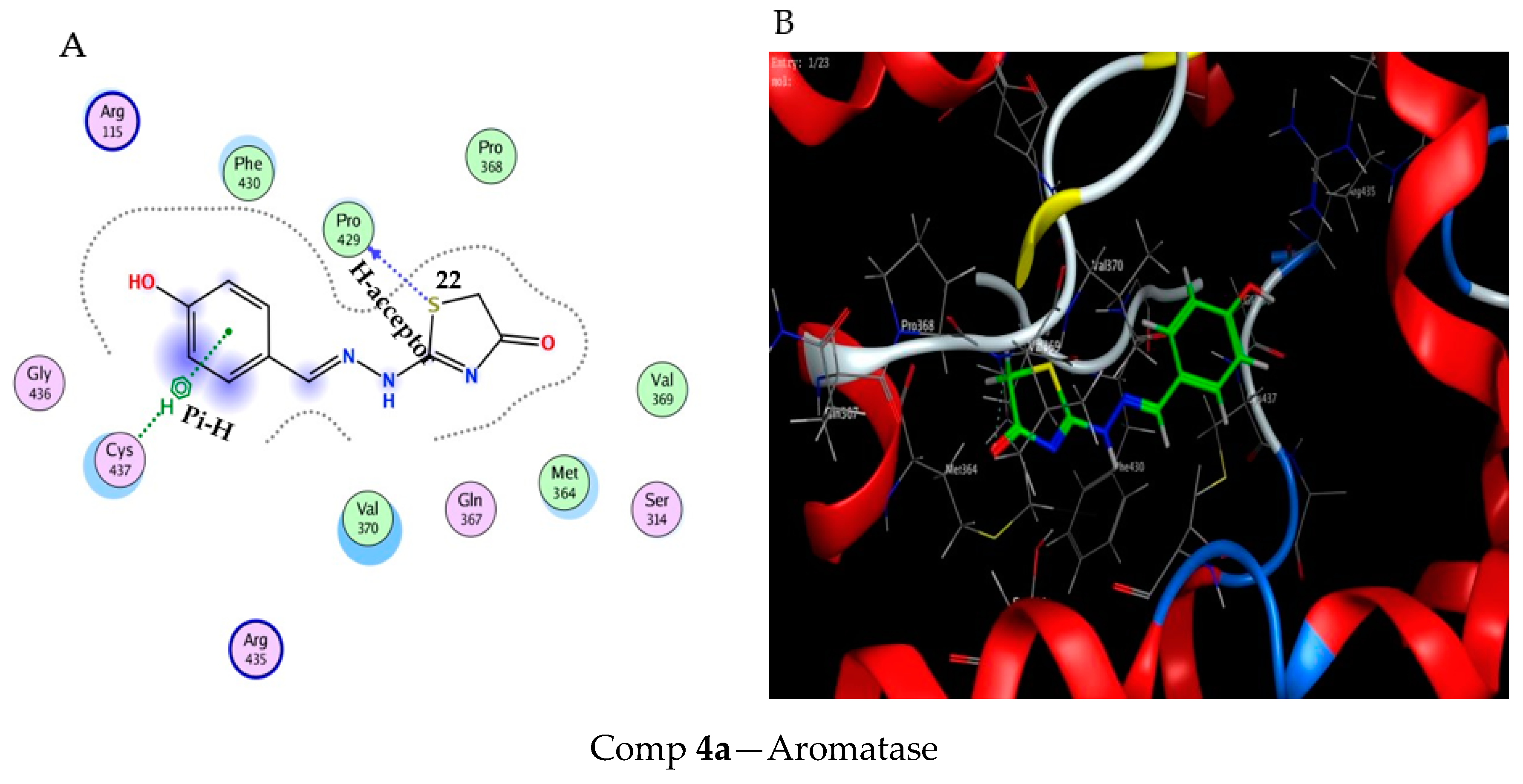
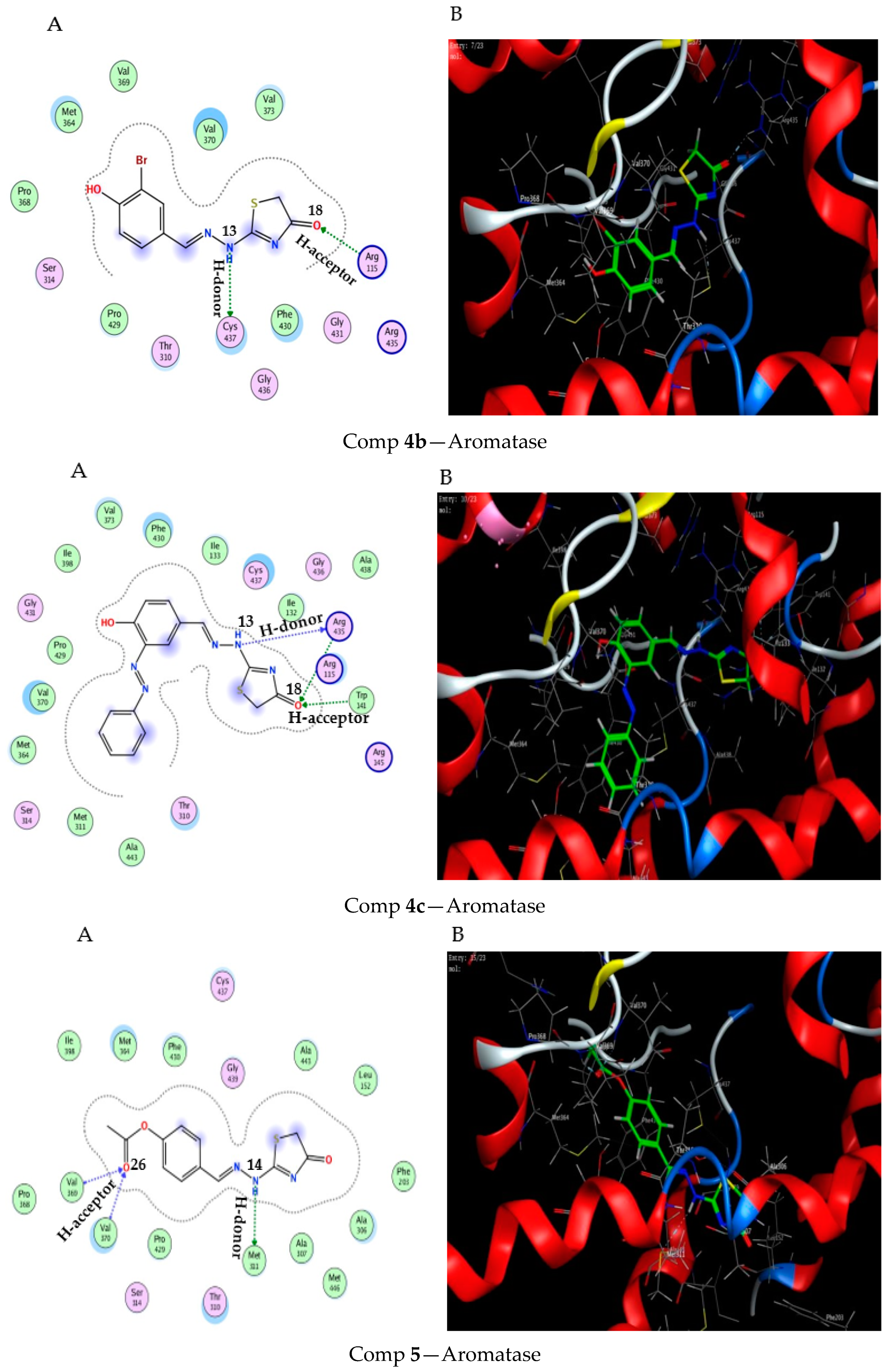
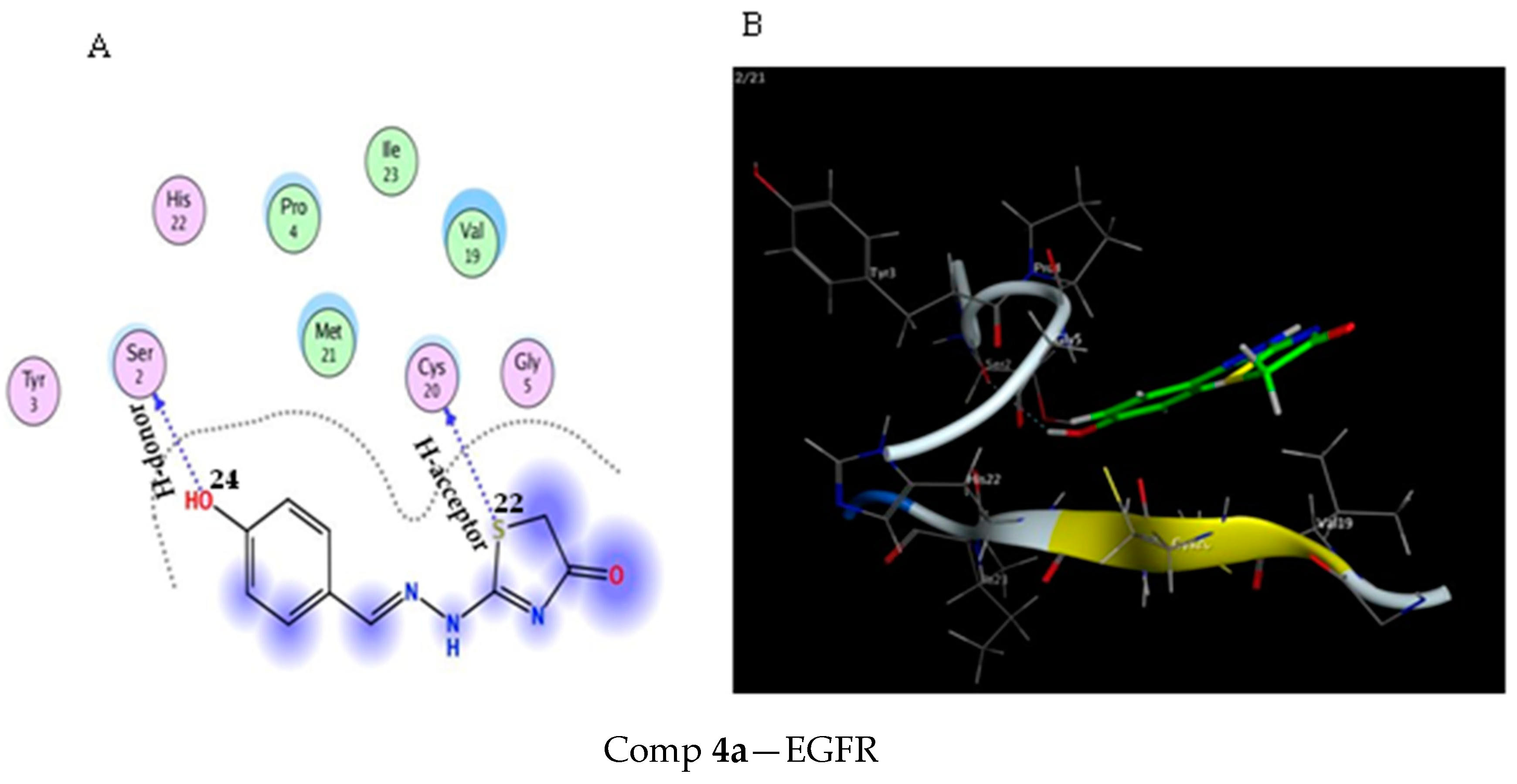
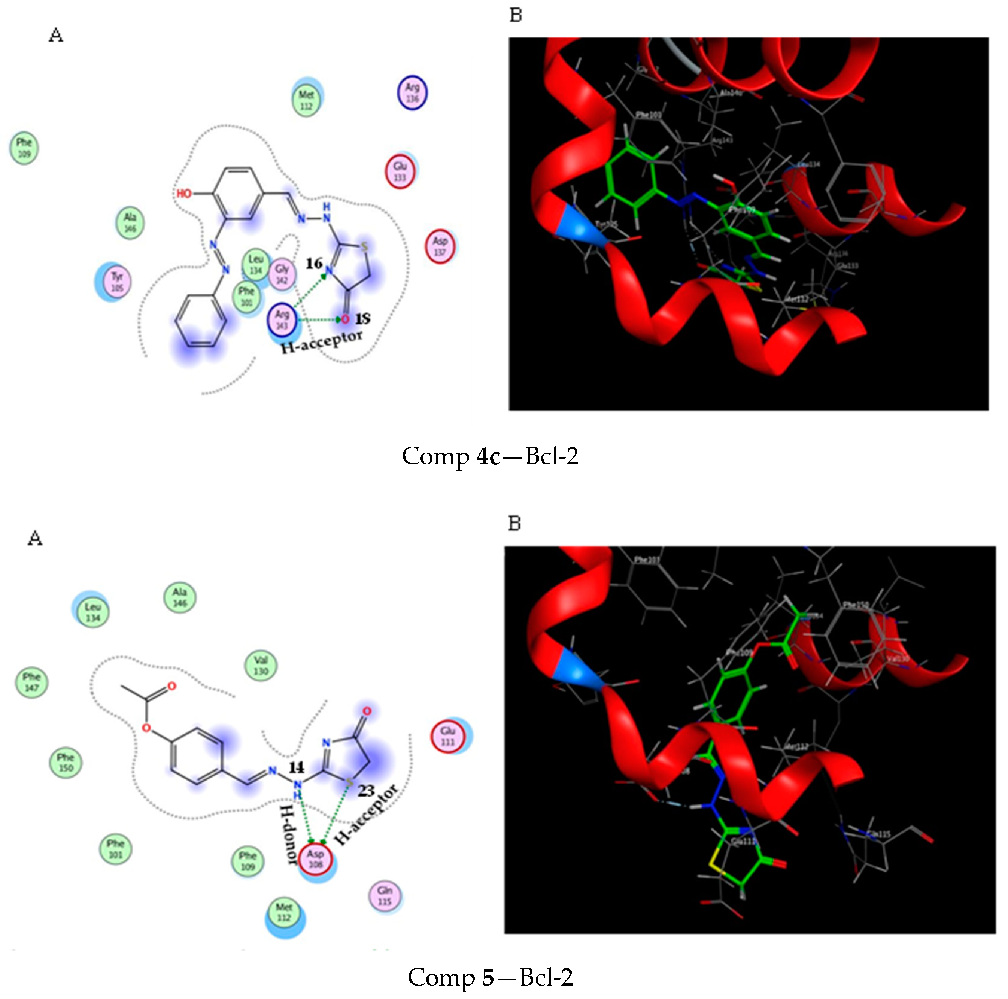
2.3. Molecular Docking Study

We believe that molecular docking could be applied to predict the binding affinity of bioactive compounds (ligands) toward a targeted protein in cancer cells (receptors). This powerful tool aids in discovering and designing a new and potent anticancer agent. We applied a molecular docking analysis to explore whether the anticancer activity of these compounds could account for their ability to inhibit target proteins that have a vital role in the growth, proliferation, and cell cycle of cancer cells, especially breast (MCF-7) cancer. In the current study, molecular docking analyses for compounds (4ac) and 5 were conducted against four key proteins, which included aromatase (PDB: 3S7S), epidermal growth factor receptor (EFGR) (PDB: 1EPG), cyclin-dependent kinase 2 (CDK2) (PDB: 1DI8), and B-cell lymphoma 2 (Bcl-2, antiapoptotic protein) (PDB: 2O2F). These proteins are proposed to stimulate the growth, proliferation, and development of cancer cells. The inhibition of estrogen production by aromatase inhibitors has been reported to efficiently suppress the proliferation of breast cancer. Therefore, examining the binding affinity of our compounds (4ac) and 5 toward aromatase and other kinds of proteins could emphasize the anticancer activity of these compounds.
The synthesized compounds (4ac) and 5 may interact with active amino acid residues of aromatase through H-bonding and Arene-H interaction. They also showed significant docking scores (binding energies) towards the receptor active key amino acids with low root mean square deviation (RMSD), as shown in Table 3. Among the docked compounds, compound 4c exhibited the best docking score (binding energies) (−7.91) kcal/mol and RMSD (1.35) (Table 3), which were comparable with FDA recommendations for the aromatase inhibitors or first-line treatments for breast cancer, anastrozole (−9.32) Kcal/mol and letrozole (−8.78) Kcal/mol [52]. Also, compound 4c formed a hydrogen bonding (H-donor and H-acceptor) with amino acids existing in the active site of the aromatase enzyme, such as ARG 435 (arginine 435) and TRP 141 (tryptophane 141), in a distance (bond length) ranging from 2.99 to 3.56 (A0), as shown in Table 3 and Figure 4. The docked compounds (4ac) and 5 were deemed to have strong docking scores by binding to the active site and displaying interactions with amino acids of the active pocket of the aromatase receptor.
In addition, the molecular docking results of synthesized compounds (4ac) and 5 against “EGFR” (PDB: 1EPG) revealed a moderate docking scores by creating H-bonding, Arene-H (pi-H), and Arene-cation (pi-cation) interactions (Table 4) compared to the interactions of these compounds with the aromatase enzyme (Table 3). Compound 4c showed a favorable docking score (−4.72) kcal/mol and RMSD (1.33) by forming a hydrogen bond with CYS 20 (cysteine 20), and pi-cation interactions with ASN 1 (asparagine 1) exist in the active site of “EGFR” (Table 4 and Figure 5) in agreement with erlotinib (−7.09 kcal/mol) as potential EGFR inhibitors [53].
In comparison to Sorafenib (−9.7) kcal/mol and kenpaullone (−9.4) kcal/mol as CDK2 enzyme inhibitors, further docking tests of the synthesized compounds (4ac) and 5 against CDK2 (cyclin-dependent kinase-2) revealed moderate docking scores [54]. Additionally, as indicated in Table 5 and Figure 6, compound 4c demonstrated the best docking score of −6.64 kcal/mol with a RMSD of 1.4 and hydrogen bonding with LEU 83 (leucine 83) and ASN 86 (asparagine 86) of CDK2.
Furthermore, the Bcl-2 protein’s active pocket was also docked by the substances (4ac) and (5). Compound 4c among the docked derivatives displayed a moderate docking score (−5.53 kcal/mol, RMSD 1.07) and formed a hydrogen bond with ARG 143 (arginine 143) at distances ranging from 2.98 to 3.52 (A0) (Table 6 and Figure 7), which is consistent with venetoclax (−9.1 kcal/mol) as an antiapoptotic Bcl-2 reference drug [55].
Detailed in silico analyses (molecular docking) against four important proteins revealed, in conclusion, that compound 4c had the best docking scores at −7.91 kcal/mol (Table 3), followed by −6.64 kcal/mol (Table 5) against the aromatase enzyme and CDK2. Additionally, compared to aromatase, compound 4c had moderate docking scores toward the antiapoptotic protein Bcl-2 (Table 6) and EGFR (Table 4). It became evident that compound 4c has anticancer activity, particularly against breast cancer (MCF-7), by combining the findings from the experimental part of our research (i.e., cytotoxicity, cell cycle assessment, and apoptosis assay) for compound 4c against MCF-7 breast cancer cells. The production of estrogen by the enzyme aromatase is necessary for the growth and development of breast cancer cells. In accordance with the antiproliferative action of compound 4c (IC50, 2.57 ± 0.16 μM) toward MCF-7 cells (Table 1), compound 4c could bind (−7.91 kcal/mol) and inhibit the aromatase enzyme (aromatase inhibitor), which, in turn, prevents the proliferation of breast cancer cells. Additionally, molecular docking demonstrated that compound 4c might function as an inhibitor for CDK2 (−6.64 kcal/mol) (Table 5), which is implicated in the cell cycle development of cancer cells, and the antiapoptotic protein Bcl-2 (−5.53 kcal/mol) (Table 6). These results of CDK2 and Bcl-2 are compatible with the cell cycle arrest (the pre-G1 phase, 37.36%) and apoptosis assays (early apoptosis, 22.39% and late apoptosis, 9.51%) for compound 4c against MCF-7 (Figure 2 and Figure 3).
A number of atoms from the compounds (4ac) and 5 in Table 3, Table 4, Table 5 and Table 6 were identified in 2D (Figure 4, Figure 5, Figure 6 and Figure 7).

3. Materials and Methods

1HNMR and 13CNHR spectra of the synthesized compounds were measured with a Bruker 400 DRX Avance NMR spectrometer (Chichago, EIK Grove Village, USA) using the DMSO- d6 as the solvent. Utilizing the Agilent MSD-5978 spectrometry instrument (Agilent technologies, Inc.5301 Stevens Greek Boulevard santa clara, CA, USA), electron ionization mass spectrometry (EI-MS) was used to determine the compounds’ molecular weights with 70 eV−. Using a Shimadzu 470 spectrometer (Shimadzu corporation, Kyoto, Japan), the IR measurements were collected. A melting point thermoelectric instrument (an electrothermal 200 digital melting point device) was used to calculate the melting points of the synthesized compounds, but the results were unreliable. A Perkin Elmer CHN elemental analyzer (Haan, Germany) was used for the microanalysis. The Aldrich Chemical Company (Sigma-Aldrich Chemie GmbH, Eschenstr. 582024 Taufkirchen Germany) was used to source all of the analytical-grade chemicals and reagents.

3.1. Chemistry

3.1.1. Synthesis Guidelines for 2-Substituted Hydrazine Carbothioamides (2ac)

A mixture of 3-substituted-4-hydroxybenzaldehyde (0.01 mol) and thiosemicarbazide (0.01 mol) in absolute ethanol (30 mL) and a few drops of acetic added as the acid catalyst was refluxed for 4 h, then cooled. The resultant precipitate was separated, ethanol washed, and dried afterward. Finally, a suitable solvent was used to crystallize the outcome, yielding the compounds (2ac).

2-(4-Hydroxybenzylidene)hydrazine Carbothioamide (2a)

Pale yellow crystals, yield 81%, m.p. 205 °C. IR (KBR) υmax 3450 (br.Oh), 3331, 3221, 3185 (NH2 and NH), 1625 (C=N), 1293 (C=S) cm−1. 1HNMR (DMSO-d6) δ: 7.01–7.89 (m, 6H, AR-H and NH2), 8.21 (s, 1H, CH=N), 9.95 (s, 1H, OH), 11.38 (s, 1H, NH) ppm.

2-(3-Bromo-4-hydroxybenzylidene)hydrazine-1-carbothioamide (2b)

As pale yellow crystals, yield 83%,m.p 221 °C. IR (KBr) υmax; 3461 (OH), 3361, 3225, 3175 (NH2 and NH), 1625 (C=N), 1296 (C=S) cm−1. 1HNMR (DMSO-d6); δ 6.94–6.961 (d, 1H, Ar-H), 7.4–7.13 (d, 1H, Ar-H), 7.45–7.48 (dd, 1H, Ar-H),7.70–7.77 (m, 3H, Ar-H, NH2),7.95–7.96 (d,1H,Ar-H) 8.03 (s, 1H, CH=N), 8.05 (s, 1H, CH=N), 9.77, 10.15 (s, 1H, OH of two isomer), 10.60, 11.54 (s, 1H, NH of two isomer) ppm.
13CNMR (DMSO-d6); δ 190.72 (C=S), 160.21 (C-O), 157.40, 155.31 (C=N), 138.65, 136.97, 131.00, 130.93, 130.63, 130.04, 128.08, 116.48, 116.62, 112.63, 110.49 (C-aromatic of two isomer) ppm.

4-Hydroxy-3-(phenyldiazenyl)benzylidene)hydrazine-1-carbothioamide (2c)

As orange crystals, yield 76%, m.p 189 °C. IR (KBr) υmax: 3420 (OH), 3369, 3220, 3180 (NH2 and NH), 1625 (C=N), 1305 (C=S) cm−1. 1HNMR (DMSO–d6) δ: 6.09–7.99 (m, 10H, Ar-H and NH2), 8.09 (s, 1H, CH=N), 9.89 (br-s, 1H, OH), 11.29 (br-s, 1H, NH) ppm.13CNMR (DMSO-d6) δ: 177.90 (C=S), 159.74 (C-O), 153.28, 152.91 (C=N), 143.32, 143.26 (C-N=N-C), 129.87, 129.65, 129.54, 125.67, 125.61, 122.20, 116.05, 113.90 (C-aromatic) ppm.

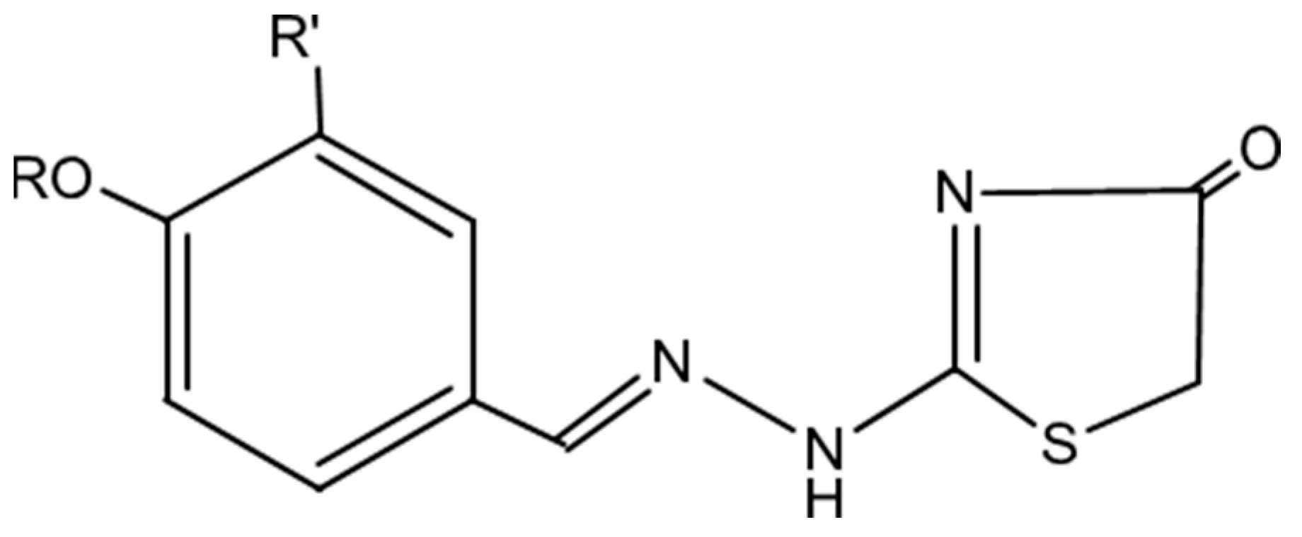
3.1.2. Synthesis of Thiazole-4 [5H]-One Derivatives (4ac): General Technique

Chloroacetyl chloride (0.01 mol) was added after compound 4 (0.01 mol) was dissolved in 100 mL of dry benzene. This mixture was allowed to heat at 60 for 1 h. The benzene was expelled, and the solid residue was refluxed in 50 mL ethanol in the presence of fused sodium acetate for 4 h. After cooling, the reaction mixture was added to the water and stirred, and the resultant solid was taken out, washed in water, dried, and recrystallized with the appropriate solvent to produce 4.

2-(2-(4-Hydroxybenzylidene)hydrazinyl)thiazole-4[5H]-one (4a)

Yield of 73% as golden crystals, mp.236 °C. IR (KBr) υmax: 3453 (br-OH), 3229 (br-s, NH), 1695 (C=O), 1625 (C=N), 1610, 1583 (C=C), 1121, 1068 (C-O) cm−1. 1HNMR (DMSO-d6): δ: 3.88 (S, 2H, CH2 of thiazole ring), 6.83–6.86 (d, 2H, Ar-H), 7.59–7.61 (d, 2H, Ar-H), 8.28 (s, 1H, CH=N), 10.04 (br.s, 1H, OH), 11.78 (br.s, 1H, NH) ppm. 13CNMR (DMSO-d6): δ 174.72 (C=O), 163.98 (S-C=N), 160.36 (C-O), 156.63 (C=N), 130.83, 125.58, 116.19 (Caromatic), 33.43 (CH2 of Thiazole ring) ppm. Ms: m/z (%): 236 (M+ +1, 1.65),235 (M+, 72.06), 234 (M+ − 1, 100), 211 (2.90), 210 (7.32), 204 (1.17), 203 (2.97), 196 (5.17), 195 (14.31), 192 (22.34), 190 (11.99), 189 (191.03), 183 (1.66), 182 (2.21), 178 (2.61), 161 (1.16), 159 (2.94), 156 (1.00), 152 (1.03), 150 (10.82), 149 (8.34), 148 (4.67), 147 (2.83), 146 (4.56), 142 (4.07), 141 (1.36), 140 (6.38), 138 (4.56), 137 (9.84), 133 (10.24), 132 (13.54), 121 (55.56), 121 (67.98), 120 (97.13), 119 (30.82), 108 (1.40), 106 (71.75), 105 (31.21), 104 (15.53), 103 (8.35), 102 (2.89), 60 (1.21), 52 (2.98), 51 (3.13). Anal. Calcd for C10H9N3O2S (M.wt. = 235): C, 51.06; H, 3.83; N, 17.87. Found: C, 50.95; H, 3.66; N, 17.71.

2-(2-(3-Bromo-4-hydroxybenzylidene)hydrazinyl)thiazole-4[5H]-one (4b)

As yellow crystals, yield 71%, m.p. 258° C. IR (KBr) υmax: 3463 (br-OH), 3271 (NH), 1641 (C=N), 1605, 1586 (C=C), 1081, 1061 (C-O) cm−1. 1H-NMR (DMSO-d6) δ: 3.91, 3.94 (s, 2H, CH2 of Thiazole ring), 7.93 (s, 1H, Ar.H), 8.13–8.21 (m, 2H, Ar-H). 8.30, 8.46 (s, 1H, CH=N of two isomers) ppm. 13CNMR (DMSO-d6). Anal. Calcd for C10H8BrN3O2S (M.wt. = 313): C, 38.34; H, 2-SS; N,13.42. Found: C, 38.11; H, 2.35; N, 13.22.

2-(2-(4-Hydroxy-3-phenyldiazenyl)benzylidene)hydrazinyl)thiazole-4[5H]-one (4c)

As orange crystals, yield 71%, m.p. 246 °C. IR (KBr) υmax: 3451 (br.OH), 3227 (NH), 1693 (C=O), 1625 (C=N), 1605, 1591 (C=C), 1081, 1062 (C-O) cm−1. 1HNMR (DMSO-d6) δ: 3.84 (s, 2H, CH2 of Thiazole ring), 6.81–7.60 (m, 8H, Ar-H), 8.29 (s, 1H, CH=N), 10.02 (br.s, 1H, OH), 11.89 (br.s, 1H, NH) ppm. 13CNMR (DMSO-d6) δ:174.62, 172.25 (C=O), 160.69, 160.40 (C-O), 156.62 (C=N), 153.33, 152.92, 145.85, 143.29 (C-N=N-C), 130.23, 129.64, 125.71, 125.66, 122.19, 116.40, 116.19, 113.86 (C of aromatic of two isomers), 33.44 (CH2 of Thiazole ring) ppm. Anal. Calcd for C16H13N5O2S (M.wt. = 339): C, 56.64; H, 3.83; N, 20.65. Found: C, 56.36; H, 3058; N,20.44.

3.1.3. Synthesis 4-((2-(4-Oxo-4,5-Dihydrothiazol-2 Yl)Hydrazinylidene)methyl)phenyl Acetate (5)

A solution of 4a (0.01 mol) in acetic anhydride (20 mL) was reflux-warmed for 2 h, then chilled alone and emptied into ice H2O. The reaction amalgam was dropped for 24 h. and the resulting solid was isolated by filtration, cleaned with water, and dehydrated; the resulting product was crystallized from ethanol to give 4 as pale yellow crystals, yield 58%, m.p. 165 °C. IR (KBr) υmax: 3225 (NH), 1753, 1693 (C=O), 1625 (C=N), 1610, 1585 (C=C), 1125, 1084 (C-O) cm−1. 1HNMR (DMSO-d6) δ: 2.30 (s, 3H, COCH3), 3.91 (s, 2H, CH2 of Thiazole ring), 7.236–7.256 (d, 2 H, J= 8.00 HZ, Ar-H), 7.802–7.821 (d, 2H, J= 8.00HZ, Ar-H), 8.428 (s, 1H, CH=N), 11.99 (br.s, 1H, NH) ppm. 13CNMR (DMSO-d6) δ:169.50, 160.40 (C=O), 155.84 (C-O), 152.64 (C=N), 132.32, 129.27, 122.88, 122.61, 116.18 (C-aromatic), 33.49 (CH2 of Thiazole ring), 21.35 (COCH3) ppm. Anal. Calcd for C12H11N3O3S (M.wt. = 277): C, 51.98; 3.97; N, 15.16. found: C, 51.77; H, 3.79; N, 14.98.

3.1.4. Synthesis of 5-(4-Hydroxy-3-methoxybenzylidene)-2-(2-4-hydroxybenzylidene) Hydrazinyl) Thiazole-4[5H]-one (6)

A mixture of compound 4a (0.01 mol) and 3-methoxy-4-hydroxybenzaldehyde (0.01 mol) in 30 mL dimethyl formamide in the incidence of fused sodium acetate (0.03 mol) was refluxed for 2 h. The outcome mixture was chilled, followed by emerging in ice water and neutralizing with HCl (1%). The resultant product constructed was separated, cleaned with dehydrated H2O, and recrystallized from ethanol to provide compound 6 as yellow crystals, yield 71%, m.p. 258 °C. IR (KBr) υmax: 3450–3420 (br.OH), 3228 (NH), 1695 (C=O) cm−1. 1HNMR (DMSO-d6) δ: 3.82 (s, 2H, CH2-N=N), 3.86 (s, 3H, OCH3), 6.87–7.88 (m, 8H, Ar-H and H-olefinic), 8.40 (s, 1H, CH=N), 10.14 (br.s, 1H, OH), 10.80 (br.s, 1H, OH), 12.43 (br.s, 1H, NH) ppm. 13CNMR (DMSO-d6) δ: 160.67 (C=O), 158.35, 157.69, 157.344, 156.46 (C-O), 150.23, 149.83 (C=N) 148.344, 148.29 (N-C=N), 134.13, 134.05, 130.73, 130.62, 130.39, 125.80, 125.41, 125.29, 124.04, 123.98,123.48, 122.05, 121.09, 120.14, 118.56, 116.31, 116.26, 10.74 (C of aromatic of two isomers), 55.92, 55.81 (OCH3 of two isomers), 44.23 (Ar-CH2-N=N) ppm. Anal. Calcd for C18H15N2O4S (M.wt. = 357): C, 57.14; H, 4.20; N, 11.76. Found: C, 56.89; H, 11.51; N, 4.04.

3.2. Biological Assessment

3.2.1. The Ability of Cytotoxicity to Kill Cancer Cell Lines

Utilizing the MTT assay, the novel synthesized compounds’ in vitro cytotoxic activity was evaluated [56,57]. Cells were grown on 96-well multi-well plates for 24 h with 104 cells per well prior to chemical treatment. The testing materials were dissolved in dimethyl sulfoxide (DMSO). The test chemical was applied to the monolayer of cells at various quantities. Each concentration was made in a separate set of three duplicate wells. Forty-eight hours were spent incubating the monolayer cells with the substance (s) at 37 °C and in a 5% CO2 environment. After 48 h, the cells were fixed, washed, and stained with 40 µL of MTT solution (5 mg/mL of MTT in 0.9% NaCl in each well was added and incubated for an additional 4 h). By adding 180 µL of acidified isopropanol to each well and shaking the plate at room temperature, the MTT crystals were made soluble. Then, an ELISA reader was used to take a photometric reading of the absorbance at 570 nm. Calculating the molar quantity required to block 50% of the cell viability (IC50) and compare it to the reference substance, STU.

3.2.2. Enzyme Assay for VEGFR-2

The manufacturer’s instructions were followed while using an ELISA kit for human VEGFR-2/KDR (RBMS# 2019R) to assess the inhibitory effects of compounds 4c and Sorafenib at various doses against VEGFR-2 [58].

3.2.3. Study of the Cell Cycle of Chemical 4c

There were 3.0 × 10⁵ MCF-7 cells per well incubated for 12 h at 37 °C. Then, for 24 h, the chemical 4c was given to the target cells at its IC50 concentration. The cells were collected after treatment, fixed with 75% ethanol at 20 °C for an overnight period, centrifuged, washed with PBS, incubated with 10 mg/mL RNase (Sigma, Ronkonkoma, NY, USA) and 5 mg/mL propidium iodide (PI, Sigma), and then subjected to flow cytometry analysis (using a FACS Calibur cytometer and cellQuest software; BD Biosciences, Franklin Lakes, NJ, USA) [59].

3.2.4. Apoptosis Assay with Annexin V FITC/PI

Apoptosis in the MCF-7 cells was investigated using a flow cytometry test and the fluorescent dye known as Annexin V-FITC/PI [60]. Briefly, the MCF-7 cells (2 × 10⁵) were administered compound 4c at its IC50 concentration for 24 h; then, the cells were harvested and stained with Annexin V-FITC/PI dye for 15 min in the dark at 37 °C. BD FACS Calibur (Becton and Dickinson, Heidelberg, Germany) detected the flow cytometry results utilizing the BD cellQuest pro program for analysis.

3.3. Molecular Docking Modeling

The most thorough understanding of ligand–receptor interactions is provided by molecular docking studies. The binding of ligands to the active site of cellular target proteins is the basis for the biological actions of chemically created substances [52,53,54,55,61,62]. For clarification of the method by which the produced chemicals conjugate with aromatase, EGFR, CDK2, and Bcl-2 proteins and act as inhibitors to prevent the growth, proliferation, and development of cancer cells by inhibiting estrogen production and onset of the cell cycle stall in cancer cells, a molecular docking program was used to determine how the synthesized compounds 4ac and 5 interacted with the selected docked proteins. The 3D structures of aromatase, EGFR, CDK2, and Bcl-2 were obtained from the RCSB Protein Data Bank (http://www.rcsb.org/PDB, accessed on 12 March 2022). The structures of the target compounds used in this study were constructed with the Chem. Draw tool. Protonation, correction, active site determination of the targeted proteins, structural preparation, energy minimization of the 3D structure of the target ligands, and, following that, docking analyses using the MOE program were completed.

4. Conclusions

In conclusion, to develop and design a safe and efficient anticancer drug, a novel series of thiazole derivatives (4ac) and their acetyl derivative (5) were synthesized through the reaction of thiosemicarbazone with chloroacetly chloride, followed by cyclization with boiling ethanol in the presence of fused sodium acetate. Acetyl derivative (5) was obtained via the acetylation of 4a with acetic anhydride. Utilizing elemental analyses and spectroscopic methods (IR and NMR), the structures of the synthesized compounds were verified. Using the MTT assay, all of the synthesized thiazole derivatives (4ac) and 5 suppressed the growth of the cancer cell lines MCF-7 and HepG2. Compound 4c was examined in vitro using the VEGFR-2 enzyme assay, cell cycle analysis, and Annexin V FITC/PI assay. Compound 4c exhibited promising and potential antiproliferative, cell cycle arrest, and apoptosis activation of MCF-7 cancer cell lines compared to the reference drug and MCF-7 control.

Supplementary Materials

The following supporting information can be downloaded at: https://www.mdpi.com/article/10.3390/cryst13111546/s1.

Author Contributions

Conceptualization, F.A.A.-S., A.H.A., M.E.B., W.M., O.A.A.A., F.G.E., E.F., F.Z.M. and A.T.K.; methodology, software, validation, formal analysis; investigation, resources, data curation, writing—original draft preparation, writing—review and editing, visualization, supervision, project administration, funding acquisition, All authors have read and agreed to the published version of the manuscript.

Funding

The authors extend their appreciation to the Deanship of Scientific Research at King Khalid University for funding this work through large group Research Project under grant number RGP2/12/44.

Data Availability Statement

Not applicable.

Acknowledgments

The authors extend their appreciation to the Deanship of Scientific Research at King Khalid University for funding this work through large group Research Project under grant number RGP2/12/44. The authors wish to thank Mostafa A. Hussein and Mohammed El Behery for their insightful discussion of the docking research, &The researchers would like to thank Fawziah A. Al-Salmi, Fahmy G. Elsaid and Eman Fayad that share in performing anticancer and biological activity part. All authors have read and agreed to the published version of the manuscript.

Conflicts of Interest

The authors declare no conflict of interest.

References

  1. Marwa, F.A.; Atiah, H.A. Design, synthesis, antiproliferative activity, and cell cycle analysis of new thiosemicarbazone derivatives targeting ribonucleotide reductase. Arab. J. Chem. 2021, 14, 102989. [Google Scholar]
  2. Mohammed, F.Z.; Rizzk, Y.W.; El Deen, I.M.; Mourad, A.A.; El Behery, M. Design, synthesis, cytotoxic screening and molecular docking studies of novel hybrid thiosemicarbazone derivatives as anticancer agents. Chem. Biodivers. 2021, 18, e2100580. [Google Scholar] [CrossRef]
  3. Waks, A.G.; Winer, E.P. Breast Cancer Treatment: A Review. JAMA 2019, 321, 288–300. [Google Scholar] [CrossRef]
  4. Loibl, S.; Poortmans, P.; Morrow, M.; Denkert, C.; Curigliano, G. Breast Cancer. Lancet. 2021, 397, 1750–1769. [Google Scholar] [CrossRef] [PubMed]
  5. Arora, S.; Narayan, P.; Osgood, C.L.; Wedam, S.; Prowell, T.M.; Gao, J.J.; Shah, M.; Krol, D.; Wahby, S.; Royce, M.; et al. U.S. FDA Drug Approvals for Breast Cancer: A Decade in Review. Clin. Cancer Res. Off. J. Am. Assoc. Cancer Res. 2022, 28, 1072–1086. [Google Scholar] [CrossRef] [PubMed]
  6. An, J.; Peng, C.; Xie, X.; Peng, F. New Advances in Targeted Therapy of HER2-Negative Breast Cancer. Front. Oncol. 2022, 12, 828438. [Google Scholar] [CrossRef]
  7. Mukhopadhyay, K.D.; Liu, Z.; Bandyopadhyay, A.; Kirma, N.B.; Tekmal, R.R.; Wang, S.; Sun, L.Z. Aromatase expression increases the survival and malignancy of estrogen receptor positive breast cancer cells. PLoS ONE 2015, 10, e0121136. [Google Scholar] [CrossRef]
  8. Wu, S.; Ye, J.; Wang, Z.; Lin, S.X.; Lu, M.; Liang, Y.; Wu, C.L. Expression of aromatase in tumor related stroma is associated with human bladder cancer progression. Cancer Biol. Ther. 2018, 19, 175–180. [Google Scholar] [CrossRef]
  9. Chumsri, S.; Howes, T.; Bao, T.; Sabnis, G.; Brodie, A. Aromatase, aromatase inhibitors, and breast cancer. J. Steroid Biochem. Mol. Biol. 2011, 125, 13–22. [Google Scholar] [CrossRef]
  10. Sasaki, T.; Hiroki, K.; Yamashita, Y. The role of epidermal growth factor receptor in cancer metastasis and microenvironment. Biomed. Res. Int. 2013, 2013, 546318. [Google Scholar] [CrossRef] [PubMed]
  11. Wee, P.; Wang, Z. Epidermal growth factor receptor cell proliferation signaling pathways. Cancers 2017, 9, 52. [Google Scholar] [CrossRef]
  12. Jonathan, C.; David, A.Q.; Troy, M.R.; Felix, Y.F.; Alan, A. Transcription-associated cyclin-dependent kinases as targets and biomarkers for cancer therapy. Cancer Discov. 2020, 10, 351–370. [Google Scholar]
  13. Johnson, L.N.; De Moliner, E.; Brown, N.R.; Song, H.; Barford, D.; Endicott, J.A.; Noble, M.E. Structural studies with inhibitors of the cell cycle regulatory kinase cyclin-dependent protein kinase 2. Pharmacol. Ther. 2002, 93, 113–124. [Google Scholar] [CrossRef]
  14. Kontopidis, G.; McInnes, C.; Pandalaneni, S.R.; McNae, I.; Gibson, D.; Mezna, M.; Fischer, P.M. Differential binding of inhibitors to active and inactive CDK2 provides insights for drug design. Chem. Biol. 2006, 13, 201–211. [Google Scholar] [CrossRef]
  15. Campbell, K.J.; Tait, S.W.G. Targeting BCL-2 regulated apoptosis in cancer. Open Biol. 2018, 8, 180002. [Google Scholar] [CrossRef]
  16. Murphy, G.J.; Holder, J.C. PPAR-γ agonists: Therapeutic role in diabetes, inflammation and cancer. Trends Pharmacol. Sci. 2000, 21, 469–474. [Google Scholar] [CrossRef]
  17. Dayam, R.; Sanchez, T.; Clement, O.; Shoemaker, R.; Sei, S.; Neamati, N. β-Diketo acid pharmacophore hypothesis. 1. Discovery of a novel class of HIV-1 integrase inhibitors. J. Med. Chem. 2005, 48, 111–120. [Google Scholar] [CrossRef]
  18. Kikkawa, R.; Hatanaka, I.; Yasuda, H.; Kobayashi, N.; Shigeta, Y.; Terashima, H.; Tsuboshima, M. Effect of a new aldose reductase inhibitor, (E)-3-carboxymethyl-5-[(2E)-methyl-3-phenylpropenylidene] rhodamine (ONO-2235) on peripheral nerve disorders in streptozotocin-diabetic rats. Diabetologia 1983, 24, 290–292. [Google Scholar] [CrossRef] [PubMed]
  19. da Rocha Junior, L.F.; Rêgo, M.J.B.; Cavalcanti, M.B.; Pereira, M.C.; Pitta, M.G.D.R.; de Oliveira, P.S.; Gonçalves, S.M.; Duarte, A.L.; de Lima Mdo, C.; Pitta Ida, R.; et al. Synthesis of a novel thiazolidinedione and evaluation of its modulatory effect on IFN-γ, IL-6, IL-17A, and IL-22 production in PBMCs from rheumatoid arthritis patients. Biomed. Res. Int. 2013, 2013, 926060. [Google Scholar] [CrossRef]
  20. Chhabria, M.T.; Patel, S.; Modi, P.; Brahmkshatriya, P.S. Thiazole: A review on chemistry, synthesis and therapeutic importance of its derivatives. Curr. Top. Med. Chem. 2016, 16, 2841–2862. [Google Scholar] [CrossRef] [PubMed]
  21. Zervosen, A.; Lu, W.P.; Chen, Z.; White, R.E.; Demuth, T.P.J.; Frere, J.M. Interactions between penicillin-binding proteins (PBPs) and two novel classes of PBP inhibitors, arylalkylidene rhodanines and arylalkylidene iminothiazolidin-4-ones. Antimicrob. Agents Chemother. 2004, 48, 961–969. [Google Scholar] [CrossRef] [PubMed]
  22. Bozdağ-Dündar, O.; Ozgen, O.; Menteşe, A.; Altanlar, N.; Atli, O.; Kendi, E.; Ertan, R. Synthesis and antimicrobial activity of some new thiazolyl thiazolidine-2,4-dione derivatives. Bioorg Med. Chem. 2007, 15, 6012–6017. [Google Scholar] [CrossRef] [PubMed]
  23. Ravi, S.; Chiruvella, K.K.; Rajesh, K.; Prabhu, V.; Raghavan, S.C. 5-Isopropylidene-3-ethyl rhodanine induce growth inhibition followed by apoptosis in leukemia cells. Eur. J. Med. Chem. 2010, 45, 2748–2752. [Google Scholar] [CrossRef] [PubMed]
  24. Aziz, H.A.; El-Saghier, A.M.M.; Badr, M.; Abuo-Rahma, G.E.D.A.; Shoman, M.E. Thiazolidine-2,4-dione-linked ciprofloxacin derivatives with broad-spectrum antibacterial, MRSA and topoisomerase inhibitory activities. Mol. Divers. 2022, 26, 1743–1759. [Google Scholar] [CrossRef] [PubMed]
  25. Alegaon, S.G.; Alagawadi, K.R. New thiazolidinedione-5-acetic acid amide derivatives: Synthesis, characterization and investigation of antimicrobial and cytotoxic properties. Med. Chem. Res. 2012, 21, 816–824. [Google Scholar] [CrossRef]
  26. Mohamed, S.F.; Flefel, E.M.; Amr, A.E.-G.; Abd El-Shafy, D.N. Anti-HSV-1 activity and mechanism of action of some new synthesized substituted pyrimidine, thiopyrimidine and thiazolopyrimidine derivatives. Eur. J. Med. Chem. 2010, 45, 1494–1501. [Google Scholar] [CrossRef] [PubMed]
  27. Barsoum, F.F.; Girgis, A.S. Facile synthesis of bis (4,5-dihydro-1H-pyrazole-1-carboxamides) and their thio-analogues of potential PGE2 inhibitory properties. Eur. J. Med. Chem. 2009, 44, 2172–2177. [Google Scholar] [CrossRef] [PubMed]
  28. Yusuf, M.; Jain, P. Synthetic and biological studies of pyrazolines and related heterocyclic compounds. Arab. J. Chem. 2014, 7, 553–596. [Google Scholar] [CrossRef]
  29. Hassan, A.S.; Moustafa, G.O.; Awad, H.M.; Nossier, E.S.; Mady, M.F. Design, synthesis, anticancer evaluation, enzymatic assays, and a molecular modeling study of novel pyrazole–indole hybrids. ACS Omega 2021, 6, 12361–12374. [Google Scholar] [CrossRef] [PubMed]
  30. Sharshira, E.M.; Hamada, N.M. Synthesis and in vitro antimicrobial activity of some pyrazolyl-1-carboxamide derivatives. Molecules 2011, 16, 7736–7745. [Google Scholar] [CrossRef] [PubMed]
  31. Hamadi, N.B.; Haouas, A.; Methamem, M.; Msaddek, M. Synthesis of new pyrazolines and their biological evaluation as antimicrobial agents. J. Chem. Res. 2012, 36, 563–565. [Google Scholar] [CrossRef]
  32. Havrylyuk, D.; Zimenkovsky, B.; Vasylenko, O.; Zaprutko, L.; Gzella, A.; Lesyk, R. Synthesis of novel thiazolone-based compounds containing pyrazoline moiety and evaluation of their anticancer activity. Eur. J. Med. Chem. 2009, 44, 1396–1404. [Google Scholar] [CrossRef] [PubMed]
  33. Sim, M.M.; Ng, S.B.; Buss, A.D.; Crasta, S.C.; Goh, K.L.; Lee, S.K. Benzylidene rhodanines as novel inhibitors of UDP-N-acetylmuramate/L-alanine ligase. Bioorg Med. Chem. Lett. 2002, 12, 697–699. [Google Scholar] [CrossRef] [PubMed]
  34. Turan-Zitouni, G.; Chevallet, P.; Kilic, F.S.; Erol, K. Synthesis of some thiazolyl-pyrazoline derivatives and preliminary investigation of their hypotensive activity. Eur. J. Med. Chem. 2000, 35, 635–641. [Google Scholar] [CrossRef] [PubMed]
  35. Haroun, M.; Tratrat, C.; Kositzi, K.; Tsolaki, E.; Petrou, A.; Aldhubiab, B.; Attimarad, M.; Harsha, S.; Geronikaki, A.; Venugopala, K.N.; et al. New benzothiazole-based thiazolidinones as potent antimicrobial agents. Design, synthesis and biological evaluation. Curr. Top. Med. Chem. 2018, 18, 75–87. [Google Scholar] [CrossRef] [PubMed]
  36. Sadek, B.; Al-Tabakha, M.M.; Fahelelbom, K.M.S. Antimicrobial prospect of newly synthesized 1, 3-thiazole derivatives. Molecules 2011, 16, 9386–9396. [Google Scholar] [CrossRef] [PubMed]
  37. Kumar, M.; Ramasamy, K.; Mani, V.; Mishra, R.K.; Majeed, A.B.A.; Clercq, E.D.; Narasimhan, B. Synthesis, antimicrobial, anticancer, antiviral evaluation and QSAR studies of 4-(1-aryl-2-oxo-1,2-dihydro-indol-3-ylideneamino)-N-substituted benzene sulfonamides. Arab. J. Chem. 2014, 7, 396–408. [Google Scholar] [CrossRef]
  38. Palekar, V.S.; Damle, A.J.; Shukla, S.R. Synthesis and antibacterial activity of some novel bis-1,2,4-triazolo [3,4-b]-1,3,4-thiadiazoles and bis-4-thiazolidinone derivatives from terephthalic dihydrazide. Eur. J. Med. Chem. 2009, 44, 5112–5116. [Google Scholar] [CrossRef] [PubMed]
  39. Hamama, W.S.; Ismail, M.A.; Shaaban, S.; Zoorob, H.H. Progress in the chemistry of 4-thiazolidinones. J. Heterocyclic Chem. 2008, 45, 939–956. [Google Scholar] [CrossRef]
  40. Kaminskyy, D.; Bednarczyk-Cwynar, B.; Vasylenko, O.; Kazakova, O.; Zimenkovsky, B.; Zaprutko, L.; Lesyk, R. Synthesis of new potential anticancer agents based on 4-thiazolidinone and oleanane scaffolds. Med. Chem. Res. 2012, 21, 3568–3580. [Google Scholar] [CrossRef]
  41. Kaur Manjal, S.; Kaur, R.; Bhatia, R.; Kumar, K.; Singh, V.; Shankar, R.; Kaur, R.; Rawal, R.K. Synthetic and medicinal perspective of thiazolidinones: A review. Bioorg Chem. 2017, 75, 406–423. [Google Scholar] [CrossRef] [PubMed]
  42. Gouda, M.A.; Abu-Hashem, A.A.; Hussein, H.A.R.; Aly, A.S. Recent Progress on Fused Thiadiazines: A Literature Review. Polycycl. Aromat. Compd. 2022, 42, 2861–2893. [Google Scholar] [CrossRef]
  43. Jilloju, P.C.; Persoons, L.; Kurapati, S.K.; Schols, D.; De Jonghe, S.; Daelemans, D.; Vedula, R.R. Discovery of (±)-3-(1 H-pyrazol-1-yl)-6,7-dihydro-5 H-[1,2,4] triazolo [3,4-b][1,3,4] thiadiazine derivatives with promising in vitro anticoronavirus and antitumoral activity. Mol. Divers. 2022, 26, 1357–1371. [Google Scholar] [CrossRef] [PubMed]
  44. Mohammed, F.Z.; Rizzk, Y.W.; El-Deen, I.M.; Gad, E.M.; El Behery, M.; Mahdy, A.R. Discovery of 2-Amino-4H-1,3,4-thiadiazine-5 (6H)-one Derivatives and Their In Vitro Antitumor Investigation. ChemistrySelect 2022, 7, e202104333. [Google Scholar] [CrossRef]
  45. Kamat, V.; Santosh, R.; Poojary, B.; Nayak, S.P.; Kumar, B.K.; Sankaranarayanan, M.; Vootla, S.K. Pyridine-and thiazole-based hydrazides with promising anti-inflammatory and antimicrobial activities along with their in silico studies. ACS Omega 2020, 5, 25228–25239. [Google Scholar] [CrossRef] [PubMed]
  46. Morigi, R.; Locatelli, A.; Leoni, A.; Rambaldi, M. Recent patents on thiazole derivatives endowed with antitumor activity. Recent. Pat. Anticancer. Drug Discov. 2015, 10, 280–297. [Google Scholar] [CrossRef] [PubMed]
  47. Ghorab, M.M.; Al-Said, M.S. Antitumor activity of novel pyridine, thiophene and thiazole derivatives. Arch. Pharm. Res. 2012, 35, 965–973. [Google Scholar] [CrossRef]
  48. Kasralikar, H.M.; Jadhavar, S.C.; Bhansali, S.G.; Patwari, S.B.; Bhusare, S.R. Design, synthesis and docking studies of novel 1,2,3-triazolyl phenylthiazole analogs as potent anti-HIV-1 NNRT inhibitors. Med. Chem. 2017, 7, 268–275. [Google Scholar]
  49. Amr, A.; Sabrry, N.M.; Abdalla, M.M.; Abdel-Wahab, B.F. Synthesis, antiarrhythmic and anticoagulant activities of novel thiazolo derivatives from methyl 2-(thiazol-2-ylcarbamoyl) acetate. Eur. J. Med. Chem. 2009, 44, 725–735. [Google Scholar] [CrossRef] [PubMed]
  50. Maghraby, M.T.; Abou-Ghadir, O.M.F.; Abdel-Moty, S.G.; Ali, A.Y.; Salem, O.I.A. Novel class of benzimidazole-thiazole hybrids: The privileged scaffolds of potent anti-inflammatory activity with dual inhibition of cyclooxygenase and 15-lipoxygenase enzymes. Bioorg Med. Chem. 2020, 28, 115403. [Google Scholar] [CrossRef] [PubMed]
  51. Ruberte, A.C.; Ramos-Inza, S.; Aydillo, C.; Talavera, I.; Encío, I.; Plano, D.; Sanmartín, C. Novel N,N′-disubstituted acylselenoureas as potential antioxidant and cytotoxic agents. Antioxidants 2020, 9, 55. [Google Scholar] [CrossRef] [PubMed]
  52. Suvannang, N.; Nantasenamat, C.; Isarankura-Na-Ayudhya, C.; Prachayasittikul, V. Molecular Docking of Aromatase Inhibitors. Molecules 2011, 16, 3597–3617. [Google Scholar] [CrossRef]
  53. Mansourian, M.; Hassanzadeh, F.; Shahlaei, M. Exploring the interaction between epidermal growth factor receptor tyrosine kinase and some of the synthesized inhibitors using combination of in-silico and in-vitro cytotoxicity methods. Res. Pharm. Sci. 2018, 13, 509–522. [Google Scholar] [CrossRef] [PubMed]
  54. Ikwu, F.A.; Isyaku, Y.; Obadawo, B.S.; Lawal, H.A.; Ajibowu, S.A. In silico design and molecular docking study of CDK2 inhibitors with potent cytotoxic activity against HCT116 colorectal cancer cell line. J. Genet. Eng. Biotechnol. 2020, 18, 51. [Google Scholar] [CrossRef] [PubMed]
  55. Ramos, J.; Muthukumaran, J.; Freire, F.; Paquete-Ferreira, J.; Otrelo-Cardoso, A.R.; Svergun, D.; Panjkovich, A.; Santos-Silva, T. Shedding Light on the Interaction of Human Anti-Apoptotic Bcl-2 Protein with Ligands through Biophysical and in Silico Studies. Int. J. Mol. Sci. 2019, 20, 860. [Google Scholar] [CrossRef] [PubMed]
  56. Mosmann, T. Rapid colorimetric assay for cellular growth and survival: Application to proliferation and cytotoxicity assays. J. Immunol. Methods 1983, 65, 55–63. [Google Scholar] [CrossRef] [PubMed]
  57. Denizot, F.; Lang, R. Rapid colorimetric assay for cell growth and survival: Modifications to the tetrazolium dye procedure giving improved sensitivity and reliability. J. Immunol. Methods 1986, 89, 271–277. [Google Scholar] [CrossRef] [PubMed]
  58. Abou-Seri, S.M.; Eldehna, W.M.; Ali, M.M.; Abou El Ella, D.A. 1-Piperazinylphthalazines as potential VEGFR-2 inhibitors and anticancer agents: Synthesis and in vitro biological evaluation. Eur. J. Med. Chem. 2016, 107, 165–179. [Google Scholar] [CrossRef] [PubMed]
  59. Amin, N.H.; Elsaadi, M.T.; Zaki, S.S.; Abdel-Rahman, H.M. Design, synthesis and molecular modeling studies of 2-styrylquinazoline derivatives as EGFR inhibitors and apoptosis inducers. Bioorg. Chem. 2020, 105, 104358. [Google Scholar] [CrossRef] [PubMed]
  60. Kuroda, K.; Fukuda, T.; Isogai, H.; Okumura, K.; Krstic-Demonacos, M.; Isogai, E. Antimicrobial peptide FF/CAP18 induces apoptotic cell death in HCT116 colon cancer cells via changes in the metabolic profile. Int. J. Oncol. 2015, 46, 1516–1526. [Google Scholar] [CrossRef] [PubMed]
  61. Mourad, A.A.; Rizzk, Y.W.; Zaki, I.; Mohammed, F.Z.; El Behery, M. Synthesis and cytotoxicity screening of some synthesized hybrid nitrogen molecules as anticancer agents. J. Mol. Struct. 2021, 1242, 130722. [Google Scholar] [CrossRef]
  62. Abu Almaaty, A.H.; Elgrahy, N.A.; Fayad, E.; Abu Ali, O.A.; Mahdy, A.R.; Barakat, L.A.; El Behery, M. Design, synthesis and anticancer evaluation of substituted cinnamic acid bearing 2-quinolone hybrid derivatives. Molecules 2021, 26, 4724. [Google Scholar] [CrossRef]
Figure 1. Design for a substituted thiazole-4[5H]ones scaffold. (a) R=R’=H. (b) R=H; R’= Br. (c) R=H; R’=N=N-Ph.(d) R= COCH3; R’= H.
Crystals 13 01546 g001
Scheme 1. Synthesis of 2-[2-[4-hydroxy -3-subtituted benzylidene] hydrazinyl]-thiazole 4[5H]-one derivatives. Reagents and reaction conditions: (a) thiosemicarbazide, EtOH,H+/reflux; (b) chloroacetly chloride, dry benzen reflux/evaporation; (c) fused sodium acetate, ethanol/reflux; (d) acetic anhydride/reflux; (e) i. dimethylformamide, sodium acetate, ii. HCl.
Crystals 13 01546 sch001
Figure 2. (a) The percentage of MCF-7 cancer cells with/and without the addition of compound 4c at its IC50 (2.57 ± 0.16 μM). (b) Flow cytometry (FACS) analyses of the most potent compound 4c at its IC50 (2.57 ± 0.16 μM) show the percentage and distribution of MCF-7 cancer cells during the cell cycle with/and without the addition of compound 4c.
Crystals 13 01546 g002aCrystals 13 01546 g002b
Figure 3. (a) Percentage of MCF-7 cancer cell death via apoptosis/or necrosis with/and without the addition of compound 4c via analysis by flow cytometry (FACS) after the double staining of MCF-7 cancer cells with Annexin V-FITC/PI. (b1,b2) Sorting of double-stained MCF-7 cells untreated/and treated with the most potent compound 4c by FACS analysis after cell double labeling with Annexin V-FITC/PI. Q1 are the necrotic cells that showed negative Annexin V-FITC (−AV) and positive propidium iodide (+PI), Q2 are the late apoptotic cells that showed positive Annexin V-FITC (+AV) and positive propidium iodide (+PI), Q3 are the normal cells that showed negative Annexin V-FITC (−AV) and negative propidium iodide (−PI), and Q4 are the early apoptotic cells that showed positive Annexin V-FITC (+AV) and negative propidium iodide (−PI).
Crystals 13 01546 g003
Figure 4. (A) The interactions of the docked molecules (4ac) and 5 with aromatase in 2D molecular docking (PDB: 3S7S). (B) Compounds’ (4ac) and 5 interactions with aromatase during 3D molecular docking (PDB: 3S7S).
Crystals 13 01546 g004aCrystals 13 01546 g004b
Figure 5. (A) The interactions between the bound molecules (4ac) and 5 in two dimensions with EGFR (epidermal growth factor receptor (PDB: 1EPG)). (B) The 3D molecular docking interactions of the docked compounds (4ac) and 5 with EGFR (epidermal growth factor receptor (PDB: 1EPG)).
Crystals 13 01546 g005aCrystals 13 01546 g005b
Figure 6. (A) The interactions between the bound molecules (4ac) and 5 in two dimensions with CDK2 (cyclin dependent kinase-2 (PDB: 1DI8)). (B) The interactions between the bound molecules (4ac) and 5 in three dimensions with CDK2 (cyclin dependent kinase-2 (PDB: 1DI8)).
Crystals 13 01546 g006aCrystals 13 01546 g006b
Figure 7. (A) The interactions of the chemicals (4ac) and 5 with the Bcl-2 protein in 2D molecular docking (PDB: 2O2F). (B) The Bcl-2 protein’s interactions with the chemicals (4ac) and 5 in 3D molecular docking (PDB: 2O2F).
Crystals 13 01546 g007aCrystals 13 01546 g007b
Table 1. In vitro cytotoxic activity (IC50) of thiazole derivatives (4ac) and 5 in the MCF-7 and HepG2 cancer cell lines when compared to STU.
IC50 Values (µM) of CompoundsMCF-7HepG2
4a12.7 ± 0.776.69 ± 0.41
4b31.5 ± 1.9151.7 ± 3.13
4c2.57 ± 0.167.26 ± 0.44
528.0 ± 1.6926.8 ± 1.62
Staurosporine6.77 ± 0.418.4 ± 0.51
Table 2. VEGFR-2 inhibitory activity in vitro of compound 4c compared to Sorafenib.
CompoundsVEGFR-2 IC50 Values (µM)
4c0.15
Sorafenib0.059
Table 3. The interactions and docking outcomes of the synthetic compounds 4ac and 5 with aromatase (PDB: 3S7S).
Score (kcal/
mol)
RMSDLigandReceptor (Key Amino
Acids)
InteractionDistance/E (kcal/mol)
Comp 4a−6.511.13S 22PRO 429H-acceptor2.96 (1.6)
6-ringCYS 437pi-H3.60 (−0.6)
Comp 4b−6.450.63NH 13CYS 437H-donor3.71 (−0.7)
O 18ARG 115H-acceptor2.96 (−2.6)
Comp 4c−7.911.35NH 13ARG 435H-donor2.99 (−4.4)
O 18TRP 141H-acceptor3.37 (−1.5)
O 18ARG 435H-acceptor3.56 (−0.6)
Comp 5−7.631.33NH 14MET 311H-donor3.70 (−1.9)
O 26VAL 369H-acceptor3.00 (−3.4)
O 26VAL370H-acceptor3.04 (−1.2)
Score: binding affinity, RMSD: root mean square deviation, Ligand: compounds (4ac) and 5, and Receptors: docked proteins.
Table 4. Docking interactions of the byproducts 4ac and 5 with EGFR (epidermal growth factor receptor (PDB: 1EPG)).
Score (kcal/mol)RMSDLigandReceptor (Key Amino
Acids)
InteractionDistance/E (kcal/mol)
Comp 4a−4.351.47S 22CYS 20H-acceptor4.05 (−0.3)
S 22CYS 20H-acceptor3.85 (−0.3)
OH 24SER 2H-donor3.02 (−0.8)
Comp 4b−4.51.21O 18GLY 5H-acceptor3.01 (−2.7)
Comp 4c−4.721.33S 22CYS 20H-acceptor pi-cation3.48 (−0.5)
6-ringASN 14.00 (−1.5)
Comp 5−4.560.92NH 14CYS 20H-donor3.00 (−3.1)
NH 14CYS 20H-donor3.95 (−2.7)
S 23CYS 20H-acceptor4.21(−0.4)
S 23CYS 20H-acceptor3.64 (−0.4)
Table 5. Docking interactions of synthetic substances 4ac and 5 with CDK2 (cyclin dependent kinase-2 (PDB: 1DI8)).
Score (kcal/mol)RMSDLigandReceptor (Key Amino Acids)InteractionDistance/E (kcal/mol)
Comp 4a−5.211.57O 18LEU 83H-acceptor3.09 (−3.3)
Comp 4b−5.441.1S 22ASP 86H-acceptor4.02 (−1.5)
OH 24ASP 145H-donor2.80 (−4.3)
O 18LEU 83H-acceptor3.17 (−2.4)
Comp 4c−6.670.93OH 24ASP 86H-donor3.08 (−2.6)
O 18LYS 33H-acceptor2.99 (−8.4)
Comp 5−6.641.4NH 14LEU 83H-donor3.16 (−2.3)
S 23ASP 86H-acceptor3.49 (−1.9)
Table 6. Docking interactions of synthetic substances 4ac and 5 with the Bcl-2 protein (PDB: 2O2F).
Score (kcal/mol)RMSDLigandReceptor (Key Amino Acids)InteractionDistance/E (kcal/mol)
Comp 4a−4.61.17N 16ARG 143H-acceptor2.99 (−5.4)
O 18ARG 143H-acceptor3.30 (−0.9)
Comp 4b−5.150.57NH 13GLU 133H-donor2.91 (−1.5)
S 22GLU 33H-acceptor3.59 (−1.1)
S 22ASP 37H-acceptor3.77 (−1.1)
O 18ARG 143H-acceptor3.14 (−1.2)
6-ringMET 112pi-H3.59 (−0.7)
Comp 4c−5.531.07N 16ARG 143H-acceptor3.52 (−0.9)
O 18ARG 143H-acceptor2.98 (−3.4)
Comp 5−5.71.29NH 14ASP 108H-donor2.93 (−7.2)
S 23ASP 108H-acceptor4.03 (−1.6)
Disclaimer/Publisher’s Note: The statements, opinions and data contained in all publications are solely those of the individual author(s) and contributor(s) and not of MDPI and/or the editor(s). MDPI and/or the editor(s) disclaim responsibility for any injury to people or property resulting from any ideas, methods, instructions or products referred to in the content.

