Article

Grossular Exsolution at Pyrope Dislocation: New Evidence for the Ultradeep Origin of Dabie Orogenic Peridotite

1
Ocean College, Hebei Agricultural University, Qinhuangdao 066000, China
2
School of Earth Sciences, China University of Geosciences, Wuhan 430074, China
3
State Key Laboratory of Geological Processes and Mineral Resources, China University of Geosciences, Wuhan 430074, China
4
Engineering Research Center of Nano-Geo Materials of Ministry of Education, China University of Geosciences, Wuhan 430074, China
5
Center for Materials Research and Analysis, Wuhan University of Technology, Wuhan 430074, China
*
Author to whom correspondence should be addressed.
Crystals 2022, 12(5), 647; https://doi.org/10.3390/cryst12050647
Submission received: 2 April 2022 / Revised: 24 April 2022 / Accepted: 25 April 2022 / Published: 1 May 2022
(This article belongs to the Special Issue Dislocation in Minerals)

Abstract

:
Exsolution and dislocation microstructures are an important basis to decipher the conditions of metamorphic deformation and evolution processes of its host minerals and rocks. The grossular-rich exsolution in pyrope-rich garnet grains of Bixiling orogenic crustal cumulative peridotite, Dabie orogen, China, was studied by electron backscatter diffraction (EBSD), conventional and high-resolution transmission electron microscopy (TEM/HRTEM) and electron-microprobe analysis (EMPA). Our results indicate that the precursor pyrope-rich host grains had undergone plastic deformation and developed numerous dislocation microstructures before the grossular precipitated. When the pressure and/or temperature decreased during the exhumation of subducted slab, the grossular-rich lamellae exsolved and precipitated at the dislocation structures of host and inherited their shapes. EBSD and TEM analyses show that the crystallographic orientation of exsolution is controlled by, and coherent with, the host grain. These exsolution textures and the chemical composition of precursor garnet correspond to a balance pressure–temperature (P–T) condition of >6 GPa and >850 °C based on the previous thermodynamic models, which indicates that the origin depth of the Bixiling garnet peridotite should be more than 200 km.

1. Introduction

Aluminosilicate garnet is an important constituent mineral in the lower crust and the upper mantle rocks, the chemical composition, dislocation patterns and exsolution textures of which are an important basis to decipher the conditions of metamorphic deformation and evolution processes [1,2,3]. The solid solutions between its main end-members (e.g., pyrope, almandine, and grossular) are stable over a wide pressure–temperature (P–T) domain and compatible with a range of phases, such as mica, quartz, pyroxene, olivine, etc., which make the knowledge of their mixing properties important for calibrating the geothermobarometers and geochronometers based on garnet to constrain a rock’s P–T history [4,5,6,7,8]. Because of the large size mismatch between divalent Mg2+ (0.89 Å) and Ca2+ (1.12 Å) in the dodecahedral site of the garnet crystal structure, the pyrope–grossular solvus is more sensitive to the temperature, pressure, and composition of the system, which makes this join an excellent sample for investigating the non-ideal mixing properties of garnet solid solutions [9,10,11,12,13,14,15]. Although there are controversies about the immiscibility P–T conditions of the complete pyrope–grossular solid solution between different thermodynamic models, there is no doubt that this solid solution would be unmixing when temperature and/or pressure decrease [8,16,17,18]. Although this immiscibility phenomenon has been found in nature, pyrope–almandine–grossular garnet from metagabbro at South Harris by Cressey [19] and ultramafic diatreme at Garnet Ridge, Arizona, by Wang et al. [20] and in laboratory experiment [8], the crystallographic and morphological relationships of these exsolutions to their host are still unclear. Thus, the formation mechanism of these phenomena and its geological significance is not yet well understood.
In this paper, we present the findings of grossular exsolution in pyrope from Bixiling garnet peridotite by electron backscatter diffraction (EBSD), transmission electron microscopy (TEM) and electron-microprobe analysis (EMPA), and we discuss the exsolution mechanism and geological implications.

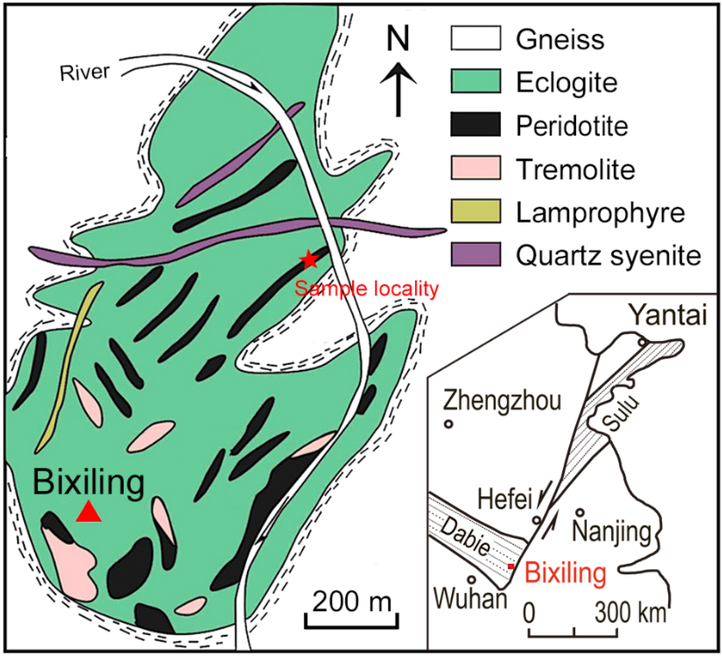
2. Geological Background and Sample Description

The garnet peridotite sample (No. B4-1-P) was collected from the Bixiling complex (Figure 1), which is the largest (~1.5 km2 in outcrop) coesite-bearing mafic-ultramafic body in the eastern part of Dabie ultrahigh-pressure (UHP) metamorphic belt of China [21]. The Bixiling complex occurs as a tectonic block enclosed within foliated quartzofeldspathic gneisses, which mainly consists of layered eclogites that contain many lenticular meta-ultramafic bodies such as peridotite, tremolite, lamprophyre, and some quartz syenite veins. The peridotite rocks can be subdivided into wehrlite and garnet peridotite, which belong to the typical crustal cumulative peridotite [22]. Previous geological, petrological and geochemical studies show that the mafic-ultramafic rocks of the Bixiling complex had equilibrated at peak P–T conditions of 4.7–6.5 GPa and 820–970 °C with an age of 230–210 Ma and experienced a fast cooling and rapid exhumation history [23,24,25]. Furthermore, the exsolution textures of rod-like titanium-chrome magnetite in olivine and the clinoenstatite lamellae in the diopside of garnet peridotite from this area were identified by Jin et al. [26] and Liu et al. [27], who indicated that the minimum subduction depth of their host rock is ~300 km (P > 9 GPa).
The meta-ultramafic rocks of the Bixiling complex are in gradational contact with the eclogite and exhibit greenish black layers as they are partially serpentinized. The studied peridotite sample is a medium grained (Figure 2), gray-black garnet peridotite consisting of serpentinized olivine (~65%), garnet (~15%), orthopyroxene (~10%), clinopyroxene (~10%), and some trace minerals (<1%). Most garnet grains are inequigranular (50–500 μm), which are smaller than that of olivine and pyroxene. Most coarse garnet grains are surrounded by the retrograde fine-grained garnet aggregate. The composition of garnet is characterized by lower grossular (2–8 mol%) and higher pyrope components (up to 65 mol%, Table 1) [21].

3. Methods

Two doubly polished thin sections of garnet peridotite sample were prepared for detailed petrology, chemistry and microstructural analyses. Crystallographic orientation data were acquired using a Quanta 450 Field Emission Gun (FEG)-SEM (FEI Company, Hillsboro, OR, USA) equipped with an HKL Nordlys EBSD Detector (Oxford Apparatus Company, Wycombe, UK) under the operating conditions of 20 kV accelerating voltage, 6 nA beam current, 20–25 mm working distance, 6 and 0.8 μm step size. Diffraction patterns were collected and indexed with a manual interactive mode using the Channel5 software (Oxford Apparatus Company, Wycombe, UK). Quantitative chemical analyses of the major element composition of minerals were obtained using a JEOL-733 EMPA (JEOL Company, Tokyo, Japan) under the operating conditions of 15 kV accelerating voltage, 20 nA beam current, 0.5 μm spot size, and 20 s/peak counting time. The EMPA standards include the following minerals: jadeite for Na, ilmenite for Ti and Fe, K-feldspar for K, wollastonite for Si and Ca, MgO for Mg, Al2O3 for Al, MnSiO3 for Mn and Cr2O3 for Cr. The above two experiments were carried out at the State Key Laboratory of Geological Processes and Mineral Resources, China University of Geosciences (GPMR-CUG-Wuhan). Five representative garnet grains were selected for TEM analysis. The TEM samples were processed by a Gatan 656 (Gatan Company, Pleasanton, CA, USA) dimple grinder, a Gatan-600 ion mill and a JEOL-JEE4X (JEOL Company, Tokyo, Japan) vacuum evaporator for physical thinning, ion thinning and carbon coating, respectively. Low-magnification observations on microstructures of garnet were carried out using a Philips CM12 TEM (Philips Company, Amsterdam, Netherlands) equipped with an EDAX PV9100 (EDAX Company, Mahwah, NJ, USA) X-ray energy dispersive spectroscopy (EDS) and a Gatan 830 (Gatan Company, Pleasanton, CA, USA) charge coupled device (CCD) camera system at GPMR-CUG-Wuhan. Selected area electron diffraction (SAED) patterns and bright field (BF) observations were carried out at 120 kV accelerating voltage. High-resolution TEM images and EDS-maps were recorded using a Talos F200S (Thermo Fisher Company, Waltham, MA, USA) scanning transmission electron microscope (STEM) under 200 kV accelerating voltage at Wuhan University of Technology.

4. Results

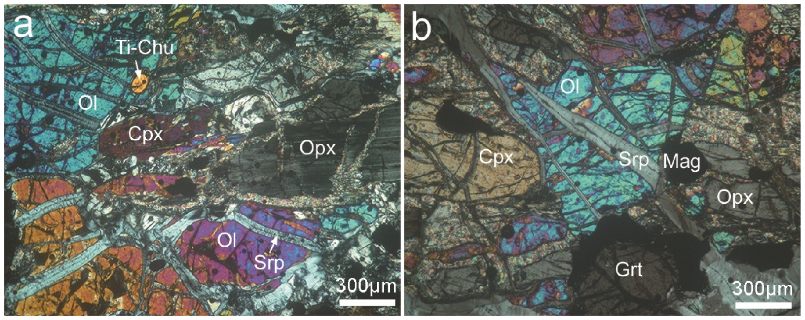
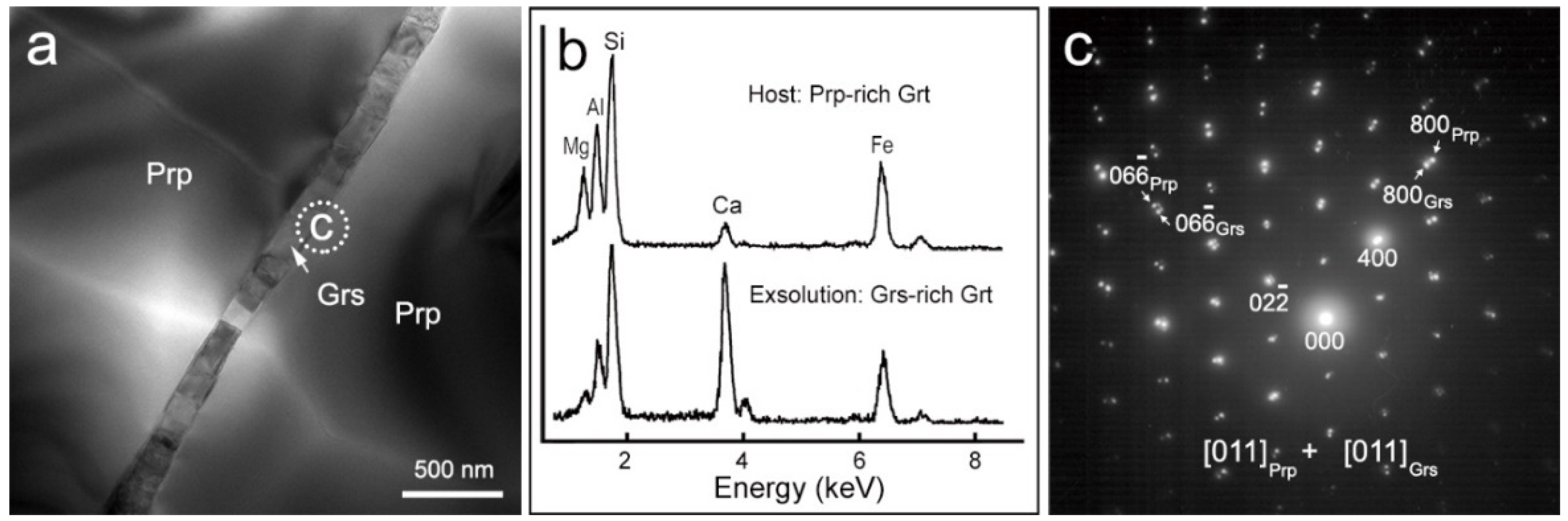
4.1. Microstructures

As shown in Figure 3a, the Prp-rich garnet grains in our sample are elongated slightly and containing numerous subgrains. The misorientation angles of almost all subgrains are less than 5° and exhibit an increase trend from the subdomain center toward subgrain margins. At the subgrain boundaries, numerous Grs-rich lamellae belts can be found. The detailed EBSD analysis (0.8 μm step size) is on the area of Figure 3b. The misorientation profiles show that the Grs-rich lamellae bear the same crystallographic orientation or exhibit small misorientation angles with their neighboring host subgrains (Figure 3c,d). These features may indicate that the Prp-rich host garnets had experienced a plastic deformation process before the Grs-rich lamellae exsolved.
After detailed TEM observation on the representative garnet grains, we have not found typical dislocation and subgrain boundary structures (e.g., dislocation arrays, networks, and walls). However, we have found numerous small lamellae belts exhibiting as shapes of dislocation or subgrain boundaries with weak shape preferred orientation (SPO) (Figure 4a and Figure 5a). The EDS spectra and EDS-maps show that these lamellae belts contain higher Ca and lower Mg, Al, Fe, and Si than that of their host grain (Figure 4b). The unit-cell parameters obtained from the SAED patterns (Figure 4c and Figure 5b) and HRTEM images (Figure 6) of these lamella and host grain agree well with the grossular and pyrope data, respectively [28,29].
The SAED patterns obtained from the areas including part of host and large-sized Grs-rich lamellae that precipitated at subgrain boundaries (Figure 4c) and dislocations (Figure 5b) show that their diffraction spots overlap each other with low indexes and separate when their index increased. This indicates that these Grs-rich lamellae bear the same crystallographic orientation as the Prp-rich host. The Grs-rich lamellae precipitated at dislocations showing almost the same image contrast imply that all of them bear the same crystallographic orientation with the host (Figure 5a). However, as shown in Figure 4a, some small-sized Grs-rich lamellae exhibit a different image contrast to the host and other lamellae, which suggests that these lamellae bear different orientations to the host.
The HRTEM analysis on the area of Figure 4a shows that most of the lamellae bear the same crystallographic orientation with the Prp-rich host except some small lamellae (Figure 6). The zone-axis of the host in Figure 6 calculated from its SAED pattern is [021], while the zone-axis of the exsolved lamella in Figure 6b inferred from its HRTEM image is [052]. The angle between these two zone-axes is about 4.7°, which is consistent with the EBSD analysis results (Figure 3c).

4.2. Chemical Compositions

The EDS-maps of major elements in exsolved Grs-rich lamellae and Prp-rich host, as shown in Figure 7, show that there is no chemical zonation both in the inner of exsolution lamellae and host grain. The anomaly color belt perpendicular to the exsolved lamellae belt in Figure 7 should be attributed to the heterogeneous thickness of the sample.
The chemical compositions of host garnet and large lamellae precipitated at the subgrain boundary are given in Table 1. The end-member proportions of garnet are calculated using the calculation scheme of Locock [30]. These data exhibit that our garnet is a solid solution of pyrope ± almandine ± spessartine + grossular + andradite + uvarovite. The host garnet is rich in pyrope and almandine components and yields a structural formula of ~Prp54Alm36Sps2Grs4Andr1Uvt3, the grossular proportion of which is lower than most of the values from Zhang et al. [21]. In contrast, the exsolution lamellae contain high grossular and andradite components and yield a structural formula of ~Prp1Grs71Andr25Uvt3. The large molar volume of grossular-rich garnet with low total FeO content would be the reason why the lamellae belt contains lower Al, Fe, and Si content than host grain, as shown in Figure 7.
The composition of precursor garnet has been calculated from the averaged chemical composition of host and exsolution in Table 1 in combination with the volume proportion of exsolved lamellae (~6.5%) in Figure 4a and Figure 5a and the densities (3.86 g/cm3 for host and 3.67 g/cm3 for exsolution) averaged from Anthony et al. [31]. This reconstruction method has been widely used in previous studies [32,33,34,35]. Consequently, the precursor garnet is a six-component solid solution and contains ~8 mol% of grossular component, which is close to the highest values reported by Zhang et al. [21], and yields a stoichiometric structural formula of ~Prp51Alm34Sps2Grs8Andr2Uvt3. Considering the exsolution’s volume proportion, this composition suggests our garnet is in chemical equilibrium before and after Grs-rich lamellae exsolved.

5. Discussion

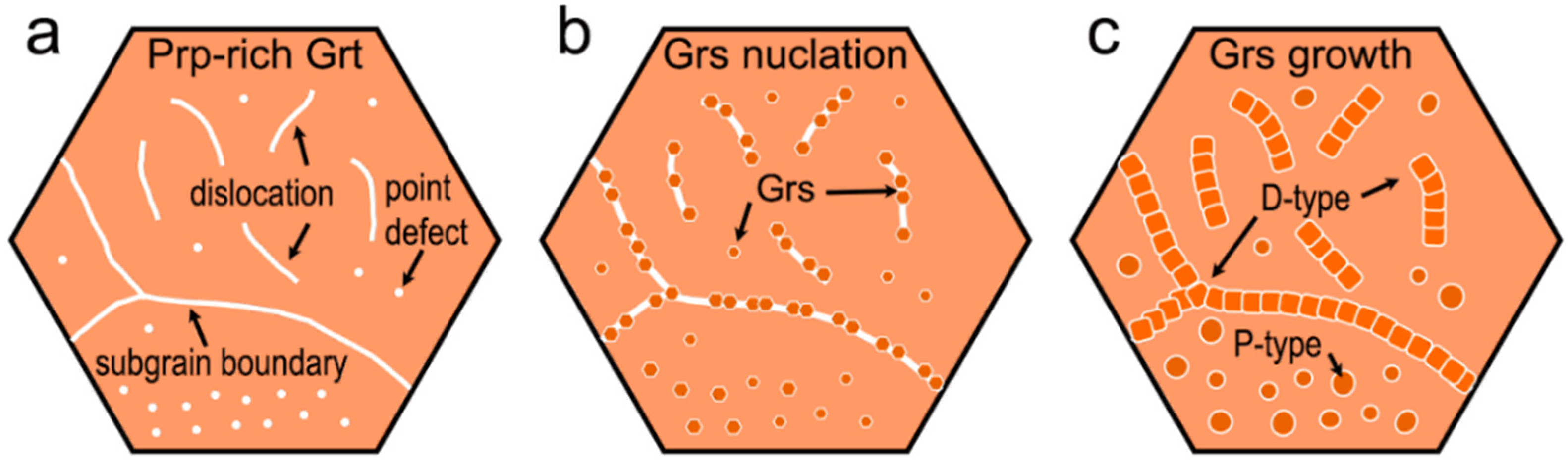
5.1. Exsolution Mechanism

Exsolution, or the unmixing of a mineral, means the transformation of an originally homogeneous solid solution that involving no less than two end-members under the subsolidus condition into a polyphase intergrowth due to the decrease in temperatures and/or pressures, which is a decomposition process in minerals caused by the decrease in Gibbs free energy and actualized by the diffusion of elements [36]. Since vacancies, dislocations, (sub)grain boundaries, inclusions, and other crystal defects are the highly strained regions, the exsolved precipitate would preferentially nucleate at these regions because that can simultaneously help reduce interfacial free energy and release the excess free energy of the defects [37,38,39,40].
The free energy needed to nucleate an exsolved precipitate phase could be expressed as: Δ G = Δ G volume + Δ G surface + Δ G strain , where Δ G volume is the chemical free energy change, Δ G surface is the interfacial free energy between the exsolution and host grain, and Δ G strain is the strain energy change [37,41,42]. The Δ G volume will be negative and act to favor nucleation. While the Δ G surface will be positive and prevent nucleation, it will be smaller for exsolved lamellae whose lattices match up across the interface (coherent precipitates) and larger for those that lack a coherent interface (incoherent precipitates). The Δ G strain term could be negative and favor the nucleation of precipitates or be positive and tend to oppose nucleation when significant distortion is required to grow a new crystal in a specific volume of space [42,43]. The exsolved lamellae nucleate and precipitate at the crystal defect sites have been found in natural olivine [44], calcite [45], and garnet [42,46,47]. The grossular lamellae precipitated at the dislocations and subgrain boundaries of the pyrope matrix in our sample (Figure 3, Figure 4 and Figure 5) should be attributed to this exsolution phenomenon.
The differences of molar volume between the six end members (~Prp51Alm34Sps2Grs8 Andr2Uvt3) in our Prp-rich precursor garnet are listed in Table 2, which are controlled by the size of divalent ion in dodecahedral site of crystal structure [13]. The high proportion of grossular, andradite, and uvarovite in exsolution (~Prp1Grs71Andr25Uvt3) should be ascribed to the large molar volume differences (>1.006 J/bar) between these components with other main end members, which would cause these components to become unstable and be exsolved from the Prp-rich matrix when the temperatures and/or pressures decreased during the exhumation of subducted slab. Similarly, the absence of almandine and spessartine in the exsolutions should be attributed to the small differences in molar volume (<0.48 J/bar) between them and pyrope-rich host.
The process of Grs-rich lamellae heterogeneously nucleating and growing at crystal defects of Prp-rich garnet is illustrated in Figure 8. Compared with the typical rod/needle-like exsolution of rutile ± apatite ± clinopyroxene ± orthopyroxene ± ilmenite in garnet [35,42,46,48], the Grs-rich exsolved lamellae show a weak SPO and occur as dislocation or subgrain boundary shapes (D-type) in this study and spherical grains, which would precipitate at point defect (P-type) with incoherently (randomly) crystallographic orientation to the matrix as reported by Cressey [19]. The spherical Grs-rich pyrope–almandine garnet inclusions in Prp-rich garnet reported by Wang et al. [20] should also be regarded as this P-type exsolution, although we do not know their topotaxy. These features should be attributed to both the exsolution and host, are cubic crystal lattices and have nearly the same unit-cell parameters, which would favor the weak SPO precipitates crystallized (Figure 3, Figure 4 and Figure 5) at crystal defects with the effects of Δ G volume , and defect-related Δ G strain outweigh those of Δ G surface . Based on our data, we have confirmed that the exsolved lamellae, especially those precipitated at the dislocations (Figure 5a), bear the same crystallographic orientation or exhibit a small misorientation angle (≤5°) with their neighboring host subgrains (Figure 3c and Figure 6b). That means that the crystallographic orientation of Grs-rich exsolution is controlled by, and coherent with, the Prp-rich host. These topotaxial relations and the shape features of exsolutions are different with that reported by Cressey [19], which might be caused by the different precipitated sites of these exsolutions (Figure 8). Theoretically, the incoherently (randomly) orientated precipitates exsolved from the matrix, as reported by Cressey [19], may not be the ideal exsolution phenomenon, which not only needs a certain topological relation but also needs a certain d-spacing relation between the exsolutions and host grain.

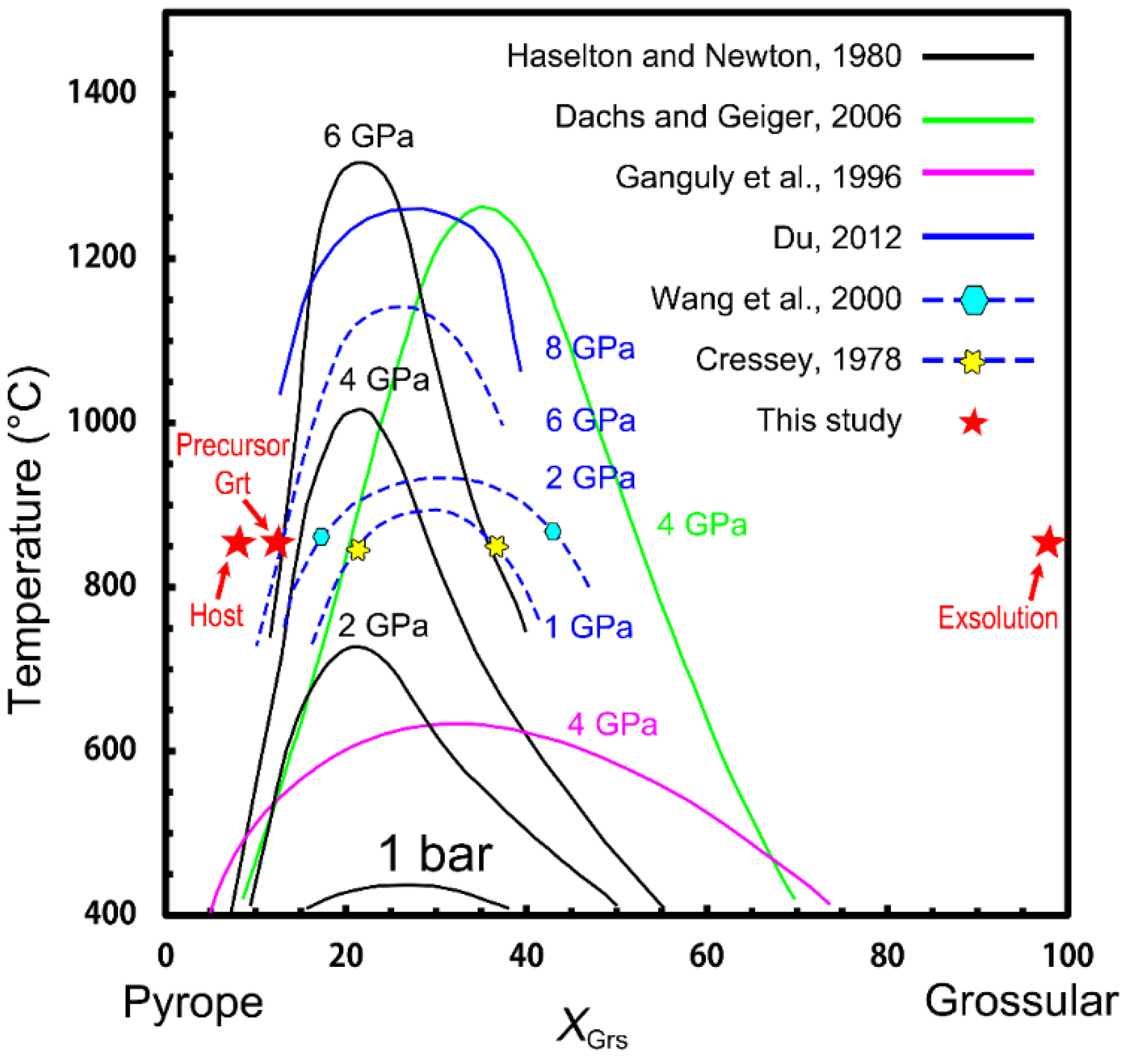
5.2. Constraints on the P–T Conditions

Based on different thermodynamic models [16,17,18] and high pressure and temperature experiment results [8], although the results from various studies do not agree well with each other, it can be concluded that the solvus of pyrope–grossular solid solution is controlled by a combination of composition, temperature and pressure (Figure 9). For example, the critical temperature (peak of the solvus) calculated by Ganguly et al. [17] is ~640 °C at 4 GPa, which about 400 and 600 °C lower than those calculated by Haselton and Newton [18] and Dachs and Geiger [16], respectively. Du [8], using the multi-anvil technique, studied the unmixing phenomenon in pyrope–grossular garnet solid solution and found the critical temperature of its solvus to be less than 1200 °C at 6 GPa, which is lower than that predicted by Haselton and Newton [18]. At 8 GPa, long-term heating experiments for both convergence and divergence showed that two garnet phases with composition around ~Prp82Gr18 and ~Prp62Gr38 were equilibrated with each other at 1200 °C, which indicating the positions of the pyrope–grossular garnet solvus’ two limbs at 1200 °C and 8 GPa. These disagreements should be attributed to the different pressure enhancement factors and thermodynamic parameters used in previous thermodynamic models [8]. The thermodynamic model established by Du [8] can explain the natural garnet exsolution found by Cressey [19] in metagabbro, which was metamorphosed in the P–T conditions of 1.0–1.3 GPa and 800–860 °C at South Harris, and by Wang et al. [20] in ultramafic diatreme at Garnet Ridge, Arizona, corresponding to the P–T conditions of 2 GPa and 600–650 °C. The later temperatures, lower than that predicted by Du [8], should be attributed to the existence of the almandine component both in host and exsolution [17].
In this study, it is hard to infer the minimum suffered P–T values of precursor garnet based on the compositions of hosts and exsolutions using previous thermodynamic models because we do not know at what temperatures the Grs-rich lamellae were exsolved from their host. However, the pyrope-rich host grains were elongated and contained numerous subgrains and Grs-rich lamellae precipitated at their dislocations and subgrain boundaries (Figure 3, Figure 4, Figure 5 and Figure 6), indicating that the precursor garnet had experienced a plastic deformation process before its decomposition. Due to the large Burgers vectors, the temperature for the brittle–ductile transition of pyrope–almandine garnet is above 850 °C (T > 0.65 Tm) in nature [49,50], which approximates to the lower limit of the peak metamorphic temperatures of 820–970 °C recorded in Bixiling mafic-ultramafic rocks [23,24]. Therefore, we infer that the minimum temperature of our precursor garnet had suffered before the Grs-rich lamellae exsolved would be higher than its brittle–ductile transition temperature (850 °C).
The EMPA data and reconstruction calculation result shows that the composition of grossular exsolution, pyrope host, and precursor garnet is a four- or six-component solid solution (Table 1). To compare with previous studies, these multi-end-member solid solutions should be simplified to the pyrope–grossular binary format [8]. Based on the size of divalent Fe2+, Mg2+, and Mn2+ being quite distinct from that of Ca2+ in dodecahedral site and the difference in molar volume between different end-members [13], we project the composition of exsolution, host, and their precursor phase to the pyrope–grossular binary format as ~Prp1(GrsAndrUvt)99, ~(PyAlmSps)92(GrsAndrUvt)8, and ~(PrpAlmSps)87 (GrsAndrUvt)13. After plotting these composition data and their minimum suffered temperature (850 °C) in the phase diagram of pyrope–grossular binary versus pressure–temperature condition (Figure 9), it can be found that the Grs-rich exsolution is a stable phase with a wide P–T stability domain, while both the equilibrium pressures of host and precursor garnet are more than 6 GPa at a temperature of 850 °C based on the model of Haselton and Newton [18], which is also above the extension line of 6 GPa from the model of Du [8]. Previous studies suggested that the equilibrium P–T condition of the reconstructed composition of precursor phase from its corresponding host-exsolution pair can be used to estimate its origin depth [32,33,34,35]. Consequently, the estimated P–T condition of >6 GPa and >850 °C would represent the minimum values that of precursor garnet suffered, which correspond to the first exhumation stage of subducted slab. Under this condition, the dislocation mobility of precursor Prp-rich garnets would be very low to favor the Grs-rich lamellae beginning to nucleate and grow at their dislocations and subgrain boundaries, because the kinetics of the exsolution process is extremely slow at natural modest pressures and lower temperatures [8]. Therefore, we infer that the origin depth of Bixiling garnet peridotite recorded by the grossular exsolution in pyrope would be more than 200 km (>6 GPa), which is consistent with the conclusions of previous studies [26,27] and should be regarded as new evidence for the ultradeep origin of Dabie orogenic crustal cumulative peridotite.

6. Conclusions

This is the first systematic structural and chemical study on the grossular-rich lamellae precipitated at the dislocations and subgrain boundaries of pyrope-rich host from the Bixiling garnet peridotite, which should be regarded as a new natural sample of the immiscibility phenomenon in pyrope–almandine–grossular solid solution. We have confirmed that the crystallographic orientation is controlled by, and coherent with, the host grain. The morphological features of exsolution lamellae exhibiting weak SPO presented here and in previous studies should be attributed to the same cubic crystal lattice. The structural features and compositions of our precursor pyrope-rich garnet correspond to a P–T condition of >6 GPa and >850 °C based on the previous thermodynamic models, which indicate that the origin depth of precursor pyrope-rich garnet would be more than 200 km.

Author Contributions

Z.X. and X.L. (Xiangwen Liu) performed sample preparation and EBSD, EMPA and TEM data collection with the help of X.L. (Xiaoqing Liu) on HRTEM analysis; Z.X. wrote the manuscript with the help of co-authors; Z.J. contributed to the interpretation of data and reviewed the manuscript. All authors have read and agreed to the published version of the manuscript.

Funding

This research was funded by the National Natural Science Foundation of China (Nos. 41272224 and 41972231) and the Natural Science Foundation of Hebei Province (No. D2021204001).

Institutional Review Board Statement

Not applicable.

Informed Consent Statement

Not applicable.

Data Availability Statement

The data presented in this study are available within the article.

Acknowledgments

The authors would like to thank Honglin Fu and Dapeng Wen for the EBSD measurement and the data analysis. We thank the two anonymous reviewers for their fruitful comments.

Conflicts of Interest

The authors declare no conflict of interest.

References

  1. Baxter, E.F.; Caddick, M.J.; Ague, J.J. Garnet: Common mineral, uncommonly useful. Elements 2013, 9, 415–419. [Google Scholar] [CrossRef]
  2. Goncalves, P.; Raimondo, T.; Paquette, J.L.; De Oliveira, J.S.D.S. Garnet as a monitor for melt–rock interaction: Textural, mineralogical, and compositional evidence of partial melting and melt-driven metasomatism. J. Metamorph. Geol. 2021, 39, 617–648. [Google Scholar] [CrossRef]
  3. Wu, J.; Zhai, M.; Hu, B.; Zhang, H.; Chu, X.; Zhou, N.; Zhang, L.; Zhang, H. Garnet perspectives on the metamorphic history and tectonic significance of Paleoproterozoic high-pressure mafic granulites from the northern Hengshan, North China Craton. Lithos 2021, 394–395, 106139. [Google Scholar] [CrossRef]
  4. Baxter, E.F.; Scherer, E.E. Garnet geochronology: Timekeeper of tectonometamorphic processes. Elements 2013, 9, 433–438. [Google Scholar] [CrossRef]
  5. Baxter, E.F.; Caddick, M.J.; Dragovic, B. Garnet: A rock-forming mineral petrochronometer. Rev. Miner. Geochem. 2017, 83, 469–533. [Google Scholar] [CrossRef] [Green Version]
  6. Beyer, C.; Rosenthal, A.; Myhill, R.; Crichton, W.A.; Yu, T.; Wang, Y.; Frost, D.J. An internally consistent pressure calibration of geobarometers applicable to the Earth’s upper mantle using in situ XRD. Geochim. Cosmochim. Acta 2018, 222, 421–435. [Google Scholar] [CrossRef] [Green Version]
  7. Caddick, M.J.; Kohn, M.J. Garnet: Witness to the evolution of destructive plate boundaries. Elements 2013, 9, 427–432. [Google Scholar] [CrossRef] [Green Version]
  8. Du, W. Excess Volume and Exsolution in Pyrope-Grossular Garnet; Columbia University: New York, NY, USA, 2012. [Google Scholar]
  9. Berman, R.G. Mixing properties of Ca-Mg-Fe-Mn garnets. Am. Miner. 1990, 75, 328–344. [Google Scholar]
  10. Dapiaggi, M.; Geiger, C.; Artioli, G. Microscopic strain in synthetic pyrope-grossular solid solutions determined by synchrotron X-ray powder diffraction at 5 K: The relationship to enthalpy of mixing behavior. Am. Miner. 2005, 90, 506–509. [Google Scholar] [CrossRef]
  11. Geiger, C.A. Thermodynamics of (Fe2+, Mn2+, Mg, Ca)3- Al2Si3O12 garnet: A review and analysis. Miner. Pet. 1999, 66, 271–299. [Google Scholar] [CrossRef]
  12. Geiger, C.A. Silicate garnet: A micro to macroscopic (re)view. Am. Miner. 2008, 93, 360–372. [Google Scholar] [CrossRef]
  13. Geiger, C.A. A tale of two garnets: The role of solid solution in the development toward a modern mineralogy. Am. Miner. 2016, 101, 1735–1749. [Google Scholar] [CrossRef]
  14. Wood, B.J. Activity measurements and excess entropy-volume relationships for pyrope-grossular garnets. J. Geol. 1988, 96, 721–729. [Google Scholar] [CrossRef]
  15. Newton, R.C.; Charlu, T.V.; Kleppa, O.J. Thermochemistry of high pressure garnets and clinopyroxenes in the system CaO-MgO-Al2O3-SiO2. Geochim. Cosmochim. Acta 1977, 41, 369–377. [Google Scholar] [CrossRef]
  16. Dachs, E.; Geiger, C.A. Heat capacities and entropies of mixing of pyrope-grossular (Mg3Al2Si3O12-Ca3Al2Si3O12) garnet solid solutions: A low-temperature calorimetric and a thermodynamic investigation. Am. Miner. 2006, 91, 894–906. [Google Scholar] [CrossRef]
  17. Ganguly, J.; Cheng, W.; Tirone, M. Thermodynamics of aluminosilicate garnet solid solution: New experimental data, an optimized model, and thermometric applications. Contrib. Miner. Pet. 1996, 126, 137–151. [Google Scholar] [CrossRef]
  18. Haselton, H.T.; Newton, R.C. Thermodynamics of pyrope-grossular garnets and their stabilities at high temperatures and high pressures. J. Geophys. Res. Solid Earth 1980, 85, 6973–6982. [Google Scholar] [CrossRef]
  19. Cressey, G. Exsolution in almandine-pyrope-grossular garnet. Nature 1978, 271, 533–534. [Google Scholar] [CrossRef]
  20. Wang, L.; Essene, E.J.; Zhang, Y. Direct observation of immiscibility in pyrope-almandine-grossular garnet. Am. Miner. 2000, 85, 41–46. [Google Scholar] [CrossRef]
  21. Zhang, R.Y.; Liou, J.G.; Cong, B.L. Talc-, magnesite- and Ti-clinohumite-bearing ultrahigh-pressure meta-mafic and ultramafic complex in the Dabie mountains, China. J. Pet. 1995, 36, 1011–1037. [Google Scholar] [CrossRef]
  22. Chen, Y.; Su, B.; Guo, S. The Dabie-Sulu orogenic peridotites: Progress and key issues. Sci. China Earth Sci. 2015, 58, 1679–1699. [Google Scholar] [CrossRef]
  23. Zhang, R.Y.; Liou, J.G.; Yang, J.S.; Yui, T.F. Petrochemical constraints for dual origin of garnet peridotites from the Dabie-Sulu UHP terrane, eastern-central China. J. Metamorph. Geol. 2000, 18, 149–166. [Google Scholar] [CrossRef]
  24. Zheng, J.P.; Sun, M.; Griffin, W.L.; Zhou, M.F.; Zhao, G.C.; Robinson, P.; Tang, H.Y.; Zhang, Z.H. Age and geochemistry of contrasting peridotite types in the Dabie UHP belt, eastern China: Petrogenetic and geodynamic implications. Chem. Geol. 2008, 247, 282–304. [Google Scholar] [CrossRef]
  25. Chavagnac, V.; Jahn, B. Coesite-bearing eclogites from the Bixiling Complex, Dabie Mountains, China: Sm-Nd ages, geochemical characteristics and tectonic implications. Chem. Geol. 1996, 133, 29–51. [Google Scholar] [CrossRef]
  26. Jin, Z.M.; Jin, S.Y.; Gao, S.; Zhao, W.X. Is the depth where UHP rocks formed in the Dabie Mountains limited to 100–150 km?―Implications from the needle-shaped Ti-Cr-bearing magnetite exsolution in olivine (in Chinese). Chin. Sci. Bull. 1998, 43, 767–771. [Google Scholar]
  27. Liu, X.; Jin, Z.; Green, H.W. Clinoenstatite exsolution in diopsidic augite of Dabieshan: Garnet peridotite from depth of 300 km. Am. Miner. 2007, 92, 546–552. [Google Scholar] [CrossRef]
  28. Ganguly, J.; Cheng, W.; O’Neill, H.S.C. Syntheses, volume, and structural changes of garnets in the pyrope-grossular join: Implications for stability and mixing properties. Am. Miner. 1993, 78, 583–593. [Google Scholar]
  29. Novak, G.A.; Gibbs, G.V. The crystal chemistry of the silicate garnets. Am. Miner. 1971, 56, 791–825. [Google Scholar]
  30. Locock, A.J. An Excel spreadsheet to recast analyses of garnet into end-member components, and a synopsis of the crystal chemistry of natural silicate garnets. Comput. Geosci. UK 2008, 34, 1769–1780. [Google Scholar] [CrossRef]
  31. Anthony, J.W.; Bideaux, R.A.; Bladh, K.W.; Nichols, M.C. Handbook of Mineralogy; Mineralogical Society of America: Chantilly, VA, USA, 2003. [Google Scholar]
  32. Dobrzhinetskaya, L.; Green, H.W.; Wang, S. Alpe Arami: A peridotite massif from depths of more than 300 km. Science 1996, 271, 1841–1845. [Google Scholar] [CrossRef]
  33. Haggerty, S.E.; Sautter, V. Ultradeep (greater than 300 km), ultramafic upper mantle xenoliths. Science 1990, 248, 993–996. [Google Scholar] [CrossRef] [PubMed]
  34. van Roermund, H.L.M.; Drury, M.R.; Barnhoorn, A.; De Ronde, A. Relict majoritic garnet microstructures from Ultra-deep orogenic peridotites in Western Norway. J. Pet. 2001, 42, 117–130. [Google Scholar] [CrossRef] [Green Version]
  35. Ye, K.; Cong, B.L.; Ye, D.I. The possible subduction of continental material to depths greater than 200 km. Nature 2000, 407, 734–736. [Google Scholar] [CrossRef] [PubMed]
  36. Liu, L.; Yang, J.X.; Zhang, J.F.; Chen, D.L.; Wang, C.; Yang, W.Q. Exsolution microstructures in ultrahigh-pressure rocks: Progress, controversies and challenges. Chin. Sci. Bull. 2009, 54, 1983–1995. [Google Scholar] [CrossRef] [Green Version]
  37. Cahn, J.W. Nucleation on dislocations. Acta Metall. 1957, 5, 169–172. [Google Scholar] [CrossRef]
  38. Gómez-Ramírez, R.; Pound, G.M. Nucleation of a second solid phase along dislocations. Metall. Mater. Trans. B 1973, 4, 1563–1570. [Google Scholar] [CrossRef]
  39. Hilliard, J.E. Nucleation within crystalline phases. Ind. Eng. Chem. 1966, 58, 18–25. [Google Scholar] [CrossRef]
  40. Militzer, M.; Sun, W.P.; Jonas, J.J. Modelling the effect of deformation-induced vacancies on segregation and precipitation. Acta Metall. Mater. 1994, 42, 133–141. [Google Scholar] [CrossRef]
  41. Lee, J.K.; Barnett, D.M.; Aaronson, H.I. The elastic strain energy of coherent ellipsoidal precipitates in anisotropic crystalline solids. Metall. Trans. A 1977, 8, 963–970. [Google Scholar] [CrossRef]
  42. Axler, J.A.; Ague, J.J. Exsolution of rutile or apatite precipitates surrounding ruptured inclusions in garnet from UHT and UHP rocks. J. Metamorph. Geol. 2015, 33, 829–848. [Google Scholar] [CrossRef]
  43. Ham, F.S. Stress-assisted precipitation on dislocations. J. Appl. Phys. 1959, 30, 915–926. [Google Scholar] [CrossRef]
  44. Kohlstedt, D.L.; Vander Sande, J.B. An electron microscopy study of naturally occurring oxidation produced precipitates in iron-bearing olivines. Contrib. Miner. Petr. 1975, 53, 13–24. [Google Scholar] [CrossRef]
  45. Snow, E.; Yund, R. The effect of ductile deformation on the kinetics and mechanisms of the aragonite-calcite transformation. J. Metamorph. Geol. 1987, 5, 141–153. [Google Scholar] [CrossRef]
  46. Axler, J.A.; Ague, J.J. Oriented multiphase needles in garnet from ultrahigh-temperature granulites, Connecticut, U.S.A. Am. Miner. 2015, 100, 2254–2271. [Google Scholar] [CrossRef]
  47. van Roermund, H.L.M.; Drury, M.R.; Barnhoorn, A.; De Ronde, A. Non-silicate inclusions in garnet from an ultra-deep orogenic peridotite. Geol. J. 2000, 35, 209–229. [Google Scholar] [CrossRef]
  48. van Roermund, H.; Drury, M.R. Ultra-high pressure (P > 6 GPa) garnet peridotites in Western Norway: Exhumation of mantle rocks from > 185 km depth. Terra Nova 1998, 10, 295–301. [Google Scholar] [CrossRef]
  49. Wang, Z.; Ji, S. Deformation of silicate garnets: Brittle-ductile transition and its geological implications. Can. Miner. 1999, 37, 525–541. [Google Scholar]
  50. Karato, S.; Wang, Z.; Liu, B.; Fujino, K. Plastic deformation of garnets: Systematics and implications for the rheology of the mantle transition zone. Earth Planet. Sci. Lett. 1995, 130, 13–30. [Google Scholar] [CrossRef]
Figure 1. Geological map of the Bixiling complex, eastern Dabie orogenic belt, China.
Crystals 12 00647 g001
Figure 2. (a,b) Optical microphotographs of the garnet peridotite sample under cross polarized light mode. Abbreviations: Ol, olivine; Cpx, clinopyroxene; Opx, orthopyroxene; Grt, garnet; Srp, serpentine; Mag, magnetite; Ti-Chu, Ti-clinohumite.
Crystals 12 00647 g002
Figure 3. EBSD images of Grs-rich exsolutions precipitated at the subgrain boundaries of Prp-rich host. (a) Texture component map shows the elongated pyrope-rich host grain containing numerous subgrains with small misorientation angles (≤5°). (b,c) Detailed texture component map and its corresponding BSE image of the outlined area in (a) show the Grs-rich exsolutions precipitated at the subgrain boundaries of Prp-rich host. (d) Misorientation profiles of the traverses marked by red lines in (b,c) show an increase in misorientation angle from the subdomain center toward subgrain margins and small misorientation angles (<3°) between the exsolutions and host.
Crystals 12 00647 g003
Figure 4. TEM images of Grs-rich exsolutions precipitated at the subgrain boundary of Prp-rich host. (a) Exsolution precipitated at the subgrain boundary of host. (b) EDS spectra of the exsolved lamellae and host in (a) show that they are grossular-rich (Grs) and pyrope-rich (Prp) garnet, respectively. (c) SAED pattern obtained from the areas marked in (a) shows this Grs-rich lamella bearing the same crystallographic orientation with Prp-rich host.
Crystals 12 00647 g004
Figure 5. TEM images of Grs-rich exsolutions precipitated at the dislocations of Prp-rich host. (a) Grs-rich exsolutions precipitated at the dislocations of Prp-rich host. (b) SAED pattern obtained from the area marked in (a) shows the exsolution bearing the same crystallographic orientation with host.
Crystals 12 00647 g005
Figure 6. HRTEM images of Grs-rich exsolutions in Prp-rich host show they are bearing both (a) coherently and (b) incoherently crystallographic orientation with host.
Crystals 12 00647 g006
Figure 7. EDS-maps of major elements in grossular exsolutions and pyrope host. (a) Bright field image of grossular lamellae precipitated at the subgrain boundary of host pyrope. (bf) Major elements distribution maps of the area in (a).
Crystals 12 00647 g007
Figure 8. Schematic diagram of Grs-rich lamellae precipitated at the crystal defects of Prp-rich garnet. (a) Point defect and dislocation as well as its substructure (subgrain boundary) in Prp-rich garnet. (b,c) Grs-rich lamellae heterogeneously nucleate and grow at point defects (P-type, shown as Cressey [19] and Wang et al. [20]) and dislocations or subgrain boundaries (D-type, this study).
Crystals 12 00647 g008
Figure 9. Phase diagram of pyrope–grossular binary versus pressure–temperature condition based on the different thermodynamic models (modified after Du [8]).
Crystals 12 00647 g009
Table 1. Chemical composition of garnet in garnet peridotite.
SpotHostExsolutionPrecursor #85 *122C *
Prp-1Prp-2GrsPrpg-105g-113g-C-50g-R-51g-60g-66
SiO241.1540.8337.5640.3942.1241.8540.4140.3540.4740.71
TiO20.000.000.000.000.040.000.010.030.050.05
Al2O322.1721.8816.2421.6223.2323.4721.1719.7122.2421.95
FeO18.2317.157.7917.2513.5714.1717.5317.8416.4815.87
MnO0.750.680.420.900.280.270.690.780.830.95
MgO14.8915.610.3013.8318.3718.0314.7013.7214.8215.03
CaO3.062.6135.684.943.453.643.694.504.403.78
Cr2O31.020.640.891.070.120.051.822.780.820.91
Na2O0.000.000.080.000.000.000.000.010.000.00
Total101.2799.4098.96100.00101.18101.48100.0299.72100.1499.25
XPrp53.0254.550.7450.9964.9363.7654.1547.6654.2254.86
XAlm36.6434.990.0033.9325.6525.7234.5135.9932.8132.81
XSps1.551.420.001.890.560.541.441.651.731.99
XGrs4.284.4571.287.586.387.322.151.817.636.27
XAndr0.760.5825.172.381.841.792.251.961.420.00
XUvt2.951.872.813.140.340.145.338.242.392.67
#: Chemical composition before Grs-rich lamellae exsolved using reconstruction calculation method. *: Data from Zhang et al. [21].
Table 2. Difference in molar volume between major end-members in garnet.
BinaryDifference in Molar Volume (J/bar)
andradite-pyrope *1.918
uvarovite-pyrope *1.760
andradite-almandine *1.710
uvarovite-almandine1.553
andradite-spessartine1.437
uvarovite-spessartine1.280
grossular-pyrope1.214
grossular-almandine1.006
grossular-spessartine0.733
andradite-grossular0.704
uvarovite-grossular0.547
spessartine-pyrope0.480
spessartine-almandine0.273
almandine-pyrope0.207
andradite-uvarovite0.157
*: Binaries have not been found in nature or synthesized in the laboratory. Data from Geiger [13].
Publisher’s Note: MDPI stays neutral with regard to jurisdictional claims in published maps and institutional affiliations.

