
Cyclohexylammonium Hexaisothiocyanatonickelate(II) Dihydrate as a Single-Source Precursor for High Surface Area Nickel Oxide and Sulfide Nanocrystals

 , , ,
, , ,
Abstract
1. Introduction
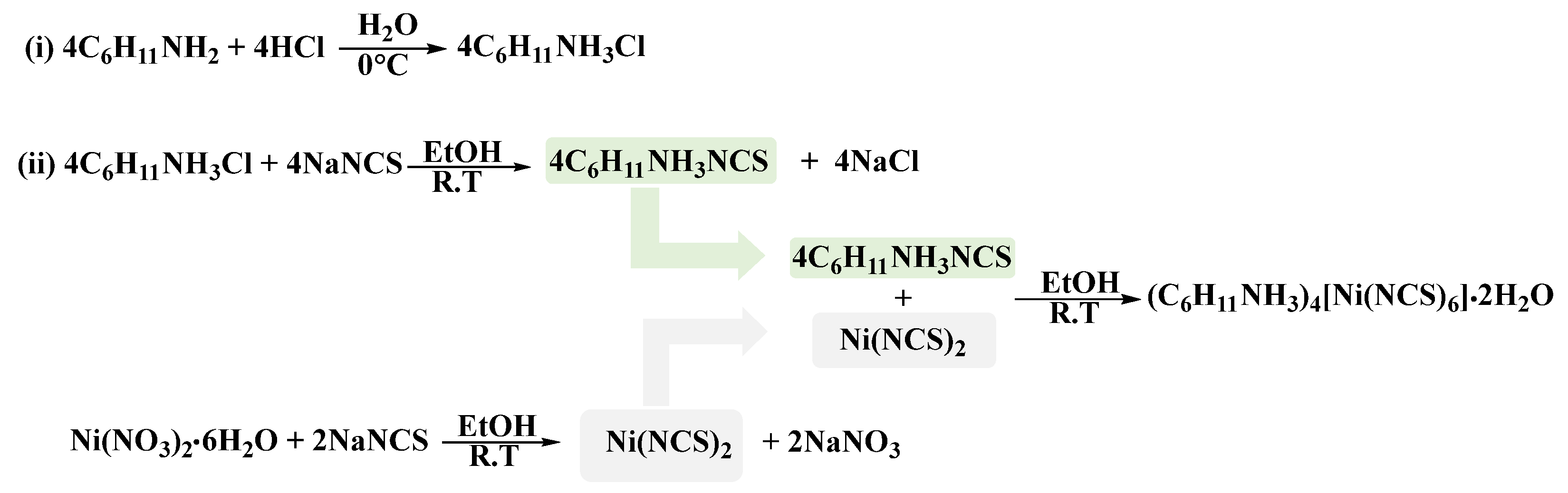
2. Material and Methods
2.1. Elemental Microanalysis
2.2. Fourier Transform Infrared (FTIR) Spectrophotometry Analysis
2.3. UV-Visible-Near Infrared Absorbance Spectrophotometry (UV-Vis-NIR)
2.4. Nuclear Magnetic Resonance (NMR) Spectroscopic Analyses
2.5. Single Crystal X-ray Diffraction Measurements
2.6. Thermal Gravimetric Analysis (TGA)
3. Results and Discussion
3.1. Elemental Analysis
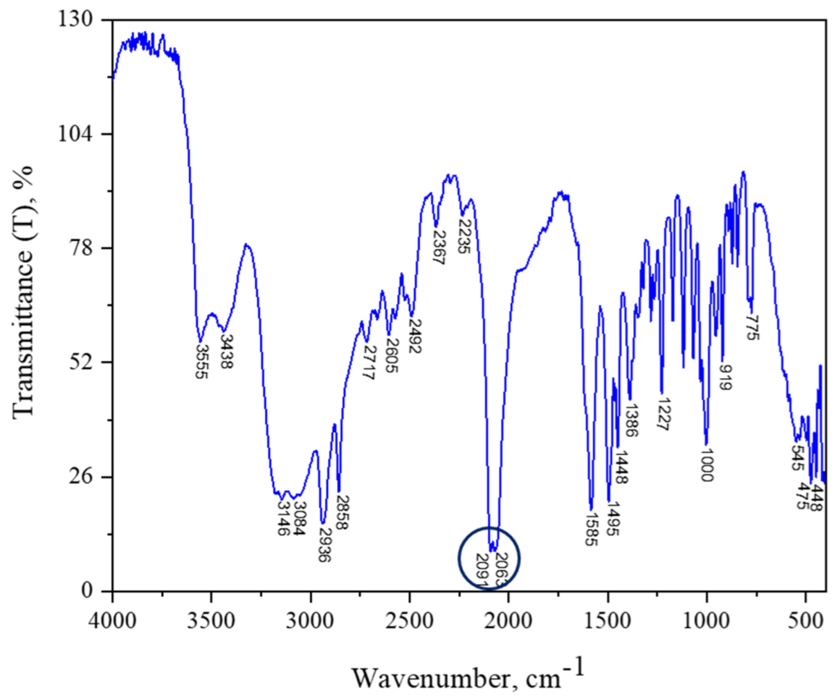
3.2. FTIR Analysis
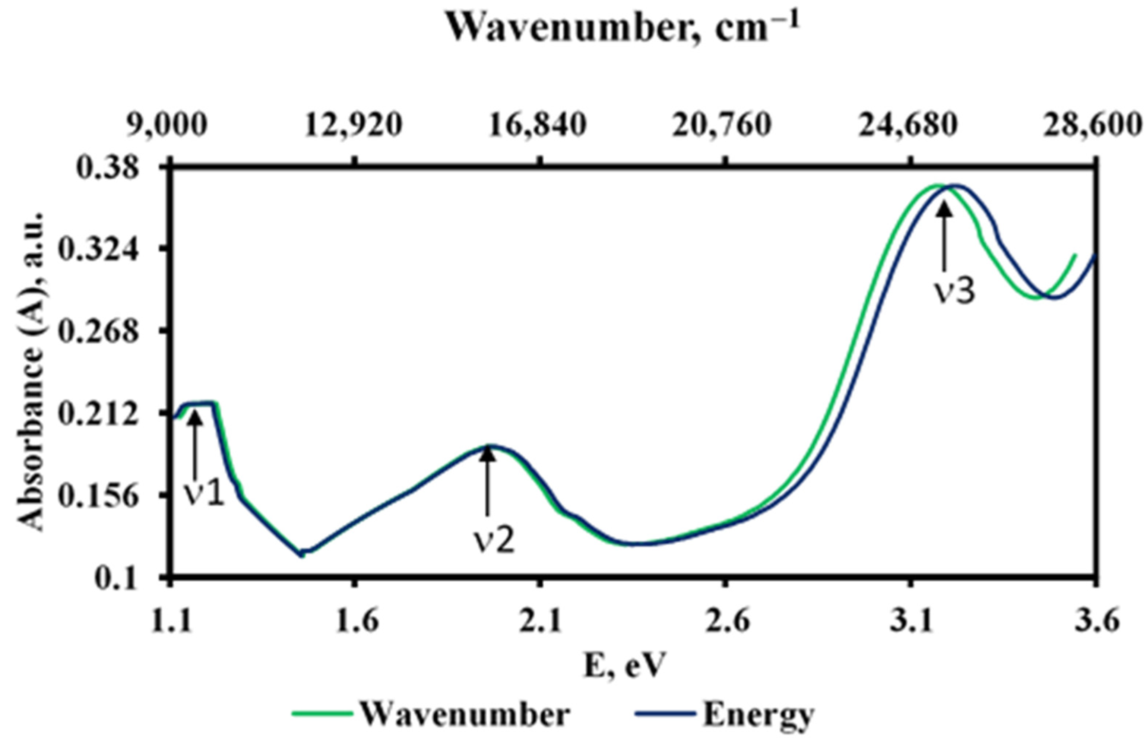
3.3. UV-Vis-NIR Analysis
3.4. NMR Spectra Analyses
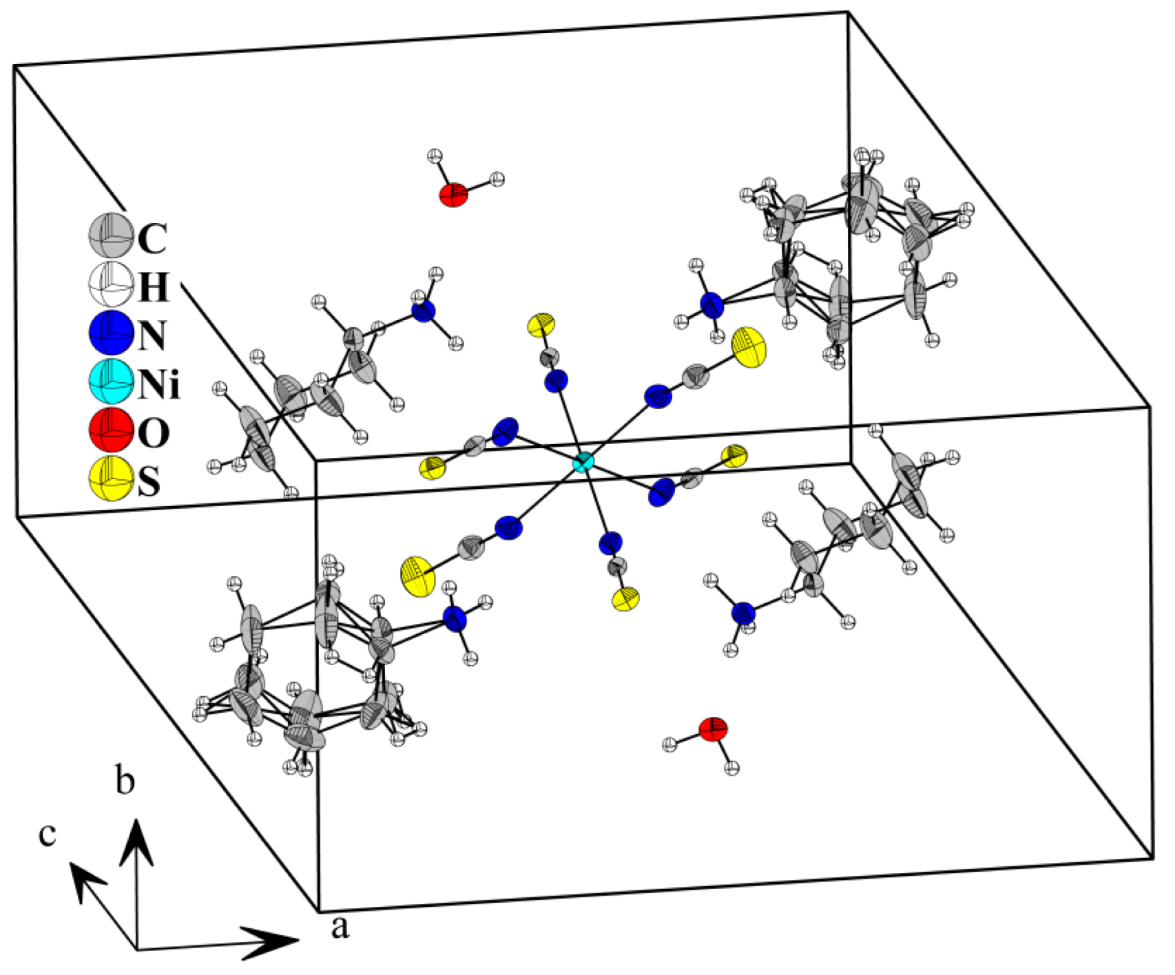
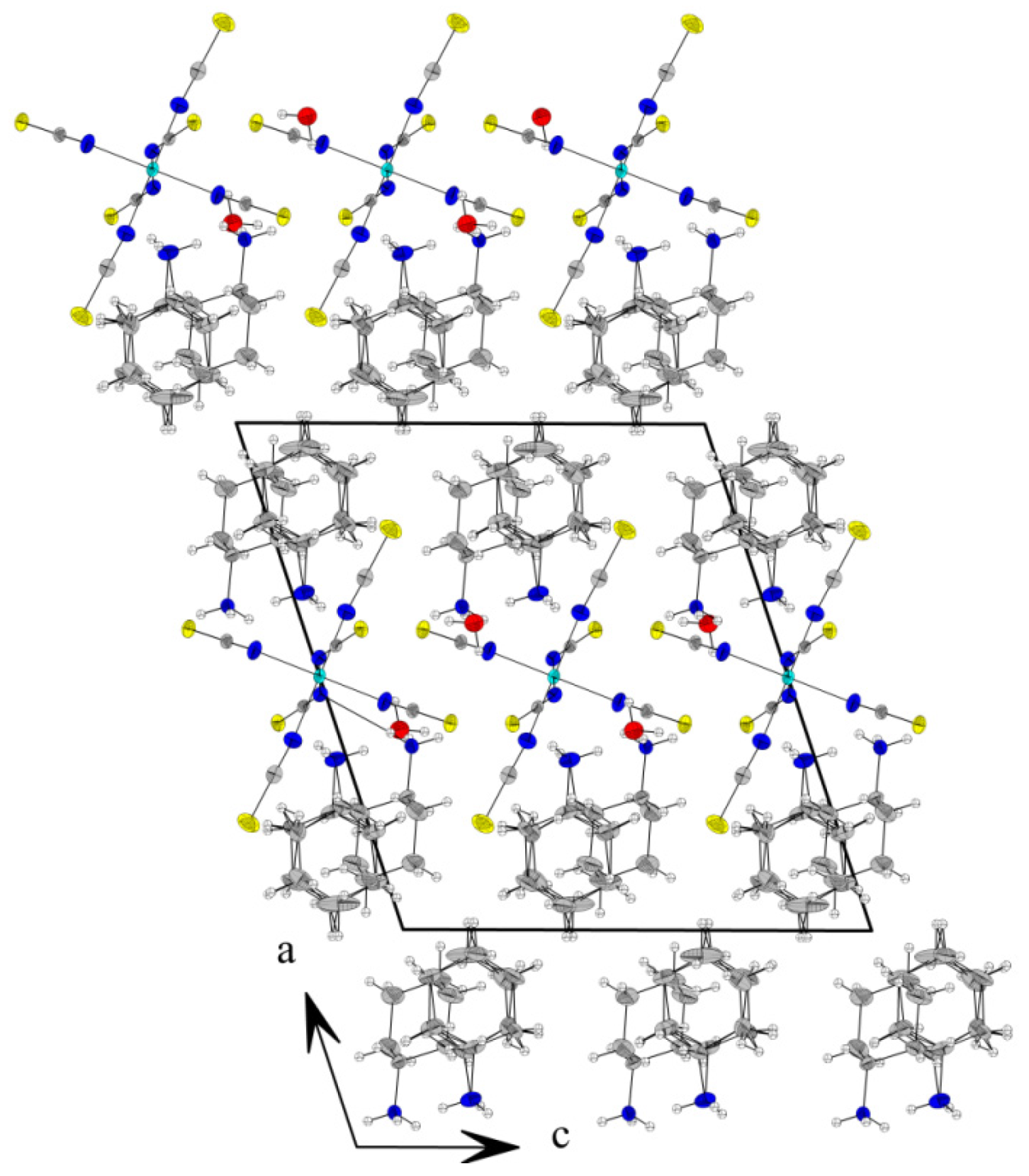
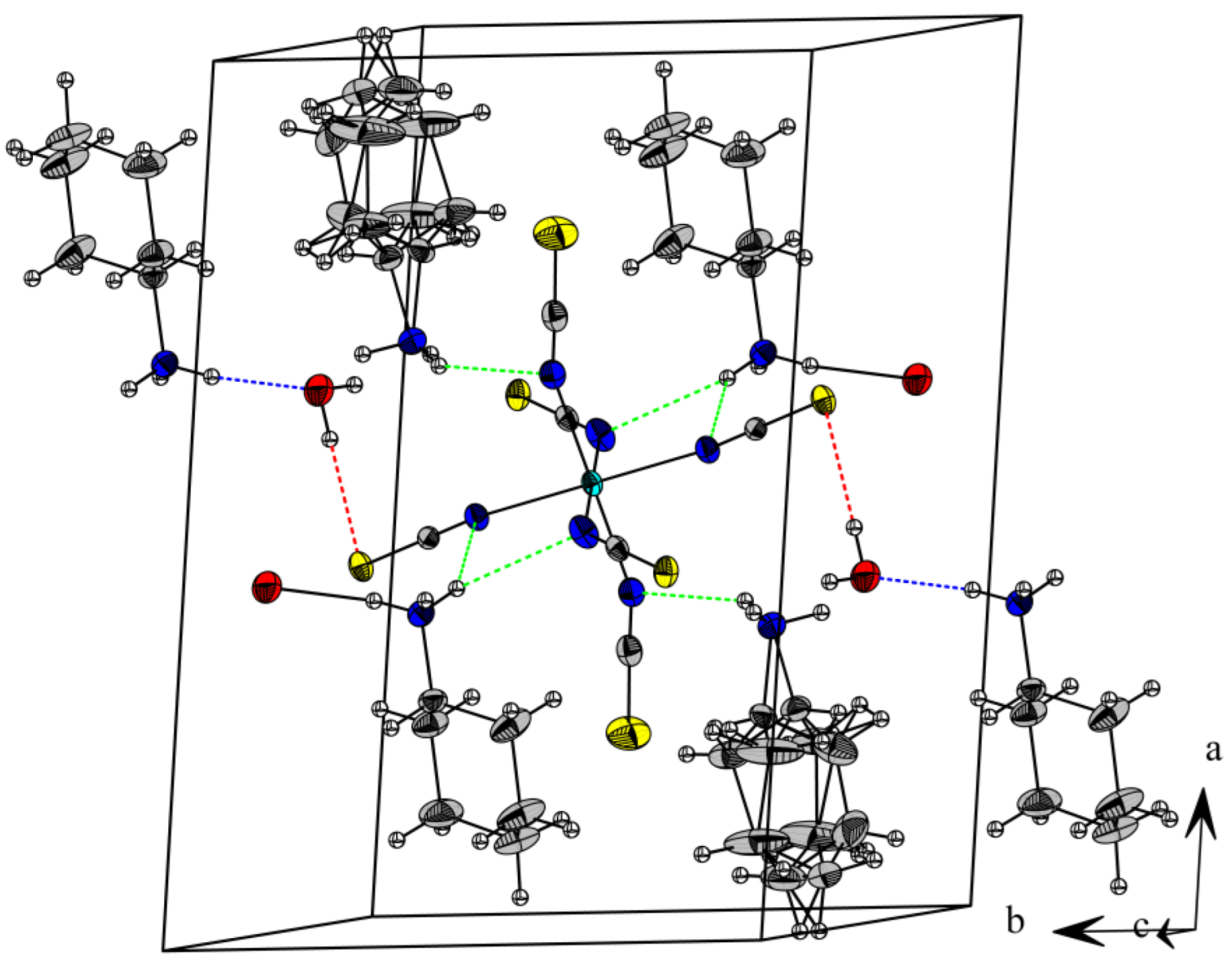
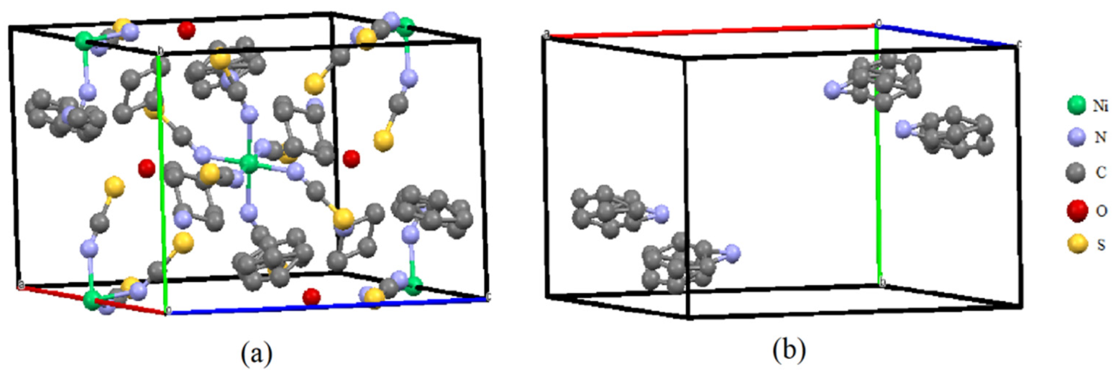
3.5. SCXRD Analyses
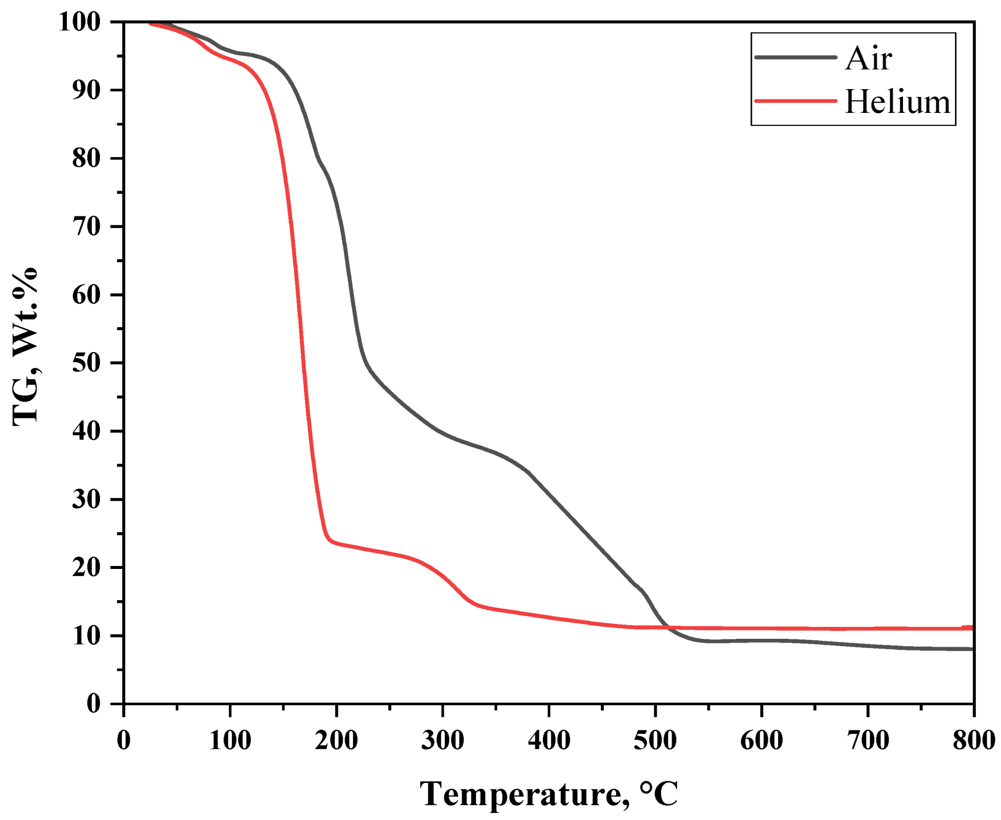
3.6. TG Analysis
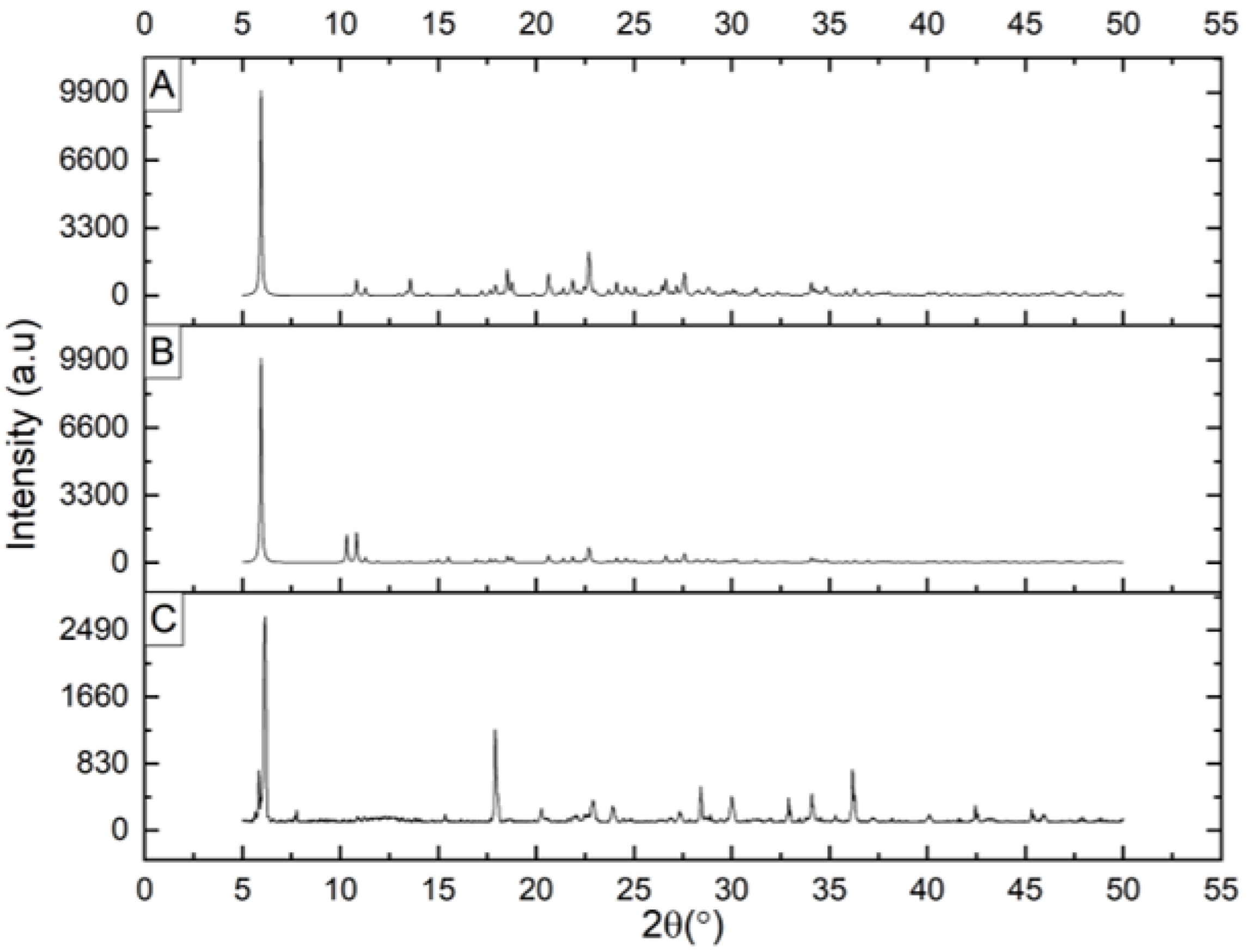
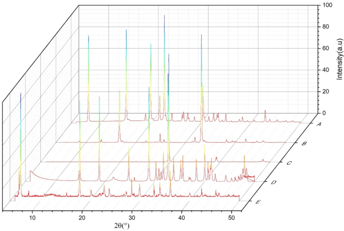
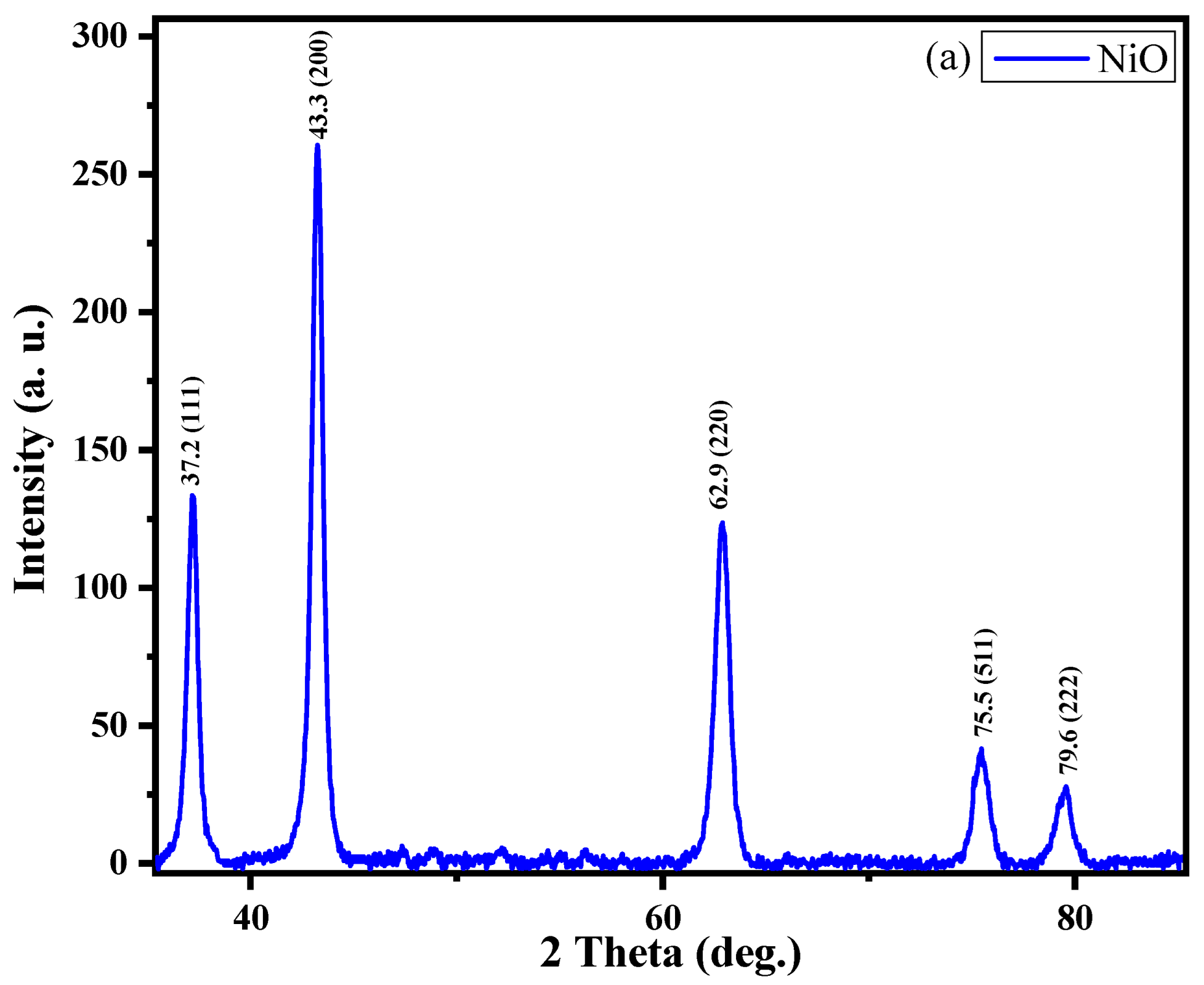
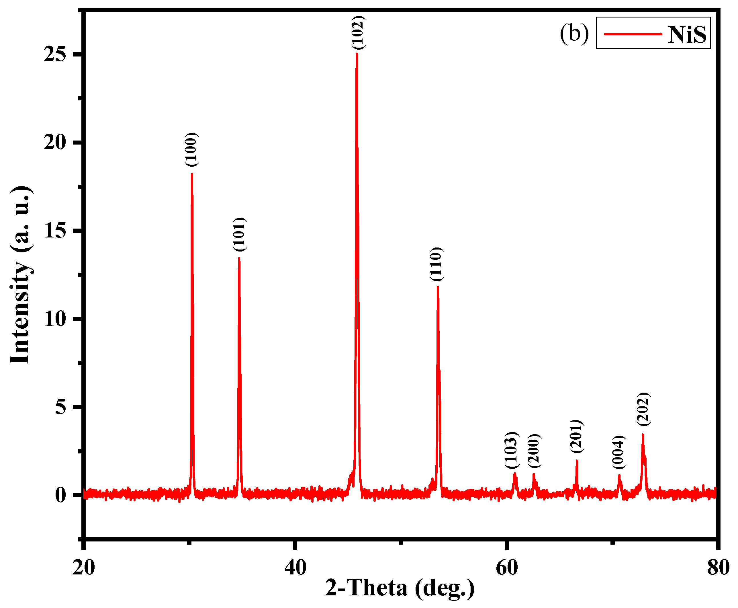
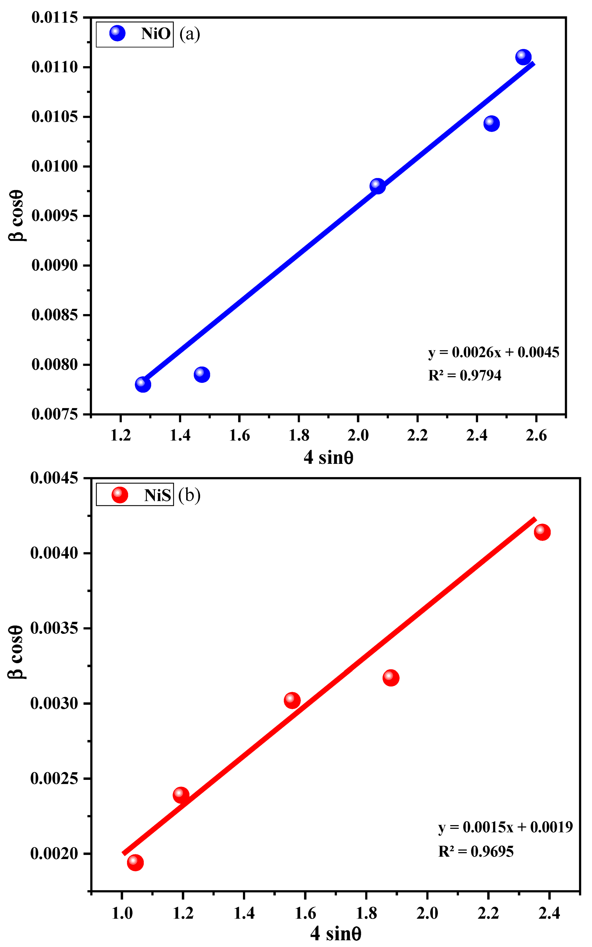
3.7. XRPD Microstructure of NiO and NiS
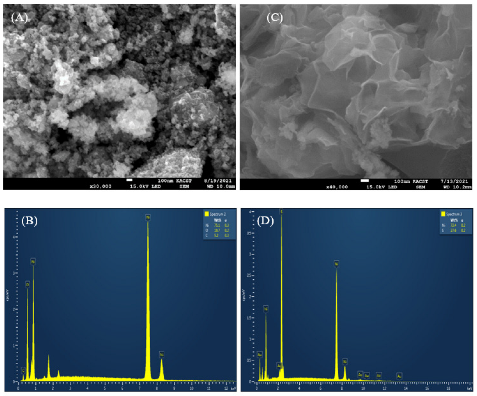
3.8. Morphological Study
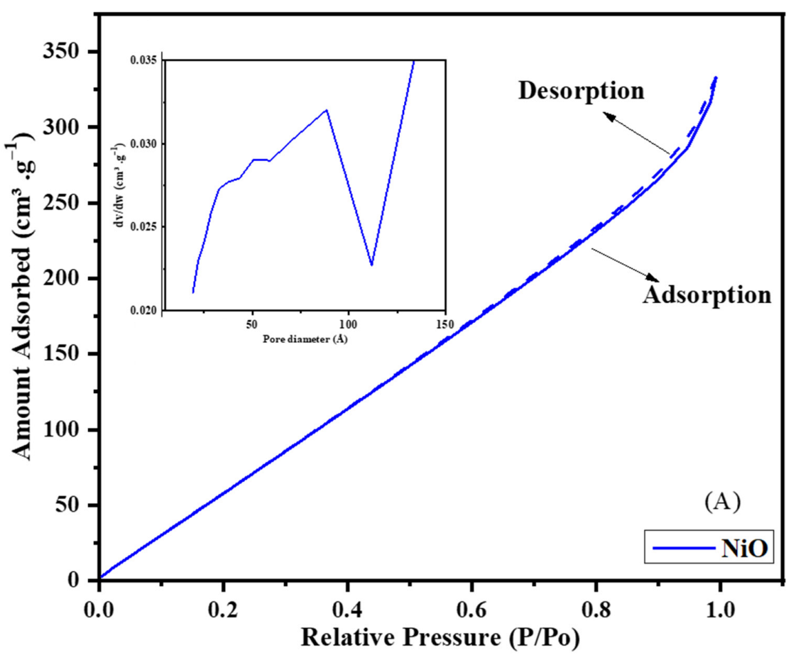
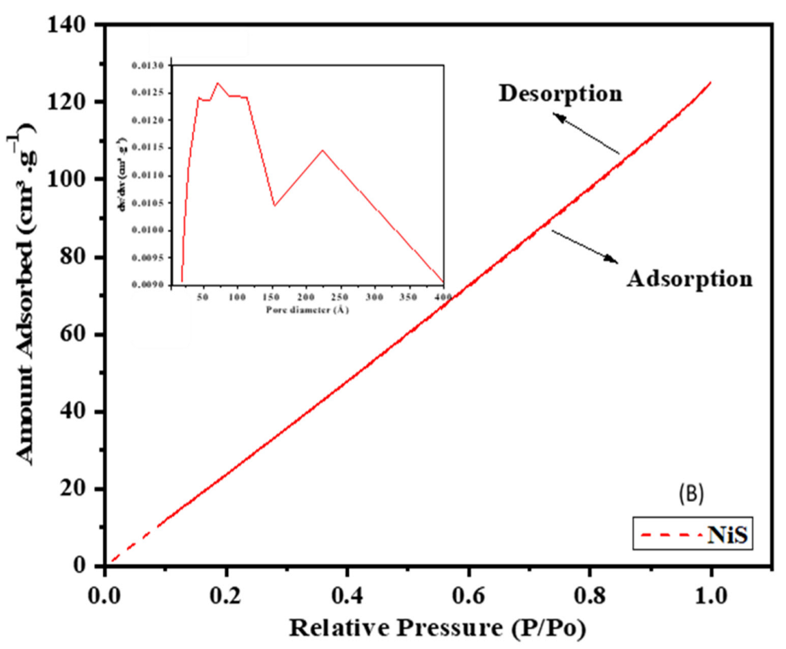
3.9. Porosity Analysis
4. Conclusions
Supplementary Materials
Author Contributions
Funding
Institutional Review Board Statement
Informed Consent Statement
Data Availability Statement
Acknowledgments
Conflicts of Interest
References
- Nazeeruddin, M.K.; Baranoff, E.; Grätzel, M. Dye-sensitized solar cells: A brief overview. Sol. Energy 2011, 85, 1172–1178. [Google Scholar] [CrossRef]
- Prananto, Y.P.; Urbatsch, A.; Moubaraki, B.; Murray, K.S.; Turner, D.R.; Deacon, G.B.; Batten, S.R. Transition metal thiocyanate complexes of picolylcyanoacetamides. Aust. J. Chem. 2017, 70, 516–528. [Google Scholar] [CrossRef]
- Kabesova, M. Structure and classification of thiocyanates and the mutual influence of their ligands. Chem. Zvesti. 1980, 34, 800–841. [Google Scholar]
- Wöhlert, S.; Runčevski, T.; Dinnebier, R.E.; Ebbinghaus, S.G.; Näther, C. Synthesis, Structures, Polymorphism, and Magnetic Properties of Transition Metal Thiocyanato Coordination Compounds. Cryst. Growth Des. 2014, 14, 1902–1913. [Google Scholar] [CrossRef]
- Hannachi, A.; Valkonen, A.; Gómez García, C.J.; Rzaigui, M.; Smirani, W. Synthesis of isomorphous cobalt and nickel thiocyanate coordination compounds: Effect of metals on compound properties. Polyhedron 2019, 173, 114122. [Google Scholar] [CrossRef]
- Lusi, M.; Barbour, L.J. Solid–vapor sorption of xylenes: Prioritized selectivity as a means of separating all three isomers using a single substrate. Angew. Chem. 2012, 124, 3994–3997. [Google Scholar] [CrossRef]
- Lago, E.L.; Seijas, J.A.; de Pedro, I.; Fernández, J.R.; Vázquez-Tato, M.P.; González, J.A.; Rilo, E.; Segade, L.; Cabeza, O.; Fernández, C.D.R. Structural and physical properties of a new reversible and continuous thermochromic ionic liquid in a wide temperature interval: [BMIM]4[Ni (NCS)6]. N. J. Chem. 2018, 42, 15561–15571. [Google Scholar] [CrossRef]
- Kruger, P.; McKee, V. Tetrakis (triethylammonium) Hexakis (isothiocyanato-N) nickel (II). Acta Crystallogr. Sect. C Cryst. Struct. Commun. 1996, 52, 617–619. [Google Scholar] [CrossRef]
- House, J., Jr.; Marquardt, L.A. Synthesis and thermal decomposition of piperidinium hexathiocyanatonickelate (II). Thermochim. Acta 1989, 153, 231–236. [Google Scholar] [CrossRef]
- Vijayakanth, T.; Ram, F.; Praveenkumar, B.; Shanmuganathan, K.; Boomishankar, R. Piezoelectric Energy Harvesting from a Ferroelectric Hybrid Salt [Ph3MeP]4[Ni(NCS)6] Embedded in a Polymer Matrix. Angew. Chem. Int. Ed. 2020, 59, 10368–10373. [Google Scholar] [CrossRef]
- Liu, J.Y.; Zhang, S.Y.; Zeng, Y.; Shu, X.; Du, Z.Y.; He, C.T.; Zhang, W.X.; Chen, X.M. Molecular Dynamics, Phase Transition and Frequency-Tuned Dielectric Switch of an Ionic Co-Crystal. Angew. Chem. 2018, 130, 8164–8168. [Google Scholar] [CrossRef]
- Larue, B.; Tran, L.-T.; Luneau, D.; Reber, C. Crystal structures, magnetic properties, and absorption spectra of nickel (II) thiocyanato complexes: A comparison of different coordination geometries. Can. J. Chem. 2003, 81, 1168–1179. [Google Scholar] [CrossRef]
- Brinzari, T.; Tian, C.; Halder, G.; Musfeldt, J.; Whangbo, M.-H.; Schlueter, J. Color properties and structural phase transition in penta-and hexacoordinate isothiocyanato Ni (II) compounds. Inorg. Chem. 2009, 48, 7650–7658. [Google Scholar] [CrossRef]
- Lis, T.; Jerzykiewicz, L.B. Potassium D-3-phosphoglycerate and cyclohexylammonium D-3-phosphoglycerate hydrate. Acta Crystallogr. Sect. C Cryst. Struct. Commun. 1995, 51, 1001–1005. [Google Scholar] [CrossRef]
- Kolev, T.; Koleva, B.; Seidel, R.W.; Spiteller, M.; Sheldrick, W.S. Cyclohexylammonium hydrogensquarate hemihydrate. Acta Crystallogr. Sect. Sect. E Struct. Rep. Online 2007, 63, o4852. [Google Scholar] [CrossRef]
- Billing, D.G.; Lemmerer, A. Inorganic–organic hybrid materials incorporating primary cyclic ammonium cations: The lead bromide and chloride series. CrystEngComm 2009, 11, 1549–1562. [Google Scholar] [CrossRef]
- Wei, X.; Li, J.; Yin, H. Bis(cyclohexylammonium) 2,2′-disulfanediyldibenzoate. Acta Crystallogr. Sect. E Struct. Rep. Online 2011, 67, o319. [Google Scholar] [CrossRef]
- Sarr, M.; Merkens, C.; Diassé-Sarr, A.; Diop, L.; Englert, U. Bis(cyclohexylammonium) tetrachloridodiphenylstannate (IV). Acta Crystallogr. Sect. Sect. E Struct. Rep. Online 2014, 70, m220–m221. [Google Scholar] [CrossRef]
- Bagabas, A.A.; Aboud, M.F.; Shemsi, A.M.; Addurihem, E.S.; Al-Othman, Z.A.; Kumar, C.; Fun, H.-K. Cyclohexylammonium nitrate. Acta Crystallogr. Sect. E Struct. Rep. Online 2014, 70, o253–o254. [Google Scholar] [CrossRef]
- Liao, W.-Q.; Ye, H.-Y.; Fu, D.-W.; Li, P.-F.; Chen, L.-Z.; Zhang, Y. Temperature-Triggered Reversible Dielectric and Nonlinear Optical Switch Based on the One-Dimensional Organic–Inorganic Hybrid Phase Transition Compound [C6H11NH3]·2CdCl4. Inorg. Chem. 2014, 53, 11146–11151. [Google Scholar] [CrossRef]
- Sathya, P.; Anantharaja, M.; Elavarasu, N.; Gopalakrishnan, R. Growth and characterization of nonlinear optical single crystals: Bis(cyclohexylammonium) terephthalate and cyclohexylammonium para-methoxy benzoate. Bull. Mater. Sci. 2015, 38, 1291–1299. [Google Scholar] [CrossRef]
- Yangui, A.; Garrot, D.; Lauret, J.-S.; Lusson, A.; Bouchez, G.; Deleporte, E.; Pillet, S.; Bendeif, E.-E.; Castro, M.; Triki, S. Optical investigation of broadband white-light emission in self-assembled organic–inorganic perovskite (C6H11NH3)2PbBr4. J. Phys. Chem. C 2015, 119, 23638–23647. [Google Scholar] [CrossRef]
- Zhao, W.; Jin, Y.; Zhang, W. Phase transitions in two organic salts based on 1,5-naphthalenedisulfonate. Sci. China Chem. 2016, 59, 114–121. [Google Scholar] [CrossRef]
- Yangui, A.; Pillet, S.; Lusson, A.; Bendeif, E.-E.; Triki, S.; Abid, Y.; Boukheddaden, K. Control of the white-light emission in the mixed two-dimensional hybrid perovskites (C6H11NH3)2[PbBr4–xIx]. J. Alloy. Compd. 2017, 699, 1122–1133. [Google Scholar] [CrossRef]
- Yangui, A.; Pillet, S.; Bendeif, E.-E.; Lusson, A.; Triki, S.; Abid, Y.; Boukheddaden, K. Broadband emission in a new two-dimensional Cd-based hybrid perovskite. ACS Photonics 2018, 5, 1599–1611. [Google Scholar] [CrossRef]
- Bagabas, A.; Alsawalha, M.; Sohail, M.; Alhoshan, S.; Arasheed, R. Synthesis, crystal structure, and characterization of cyclohexylammonium tetraisothiocyanatocobaltate (II): A single-source precursor for cobalt sulfide and oxide nanostructures. Heliyon 2019, 5, e01139. [Google Scholar] [CrossRef]
- Jones, P.; Sheldrick, G.; Kirby, A.; Abell, K. Bis(cyclohexylammonium) propargyl phosphate dihydrate, 2C6H14N+·C3H3O4P2−·2H2O. Acta Crystallogr. Sect. C Cryst. Struct. Commun. 1984, 40, 547–549. [Google Scholar] [CrossRef]
- Gomathi, R.; Madeswaran, S.; Babu, D.R. Bulk growth, electrical, linear, third order nonlinear optical and optical limiting properties on bis(cyclohexylammonium) succinate succinic acid crystal. Mater. Chem. Phys. 2018, 207, 84–90. [Google Scholar] [CrossRef]
- Dupont, N.; Barbey, C.; Pfund, E.; Lequeux, T.; Navaza, A. Crystal Structure of a Cyclohexylammonium Salt of the N-(2,2-Difluoro-2-phosphonoethanethioyl) aspartate: A Difluorinated N-(Phosphonoacetyl)-L-aspartate (PALA) Thio Analogue. Anal. Sci. X-ray Struct. Anal. Online 2008, 24, x293–x294. [Google Scholar] [CrossRef][Green Version]
- Senthil, K.; Senthil, A.; Elangovan, K. Crystal structure, growth and physiochemical properties of nonlinear optical single crystal: Bis(cyclohexylammonium) dioxalate hydrate. J. Mol. Struct. 2020, 1209, 127926. [Google Scholar] [CrossRef]
- Gomathi, R.; Ramasamy, G.; Vinitha, G.; Ramki, S.; Chen, S.M. Cyclohexylammonium Cinnamate Single Crystal for Nonlinear Optical Applications. J. Electron. Mater. 2020, 49, 3350–3356. [Google Scholar] [CrossRef]
- Wang, J.-P.; Cheng, X.-X.; Wang, J.-G.; Chen, Q.-H. Cyclohexylammonium dichloroacetate. Acta Crystallogr. Sect. E Struct. Rep. Online 2005, 61, o4006–o4007. [Google Scholar] [CrossRef]
- Cuquejo-Cid, A.; Garcia-Fernandez, A.; Garcia-Ben, J.; Senaris-Rodriguez, M.A.; Castro-Garcia, S.; Sanchez-Andujar, M.; Vazquez-Garcia, D. Photoluminescent and vapochromic properties of the Mn (II)-doped (C6H11NH3)2PbBr4 layered organic–inorganic hybrid perovskite. Polyhedron 2021, 193, 114840. [Google Scholar] [CrossRef]
- Bagabas, A.A.; Alhoshan, S.B.; Ghabbour, H.A.; Kumar, C.; Fun, H.-K. Crystal structure of cyclohexylammonium thiocyanate. Acta Crystallogr. Sect. E Crystallogr. Commun. 2015, 71, o62–o63. [Google Scholar] [CrossRef]
- Yun, S.-S.; Moon, H.-S.; Kim, C.-H.; Lee, S.-G. A novel, wave-like, two-dimensional anionic host with Cationic and neutral guests: Crystal structure of [C6H11NH3][Cu2(CN)3]·2C6H5NH2. J. Coord. Chem. 2004, 57, 321–327. [Google Scholar] [CrossRef]
- Saminathan, K.; SethuSankar, K.; Muthamizhchelvan, C.; Sivakumar, K. Cyclohexylammonium picrate. Acta Crystallogr. Sect. E 2005, 61, o3605–o3607. [Google Scholar]
- Kumara Swamy, K.; Said, M.A. Cyclohexylammonium 2′-hydroxy-2-biphenyl phosphonate. Acta Crystallogr. Sect. C Cryst. Struct. Commun. 2001, 57, 491–492. [Google Scholar] [CrossRef]
- Bagabas, A.A.; Alsawalha, M.; Taha, K.K.; Albaid, A.; Sohail, M.; Alqahtani, M.; Alrasheed, R.; Alqarn, A.; Ashamari, B.; Parkin, I.P. High Surface Area of Polyhedral Chromia and Hexagonal Chromium Sulfide by the Thermolysis of Cyclohexylammonium Hexaisothiocyanatochromate (III) Sesquihydrate. Chem. Sel. 2021, 6, 4298–4311. [Google Scholar] [CrossRef]
- Alshammari, A.; Mokhtar, M.; Arasheed, R.; Asayegh, A.; Bagabas, A. Acetone Reaction with Hydrogen over Mesoporous Magnesium Oxide-Supported Rhodium Nanoparticles. Top. Catal. 2019, 62, 795–804. [Google Scholar] [CrossRef]
- Hogarth, G. Transition metal dithiocarbamates: 1978–2003. Prog. Inorg. Chem. 2005, 53, 71–561. [Google Scholar]
- Coucouvanis, D. The chemistry of the dithioacid and 1,1-dithiolate complexes, 1968–1977. Prog. Inorg. Chem 1979, 26, 301–469. [Google Scholar]
- Bond, A.; Martin, R. Electrochemistry and redox behaviour of transition metal dithiocarbamates. Coord. Chem. Rev. 1984, 54, 23–98. [Google Scholar] [CrossRef]
- Heard, P.J. Main Group Dithiocarbamate Complexes. Prog. Inorg. Chem. 2005, 53, 1–69. [Google Scholar] [CrossRef]
- Coucouvanis, D. The Chemistry of the Dithioacid and 1,1-Dithiolate Complexes. Prog. Inorg. Chem. 1970, 4, 233–371. [Google Scholar] [CrossRef]
- Afzaal, M.; Malik, M.A.; O’Brien, P. Chemical routes to chalcogenide materials as thin films or particles with critical dimensions with the order of nanometres. J. Mater. Chem. 2010, 20, 4031–4040. [Google Scholar] [CrossRef]
- Hollingsworth, N.; Roffey, A.; Islam, H.-U.; Mercy, M.; Roldan, A.; Bras, W.; Wolthers, M.; Catlow, C.R.A.; Sankar, G.; Hogarth, G. Active nature of primary amines during thermal decomposition of nickel dithiocarbamates to nickel sulfide nanoparticles. Chem. Mater. 2014, 26, 6281–6292. [Google Scholar] [CrossRef]
- Baer, C.; Pike, J. Infrared spectroscopic analysis of linkage isomerism in metal–thiocyanate complexes. J. Chem. Educ. 2010, 87, 724–726. [Google Scholar] [CrossRef]
- Forster, D.; Goodgame, D. Isothiocyanato complexes of nickel (II) and copper (II). Inorg. Chem. 1965, 4, 823–829. [Google Scholar] [CrossRef]
- Cabeza, O.; Segade, L.; Domínguez-Pérez, M.; Rilo, E.; Ausín, D.; Seijas, J.A.; Vazquez-Tato, M.P.; Matleev, V.; Ievlev, A.; Salgado, J. Strange behaviour of transport properties in novel metal thiocyanate based ionic liquids. J. Mol. Liq. 2021, 340, 117164. [Google Scholar] [CrossRef]
- Kauffman, G.B.; Beech, G. Thermal decomposition of solid isothiocyanate complexes: Part II. Kinetic parameters. Thermochim. Acta 1970, 1, 99–102. [Google Scholar] [CrossRef]
- Abdu, B. Crystal Structure of Rubidium Nickel Thiocyanate Tetrahyderate Rb4Ni(SCN)6·4H2O. Int. J. Chem. Environ.Biol. Sci. (IJCEBS) 2016, 4, 12–15. [Google Scholar]
- Forster, D.; Goodgame, D. Infrared Spectra (400-200 Cm.-1) of Some Thiocyanate and Isothiocyanate Complexes. Inorg. Chem. 1965, 4, 715–718. [Google Scholar] [CrossRef]
- Vicente, R.; Escuer, A.; Solans, X.; Font-Bardía, M. Aqueous syntheses and crystal structures of the hexa- and pentacoordinated nickel(II) isothiocyanato derivatives of bulky triamines: (H2Et5dien)2[Ni(NCS)6], [Ni(Me5dien)(NCS)2] and [Ni(Me4Etdien)(NCS)2]. Inorg. Chim. Acta 1996, 248, 59–65. [Google Scholar] [CrossRef]
- Petříček, V.; Dušek, M.; Palatinus, L. Crystallographic computing system JANA2006: General features. Z. Für Krist.-Cryst. Mater. 2014, 229, 345–352. [Google Scholar] [CrossRef]
- Balachandran, V.; Murali, M.K. FT-IR and FT-Raman spectral analysis of 3-(trifluromethyl) phenyl isothiocyanate. Elixir Vib. Spec. 2011, 40, 5105–5107. [Google Scholar]
- Silverstein, R.M.; Bassler, G.C. Spectrometric identification of organic compounds. J. Chem. Educ. 1962, 39, 546. [Google Scholar] [CrossRef]
- Sundaraganesan, N.; Ayyappan, S.; Umamaheswari, H.; Joshua, B.D. FTIR, FT-Raman spectra and ab initio, DFT vibrational analysis of 2,4-dinitrophenylhydrazine. Spectrochim. Acta Part A Mol. Biomol. Spectrosc. 2007, 66, 17–27. [Google Scholar] [CrossRef]
- Mani, P.; Umamaheswari, H.; Joshua, B.D.; Sundaraganesan, N. Molecular structure, vibrational spectra and NBO analysis of phenylisothiocyanate by density functional method. J. Mol. Struct. THEOCHEM 2008, 863, 44–49. [Google Scholar] [CrossRef]
- Kauffman, K.L.; Culp, J.T.; Goodman, A.; Matranga, C. FT-IR study of CO2 adsorption in a dynamic copper (II) benzoate– pyrazine host with CO2–CO2 interactions in the adsorbed state. J. Phys. Chem. C 2011, 115, 1857–1866. [Google Scholar] [CrossRef]
- Lex, A.; Trimmel, G.; Kern, W.; Stelzer, F. Photosensitive polynorbornene containing the benzyl thiocyanate group—Synthesis and patterning. J. Mol. Catal. A Chem. 2006, 254, 174–179. [Google Scholar] [CrossRef]
- Coenen, K.; Gallucci, F.; Mezari, B.; Hensen, E.; Van Sint Annaland, M. An in-situ IR study on the adsorption of CO2 and H2O on hydrotalcites. J. CO2 Util. 2018, 24, 228–239. [Google Scholar] [CrossRef]
- Clark, R.; Goodwin, A. Infrared and laser Raman spectra of metal hexaisothiocyanate ions. Spectrochim. Acta Part A Mol. Spectrosc. 1970, 26, 323–330. [Google Scholar] [CrossRef]
- Chamberlain, M.M.; Bailar, J.C., Jr. The infrared spectra of some thiocyanatocobalt ammines. J. Am. Chem. Soc. 1959, 81, 6412–6415. [Google Scholar] [CrossRef]
- Groom, C.R.; Bruno, I.J.; Lightfoot, M.P.; Ward, S.C. The Cambridge Structural Database. Acta Crystallogr. Sect. B 2016, 72, 171–179. [Google Scholar] [CrossRef] [PubMed]
- Wang, X.; Yu, L.; Hu, P.; Yuan, F. Synthesis of single-crystalline hollow octahedral NiO. Cryst. Growth Des. 2007, 7, 2415–2418. [Google Scholar] [CrossRef]
- Motazedian, F.; Wu, Z.; Zhang, J.; Samsam Shariat, B.; Jiang, D.; Martyniuk, M.; Liu, Y.; Yang, H. Determining intrinsic stress and strain state of fibre-textured thin films by X-ray diffraction measurements using combined asymmetrical and Bragg-Brentano configurations. Mater. Des. 2019, 181, 108063. [Google Scholar] [CrossRef]
- Karthikeyan, R.; Thangaraju, D.; Prakash, N.; Hayakawa, Y. Single-step synthesis and catalytic activity of structure-controlled nickel sulfide nanoparticles. Cryst. Eng. Comm. 2015, 17, 5431–5439. [Google Scholar] [CrossRef]
- Elamin, N.; Modwi, A.; Aissa, M.B.; Taha, K.K.; Al-Duaij, O.K.; Yousef, T. Fabrication of Cr–ZnO photocatalyst by starch-assisted sol–gel method for photodegradation of congo red under visible light. J. Mater. Sci. Mater. Electron. 2021, 32, 2234–2248. [Google Scholar] [CrossRef]
- Brandstetter, S.; Derlet, P.; Van Petegem, S.; Van Swygenhoven, H. Williamson–Hall anisotropy in nanocrystalline metals: X-ray diffraction experiments and atomistic simulations. Acta Mater. 2008, 56, 165–176. [Google Scholar] [CrossRef]
- Mote, V.; Purushotham, Y.; Dole, B. Williamson-Hall analysis in estimation of lattice strain in nanometer-sized ZnO particles. J. Theor. Appl. Phys. 2012, 6, 1–8. [Google Scholar] [CrossRef]
- Xia, H.; Xiahou, Y.; Zhang, P.; Ding, W.; Wang, D. Revitalizing the Frens method to synthesize uniform, quasi-spherical gold nanoparticles with deliberately regulated sizes from 2 to 330 nm. Langmuir 2016, 32, 5870–5880. [Google Scholar] [CrossRef]
- Vijaya, J.J.; Sekaran, G.; Bououdina, M. Effect of Cu2+ doping on structural, morphological, optical and magnetic properties of MnFe2O4 particles/sheets/flakes-like nanostructures. Ceram. Int. 2015, 41, 15–26. [Google Scholar] [CrossRef]
- Rouquerol, J.; Rouquerol, F.; Llewellyn, P.; Maurin, G.; Sing, K.S. Adsorption by Powders and Porous Solids: Principles, Methodology and Applications; Academic Press: Cambridge, MA, USA, 2013. [Google Scholar]
- Harraz, F.; Mohamed, R.; Shawky, A.; Ibrahim, I. Composition and phase control of Ni/NiO nanoparticles for photocatalytic degradation of EDTA. J. Alloy. Compd. 2010, 508, 133–140. [Google Scholar] [CrossRef]
- Xing, W.; Li, F.; Yan, Z.-f.; Lu, G. Synthesis and electrochemical properties of mesoporous nickel oxide. J. Power Sources 2004, 134, 324–330. [Google Scholar] [CrossRef]
- Juang, R.-S.; Wu, F.-C.; Tseng, R.-L. Characterization and use of activated carbons prepared from bagasses for liquid-phase adsorption. Colloids Surf. A Physicochem. Eng. Asp. 2002, 201, 191–199. [Google Scholar] [CrossRef]
- Bruschi, L.; Mistura, G.; Nguyen, P.T.; Do, D.D.; Nicholson, D.; Park, S.-J.; Lee, W. Adsorption in alumina pores open at one and at both ends. Nanoscale 2015, 7, 2587–2596. [Google Scholar] [CrossRef]


| Element | Theoretical (wt/wt%) | Experimental (wt/wt%) |
|---|---|---|
| C | 42.70 | 42.57 |
| H | 7.17 | 7.12 |
| N | 16.60 | 16.58 |
| S | 22.80 | 22.65 |
| O | 3.79 | 3.68 |
| Ni | 6.95 | 6.86 |
| Crystal Data | ||
|---|---|---|
| Chemical formula | (C6H11NH3)4[Ni(NCS)6]·2H2O | (C6H11NH3)4[Ni(NCS)6]·2H2O |
| Mr | 843.9 | 843.9 |
| Crystal system, space group | Monoclinic, P21/c | Monoclinic, P21/c |
| Temperature (K) | 293 | 120 |
| a, b, c (Å) | 15.8179 (5), 10.6222 (3), 13.8751 (4) | 15.6486 (5), 10.4641 (3), 13.7387 (4) |
| β (°) | 109.362 (1) | 108.393 (1) |
| V (Å3) | 2199.45 (11) | 2134.77 (11) |
| Z | 2 | 2 |
| Radiation type | Mo Kα | Mo Kα |
| µ (mm−1) | 0.76 | 0.79 |
| Crystal size (mm) | 0.23 × 0.19 × 0.07 | 0.23 × 0.19 × 0.07 |
| Data collection | ||
| Diffractometer | D8 venture diffractometer | D8 venture diffractometer |
| Absorption correction | Multi-scan SADABS | Multi-scan SADABS |
| Tmin, Tmax | 0.694, 0.747 | 0.695, 0.746 |
| No. of measured, independent and observed [I > −4σ(I)] reflections | 39757, 6892, 6892 | 31049, 5537, 5537 |
| Rint | 0.028 | 0.026 |
| (sin θ/λ)max (Å−1) | 0.722 | 0.677 |
| Refinement | ||
| R[F2 > 2σ(F2)], wR(F2), S | 0.072, 0.168, 1.54 | 0.036, 0.096, 1.18 |
| No. of reflections | 6892 | 5537 |
| No. of parameters | 230 | 277 |
| No. of restraints | 3 | 2 |
| H-atom treatment | H atoms treated by a mixture of independent and constrained refinement | H atoms treated by a mixture of independent and constrained refinement |
| (Δ/σ)max | 0.107 | |
| Δρmax, Δρmin (e Å−3) | 0.50, −0.39 | 0.18, −0.23 |
| Bond Length (Å) | Angles Degree | |||||
|---|---|---|---|---|---|---|
| Ni1 | N1 | 2.092 | N1 | Ni1 | N2 | 86.77 |
| Ni1 | N2 | 2.099 | N1 | Ni1 | N3 | 89.30 |
| Ni1 | N3 | 2.082 | N1 | Ni1 | N1 | 180.00 |
| Ni1 | N1 | 2.092 | Ni1 | N1 | C1 | 146.4 |
| Ni1 | N2 | 2.099 | Ni1 | N2 | C2 | 153.4 |
| Ni1 | N3 | 2.082 | Ni1 | N3 | C3 | 156.4 |
| N1 | C1 | 1.155(2) | N1 | C1 | S1 | 179.0(1) |
| N2 | C2 | 1.154(2) | N2 | C2 | S2 | 178.6(1) |
| N3 | C3 | 1.153(2) | N3 | C3 | S3 | 178.4(1) |
| C1 | S1 | 1.630(1) | ||||
| C2 | S2 | 1.637(1) | ||||
| C3 | S3 | 1.620(1) | ||||
| Donor | Hydrogen | Acceptor | D-H Distance, Å | H…A Distance, Å | D-A Distance, Å | A-H…D Angle, ° |
|---|---|---|---|---|---|---|
| O1 | H1O1 | S1 | 0.866(15) | 2.459(19) | 3.3043(14) | 166(3) |
| N5 | H2N5 | N1 | 0.87 | 2.35 | 3.0924(19) | 143.14 |
| N5 | H2N5 | N2 | 0.87 | 2.39 | 3.0791(17) | 135.97 |
| N5 | H3N5 | O1 | 0.87 | 1.97 | 2.8375(15) | 172.00 |
| N4 | H2N4 | N3 | 0.87 | 2.31 | 3.0838(18) | 148.52 |
| Atom | Standard | Disorder |
|---|---|---|
| O | 4 | 3 |
| C (radial) | 12 | 12 |
| C (ring) | 44 | 48 |
| Ni | 2 | 2 |
| N | 20 | 20 |
| S | 12 | 12 |
| Sample | β | D (nm) | D* (nm) | d (Å) | L. Parameters | ε | ε* | |
|---|---|---|---|---|---|---|---|---|
| a | c | |||||||
| NiO | 0.5027 | 16.34 | 30.80 | 2.2530 | 4.180 | 0.0055 | 0.0026 | |
| NiS | 0.2077 | 53.79 | 72.98 | 2.3050 | 3.421 | 5.322 | 0.0021 | 0.0015 |
| Sample | SBET (m2/g) | Pore Volume (cm3/g) | Pre Size, (Å) |
|---|---|---|---|
| NiO | 451.45 | 0.472 | 53.33 |
| NiS | 219.92 | 0.170 | 45.96 |
Publisher’s Note: MDPI stays neutral with regard to jurisdictional claims in published maps and institutional affiliations. |
© 2022 by the authors. Licensee MDPI, Basel, Switzerland. This article is an open access article distributed under the terms and conditions of the Creative Commons Attribution (CC BY) license (https://creativecommons.org/licenses/by/4.0/).
Share and Cite
Aboud, M.F.A.; BinTaleb, A.M.; Yahia, H.B.; Albaid, A.; Albishi, S.; Arasheed, R.; Albinali, I.; Albaqi, F.; Anojaidi, K.; Alqahtani, F.; et al. Cyclohexylammonium Hexaisothiocyanatonickelate(II) Dihydrate as a Single-Source Precursor for High Surface Area Nickel Oxide and Sulfide Nanocrystals. Crystals 2022, 12, 315. https://doi.org/10.3390/cryst12030315
Aboud MFA, BinTaleb AM, Yahia HB, Albaid A, Albishi S, Arasheed R, Albinali I, Albaqi F, Anojaidi K, Alqahtani F, et al. Cyclohexylammonium Hexaisothiocyanatonickelate(II) Dihydrate as a Single-Source Precursor for High Surface Area Nickel Oxide and Sulfide Nanocrystals. Crystals. 2022; 12(3):315. https://doi.org/10.3390/cryst12030315
Chicago/Turabian StyleAboud, Mohamed F. Aly, Abdulmalik M. BinTaleb, Hamdi Ben Yahia, Abdelhamid Albaid, Sultan Albishi, Rasheed Arasheed, Ibrahim Albinali, Fahad Albaqi, Khalid Anojaidi, Fahad Alqahtani, and et al. 2022. "Cyclohexylammonium Hexaisothiocyanatonickelate(II) Dihydrate as a Single-Source Precursor for High Surface Area Nickel Oxide and Sulfide Nanocrystals" Crystals 12, no. 3: 315. https://doi.org/10.3390/cryst12030315
APA StyleAboud, M. F. A., BinTaleb, A. M., Yahia, H. B., Albaid, A., Albishi, S., Arasheed, R., Albinali, I., Albaqi, F., Anojaidi, K., Alqahtani, F., Alenad, A. M., Taha, K., & Bagabas, A. (2022). Cyclohexylammonium Hexaisothiocyanatonickelate(II) Dihydrate as a Single-Source Precursor for High Surface Area Nickel Oxide and Sulfide Nanocrystals. Crystals, 12(3), 315. https://doi.org/10.3390/cryst12030315