Article

(Z)-2-(2-(Anthracen-9-ylmethylene)hydrazineyl)pyrimidine-rhodium(III): Crystal Structure and DNA Binding Property

1
School of Material Science and Energy Engineering, Foshan University, Foshan 528000, China
2
School of Chemistry & Pharmaceutical Sciences, Guangxi Normal University, Guilin 541004, China
*
Authors to whom correspondence should be addressed.
These authors contributed equally to this work.
Crystals 2022, 12(11), 1664; https://doi.org/10.3390/cryst12111664
Submission received: 1 November 2022 / Accepted: 13 November 2022 / Published: 18 November 2022

Abstract

:
A new rhodium(III) complex of a 9-anthrahydrazone ligand ((Z)-2-(2-(Anthracen-9-ylmethylene)hydrazineyl)pyrimidine, 9-PMAH) is six-coordinated respectively by one bidentate ligand, one solvent DMSO, and three chlorides, to form a distorted octahedral coordination geometry. The ligand chelates to Rh(III) via the hydrazone N atom (N1) and pyrimidine N atom (N3), whereas DMSO coordinates to Rh(III) via the S atom. The three chloride ligands (Cl1, Cl2, and Cl3) are in a mer-configuration of the octahedron, with the DMSO cis- to the pyrimidine nitrogen. In solution, the complex may exist in the positively charged species, [RhIIICl2(DMSO)(9-PMAH)]+, but can be stable in this mode for no less than 48 h. It was suggested by the fluorescence spectroscopic analysis that the complex showed much higher intercalative binding ability with ct-DNA than the typical DNA intercalator, EtBr.

1. Introduction

Anthracene is a kind of typical pharmacogroup of polycyclic aromatic hydrocarbons. The drugs based on the anthracene structure can be represented by anthracyclines, such as the classic anticancer drugs, doxorubicin, mitoxantrone, etc. [1,2]. The potential pharmacological activity of the anthracene ring is directly related to its conjugated aromatic planar structure. This structural feature helps it to penetrate the cell membrane and bind to DNA by intercalation, and this hinders the replication and transcription of nuclear chromosomes, and effectively inhibits cell proliferation [3]. Anthrahydrazone is a derivative of anthracene and a potential pharmacophore with pharmacological activity. For example, bisantrene is a compound with the structure of anthrahydrazone that has been developed to be a clinical anticancer drug [4]. However, up to now, the study on the activity and function of the anthrahydrazone structure has not been sufficient or in-depth. As metal complexes with anthrahydrazone as the ligand are rarely reported, it is necessary to expand the research in this field [5].
The successful clinical application of cisplatin and its derivatives, such as carboplatin and oxaliplatin, as effective metal-based anticancer drugs have greatly promoted the development of metal complexes in the field of anticancer therapy. Until now, thousands of platinum complexes have been designed and synthesized to explore their anticancer activities. However, their neurotoxicity, nephrotoxicity, and other side effects, accompanied by their widespread use in clinics, have seriously limited further clinical applications [6,7]. Therefore, researchers have turned their attentions to other platinum-group elements, such as Ru, Rh, Pd, Ir, etc. [8,9,10,11]. In the 1970s, rhodium carboxylate complexes were found to have significant antitumor activity with limited side effects, especially against L1210, S180, P388, and Ehrlich ascites tumors in vivo [9,10]. Further results demonstrated that these complexes were similar to cisplatin, and inhibited DNA, RNA, and protein synthesis, which showed us the prospect of designing rhodium complexes as promising anticancer agents [9,10].
In this study, we reported a previously unreported rhodium(III) complex of a novel anthrahydrazone ligand, (Z)-2-(2-(anthracen-9-ylmethylene)hydrazineyl)pyrimidine, also known as 9-pyrimidine anthrahydrazone (9-PMAH) (Scheme 1). It was structurally characterized by IR spectroscopy, elemental analysis, and single crystal X-ray diffraction analysis. Furthermore, its antiproliferative activity toward a series of human tumor cell lines, as well as the related DNA binding properties, were examined by means of MTT assay and fluorescent spectroscopic methods. Until now, the rhodium complexes of anthrahydrazone have not yet been reported or studied. This report will help further explore and understand these kinds of metal complexes, as well as their potential biological activities.

2. Results and Discussion

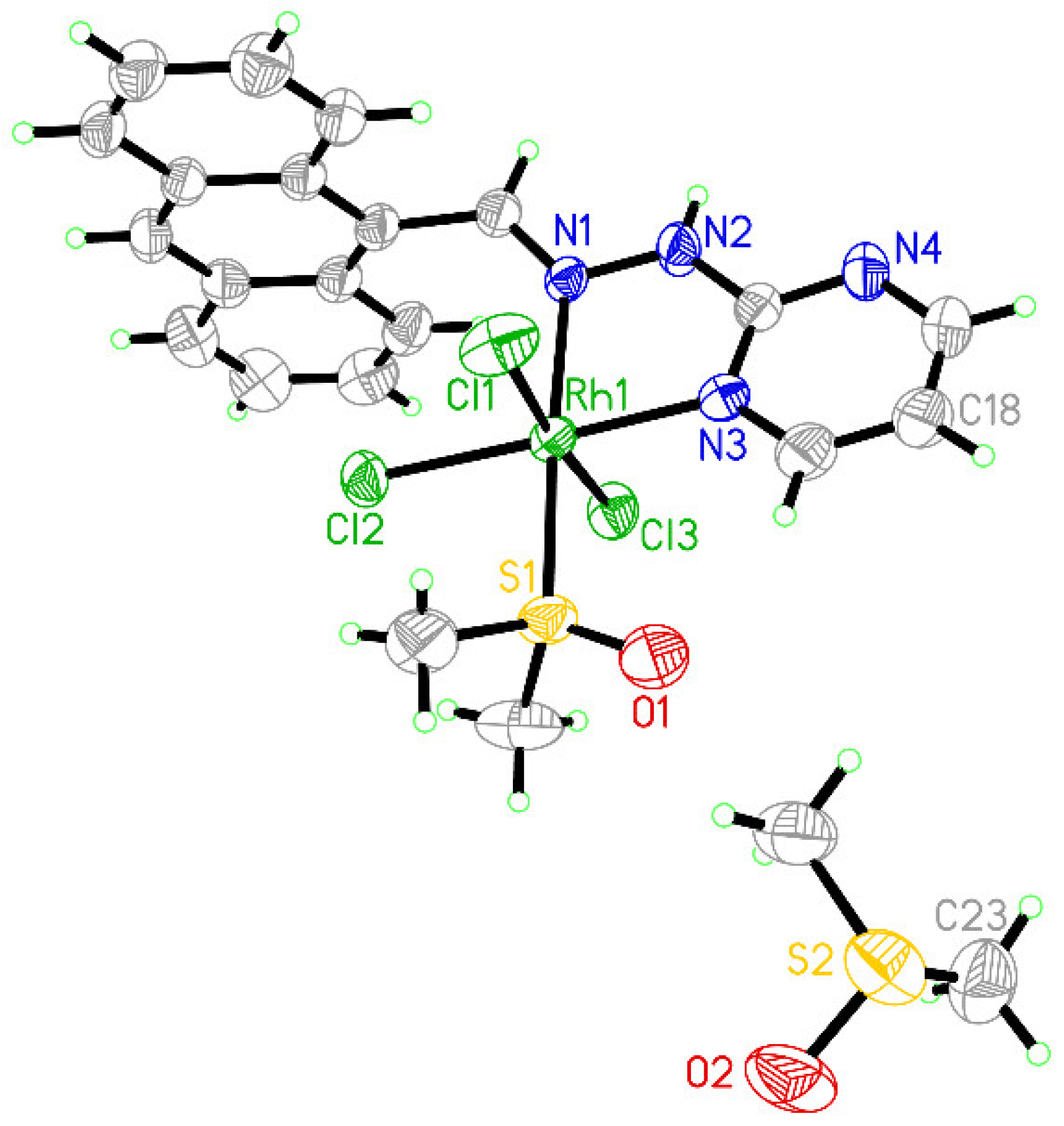
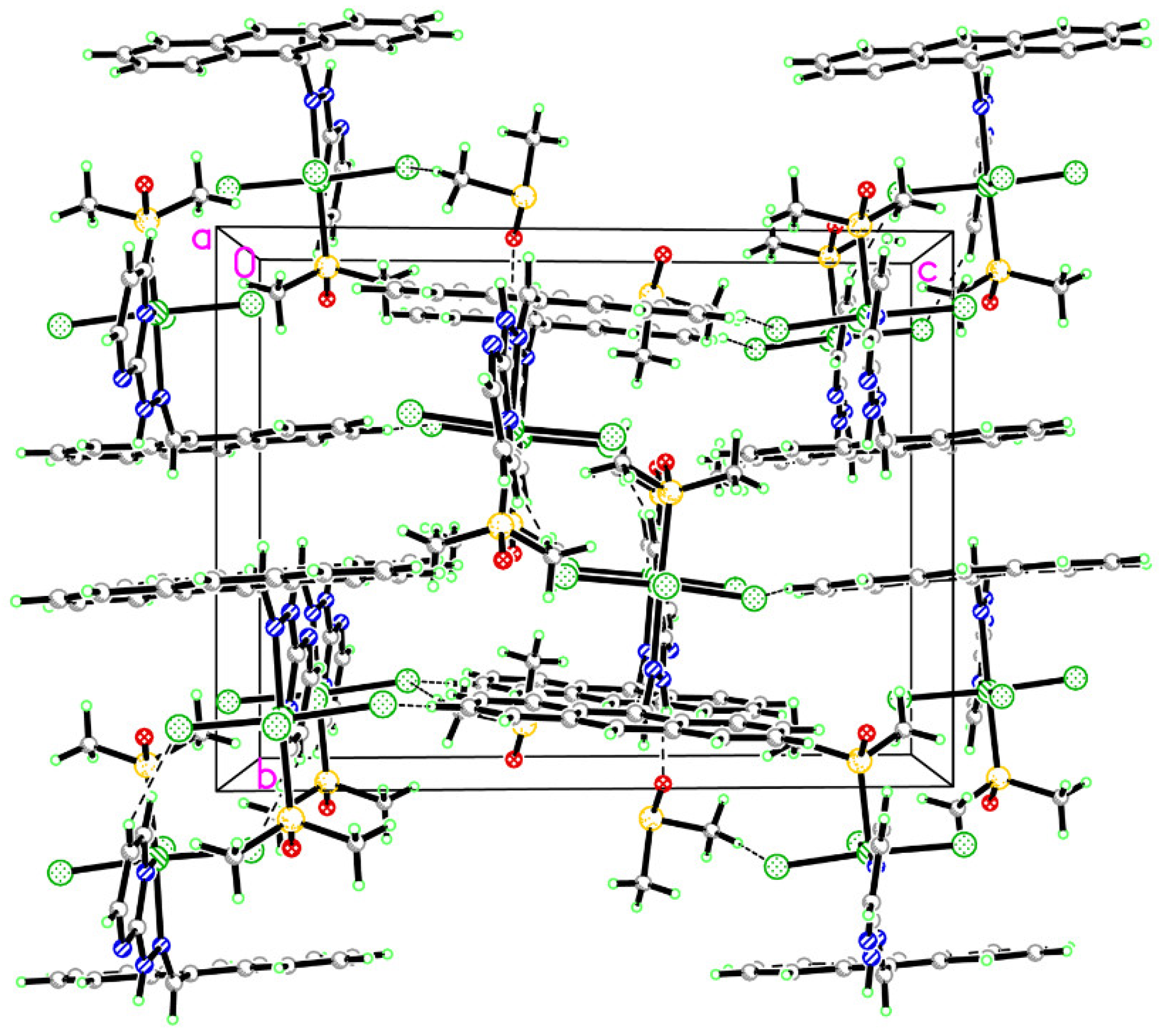
2.1. Crystal Structure Refinement and Analysis

An orange-red block single crystal with dimensions of 0.41 × 0.23× 0.12 mm3 was selected and mounted on a glass fiber. It was noted that room temperature was suited for the crystal quality of this complex, at which it would not shatter or weatherize. Thus, the data were collected, with the structure was routinely solved and refined, [12,13]. A total of 29,946 reflections were collected in the range of 1.92° < θ < 26.37° by using an ω-2θ scan mode, of which 5478 were unique, with Rint = 0.0310, and used in the succeeding structure calculations. Data intensity was corrected for Lorentz-polarization effects and empirical absorption. The structure was solved by direct methods and expanded with Fourier techniques. The non-hydrogen atoms were anisotropically refined. The final cycle of full-matrix least-squares refinement was based on 4711 observed reflections [I > 2σ(I)] and 323 variable parameters, converged with weighted agreement factors. The final R = 0.0285 and wR = 0.0684 (w = 1/[σ2(F02) + (0.0326P)2 + 1.6204P], where P = (F02 + 2Fc2)/3), with a goodness-of-fit of 1.082, and maximum and minimum peaks in the final difference Fourier map of 0.77 and –0.33 eÅ−3, respectively. The crystallographic data and refinement details of the structure analyses are summarized in Table 1, Table 2 and Table 3.
A view of the crystal structure of the title complex is shown in Figure 1, and shows that the central rhodium(III) adopts an octahedral configuration with six-coordination by one 9-PMAH ligand, one solvent DMSO, and three chloride ions, respectively. In this octahedron geometry, the 9-PMAH bidentate ligand chelates to Rh(III) via the N atom of the imine C=N (N1) and the N atom of pyrimidine (N3), whereas the three chloride ligands (Cl1, Cl2, and Cl3) are in a mer- configuration, with the DMSO cis- to the pyrimidine nitrogen. The DMSO coordinates to Rh(III) via the S atom, which is more familiar to the softer Rh center, as a fifth period metallic element.
In view of the bond angles of one of the equatorial planes (N1, N3, S1, and Cl2) of the octahedron, there is an indication that the octahedral configuration is slightly distorted. As to the bond lengths and bond angles around the central Rh(III), it is found that the bond lengths of Rh1–N1 and Rh1–N3 are comparable with the reported bond lengths of Rh–N [14,15]. A packing diagram of this complex, together with the solvent DMSO, shows that there exist two types of hydrogen bonds, N–H···O and C–H···Cl, contributing to the construction of the crystal structure, which are formed between the Cl1 and C–H bond on the pyrimidine ring of 9-PMAH, the Cl3 and C–H bond on the methyl group of DMSO, and the O2 of DMSO and N–H bond of 9-PMAH (Figure 2). On the other hand, there also exist a series of π–π interactions between the neighboring anthracene rings of each 9-PMAH ligand, the shortest distance of which was measured to be 3.6442(13) Å by the Olex2 program, which is comparable to reported values [16,17]. The whole crystal structure was thus stabilized by these hydrogen bonds and π–π interactions, to form a three-dimensional supramolecular structure.

2.2. In Vitro Cytotoxicity Activities

The rhodium(III/II) complexes are regarded as promising antitumor agents, considering their similar elemental properties and coordination mode to ruthenium(III/II) [18,19]. Thus, the in vitro inhibition ratios (%) of the 9-PMAH-Rh toward several typical human tumor cell lines were primarily screened by MTT assay, compared with cisplatin as a positive control. The experimental results are shown in Figure 3, indicating that the rhodium(III) complex, 9-PMAH-Rh, showed comparable inhibition ability on cell proliferation with cisplatin against most of the tested cell lines under the same concentration of 10 µM. Although 9-PMAH-Rh did not show much sensitivity toward some of the tested tumor cell lines, such as HeLa-229 and NCI-H460, with only ca. 30% of inhibition ratios, it indeed exhibited higher growth inhibition ratios than cisplatin toward T-24 and MGC-803 tumor cell lines. For the T-24 and MGC-803 tumor cell lines, the inhibition ratios of 9-PMAH-Rh was 41.26 ± 3.56% and 45.45 ± 1.33%, whereas those for cisplatin were only 36.32 ± 1.29 and 37.83 ± 2.00%, respectively. Compared with cisplatin, 9-PMAH-Rh showed much lower toxicity towards human normal liver cell line HL-7702. At 10 µM, the inhibition ratio of 9-PMAH-Rh on HL-7702 was 47.08 ± 0.14%, whereas that of cisplatin was up to 60.49 ± 2.29%, which strongly suggested that 9-PMAH-Rh was of higher drug safety than cisplatin in terms of its in vitro activity, considering that the liver is the primary metabolic organ for such anthracycline derivatives [20].

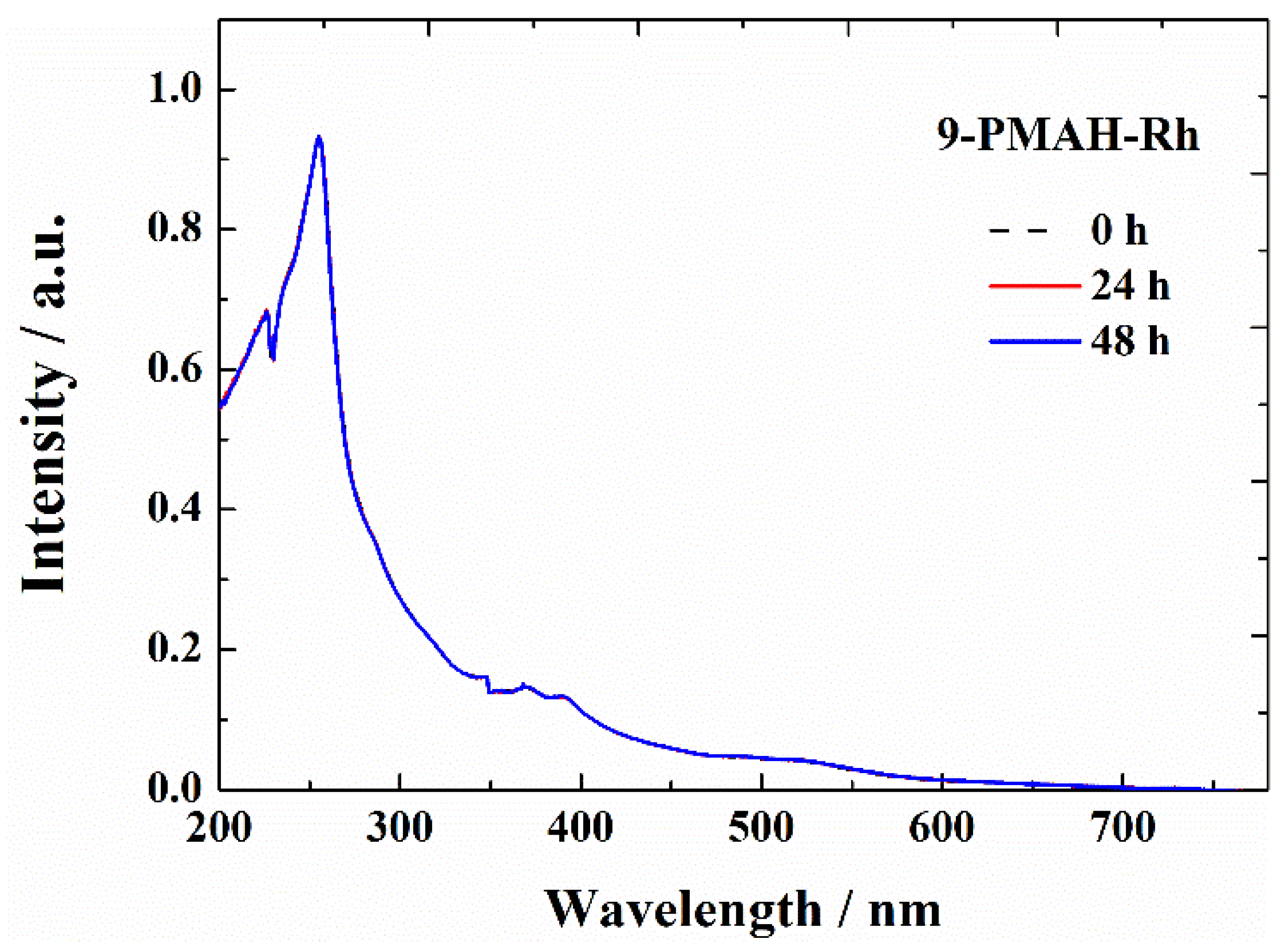
2.3. The Stability of the Complex in Solution

For antitumor metal complexes, not only are their solid crystal structure characteristics important, but also their species presence in solution. Does the metal complex remain in coordination after being dissolved in solution? It is essential to understand the antitumor activity and structure–activity relationship of metal complexes [21]. Therefore, the structural characteristics of 9-PMAH-Rh in solution were investigated by UV–Vis and ESI-MS to discuss its coordinating stability in solution.
The result of ESI-MS analysis indicated that 9-PMAH-Rh existed as the single positively charged species, [RhIIICl2(9-PMAH)]+ (+m/z, 469.97), in methanol solution, as shown in Figure 4. Thus, one of the coordinated chloride ions, as well as the co-crystallized solvent DMSO, might dissociate from the whole coordination moiety, as indicated from its crystallized state, [RhIIICl3(DMSO)(9-PMAH)]∙DMSO. In addition, the examined species, [RhIIICl2(9-PMAH)]+, may also form when under the ionization condition of ESI-MS, which means that the whole structure of this complex could be retained in the working solution of methanol. Nevertheless, if the complex is used as a potential antitumor agent, which needs to dissolve in physiological conditions, it would more easily partly dissociated, considering the easier dissociation of chloride in aqueous solution. Furthermore, the stability of 9-PMAH-Rh in aqueous solution (Tris buffer) was also examined by means of UV–Vis spectral analysis, as shown in Figure 5. The maximal absorption (Amax) of 9-PMAH-Rh was 0.9313, as shown at 255 nm with εmax = 4.66 × 10⁴ L∙mol–1. After it was kept in Tris buffer solution for 24 and 48 h, neither changes in the absorbance nor absorption shifts (red- or blue- shift) were found in the UV–Vis spectrum of 9-PMAH-Rh. This indicated that the complex would not dissociate into an anthrahydrazone ligand and aqua-rhodium(III) species, which would lead to obvious absorption shifts, and also would not precipitate or aggregate, indicated by the possible hypochromicity. The UV–Vis spectrum shown in Figure 5 may correspond to the positive-charged species, [RhIIICl2(9-PMAH)]+, suggested by the ESI-MS results.

2.4. GR-Competitive Binding Studies

The antitumor mechanism of anthracyclines, represented by Adriamycin, is to inhibit the replication and transcription of DNA by binding to DNA, thus effectively inhibiting the proliferation of tumor cells with vigorous DNA replications [22]. Therefore, when discussing the antitumor mechanisms of anthracyclines and their metal complexes, DNA will be considered the primary intracellular target. Thus, the DNA binding property of 9-PMAH-Rh was further studied by means of fluorescence spectral analysis in a competitive binding mode with GelRed, which is generally used as a bio-safe intercalative DNA binding probe.
In this study, when GelRed was incubated and then bound with ct-DNA in a [GelRed]/[DNA] ratio of 1:10, it emitted characteristic fluorescence at ca. 600 nm under excitation at 523 nm, as indicated by the black dashed line in Figure 6. Then, the addition of increasing amounts of 9-PMAH-Rh was found to efficiently quench the fluorescence emission of GelRed. Even at an extremely low [GelRed]/[DNA]/[9-PMAH-Rh] ratio of 1:10:0.01, it showed 13.57% fluorescence quenching on GelRed. The final cumulative addition of 9-PMAH-Rh led to a total 52.65% fluorescence quenching on GelRed, when the [GelRed]/[DNA]/[9-PMAH-Rh] ratio reached 1:10:0.08. It should be noted that the [GelRed]/[DNA] ratio was set to be 1:10 to ensure sufficient DNA binding sites for GelRed. However, such low concentrations of 9-PMAH-Rh still effectively quenched the fluorescence, which demonstrated the competitive DNA binding property between 9-PMAH-Rh and GelRed, further suggested the intercalative binding mode of 9-PMAH-Rh to DNA. According to the classic Stern–Volmer quenching equation, the quenching constant, Kq, of 9-PMAH-Rh. compared with GelRed to bind DNA, was calculated to be 1.45 × 10⁶ L∙mol−1, which was much higher than those of some reported rhodium complexes, comparable with the classical DNA intercalator, EtBr [23,24].

3. Experimental Section

3.1. Synthesis and Crystallization of 9-PMAH-Rh(III)

The synthesis of the anthrahydrazone ligand, 9-PMAH, has just been recently reported [25]. For the synthesis of the title complex, RhCl3 (0.1 mmol) and 9-PMAH (0.1 mmol) were mixed with methanol (3.0 mL), acetone (1.0 mL), and DMSO (0.5 mL) in a long, thick Pyrex glass tube, in which RhCl3 and 9-PMAH were only partly dissolved. Then the orange mixture reacted at 323 K for one week until the orange-red block crystals of the 9-PMAH-Rh(III) were slowly harvested from the heated orange solution. Further selection of single crystals were made for X-ray diffraction analysis (yield: 0.0463 g, 70%). IR (KBr): 3436, 3018, 1599, 1549, 1433, 1138, 1015, 778, 734, and 599 cm−1, recorded on the PerkinElmer Spectrum One FT-IR spectrometer. ESI-MS m/z: 469.97, [RhIIICl2(9-PMAH)]+; Elemental analysis: (%) for C23H26Cl3N4O2RhS2 ([RhIIICl3(DMSO)(9-PMAH)]∙DMSO): calculated: C 41.61, H 3.95, N 8.44%; found: C 41.32, H 3.80 N 8.65%, recorded on the PerkinElmer Series II CHNS/O Analyzer-2400. The determined coordination structure of the title complex, 9-PMAH-Rh(III), without the co-crystalized DMSO, is demonstrated in Scheme 1.

3.2. X-ray Crystallography

The data collection of single crystal of complex 1 was performed on a Bruker APEX-II CCD diffractometer equipped with graphite monochromator Mo-Kα radiation (λ = 0.71073 Å) at 296 K (room temperature). The structure was solved with direct methods and refined using OLEX2 and SHELXL-97 programs. The non-hydrogen atoms were located in successive difference Fourier syntheses. The final refinement was performed by full-matrix least-squares methods with anisotropic thermal parameters for the non-hydrogen atoms on F2. The hydrogen atoms were theoretically added and riding on the concerned atoms. All crystallographic data were summarized in Table 1, Table 2 and Table 3.

3.3. Biological Evaluation

3.3.1. Cell Culture

Several selected human tumor cell lines (T-24, Hep-G2, MGC-803, SK-OV-3, and NCI-H460) and one normal human cell line, HL-7702, were all obtained from the Shanghai Cell Bank in Chinese Academy of Sciences. Cells were cultured in DMEM or 1640 Gibco Medium, mixed with 10% fetal bovine serum, 100 U/mL of penicillin and 100 μg/mL of streptomycin, at 37 °C under 5% CO2/95% air atmosphere.

3.3.2. Cytotoxicity Analysis against Tumor Cell Lines

Cells were transplanted and incubated into 96-wells for 24 h. Then, we diluted the tested compound and added them into each of the 96 wells, with the final concentrations of all tested compounds set to 1.25, 2.5, 5, 10, and 20 μM, respectively. After 48 h of cultivation, the MTT solution (10 μL, 5 mg/mL) was added to each of the 96 wells and then incubated for another 4–6 h. Finally, the formed formazan crystals were dissolved in 100 μL of DMSO and measured by a multi-functional enzyme marker. The IC50 values were independently performed at least three times [26].

3.4. The Solution Chemistry Study of the Complex Examined by Spectroscopic Analysis

The solution chemistry of 9-PMAH-Rh was studied by ESI-MS (electrospray ionization mass spectrometry) and UV–Vis (Ultraviolet–visible) spectral analysis to analyze and confirm its stability and proposed species in solution. ESI-MS and UV–Vis spectra was performed on a Thermo-Finnigan LCQ/AD Quadrupole Ion Trap ESI-MS and a PerkinElmer Lambda 25 UV–Vis spectrometer, respectively. For the ESI-MS analysis, the stock solution of 9-PMAH-Rh was used before it was diluted with methanol to form a working solution. Then, it was injected into the mass spectrometer to record the existing species under the ionized mode. For the UV–Vis absorption experiment, the working solution of 9-PMAH-Rh was prepared to 2 mM in a 1 cm quartz cuvette, by dilution in DMSO stock solution with Tris buffer. Then, the solution was respectively scanned and recorded at 0, 24, and 48 h under the same conditions.

3.5. DNA Binding Study by Fluorescent Spectral Analysis

The working solution for the GR-DNA competitive experiment was prepared according to a concentration ratio of [GR]/[DNA] = 1:10, in which [GR] and [DNA] in the mixed solution were set to 1 × 10⁻⁵ and 1 × 10⁻⁴ mol·L−1, respectively. The Tris buffer solution was added to fix the volume of the working solution to be 3.0 mL. It was then stored in a refrigerator overnight to make sure that GR and DNA were fully bound. The stock solution of 9-PMAH-Rh (2 × 10⁻³ mol·L−1 in DMSO) was then added gradually into the working solution by a microinjector, to achieve the [GelRed]/[DNA]/[9-PMAH-Rh] ratio, step-to-step, from 1:10:0.01 to 1:10:0.08, where it reached a nearly saturated state. The fluorescence emission spectra of Ex (350 nm)/Em (590 nm) were recorded at the slit width of 5 nm/5 nm. For each step, the maximum fluorescence emission intensity was recorded, and the emission curve was saved, to form the GR-DNA complex competitive binding plot and calculate the quenching constant, Kq. Kq was obtained by a linear fit of plotting I0/I versus [Q], using the classic Stern–Volmer equation: I0/I = 1 + Kq × [Q], in which I0 and I refer to the maximum emission intensity of the GR-DNA system in the absence and presence of 9-PMAH-Rh, as both the quencher and competing binding agent, whereas [Q] is the concentration of the quencher.

4. Conclusions

In this work, we report a new rhodium(II) complex of a new single-armed anthrahydrazone ligand, [RhIIICl3(DMSO)(9-PMAH)]. This is a new exploration on the structural design and properties of the metal complexes of anthrahydrazone, which have been rarely and studied until now. Although this complex does not show significant antitumor activity as expected against a panel of tested human tumor cell lines in vitro, it indeed shows very high intercalative binding ability with DNA, compared with the typical DNA intercalator, GelRed. This contrast is most likely related to the hydrophilicity of the complex in solution under physiological conditions, as suggested by the results of ESI-MS, resulting in its inadequate ability to penetrate the cell membrane. Future studies should focus on methods to improve or modulate lipophilicity of these metal complexes. However, this study strongly suggests that the transition metal complexes of anthrahydrazone ligands under a rational design may be a promising pathway to develop new metal-based antitumor candidates, thanks to the bioactivity of anthracyclines such as Adriamycin.

Author Contributions

Conceptualization, Y.L. (Yancheng Liu); Methodology, Z.T.; Formal analysis, H.L.; Investigation, Q.D.; Data curation, Z.T. and Y.L. (Yuan Liu); Writing – original draft, R.L.; Writing – review & editing, Z.T. and Q.D.; Supervision, Y.L. (Yancheng Liu); Funding acquisition, Y.L. (Yancheng Liu) and Q.D. All authors have read and agreed to the published version of the manuscript.

Funding

This research was funded by the National Natural Science Foundation of China [21561005]; the Key Project of Biomedicine and Health in Colleges and Universities of Guangdong Province [2021ZDZX2055] and the Medical Science and Technology Research Fund of Guangdong Province [A2022004].

Acknowledgments

We thank Fuping Huang for crystallographic directions and Mingguang Yu for syntax checking and proofreading.

Conflicts of Interest

The authors declare no conflict of interest.

References

  1. Panaretakis, T.; Pokrovskaja, K.; Shoshan, M.C.; Grandér, D. Activation of Bak, Bax, and BH3-only proteins in the apoptotic response to doxorubicin. J. Biol. Chem. 2002, 277, 44317–44326. [Google Scholar] [CrossRef] [PubMed] [Green Version]
  2. Shenkenberg, T.D.; Von Hoff, D.D. Mitoxantrone: A new anticancer drug with significant clinical activity. Ann. Intern. Med 1986, 105, 67–81. [Google Scholar] [CrossRef] [PubMed]
  3. Agudelo, D.; Bourassa, P.; Bérubé, G.; Tajmir-Riahi, H.A.J. Review on the binding of anticancer drug doxorubicin with DNA and tRNA: Structural models and antitumor activity. Photochem. Photobiol. B. 2016, 158, 274–279. [Google Scholar] [CrossRef] [PubMed]
  4. Coltman, C.A., Jr.; Osborne, C.K. Bisantrene, biological and clinical effects. Cancer Treat. Rev. 1984, 11, 285–288. [Google Scholar] [CrossRef]
  5. Qin, Q.-P.; Liu, Y.-C.; Wang, H.-L.; Qin, J.-L.; Cheng, F.-J.; Tang, S.-F.; Liang, H. Synthesis and antitumor mechanisms of a copper(ii) complex of anthracene-9-imidazoline hydrazone (9-AIH). Metallomics 2015, 7, 1124–1136. [Google Scholar] [CrossRef]
  6. Karasawa, T.; Steyger, P.S. An integrated view of cisplatin-induced nephrotoxicity and ototoxicity. Toxicol. Lett. 2015, 237, 219–227. [Google Scholar] [CrossRef] [Green Version]
  7. Zhong, Y.; Jia, C.; Zhang, X.; Liao, X.; Yang, B.; Cong, Y.; Pu, S.; Gao, C. Targeting drug delivery system for platinum (IV)-Based antitumor complexes. Eur. J. Med. Chem. 2020, 194, 112229. [Google Scholar] [CrossRef]
  8. Štarha, P. Multinuclear biologically active Ru, Rh, Os and Ir arene complexes. Coord. Chem. Rev. 2021, 431, 213690. [Google Scholar] [CrossRef]
  9. Bear, J.L.; Gray, H.B., Jr.; Rainen, L.; Chang, I.M.; Howard, R.; Serio, G.; Kimball, A.P. Interaction of Rhodium (II) Carboxylates with Molecules of Biologic. Cancer Chemother. Rep. 1975, 59, 611–620. [Google Scholar]
  10. Howard, R.A.; Kimball, A.P.; Bear, J.L. Mechanism of action of tetra-µ-carboxylatodirhodium (II) in L1210 tumor suspension culture. Cancer Res. 1979, 39, 2568–2573. [Google Scholar]
  11. Peng, Y.B.; Tao, C.; Tan, C.P.; Zhao, P. Mitochondrial targeted rhodium (III) complexes: Synthesis, characterized and antitumor mechanism investigation. J. Inorg. Biochem. 2021, 218, 111400. [Google Scholar] [CrossRef] [PubMed]
  12. Dolomanov, O.V.; Bourhis, L.J.; Gildea, R.J.; Howard, J.A.K.; Puschmann, H.J. iotbx. cif: A comprehensive CIF toolbox. Appl. Cryst. 2009, 42, 339–341. [Google Scholar] [CrossRef]
  13. Sheldrick, G.M. Redetermination of cyclo-trimethylenetrinitramine. Acta Cryst. 2008, A64, 112–122. [Google Scholar] [CrossRef] [PubMed] [Green Version]
  14. Dkhar, L.; Kaminsky, W.; Poluri, K.M.; Kollipara, M.R.J. Versatile coordination modes of benzothiazole hydrazone derivatives towards Ru(II), Rh(III) and Ir(III) complexes and their reactivity studies with azides and activated alkynes. Organomet. Chem. 2019, 891, 54–63. [Google Scholar] [CrossRef]
  15. Palepu, N.R.; Premkumar, J.R.; Verma, A.K.; Bhattacharjee, K.; Joshi, S.R.; Forbes, S.; Mozharivskyj, Y.K.; Rao, M. Antibacterial, in vitro antitumor activity and structural studies of rhodium and iridium complexes featuring the two positional isomers of pyridine carbaldehyde picolinic hydrazone ligand. Arab. J. Chem. 2018, 11, 714–728. [Google Scholar] [CrossRef] [Green Version]
  16. Pal, P.; Das, K.; Hossain, A.; Frontera, A.; Mukhopadhyay, S. Supramolecular and theoretical perspectives of 2,2′:6′,2′′-terpyridine based Ni(II) and Cu(II) complexes: On the importance of C–H⋯Cl and π⋯π interactions. New J. Chem. 2020, 44, 7310–7318. [Google Scholar] [CrossRef]
  17. Pramanik, S.; Pathak, S.; Frontera, A.; Mukhopadhyay, S. Syntheses, crystal structures and supramolecular assemblies of two Cu(II) complexes based on a new heterocyclic ligand: Insights into C–H⋯Cl and π⋯π interactions. Cryst. Eng. Comm. 2022, 24, 1598–1611. [Google Scholar] [CrossRef]
  18. Geldmacher, Y.; Kitanovic, I.; Alborzinia, H.; Bergerhoff, K.; Rubbiani, R.; Wefelmeier, P.; Prokop, A.; Gust, R.; Ott, I.; Wölfl, S.; et al. Cellular Selectivity and Biological Impact of Cytotoxic Rhodium (III) and Iridium (III) Complexes Containing Methyl-Substituted Phenanthroline Ligands. Chem. Med. Chem. 2011, 6, 429–439. [Google Scholar] [CrossRef]
  19. Khan, T.M.; Gul, N.S.; Lu, X.; Kumar, R.; Choudhary, M.I.; Liang, H.; Chen, Z.-F. Rhodium (III) complexes with isoquinoline derivatives as potential anticancer agents: In vitro and in vivo activity studies. Dalton Trans. 2019, 48, 11469–11479. [Google Scholar] [CrossRef]
  20. Skarka, A.; Škarydová, L.; Štambergová, H.; Wsól, V. Anthracyclines and their metabolism in human liver microsomes and the participation of the new microsomal carbonyl reductase. Chem-Biol. Interact. 2011, 191, 66–74. [Google Scholar] [CrossRef]
  21. Gothe, Y.; Marzo, T.; Messori, L.; Metzler-Nolte, N. Iridium(I) Compounds as Prospective Anticancer Agents: Solution Chemistry, Antiproliferative Profiles and Protein Interactions for a Series of Iridium(I) N-Heterocyclic Carbene Complexes. Chem. Eur. J. 2016, 22, 12487–12494. [Google Scholar] [CrossRef] [PubMed]
  22. Bachur, N.R. Encyclopedia of Cancer, 2nd ed.; Elsevier: Amsterdam, The Netherlands, 2002; pp. 57–61. [Google Scholar]
  23. Zhang, G.; Guo, J.; Zhao, N.; Wang, J. Study of interaction between kaempferol–Eu3+ complex and DNA with the use of the Neutral Red dye as a fluorescence probe. Sensor Actuat. B Chem. 2010, 144, 239–246. [Google Scholar] [CrossRef]
  24. Lakowicz, J.R. Principles of Fluorescence Spectroscopy, 3rd ed.; Springer: Boston, MA, USA, 2006; pp. 278–280. [Google Scholar]
  25. Liu, R.-X.; Wu, Y.-S.; Liu, Y.-C.; Luo, R.-Y.; Yang, L.-D.; Tang, M.-T.; Chen, Z.-F.; Liang, H. New anthrahydrazone derivatives and their cisplatin-like complexes: Synthesis, antitumor activity and structure–activity relationship. New J. Chem. 2019, 43, 18685–18694. [Google Scholar] [CrossRef]
  26. The MTT Assay Protocol for Tumor Cell Proliferation Is Available on the Web Site. Available online: https://www.thermofisher.cn/cn/zh/home/references/protocols/cell-and-tissue-analysis/protocols/vybrant-mtt-cell-proliferation-assay-protocol.html (accessed on 15 October 2022).
Scheme 1. The chemical structure of the title complex, [RhCl3(DMSO)(9-PMAH)]∙DMSO.
Crystals 12 01664 sch001
Figure 1. An ORTEP view of the title complex, 9-PMAH-Rh, showing displacement ellipsoids at the 50% probability level (co-crystalized solvent DMSO omitted for clarity).
Crystals 12 01664 g001
Figure 2. The packing diagram of the crystal structure of 9-PMAH-Rh viewed parallel to the a-axis, showing the C–H···Cl and N–H···O hydrogen bonds (shown as dashed lines).
Crystals 12 01664 g002
Figure 3. In vitro inhibition ratios (%) of 9-PMAH-Rh toward a series of human tumor cell lines.
Crystals 12 01664 g003
Figure 4. The ESI-MS analysis of 9-PMAH-Rh in methanol solution.
Crystals 12 01664 g004
Figure 5. UV–Vis spectrum of 9-PMAH-Rh, showing its stability over 48 h in Tris buffer solution at room temperature.
Crystals 12 01664 g005
Figure 6. The intercalative binding ability of 9-PMAH-Rh to ct-DNA when competing with GelRed, examined by means of fluorescence spectral analysis.
Crystals 12 01664 g006
Table 1. Crystal data and structure refinement parameters for the complex 9-PMAH-Rh.
Empirical FormulaC23H26Cl3N4O2RhS2
Formula weight663.86
Temperature/K296.15
Crystal systemmonoclinic
Space groupP21/n
a/Å, b/Å, c11.573(5), 13.309(5), 17.577(7)
α/°, β/°, γ90.00, 94.955(6), 90.00
Volume/Å32697.2(18)
Z4
ρcalcg/cm31.635
μ/mm−11.114
F(000)1344.0
Crystal size/mm30.41 × 0.23 × 0.12
RadiationMoKα (λ = 0.71073)
2θ range for data collection/°3.84 to 52.74
Index ranges−14 ≤ h ≤ 14, −16 ≤ k ≤ 16, −20 ≤ l ≤ 21
Reflections collected29946
Independent reflections5478 [Rint = 0.0310, Rsigma = 0.0225]
Data/restraints/parameters5478/0/323
Goodness-of-fit on F21.082
Final R indexes [I >= 2σ (I)]R1 = 0.0285, wR2 = 0.0684
Final R indexes [all data]R1 = 0.0363, wR2 = 0.0729
Largest diff. peak/hole / e Å−30.77/−0.33
Table 2. Selected bond lengths and angles for 9-PMAH-Rh.
Selected bond (Å)
Rh1-Cl12.3437(12)Rh1-S12.2869(11)
Rh1-Cl22.3437(12)Rh1-N12.118(2)
Rh1-Cl32.3320(11)Rh1-N32.063(2)
Selected angles (°)
Cl2-Rh1-Cl193.45(3)S1-Rh1-Cl191.07(3)
Cl3-Rh1-Cl1174.79(3)S1-Rh1-Cl284.14(3)
Cl3-Rh1-Cl290.62(3)S1-Rh1-Cl392.58(3)
N1-Rh1-Cl190.22(6)N1-Rh1-Cl385.95(6)
N1-Rh1-Cl298.47(6)N1-Rh1-S1177.01(6)
Table 3. Hydrogen-bond geometry (Å, °) existing in 9-PMAH-Rh.
D—H···AD—HH···AD···AD—H···A
N2—H2···O2 i0.861.882.733(3)174
C18—H18···Cl1 ii0.932.783.600(4)148
C23—H23C···Cl3 iii0.962.833.713(4)154
Symmetry codes: i x, y + 1, z; ii −x + 1, −y + 1, −z + 1; iii −x + 3/2, y−1/2, −z + 3/2.
Publisher’s Note: MDPI stays neutral with regard to jurisdictional claims in published maps and institutional affiliations.

