Abstract
Single crystals of Cu-8.% at. Al were subjected to cold drawing up to 1.1 true strain. Development of the crystallographic texture was analysed at various stages of the drawing process. Investigations revealed significant crystal rotation and reorientation due to a twinning process, while general 4-fold symmetry around the drawing direction was preserved. Local texture analysis performed on cross-sections perpendicular to the drawing direction showed that due to crystal rotation, the original crystal was initially fragmented into four quadrants. However, within the volume near the central axis the initial cubic orientation was preserved. Subsequent deformation within dominating twinning systems led to further fragmentation which divided the crystal into sectors one eighth the size of the full circular cross-section. The obtained results also indicate that approximation of the drawing process stress state by the tensor having only normal components cannot be used during analysis of the activation of deformation systems.
1. Introduction
Evolution of the microstructure of metallic materials during deformation processes is fundamental to determining their physical and mechanical parameters. The final microstructure state as well as the resulting properties depend on both the deformation path and the initial state of the material. Use of single crystals allows for precise control of the starting conditions prior to the deformation process. In particular, perfect or near perfect initial orientation and lack of grain boundaries allow for tracking microstructure evolution at the level of operating deformation mechanisms. Face-centred cubic (FCC) single crystals have been used for years to test material response in the various deformation schemes beginning from uniaxial stress conditions through tests with complex stress tensors. These include channel die tests [,,] and very complex deformation path tests using several plastic deformations, such as equal channel angular pressing (ECAP) [,] or high-pressure torsion (HPT) [].
Special cases are axisymmetric deformation routes such as wire drawing, which can achieve high cumulative deformation. At the same time, the applied load can be approximated by a simplified stress tensor, enabling analysis of deformation mechanisms. There are many papers about evolution of the microstructure, crystallographic texture, and mechanical properties during the drawing process in FCC materials, however, most of them are performed on polycrystalline samples where macroscopic fibre components of the texture were analysed using both X-ray and electron backscatter diffraction (EBSD) techniques [,,]. The most frequently cited work on the analysis of the deformation texture was published by English and Chin []. Duplex fibre textures of <111> and <100> components parallel to the axis direction in drawn FCC samples were found. Non-monotonic change in the strength of the <100> component with respect to the material stacking fault energy (SFE) value was shown. The authors concluded that it may be caused by the activation of the deformation twinning mechanism leading to formation of new reoriented volumes in the drawn samples.
More detailed information about SFE’s impact on texture formation is presented in the work of Kuffmann et al., where Cu and Cu-Al polycrystalline samples were subjected to the drawing process at room and cryogenic temperatures []. By varying the SFE parameter, the authors found that activation of the twinning mechanism resulted in additional <115> fibre texture components over standard <100> and <111> fibre components in non-twinning cases. The new <115> fibre component was attributed to the activation of the twinning process within grains having the <111> texture component. It is important to mention that formation of the new twin orientation was attributed to the grains characterised by the <111> component only. On the contrary, grains with the <100> component were considered as twin-free areas due to unfavourable orientation with respect to the applied tensor stress.
As mentioned above, although analysis of polycrystalline material gave important insights about macroscopic texture evolution, detailed analysis of the activation of the deformation systems is difficult if not impossible. Evolution of the microstructure of single crystalline Cu with <100>, <110> and <111> initial orientations was studied by the group of Chen []. The EBSD technique was employed to measure variations in the texture component during various steps of drawing. The authors reported stability of <100> and <111> components as well as inhomogeneity of the orientation distribution along the radial direction of the samples. However, mechanism of texture evolution by means of operating systems were not analysed. All investigations were performed on the cross-section parallel to the drawing axis. Such selection hinders the possibility for analysis of active deformation mechanisms with respect to the axisymmetric stress tensor. In the light of the work of Borbely, selected cross-sections can lead to false inconsistent results []. In particular, the paper presents detailed analysis of the texture evolution during drawing of <100> Cu performed by means of experimental X-ray measurements as well as simulations. All data gathered/acquired and presented on the cross-section perpendicular to the drawing axis showed fragmentation of the crystal into four quadrants. The most important conclusion from the work was that the macroscopic 4-fold pseudo-symmetry along the drawing axis was preserved during deformation. In each crystal quadrant, rotation of the <100> axis towards the corresponding <111> direction was observed, giving a characteristic cross pattern in the centre of {200} pole figure. The authors concluded that such microstructure/texture is stable for deformations approaching 0.26 of a true strain. Cu has relatively high SFE at room temperature, thus deformation progresses in slip systems only. By adding Al and thus decreasing SFE, the deformation path of the alloy will change. Low SFE will affect plane slip phenomena, and additionally a twinning mechanism will be activated, leading to the formation of the new orientations in the material. To our best knowledge, there is no information in the literature about texture evolution of a single crystal with high propensity to twinning. In the current work, Cu-8%Al single crystals with initial orientation of <100> were used to qualitatively show the sequence of deformation system activation and their impact on the texture evolution.
2. Material and Methods
A single crystal Cu-Al alloy (Cu-8.5% at. Al) with an initial crystallographic orientation of <001> was used for the experiments. Single crystals were obtained using the Bridgman–Stockbarger technique where the temperature gradient change was enforced by three separately controlled heating zones. Crystals were grown in a rectangular 12 mm × 12 mm × 120 mm crucible made from high-purity graphite. At the bottom of the crucible, an additional groove for a small seed (4 mm × 4 mm × 20 mm) with selected crystallographic orientation was prepared. The crucible height position was aligned to set the seed just below lowest heating zone. The heater temperature was set to create a temperature gradient inside the reaction chamber and to melt the seed in approximately half of its height. During crystal growth, the temperature was then continuously decreased with the rate 25 °C per hour. After the 3rystallization process, rectangular prisms with dimensions of 12 mm × 12 mm × 120 mm were obtained. Then, a wire electrical discharge machine was used to cut samples 9 mm in diameter and 120 mm in length. The prepared samples were subjected to the drawing process at room temperature with three die diameters of 7.8 mm, 6.4 mm and 5.2 mm. Reduction in diameter with corresponding cumulative strain is presented in Table 1. Texture as well as EBSD analysis was performed for selected deformation steps (Table 1) on the cross-sections perpendicular to the drawing direction. Samples were cut using a precise mechanical saw, ground on successive paper grades and finally electropolished using a D2 Struers electrolyte. Electropolishing process conditions were as follows: the voltage was 7 V; the electrolyte temperature was 283 K. For the calculation of true strain ε, following formula was used:
where: h0 and h are the initial and actual diameter of the specimen, respectively.

Table 1.
Summary of die diameters used during drawing process together with cumulative deformation applied at given processing step.
Texture measurements were carried out using a Panalytical Empyrean X-ray diffractometer equipped with a Cu anode X-ray source. Pole figures were measured for {111} and {200} planes with an angle step of 1° and are presented without any correction.
EBSD analysis was performed using a Versa 3D FEG-SEM equipped with a Hikari EDAX camera. Maps were acquired with 20 kV accelerating voltage, 16 nA beam current and 4 × 4 binning mode, resulting in resolution of 120 × 120 pixels. Depending on the magnification during map acquisition, the step size varied between 200 nm and 2.5 µm. Large area maps were acquired in several subregions and subsequently stitched together using OIM software from EDAX. All maps are presented in this form without any cleaning procedures applied. Direction for colour coding of the inverse pole figure maps was parallel to the drawing direction of the crystal. Poles figures within selected EBSD map subregions were reconstructed using OIM software.
3. Results
3.1. Macro-Texture Analysis
Figure 1 presents {111} and {200} pole figures at three deformation steps of 0.29, 0.68 and 1.1 of true strain. All samples were aligned on the goniometer of the X-ray diffractometer in such a way as to preserve the reference frame corresponding to the initial cubic orientation of the crystal. After the strain of 0.29, broadening and splitting of the {111} poles around the initial position (marked by the triangles) is clearly visible (Figure 1a). Additionally, new {111} poles appeared near the centre of the stereographic projection, suggesting formation of the new orientation. Similarly, the {200} pole figure showed symmetric splitting of the central pole (marked by a square on the projection). Both effects can be ascribed to the fragmentation of the drawn sample due to local crystal rotation and mechanical twinning mechanisms. Details will be presented in the following EBSD analysis section. As the deformation progressed to 0.68 true strain, {111} crystal fragmentation caused significant texture weakening (Figure 1b). Poles close to the initial cubic orientation are less localised, while four previously created {111} poles near the projection centre are transformed by splitting into two separate peaks each. Central {200} poles are more distant from the projection centre and new weak peaks are formed in the vicinity of the initial cubic {111} pole orientation. Besides quantitative changes, direct correspondence between pole figures for the deformation steps of 0.29 and 0.68 is clearly seen. However, pole figures obtained for the crystal drawn to 1.1 true strain are qualitatively completely different (Figure 1c) in comparison to the deformation of 0.68 (Figure 1b). Initial {111} poles are almost invisible in the pole figure background, while texture intestines in the centre of the figure evolved from v-shaped features of the localised peaks. The {200} pole figure is showing much stronger peaks positioned at approximately 45° with respect to the horizontal/vertical direction. Besides substantial figure changes, the 4-fold pseudo-symmetry is preserved, showing that in each quadrant crystal fragmentation is similar. This pseudo-symmetry allowed us to focus only on a quarter of the sample cross-section during the following EBSD analysis.
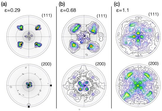
Figure 1.
The {111} and {200} pole figures for texture after drawing to cumulative strains of 0.29 (a), 0.68 (b) and 1.1 (c). Initial position of the {111} and {200} poles are marked by the black triangles and squares for the strain of 0.29.
3.2. EBSD Measurements
An inverse pole figure map showing a quarter of the cross-section of the wire drawn to deformation 0.68 is presented in Figure 2. Three distinctive regions with different dominating colours can be distinguished: centre sample region (1) dominated by red colours, magenta in the outer sample areas (2 and 2’) and yellow (3) in the transition zone aligned 45° to the horizontal direction. For each region, pole figures were recalculated and are represented in the form of stereographic projections in Figure 2b,c. Major orientation components are shown in the form of large partially reconstructed circles in the stereographic projection connecting {111} poles. Region 1 in the crystal centre preserves the initial 001 single crystal orientation, where imposed deformation causes pole broadening around the initial crystal orientation. Moving towards outer crystal regions, the magenta colour becomes dominant (regions 2 and 2’). Corresponding {111} and {200} pole figures show effects of significant crystal rotation and mechanical twinning effects. Three distinctive orientations can be seen, with red representing rotated matrix orientation while twin orientations are marked by blue and magenta. Intensity of the poles belonging to each twin orientation shows that one twinning variant is dominant within each area, 2 or 2’. EBSD analysis performed at the higher magnification confirms the above conclusion. Figure 3a shows a closer look at the twinned structure within region 2. For clarity of presentation, a map is presented in the Euler angles coding scheme. Matrix and two different twin orientations are visible, where the blue one is dominant among them. A trace of the blue twin plane corresponds to the habit plane of the strongest twin in the pole figure for region 2 (Figure 2b).
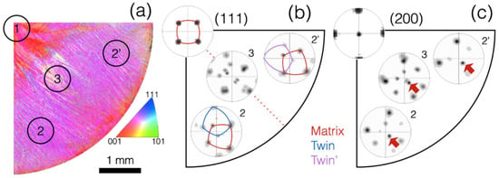
Figure 2.
EBSD analysis of one crystal quadrant deformed to 0.68 of the true strain. IPF map showing 4 distinctive regions (a). Pole figures from the regions marked in IPF (b,c), corresponding to the (111) and (200) planes reconstruction, respectively. Red arrows show direction with respect to which matrix lattice rotates while red dotted line is showing additional symmetry in the formed microstructure.
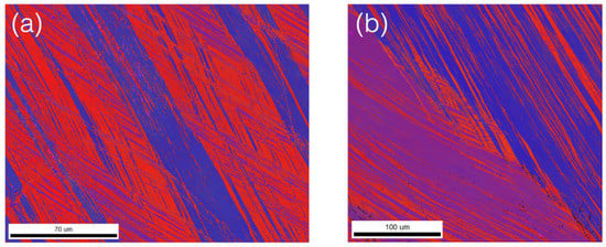
Figure 3.
EBSD maps of magnified regions 2 (a) and 3 (b) from Figure 2 displayed in the Euler coding.
A magnified EBSD map acquired in region 3 (Figure 3b) shows the transition area between regions 2 and 2’. The upper right of the map and bottom left region show dominance of different twinning orientations (highlighted in blue and magenta).
The presented structure in the analysed quarter of the examined cross-section is thus symmetric along the dotted line (Figure 2b), where active twinning mechanisms in region 2 and 2’ have a mirror-like relationship along the () plane as defined in the initial crystal reference frame. Following this result, subsequent EBSD analysis for crystal deformed to 1.1 true strain was performed only in a 1/8 section of the crystal.
Figure 4 shows the IPF map together with pole figures measured for the crystal deformed to 1.1 true strain. Three separate zones discussed previously are still preserved, however, intensity of the twin lattice presented in the (111) and (200) pole figures (highlighted in magenta) prevails over intensity of the matrix lattice (red). It is also evident that formation of the new orientation changed the lattice rotation in the opposite direction than discussed previously. This change can be ascribed to the activation of the slip systems connected to the twin lattice, which gradually become the dominant orientation within the material volume.
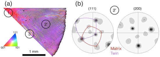
Figure 4.
EBSD analysis of one 1/8 crystal section deformed to the 1.1 of the true strain: IPF map with marked characteristic regions (a), (111) and (200) pole figures from the regions 2’ (b).
4. Discussion
Stability of the given crystal orientation during plastic deformation depends on the alignment of possible deformation systems with respect to the applied loads. Calculation of highly stressed systems can be easily performed by knowing the stress tensor. In the case of uniaxial stress conditions, the active deformation system is usually determined by the calculation of the Schmid factor [,]. However, the drawing process is characterised by a more complex stress tensor, which is often approximated by three normal components. These are positive S1 in the drawing direction, along with two others, S2 and S3, which are negative. Due to symmetry of the drawing the approximation, S2 = S3 is often made (Figure 5). Calculation of the maximum shear stress (resolved shear stress) and thus activation of a particular deformation system can be carried out with the full tensor transformation. However, under constraints of S1 being parallel to the drawing direction and S2 = S3, the selection ratios of the resolved shear stresses in particular deformation systems are the same as in the tensile test case []. This result means that selection of the active deformation systems will be the same. Geometrical representation of the available slip systems in cubic configuration is presented in Figure 5a. Assuming accurate [001] orientation, eight slip systems are equally stressed (four {111} planes with two {110} directions each). At the same time, because of the twin shear polarisation phenomena, twin systems should be inactive []. Tensile tests of FCC crystals showed that the <001> initial orientation is not stable and is rotating towards the <112> direction, following the path of the one temporarily active slip system []. Contrary to this observation, during the drawing process of the <100> single crystal, fragmentation of the orientation is observed. It led to characteristic 4-fold pseudo-symmetry where the initially split <100> direction was rotated toward the <112> direction in each crystal quadrant.
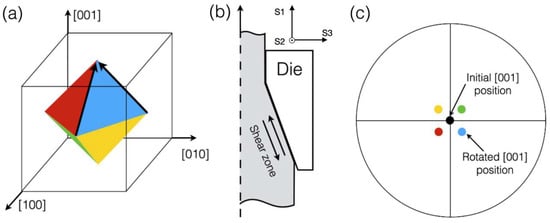
Figure 5.
The diagram showing additional shear zone during drawing process (a,b), stereographic projection with marked poles after rotation in each crystal quadrant (c).
The major differences between tensile and drawing tests are the constraints imposed by the tooling. In particular, material flowing through a cone-shaped die is subjected to an additional shear component (Figure 5b), the magnitude of which changes monotonically along the wire radius with the highest value at the sample surface []. The resulting additional shear component is responsible for the local rotation of the lattice, leading to crystal fragmentation into four quadrants. A geometrical representation of the local crystal rotation is shown in Figure 5a. Among the possible slip planes, for the quadrant presented in Figure 2a, the most favourable slip plane is highlighted in blue. Shearing material along slip directions marked with the black arrows will cause relative lattice rotation around direction [10]. Once again, the described situation can be referenced to the tensile test experiment where, due to external constraints, the tensile axis rotates in the opposite direction to the shear in an active slip system [,]. Complete split and rotation of the (200) pole in all quadrants is presented in Figure 5c where each pole colour corresponds to the slip plane from Figure 5a. Pole figures presented in Figure 1 clearly support the above analysis. Obtained texture for the sample drawn to 0.29 true strain shows four distinctive poles in the centre of the (200) pole figure, each being the result of the lattice rotation toward the respective <112> direction. While cumulative deformation increases, lattice rotation and thus pole rotation away from the initial <001> position also increase.
A schematic of the lattice rotation within one crystal quadrant is also evident from EBSD analysis (Figure 2). Similar behaviour was observed for the drawn <001> Cu single crystals performed by Borbely [], who observed formation of the central cross in the (200) pole figure. The distinctive feature of the pure Cu case was the continuously increasing rotation of the lattice from the centre of the sample outward. As the result, continuous spread of the <001> pole was observed, forming the cross shape. On the contrary, the Cu-Al alloy shows sharp peak positions, suggesting relatively uniform rotation within large volumes of the material. This situation is clearly visible on the IPF maps acquired for crystal deformed to 0.29 true strain (Figure 2). Large parts of the image have the same magenta colour with a sharp transition to the red colour zone in the central region. The difference between the behaviour of the pure Cu and Cu-Al alloy can be explained by lower SFE of the former. With the decrease in the SFE, cross slip activity is reduced [,].
An additional characteristic of low-SFE materials is propensity to mechanical twinning. Due to polarisation of the twinning shear [], the <001> orientation during tensile load makes mechanical twinning an unfavourable deformation mode. Additionally, critical resolved shear stress for mechanical twinning is higher, which delays activation of the twinning mode to the later stages of the deformation []. Confirmation of such behaviour can be found in Kaufmann’s work where active twinning in <001> oriented grains in polycrystalline samples was not observed. Nonetheless, in the <001> crystal, the first evidence of twinning in the orientation is seen in the crystal deformed to 0.29 true strain (Figure 1). The (111) pole figure clearly shows newly formed poles close to the centre of the stereographic projection. Such discontinuous lattice reorientation can be explained only by an active twinning mechanism. Results are also supported by the EBSD measurements performed at the later stages of deformation, showing both matrix and twinned lattice with the common habit planes/poles (Figure 2 and Figure 4). At a larger deformation of 0.69 true strain, additional splitting of the (111) poles in the projection centre is evident. At the same time, centrally positioned (200) poles are preserved. Explanation of this effect can be made by analysis of the local crystal lattice rotation performed within one wire quadrant (Figure 2). By comparison of the pole figures coming from the 2 and 2’ areas, opposite rotation of the matrix as well as the twin lattice is observed. The rotation axis coincides with the (200) pole marked by the red arrow in Figure 2c. This configuration leads to splitting of central (111) poles, while preserving the position of the central (200) poles. It is also clear that the described rotations cause additional quadrant fragmentation into two parts. Figure 3 shows the orientation map for one of the symmetric parts corresponding to area 2. Euler colour coding allows for easy discrimination of twins belonging to the two different {111} planes, of which one is dominant over the other. The same result can be seen in the EBSD pole figures (Figure 2b) where the strong twin orientation is highlighted with continuous magenta lines, while weak peaks coming from second twin are almost invisible. Deformation mechanisms operating in area 2’ are mirror-like with respect to area 2, and are especially visible within area 3, showing magnified transition zones between the two mirror-like parts. Similarly, the pole figure reconstructed from region 3 can be qualitatively described as a combination of the pole figures of 2 and 2’ (Figure 2b).
While areas 2 and 3 are subjected to significant rotation and reorientation caused by twinning, the centre crystal region still shows the original cubic orientation without signs of twinning mechanism activation. As opposed to the outer region of the sample, there is no additional shear component in the centre of examined sample and the argument of twinning polarisation is valid. The results also confirm that the additional shearing component cannot be neglected during analysis of the active deformation systems operating in the outer parts of the drawn material.
Further deformation leads to the intensification of the twinning deformation. In the centre, the red colour dominates, meaning that the <001> direction is preserved. Pole figures extracted from area 2’ show two lattices (drawn in blue) with a twinning relationship with the matrix (red). Presentation of the same pole figures with a linear scale showed relative intensities of each orientation. One twin lattice prevails while the matrix and the second twin are barely marked. At this stage of deformation, almost all matrix volume was consumed by the dominant twinning system. Excluding the weak twin orientation, the presented pole figure can be obtained by rotation of the corresponding pole figure around the <001> axis.
5. Conclusions
We have described low-SFE single crystal fragmentation during a drawing process. Crystals with cubic orientation and <001> axis set parallel to the drawing direction were divided into four distinctive areas, following symmetric arrangement of slip systems with respect to the sample frame. These results are in contrast to tensile test results to which the drawing process is often compared during analysis of deformation system activation. At the initial deformation stage, additional shear components of the stress tensor imposed by drawing process constraints were responsible for the activation of separate slip systems. It caused significant lattice rotation within outer areas of the drawn sample. Rotation axis position in each crystal part followed a 4-fold symmetry, creating a pseudo-stable orientation. All subsequent activations of the mechanical twin systems were subjected to the 4-fold symmetry. Creation of the new, twinned orientation caused additional subdivision of the crystal volume. This new situation can be described by two symmetry planes parallel to the drawing direction having indices of the {110} type in the initial crystal orientation. During all deformation processes up to 1.1 true strain, orientation of the inner volume of the crystal was stable. As a result of a lack of additional shear components, no twinning was observed, with following twin shear polarisation phenomena. The results also show that approximation of the drawing process stress state by the tensor having only normal components cannot be used during analysis of the activation of deformation systems.
Author Contributions
Conceptualisation, J.M. and D.M.; methodology, T.T. and D.M.; formal analysis, D.M., M.K. and B.A.-C.; investigation, T.T. and D.M.; writing—original draft preparation, T.T. and G.C.; writing—review and editing, D.M., M.K. and B.A.-C.; project administration, J.M. All authors have read and agreed to the published version of the manuscript.
Funding
The research was funded by the Polish National Science Centre UMO-2016/21/B/ST8/01183.
Institutional Review Board Statement
Not applicable.
Informed Consent Statement
Not applicable.
Conflicts of Interest
The authors declare no conflict of interest.
References
- Paul, H. Nucleation of recrystallization in channel-die compressed Al single crystals. Mater. Chem. Phys. 2003, 81, 531–534. [Google Scholar] [CrossRef]
- Chidambarrao, D.; Havner, K. On finite deformation of f.c.c crystals in (110) channel die compression. J. Mech. Phys. Solids 1988, 36, 285–315. [Google Scholar] [CrossRef]
- Miszczyk, M.M.; Paul, H.; Driver, J.H.; Drzymala, P. Recrystallization nucleation in stable aluminium-base single crystals: Crystallography and mechanisms. Acta Mater. 2017, 125, 109–124. [Google Scholar] [CrossRef]
- Miyamoto, H.; Erb, U.; Koyama, T.; Mimaki, T.; Vinogradov, A.; Hashimoto, S. Microstructure and texture development of copper single crystals deformed by equal-channel angular pressing. Philos. Mag. Lett. 2004, 84, 235–243. [Google Scholar] [CrossRef]
- Tohidlou, E.; Bertram, A. Effect of initial orientation on subgrain formation in nickel single crystals during equal channel angular pressing. Mech. Mater. 2017, 114, 30–39. [Google Scholar] [CrossRef]
- Peitang, W.; Cheng, L.; Kiet, T.; Lihong, S.; Guanyu, D.; Wenbin, H. A study on the texture evolution mechanism of nickel single crystal deformed by high pressure torsion. Mat. Sci. Eng. A 2017, 684, 239–248. [Google Scholar] [CrossRef]
- Kauffmann, A.; Freudenberger, J.; Geissler, D.; Yin, S.; Schillinger, W.; Subramany Sarma, V.; Bahmanpour, H.; Scattergood, R.; Khoshkhoo, M.S.; Wendrock, H.; et al. Severe deformation twinning in pure copper by cryogenic wire drawing. Acta Mater. 2011, 59, 7816–7823. [Google Scholar] [CrossRef]
- Shih-Ju, H.; Liuwen, C.; Tien-Wei, S. Characterization of microtexture of 316L stainless steel fiber after multi-pass drawing by electron backscatter diffraction. Mater. Charact. 2014, 141, 338–347. [Google Scholar] [CrossRef]
- Chen, J.; Yan, W.; Li, W.; Miao, J.; Fan, X. Texture evolution and its simulation of cold drawing copper wires produced by continuous casting. Trans. Nonferrous Met. Soc. China 2011, 21, 152–158. [Google Scholar] [CrossRef]
- English, A.T.; Chin, G.Y. On the variation of wire texture with stacking fault energy in fcc metals and alloys. Acta Metall. 1965, 13, 1013–1016. [Google Scholar] [CrossRef]
- Kauffmann, A.; Freudenberger, J.; Klauß, H.; Schillinger, W.; Sarma, V.S.; Schultz, L. Efficiency of the refinement by deformation twinning in wire drawn single phase copper alloys. Mater. Sci. Eng. A 2015, 624, 71–78. [Google Scholar] [CrossRef]
- Chen, J.; Yan, W.; Liu, C.X.; Ding, R.G.; Fan, X.H. Dependence of texture evolution on initial orientation in drawn single crystal copper. Mater. Charact. 2011, 62, 237–242. [Google Scholar] [CrossRef]
- Borbely, A.; Hoffmann, G.; Aernoudt, E.; Ungar, T. Dislocation arrangement and residual long-range internal stresses in copper single crystals at large deformations. Acta Mater. 1996, 45, 89–98. [Google Scholar] [CrossRef]
- Moszczyńska, D.; Adamczyk-Cieślak, B.; Osiak, B.; Lipiec, R.; Kulczyk, M.; Mizera, J. Microstructure and texture development in a polycrystal and different aluminium single crystals subjected to hydrostatic extrusion. Bull. Mater. Sci. 2019, 42, 1–8. [Google Scholar] [CrossRef]
- Winther, G.; Huang, X. Dislocation structures. Part II. Slip system dependence. Philos. Mag. 2007, 87, 5215–5235. [Google Scholar] [CrossRef]
- Berger, A.; Wilbrandt, P.J.; Ernst, F.; Klement, U.; Haasen, P. On the generation of new orientations during recrystallization: Recent results on the recrystallization of tensile-deformed fcc Single Crystals. Prog. Mater. Sci. 1988, 32, 1–95. [Google Scholar] [CrossRef]
- Leffers, T.; Ray, R.K. The brass-type texture and its deviation from the copper-type texture. Prog. Mater. Sci. 2009, 54, 351–396. [Google Scholar] [CrossRef]
- Kocks, U.F. Polyslip in single crystals. Acta Metall. 1960, 8, 345–352. [Google Scholar] [CrossRef]
- Ranming, N.; Xianghai, A.; Linlin, L.; Zhefeng, Z.; Yiu-Wing, M.; Xiaozhou, L. Mechanical properties and deformation behaviours of submicron-sized Cu–Al single crystals. Acta Mater. 2022, 223, 1–13. [Google Scholar] [CrossRef]
- Vo, H.T.; Still, E.K.; Lam, K.; Drnovšek, A.; Capolungo, L.; Maloy, S.A.; Chou, P.; Hosemann, P. Experimental methodology and theoretical framework in describing constrained plastic flow of FCC microscale tensile specimens. Mater. Sci. Eng. A 2021, 779, 1–9. [Google Scholar] [CrossRef]
- Rohatgi, A.; Vecchio, K.S.; Gray, G.T. The influence of stacking fault energy on the mechanical behavior of Cu and Cu-Al alloys: Deformation twinning, work hardening, and dynamic recovery. Metall. Mater. Trans. A 2001, 32, 135–145. [Google Scholar] [CrossRef]
- Grace, F.I.; Inman, M.C. Influence of stacking fault energy on dislocation configurations in shock-deformed metals. Metallography 1970, 3, 89–98. [Google Scholar] [CrossRef]
- Szczerba, M.S.; Kopacz, S.; Szczerba, M.J. A study on crystal plasticity of face-centered cubic structures induced by deformation twinning. Acta Mater. 2020, 197, 146–162. [Google Scholar] [CrossRef]
- Szczerba, M.S.; Bajor, T.; Tokarski, T. Is there a critical resolved shear stress for twinning in face-centred cubic crystals? Philos. Mag. 2004, 84, 3–5. [Google Scholar] [CrossRef]
Publisher’s Note: MDPI stays neutral with regard to jurisdictional claims in published maps and institutional affiliations. |
© 2022 by the authors. Licensee MDPI, Basel, Switzerland. This article is an open access article distributed under the terms and conditions of the Creative Commons Attribution (CC BY) license (https://creativecommons.org/licenses/by/4.0/).