Article

Growth of Freestanding Gallium Nitride (GaN) Through Polyporous Interlayer Formed Directly During Successive Hydride Vapor Phase Epitaxy (HVPE) Process

1
State Key Lab of Crystal Materials, Shandong University, Jinan 250100, China
2
Department of Materials Science and Engineering, Qilu University of Technology, (Shandong Academy of Science), Jinan 250353, China
*
Authors to whom correspondence should be addressed.
Crystals 2020, 10(2), 141; https://doi.org/10.3390/cryst10020141
Submission received: 11 February 2020 / Revised: 18 February 2020 / Accepted: 21 February 2020 / Published: 24 February 2020
(This article belongs to the Special Issue Semiconductor Heteroepitaxy)

Abstract

:
The progress of nitride technology is widely limited and hindered by the lack of high-quality gallium nitride (GaN) wafers. Therefore, a large number of GaN epitaxial devices are grown on heterogeneous substrates. Although various additional treatments of substrate have been used to promote crystal quality, there is still plenty of room for its improvement, in terms of direct and continuous growth based on the hydride vapor phase epitaxy (HVPE) technique. Here, we report a three-step process that can be used to enhance the quality of GaN crystal by tuning V/III rate during successive HVPE process. In the growth, a metal-organic chemical vapor deposition (MOCVD) grown GaN on sapphire (MOCVD-GaN/Al2O3) was employed as substrate, and a high-quality GaN polyporous interlayer, with successful acquisition, without any additional substrate treatment, caused the growth stress to decrease to 0.06 GPa. Meanwhile the quality of GaN improved, and the freestanding GaN was directly obtained during the growth process.

1. Introduction

Gallium nitride (GaN), a III-V compound semiconductor, has a very wide range of applications in optoelectronic devices, including light-emitting diodes (LEDs) [1,2], laser diodes (LDs) [3,4], high-power and high-temperature electronic devices [5,6], and visible-blind photodetectors [7], thanks to its wide bandgaps. Several growth techniques have been developed to grow GaN crystal, such as the hydride vapor phase epitaxy (HVPE) technique, the ammonothermal method, the high-pressure solution growth method, and the Na flux method [8,9,10,11,12]. However, HVPE is generally accepted as the most promising growth technique, due to the simple equipment and the flexibility of high growth rates [13]. However, lack of homoepitaxial substrate becomes the main reason that major GaN crystals are grown on foreign substrates, such as sapphire (Al2O3) [14], gallium arsenide (GaAs) [15], and silicon (Si) [16]. Heteroepitaxial growth will generate high dislocation density, about 108 cm−2 caused by lattice mismatch, which is bad for the manufacturing of GaN-based devices with reliability and long lifetime [17]. Therefore, greater efforts for the improvement of GaN crystal quality need to be done.
In order to block the propagation of defects from substrate, as possible, many methods have been explored, such as GaN nanorods [18], TiN films [19], 2D nanosheet material coating [8,13], and aluminum nitride (AlN) buffer layer [20]. To a certain extent, these methods are conducive to the improvement of GaN crystal quality. However, pretreating the substrate in a variety of ways before growing the crystal is inevitable, and will increase the possibility of the introduction of impurities, and the growth cost.
For direct growth without pretreatment, the thickness of GaN is limited, due to the lattice and thermal mismatch [21,22]. Meanwhile, the separation of GaN from substrates requires the help of external tools, such as mechanical polish or laser [23,24,25]. Therefore, it is significant to develop a direct method to grow a high-quality and self-separated GaN crystal based on the foreign substrates.
In this work, a three-step growth through tuning V/III rate, by controlling GaN growth mode, to develop a GaN polyporous buffer interlayer without additional processing during HVPE, was invented. The V/III ratio of 83.3 was used to grow the high-quality GaN epilayer, while the V/III ratio of 27.0 tended to form a porous structure. Thus, we have successfully obtained a high-quality GaN polyporous buffer interlayer by direct growth. Based on this, a high-quality freestanding GaN crystal with fast growth rate, low dislocation density, and good optical properties was successfully acquired by HVPE, with the V/III ratio of 41.6 as the three-step growth. By the three-step growth method, we can get freestanding GaN more easily and cheaply than other methods, and the as-grown GaN can be used for further homogeneous growth. The multi-step growth method paves a way toward significantly improving the crystalline quality, and acquiring a freestanding GaN crystal by direct growth on the MOCVD-GaN/Al2O3 (MGA) substrate.

2. Materials and Methods

A MGA template with 3 μm GaN film fabricated by MOCVD on a c-plane sapphire substrate was employed in the subsequent growth. The freestanding GaN was grown in a home-build vertical HVPE reactor. Liquid Ga and NH3 gas were employed as gallium and nitrogen source, respectively. High-purity N2 was selected as carrier gas. HCl gas was reacted with liquid Ga at approximately 850 °C to produce GaCl, which was carried by N2 and reacted with NH3 at approximately 1070 °C to form GaN, and deposited on the substrate (HVPE-GaN). Keeping these above growth conditions unchanged, the aim of consecutive growth, with a three-stage step, was employed by adjusting the flow rate of NH3 and HCl during this growth. In the first step of growth, a high V/III ratio of 83.3 (NH3: 2500 sccm, HCl: 30 sccm) was used to reduce dislocation while providing a high-quality buffer layer for the second step of growth. In the next, the V/III ratio rapidly decreased to 27.0 (NH3: 2700 sccm, HCl: 100 sccm) and we continued growth to obtain a layer of porous epilayer. Finally, the optimal V/III ratio of 41.6 (NH3: 2500 sccm, HCl: 60 sccm) was used as the last step for growth to achieve rapid (about 60 µm/h) and high quality. With the prolongation of the third step growth time, the crystal can be self-separated. The morphology of the cross-section and surface for HVPE-GaN was observed by field emission scanning electron microscopy (FE-SEM, Japan, Hitachi S4800). High-resolution TEM (HRTEM, Japan, JEOL, JEM-2100F) images were obtained using a Philips Tecnai 20U-Twin microscope at an acceleration voltage of 200 kV. Raman spectra (Japan, Horiba, LabRAM HR800) were obtained at room temperature using a 532 nm solid-laser as the exciting source to research the internal stress of substrate. The crystal quality of HVPE-GaN was characterized by high-resolution X-ray diffraction (HRXRD, Germany, Bruker AXS, D5005HR). Photoluminescence (PL, Japan, HORIBA, iHR320) measurements were carried out at room temperature using 325 nm He–Cd lasers as the excitation source.

3. Results

Figure 1 shows the scanning electron microscopy (SEM) images of the surface and cross-section morphology of the GaN crystals grown on the MGA with a different process. Sample 1 (S1) was grown with high V/III ratio (83.3), its surface and cross-section morphology were shown on Figure 1a,e. There is scarcely any defect, both on surface (Figure 1a) and cross-section (Figure 1e). Different from the high V/III ratio growth, Sample 2 (S2) was grown under a lower V/III ratio of 27.0, many hexagonal tunnel structures appear on the surface, shown as Figure 1b. Its cross-section image (Figure 1f) reveals that the tunnel is continuous, produced from the beginning of growth. Thus, we can know that the lower V/III ratio produces many hexagonal tunnel structures. In order to verify whether the higher V/III ratio can effectively reduce the number of dislocation density. Cross-sectional transmission electronic microscopy (TEM) of S1 and S2 were tested and shown on Figure 2. Figure 2a shows the cross-sectional TEM image of GaN grown on MGA with V/III ratio of 83.3. The dislocation annihilation phenomenon is directly observed on Figure 2a. In contrast, Figure 2b shows that the dislocations penetrate the HVPE epilayer when the V/III ratio is 27.0. The reason for this phenomenon is that N atoms reach the equilibrium positions more quickly than Ga atoms and the amplitude excursion of Ga atoms is bigger than that of N atoms. [26] Therefore, different V/III ratio will result in a different growth mode. The transverse epitaxial velocity of the higher V/III ratio is faster than that of longitudinal growth. Thus, a nearly atomic level of low-dislocation surface is formed by epitaxial lateral overgrowth; therefore, a 2D (two-dimensional) growth mode is formed and makes the extension of the dislocation inhibited. On the contrary, a lower V/III ratio with a faster longitudinal growth rate, which leads the whole growth process, is termed as a 3D growth mode. It cannot effectively prevent dislocation extension. In other words, a higher V/III ratio on MGA substrate is more beneficial to the growth of GaN by HVPE.
Combined with our team’s early research, the voids or porous structure can release thermal stressed and assist in separating GaN from the base substrate [27]. On the basis of the aforementioned conclusion, and early works, we designed a three-step growth process. The first step was to grow at a higher V/III ratio (83.3) for 15 min to form a high quality HVPE epitaxial film. After that, a lower V/III ratio (27.0) was used to continuous grow on the epitaxial film, and achieve a high-quality GaN polyporous buffer interlayer. The GaN obtained by this two-step HVPE growth was called sample 3 (S3). The surface and cross-section SEM images of S3 are shown in Figure 1c,g. It can be seen that the number and size of the hexagonal structures are reduced compared with S2. That is because the GaN acquired in the first step has a low dislocation density and a high crystal quality. Therefore, the same V/III ratio for further growth could get a higher quality, and smaller, new tunnels came into being. At this time, it is testified that a high-quality polyporous GaN can be directly obtained by HVPE growth. Therefore, an extra rapid growth was added on the basis of the two-step growth. In the third step, a V/III ratio of 41.6 was employed, throughout the successive growth process, none of the other conditions were changed. Then sample 4 (S4) was obtained by the three-step process HVPE growth. From the cross-section images (Figure 1h), we observe that the tunnels in the previous step were partially filled and continued to grow, which indicated that a high-quality GaN polyporous buffer interlayer was successfully formed in the crystal with a cavity structure. Figure 1d shows the surface SEM image of S4, where the overall surface is smooth, although a small number of hexagonal pits have arisen with the further increase of growth time. The schematic diagram of interlayer formation is shown as Figure 3. It can be seen that samples 1 or 2 were grown with only one step, with the V/III ratio of 83.3 or 27.0, and sample 3 was gown with two steps, associated with the V/III ratio from 83.3 to 27.0. Sample 4 was grown with three steps covering all proportions.
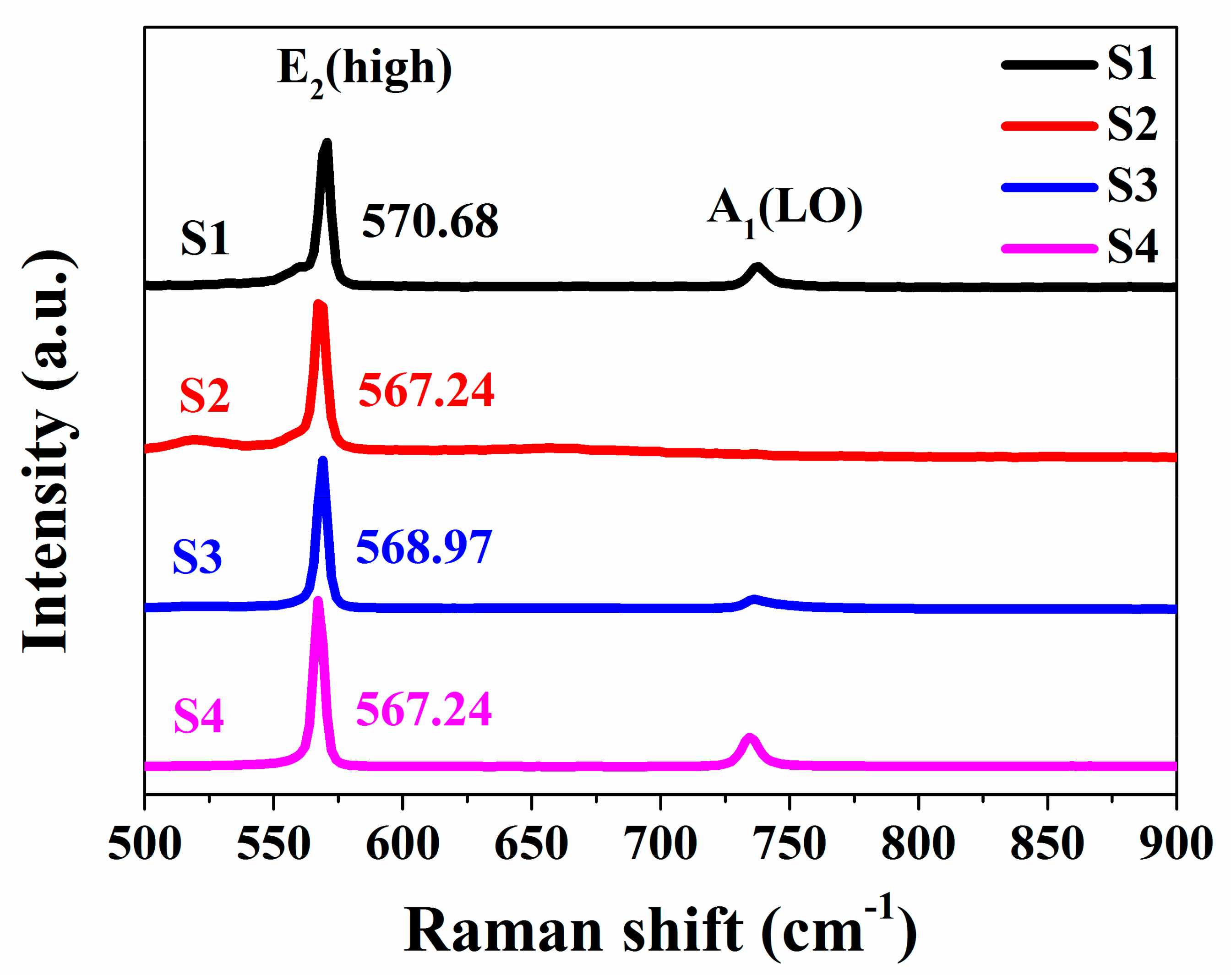
The stress of GaN grown by HVPE was analyzed by Raman spectroscopy, tested on the surface [28]. The E2 (high) phonon mode peak position can reveal the internal stress clearly. The E2 (high) phonon peak position is used to estimate the stress by the following equation [29]:
σ = Δω/4.3 (cm−1 GPa−1)
where the σ is the stress and the Δω is the E2 (high) peak shift compared to the stress-free GaN. The E2 (high) phonon peak of stress-free GaN is believed to be 567.00 cm−1. [30] Figure 4 shows the Raman spectra of GaN grown by HVPE with a different process. As can be seen from the Figure 4, the E2 (high) peak positions of GaN are 570.68, 567.24, 568.97, and 567.24 cm−1, respectively. According to the theoretical formula, the stresses of GaN crystals are calculated to be 0.86, 0.06, 0.46, and 0.06 GPa. The Raman results indicate that the GaN crystal with higher V/III ratio, not only has a better surface quality, but also has larger internal stress compared with that of others. Simultaneously, in the actual crystal growth experiment, a serious fragmentation occurred when the crystal thickness was more than 300 µm. In the contrary, the GaN with lower V/III ratio has a poor surface, but a small internal stress. The growth thickness could exceed 500 µm, but there was a macroscopic deterioration on the surface of the crystal. Compared with S4, the polyporous buffer interlayer (Figure 1h) effectively reduced the internal stress of the crystal. Therefore, it is very important to use three-step growth, which can provide not only relatively good surface (Figure 1d) but also low stress (0.06 GPa). At the same time, when the growth time increased to 20 h, self-separating occurred at the polyporous buffer interlayer, and the freestanding GaN was obtained.
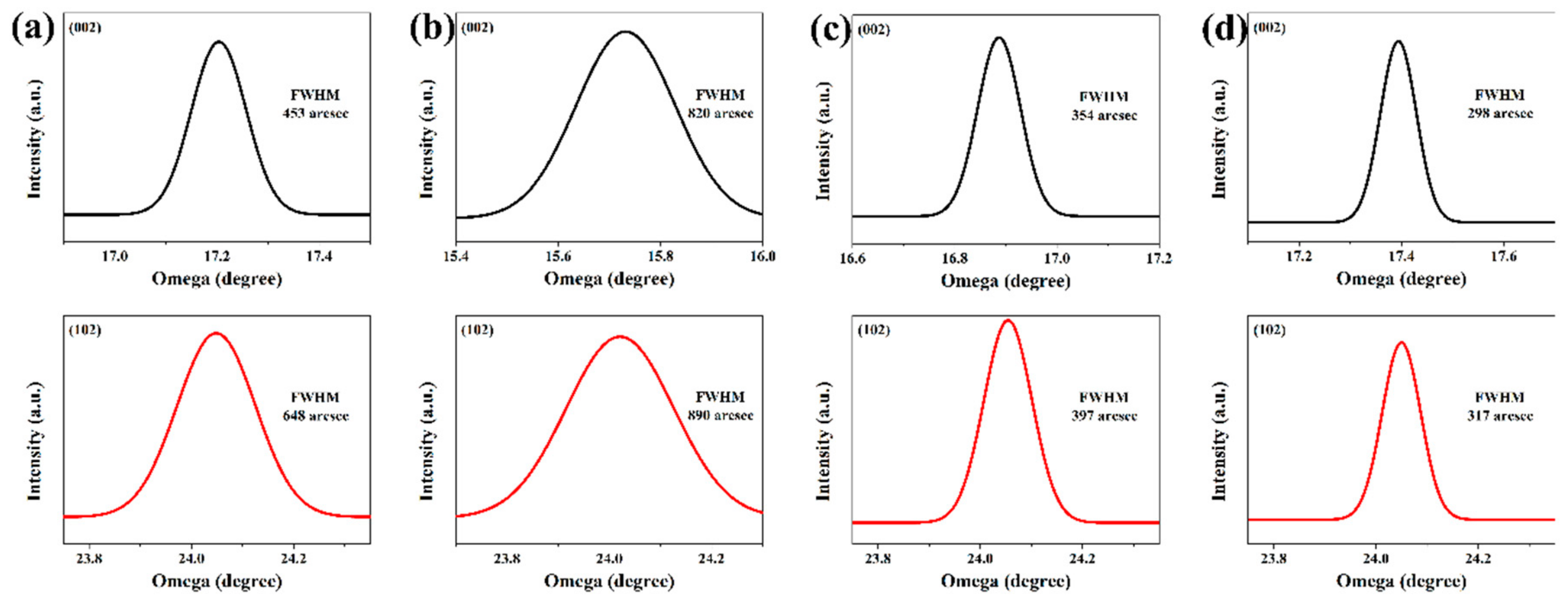
The crystalline quality of the GaN crystals was characterized by high-resolution X-ray diffraction (HRXRD) rocking curve [31]. Figure 5 exhibits the ω-scan spectra of the (002) symmetry planes, and the (102) asymmetry planes of GaN grown by HVPE with different steps. When the growth process was directly with one-step process: higher (83.3) or lower (43.3) V/III ratio, the HRXRD is shown as Figure 5a,b, corresponding to S1 and S2, respectively. The full width at half maxima (FWHM) of (002) peak is 453 and 820 arcsecond (arcsec), respectively. For (102) plane, the peaks locate at 648 and 890 arcsec, respectively. These results indicate that a higher V/III ratio can effectively improve the crystal quality of epitaxial layer. This is consistent with aforementioned tested results. Besides, the rocking curves of (002) are shifted for S2 and S3. The main reason may be that the surfaces of S2 and S3 have a number of large hexagonal, which makes the more sensitive (002) peak shift. In addition, the peak of S2 is much more remote from the standard due to its much bigger pits. Figure 5c shows the FWHM of (002) and (102) are 354 and 397 arcsec. It indicates that the lower V/III ratio is employed as the second step of growth based on the first step, which can appropriately improve the crystal crystallinity, compared with S2 (Figure 5b), although the hexagonal tunnel structures still existed. This also provided support for the third step of growth. As shown in Figure 5d, the GaN via three-step growth (S4) with 298,317 arcsec of (002) and (102), respectively. Previous researches have demonstrated that the screw dislocation density and the edge dislocation density are indirectly represented, respectively, by the FWHM of the HRXRD peaks, for the (002) and (102) planes. [13] The dislocation density can be calculated from equation [31].
D = β 2 2 π ln 2 b 2
where β is the FWHM and b represents the Burgers vector. The calculated TD density in S4 is 1.9 × 108 cm−2, a great improvement compared with the 8.1 × 108 cm−2.This result shows that the three-step growth method can effectively improve the crystal quality.
Figure 6 illustrates the room temperature photoluminescence (PL) spectra of GaN with a different process. Strong band edge emission peaks are observed at 366, 364, 365, and 367 nm for GaN grown with different V/III ratio. However, for S2 and S3, the luminous intensity is relatively weak, which may be due to many pits that existed on the surface. The defect causing the yellow luminescence band appears, mostly, surface oriented, mainly on the N face, and can be easily saturated, increasing the excitation intensity. The defect causing the green luminescence band is uniformly distributed in bulk GaN [32]. In additional, due to a large number of tunnels and the poor crystal quality, the defects of sample S2 increased. Hence, a yellow band appeared and located at 500–600 nm. The results of the PL spectra at room temperature demonstrate the optical quality of GaN grown by three-step HVPE growth can be improved.
Figure 7 shows a digital camera image of the freestanding GaN crystal produced by the three-step process and the vestigial substrate. The thickness of GaN crystal was 2 mm, and the residual GaN can be seen on the vestigial substrate. It is also powerful proof that the GaN was separated from the polyporous buffer interlayer. Although we have a little gap, with state-of-the-art, this method can be used to obtain freestanding GaN, simply and cheaply, and the GaN can be used as substrate for further homogeneous growth.

4. Conclusions

A three-step growth method was designed for HVPE growth. During the growth, a high-quality GaN polyporous buffer interlayer structure was formed directly. This interlayer played an important role in the separation of the GaN crystal from substrate and reducing growth stress. High quality freestanding GaN was obtained. In addition, the Raman spectroscopy indicated that the stress of GaN was 0.06 GPa. The FWHM of (002) and (102) peaks were 298 and 317 arcsec, indicating that the quality of GaN with three-step was improved. The PL spectra also proved the GaN had good optical quality. Therefore, the multi-step growth method can effectively improve the crystal quality and obtain the high-quality freestanding GaN.

Author Contributions

X.H. and Y.W. designed experiment. Y.S. and H.H. wrote the main manuscript text. B.Z., L.L., and D.X. helped with the sample fabrication. All of the authors participated to discussion and revision of the manuscript. All authors have read and agreed to the published version of the manuscript.

Funding

This work was supported by National Natural Science Foundation of China (Contract No. 51872162, 11890700).

Conflicts of Interest

The authors declare no conflict of interest.

References

  1. Pankove, J.I.; Miller, E.A.; Berkeyheiser, J.E. GaN blue light-emitting diodes. J. Lumin. 1972, 5, 84–86. [Google Scholar] [CrossRef]
  2. Lee, H.E.; Choi, J.; Lee, S.H.; Jeong, M.; Shin, J.H.; Joe, D.J.; Kim, D.H.; Kim, C.W.; Park, J.H.; Lee, J.H.; et al. Monolithic Flexible Vertical GaN Light-Emitting Diodes for a Transparent Wireless Brain Optical Stimulator. Adv. Mater. 2018, 30, 1800649. [Google Scholar] [CrossRef] [PubMed]
  3. Nakamura, S.; Senoh, M.; Nagahama, S.I.; Iwasa, N.; Yamada, T.; Matsushita, T.; Kiroyuki, K.; Sugimoto, Y.; Kozaki, T.; Umemoto, H.; et al. InGaN/GaN/AlGaN-based laser diodes with modulation-doped strained-layer superlattices grown on an epitaxially laterally overgrown GaN substrate. Appl. Phys. Lett. 1998, 72, 211–213. [Google Scholar] [CrossRef] [Green Version]
  4. Tyagi, A.; Zhong, H.; Chung, R.B.; Feezell, D.F.; Saito, M.; Fujito, K.; Speck, J.S.; DenBaars, S.P.; Nakamura, S. Semipolar (1011) InGaN/GaN laser diodes on bulk GaN substrates. Jpn. J. Appl. Phys. 2007, 46, L444. [Google Scholar] [CrossRef]
  5. Pearton, S.J.; Ren, F.; Zhang, A.P.; Dang, G.; Cao, X.A.; Lee, K.P.; Cho, H.; Gila, B.P.; Johnson, J.W.; Monier, C.; et al. GaN electronics for high power, high temperature applications. Mater. Sci. Eng. B Adv. 2001, 82, 227–231. [Google Scholar] [CrossRef] [Green Version]
  6. Cui, M.; Bu, Q.; Cai, Y.; Sun, R.; Liu, W.; Wen, H.; Lam, S.; Liang, Y.C.; Mitrovic, I.Z.; Taylor, S.; et al. Monolithic integration design of GaN-based power chip including gate driver for high-temperature DC–DC converters. Jpn. J. Appl. Phys. 2019, 58, 056505. [Google Scholar] [CrossRef]
  7. Ren, B.; Liao, M.; Sumiya, M.; Huang, J.; Wang, L.; Koide, Y.; Sang, L. Vertical-Type Ni/GaN UV Photodetectors Fabricated on Free-Standing GaN Substrates. Appl. Sci. 2019, 9, 2895. [Google Scholar] [CrossRef] [Green Version]
  8. Zhang, B.; Wu, Y.; Zhang, L.; Huo, Q.; Hu, H.; Ma, F.; Yang, M.; Shi, D.; Shao, Y.; Hao, X. Growth of high-quality GaN crystals on a BCN nanosheet-coated substrate by hydride vapor phase epitaxy. CrystEngComm 2019, 21, 1302–1308. [Google Scholar] [CrossRef]
  9. Andre, Y.; Trassoudaine, A.; Tourret, J.; Cadoret, R.; Gil, E.; Castelluci, D.; Aoude, O.; Disseix, P. Low dislocation density high-quality thick hydride vapour phase epitaxy (HVPE) GaN layers. J. Cryst. Growth 2007, 306, 86–93. [Google Scholar] [CrossRef]
  10. Li, T.; Ren, G.; Su, X.; Yao, J.; Yan, Z.; Gao, X.; Xu, K. Growth behavior of ammonothermal GaN crystals grown on non-polar and semi-polar HVPE GaN seeds. CrystEngComm 2019, 21, 4774–4879. [Google Scholar] [CrossRef]
  11. Inoue, T.; Seki, Y.; Oda, O.; Kurai, S.; Yamada, Y.; Taguchi, T. Pressure–Controlled Solution Growth of Bulk GaN Crystals under High Pressure. Phys. Status Solidi B 2001, 223, 15–27. [Google Scholar] [CrossRef]
  12. Mori, Y.; Imanishi, M.; Murakami, K.; Yoshimura, M. Recent progress of Na-flux method for GaN crystal growth. Jpn. J. Appl. Phys. 2019, 58, SC0803. [Google Scholar] [CrossRef]
  13. Zhang, L.; Li, X.; Shao, Y.; Yu, J.; Wu, Y.; Hao, X.; Yin, Z.; Dai, Y.; Tian, Y.; Huo, Q.; et al. Improving the quality of GaN crystals by using graphene or hexagonal boron nitride nanosheets substrate. ACS Appl. Mater. Inter. 2015, 7, 4504–4510. [Google Scholar] [CrossRef] [PubMed]
  14. Ramesh, C.; Tyagi, P.; Singh, S.; Singh, P.; Gupta, G.; Maurya, K.K.; Srivatsa, K.M.K.S.; Kumar, M.S.; Kushvaha, S.S. Influence of growth temperature on structural and optical properties of laser MBE grown epitaxial thin GaN films on a-plane sapphire. J. Vac. Sci. Technol. B 2018, 36, 04G102. [Google Scholar] [CrossRef]
  15. Ubukata, A.; Sodabanlu, H.; Watanabe, K.; Koseki, S.; Yano, Y.; Tabuchi, T.; Takeyoshi, S.; Koh, M.; Yoshiaki, N.; Masakazu, S. Accelerated GaAs growth through MOVPE for low-cost PV applications. J. Cryst. Growth 2018, 489, 63–67. [Google Scholar] [CrossRef]
  16. Wang, K.; Li, M.; Yang, Z.; Wu, J.; Yu, T. Stress control and dislocation reduction in the initial growth of GaN on Si (111) substrates by using a thin GaN transition layer. CrystEngComm 2019, 21, 4792–4797. [Google Scholar] [CrossRef]
  17. Huang, H.H.; Chao, C.L.; Chi, T.W.; Chang, Y.L.; Liu, P.C.; Tu, L.W.; Tsay, J.D.; Kuo, H.C.; Cheng, S.J.; Lee, W.I. Strain-reduced GaN thick-film grown by hydride vapor phase epitaxy utilizing dot air-bridged structure. J. Cryst. Growth 2009, 311, 3029–3032. [Google Scholar] [CrossRef]
  18. Moram, M.A.; Kappers, M.J.; Barber, Z.H.; Humphreys, C.J. Growth of low dislocation density GaN using transition metal nitride masking layers. J. Cryst. Growth 2007, 298, 268–271. [Google Scholar] [CrossRef]
  19. Amano, H.; Sawaki, N.; Akasaki, I.; Toyoda, Y. Metalorganic vapor phase epitaxial growth of a high quality GaN film using an AlN buffer layer. Appl. Phys. Lett. 1986, 48, 353–355. [Google Scholar] [CrossRef] [Green Version]
  20. Kim, C.; Robinson, I.K.; Myoung, J.; Shim, K.; Yoo, M.C.; Kim, K. Critical thickness of GaN thin films on sapphire (0001). Appl. Phys. Lett. 1996, 69, 2358–2360. [Google Scholar] [CrossRef]
  21. Lahrèche, H.; Nataf, G.; Feltin, E.; Beaumont, B.; Gibart, P. Growth of GaN on (1 1 1) Si: A route towards self-supported GaN. J. Cryst. Growth 2001, 231, 329–334. [Google Scholar] [CrossRef]
  22. Chu, C.F.; Lai, F.I.; Chu, J.T.; Yu, C.C.; Lin, C.F.; Kuo, H.C.; Wang, S.C. Study of GaN light-emitting diodes fabricated by laser lift-off technique. J. Appl. Phys. 2004, 95, 3916–3922. [Google Scholar] [CrossRef]
  23. Kim, S.J.; Lee, H.E.; Choi, H.; Kim, Y.; We, J.H.; Shin, J.S.; Lee, K.J.; Cho, B.J. High-performance flexible thermoelectric power generator using laser multiscanning lift-off process. ACS Nano 2016, 10, 10851–10857. [Google Scholar] [CrossRef] [PubMed]
  24. Kim, H.M.; Oh, J.E.; Kang, T.W. Preparation of large area free-standing GaN substrates by HVPE using mechanical polishing liftoff method. Mater. Lett. 2001, 47, 276–280. [Google Scholar] [CrossRef]
  25. Chen, Z.; Yu, Z.; Lu, P.; Liu, Y. Molecular dynamics simulations of atomic assembly in the process of GaN film growth. Physica B 2009, 404, 4211–4215. [Google Scholar] [CrossRef]
  26. Zhang, L.; Dai, Y.; Wu, Y.; Shao, Y.; Tian, Y.; Huo, Q.; Hao, X.; Shen, Y.; Hua, Z. Epitaxial growth of a self-separated GaN crystal by using a novel high temperature annealing porous template. CrystEngComm 2014, 16, 9063–9068. [Google Scholar] [CrossRef]
  27. Zhang, H.; Shao, Y.; Zhang, L.; Hao, X.; Wu, Y.; Liu, X.; Dai, Y.; Tian, Y. Growth of high quality GaN on a novel designed bonding-thinned template by HVPE. CrystEngComm 2012, 14, 4777–4780. [Google Scholar] [CrossRef]
  28. Hu, H.; Chang, B.; Sun, X.; Huo, Q.; Zhang, B.; Li, Y.; Shao, Y.; Zhang, L.; Wu, Y.Z.; Hao, X. Intrinsic properties of macroscopically tuned gallium nitride single crystalline facets for electrocatalytic hydrogen evolution. Chem. Eur. J. 2019, 25, 10420–10426. [Google Scholar] [CrossRef]
  29. Li, H.D.; Zhang, S.L.; Yang, H.B.; Zou, G.T.; Yang, Y.Y.; Yue, K.T.; Wu, X.H.; Yan, Y. Raman spectroscopy of nanocrystalline GaN synthesized by arc plasma. J. Appl. Phys. 2002, 91, 4562–4567. [Google Scholar] [CrossRef]
  30. Pandian, M.S.; In, U.C.; Ramasamy, P.; Manyum, P.; Lenin, M.; Balamurugan, N. Unidirectional growth of sulphamic acid single crystal and its quality analysis using etching, microhardness, HRXRD, UV–visible and Thermogravimetric-Differential thermal characterizations. J. Cryst. Growth 2010, 312, 397–401. [Google Scholar] [CrossRef]
  31. Tian, Y.; Shao, Y.; Wu, Y.; Hao, X.; Zhang, L.; Dai, Y.; Huo, Q. Direct growth of freestanding GaN on C-face SiC by HVPE. Sci. Rep. 2015, 5, 10748. [Google Scholar] [CrossRef] [PubMed] [Green Version]
  32. Reshchikov, M.A.; Morkoç, H.; Park, S.S.; Lee, K.Y. Yellow and green luminescence in a freestanding GaN template. Appl. Phys. Lett. 2001, 78, 3041–3043. [Google Scholar] [CrossRef] [Green Version]
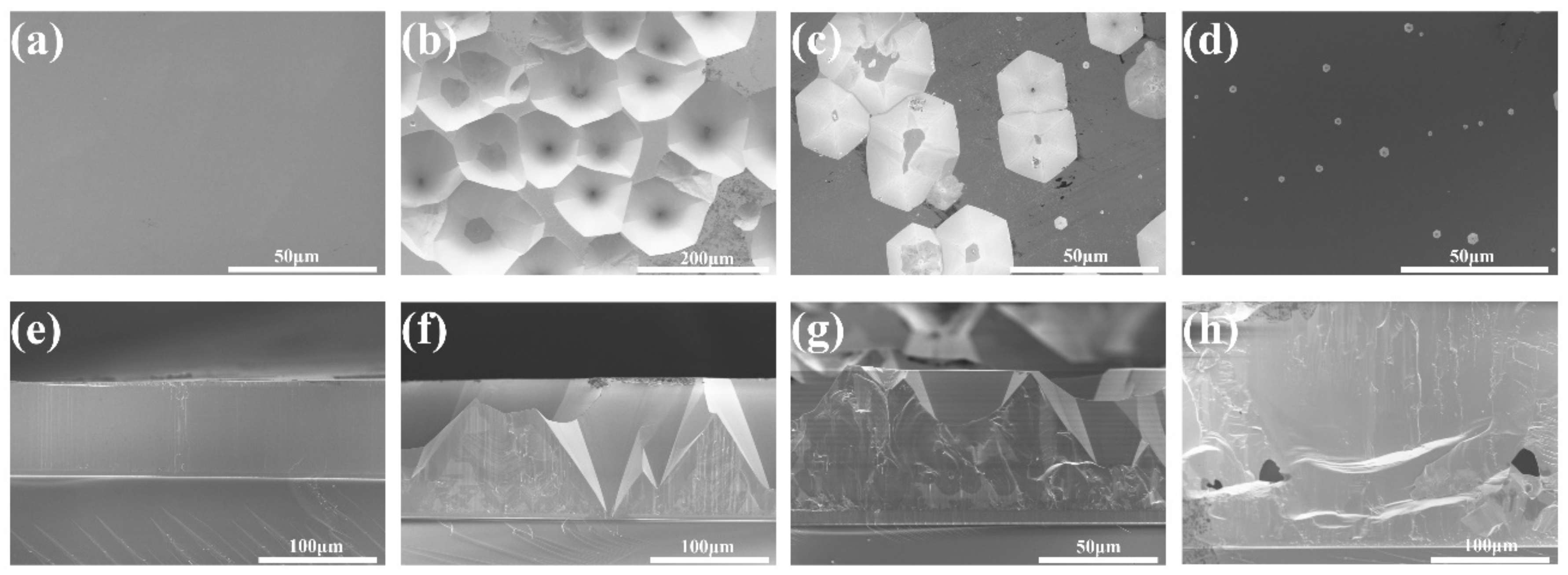
Figure 1. Surface SEM images of gallium nitride (GaN) crystals grown with a different process: S1 (a), S2 (b), S3 (c), S4 (d); Cross-section SEM images: S1 (e), S2 (f), S3 (g), S4 (h).
Crystals 10 00141 g001
Figure 2. The cross-sectional TEM images of GaN grown by hydride vapor phase epitaxy (HVPE) with V/III ratio of 83.3 (a) and 27.0 (b).
Crystals 10 00141 g002
Figure 3. (a) Schematic diagram of high-quality GaN polyporous buffer interlayer formation; (b) Table shows the growth conditions for different samples.
Crystals 10 00141 g003
Figure 4. Raman spectra of GaN grown by HVPE for S1, S2, S3, and S4.
Crystals 10 00141 g004
Figure 5. High-resolution X-ray diffraction (HRXRD) rocking curves of (002) and (102) of HVPE grown GaN crystals: S1 (a), S2 (b), S3 (c), S4 (d).
Crystals 10 00141 g005
Figure 6. PL spectra of different GaN samples grown by HVPE.
Crystals 10 00141 g006
Figure 7. (a) Digital camera image of the freestanding GaN crystal produced using three-step process; (b) vestigial substrate.
Crystals 10 00141 g007